Article

Implementation of Fluorescent-Protein-Based Quantification Analysis in L-Form Bacteria

Laboratory of Biology and Information Science, School of Life Sciences, East China Normal University, Shanghai 200062, China
*
Authors to whom correspondence should be addressed.
Bioengineering 2024, 11(1), 81; https://doi.org/10.3390/bioengineering11010081
Submission received: 19 December 2023 / Revised: 9 January 2024 / Accepted: 12 January 2024 / Published: 15 January 2024
(This article belongs to the Section Cellular and Molecular Bioengineering)

Abstract

:
Cell-wall-less (L-form) bacteria exhibit morphological complexity and heterogeneity, complicating quantitative analysis of them under internal and external stimuli. Stable and efficient labeling is needed for the fluorescence-based quantitative cell analysis of L-forms during growth and proliferation. Here, we evaluated the expression of multiple fluorescent proteins (FPs) under different promoters in the Bacillus subtilis L-form strain LR2 using confocal microscopy and imaging flow cytometry. Among others, Pylb-derived NBP3510 showed a superior performance for inducing several FPs including EGFP and mKO2 in both the wild-type and L-form strains. Moreover, NBP3510 was also active in Escherichia coli and its L-form strain NC-7. Employing these established FP-labeled strains, we demonstrated distinct morphologies in the L-form bacteria in a quantitative manner. Given cell-wall-deficient bacteria are considered protocell and synthetic cell models, the generated cell lines in our work could be valuable for L-form-based research.

1. Introduction

Cell-wall-less (L-form) bacteria can grow and reproduce after long-term adaption to various stressors, such as inhibitors of peptidoglycan biosynthesis, high temperature and nutrient starvation [1,2,3]. The L-forms induced from rod-shaped bacteria, e.g., Escherichia coli and Bacillus subtilis, exhibit spherical or pleomorphic shapes due to the lack of cell walls that maintain their original morphology [1,4]. It was described that the shape of the E. coli L-form strain NC-7 varies between spherical, angular and cylindrical [5], with intracellular vesicles and small membrane particles easily detectable [4]. Therefore, quantitative analysis is more challenging for L-forms due to their high complexity and heterogeneity, unlike the wild type with intact cell walls.
Advanced microscopy and flow cytometry (FCM)-based platforms have been extensively utilized for quantitative cell analysis, both at the single-cell and population levels. The resulting high-resolution microscopic cell images can be further processed using analytical tools like ImageJ, which offers various convenient ready-to-use plugins [6,7]. Additionally, fluorescence-activated cell sorting (FACS) and imaging flow cytometry (IFC) have been developed for applications including cell sorting [8,9,10], quantification [11,12], bacterial viability assessment [13,14], dynamic monitoring of bacterial morphology [15,16] and analysis of the heterogeneity in bacterial communities [16,17]. However, the quality and reproducibility of the results of these tools primarily rely on the employed fluorescent labeling markers for targeted cells [18,19]. Fluorescent proteins (FPs) can be expressed in specific subcellular locations, enabling effective cell tracing and imaging without extra staining procedures, potential cell toxicity and the insufficient selectivity of the commercially available dyes [18,20,21,22].
A robust promoter, either constitutive or inducible, is critical for efficient protein expression in host cells, including bacteria [23]. The promoters used in E. coli and B. subtilis have been extensively studied for fundamental and practical purposes, rendering them the most employed bacterial models [24]. For example, the available options in B. subtilis include inducible promoters (e.g., Pspac, PxylA, PsacB) [25,26,27,28,29], constitutive promoters (e.g., P43, Pveg, Pshuttle-09) [30,31,32], auto-inducible promoters (e.g., PsrfA and Pylb) [33,34,35,36,37] and phase-specific promoters (e.g., PrpsF and PaprE) [38,39]. Auto-inducible promoters allow for protein expression without inducers, making them ideal for industrial applications while mitigating the risks associated with prolonged chemical inducer exposure (e.g., arabinose, xylose, maltose and IPTG) [36,40].
Although FP labeling in wild-type bacteria has been well investigated, only limited reports have described the expression of fluorescent proteins in cell-wall-less (L-form) strains. Currently, only green fluorescent protein (GFP) and mCherry have been tested in B. subtilis L-form strains as fluorescent labels or fusion proteins for studying gene function and regulation [41,42,43]. Furthermore, most of the available promoter activities remain unexplored in a cell-wall-less scenario. Thus, L-form bacteria with stable and efficient FP expression are highly valuable for quantitative cell analysis in growth and proliferation, particularly as models for synthetic cells or protocells [44].
In this study, we performed a small-scale screening for multiple FPs (GFP+, EGFP, Staygold, mKO2, mCherry, DsRed and tdTomato) expressed under different promoters (Pspac, P43, Pveg and NBP3510) in the B. subtilis L-form strain LR2 [45]. The results indicated that NBP3510 performed better in driving fluorescent gene expressions in a growth-phase-dependent manner compared to the other tested promoters. Both the wild-type and L-form strains exhibited brighter fluorescent signals for EGFP and mKO2, among others. Surprisingly, we also identified a significant promoter activity for NBP3510 in both E. coli MG1655 and the L-form strain NC-7, emphasizing its broad applicability. Quantitative analysis of the constructed fluorescent strains was conducted using microscopic and IFC techniques under various culture conditions, providing valuable insights for future investigations of L-form bacteria as early life form and synthetic cell models.

2. Materials and Methods

2.1. Bacterial Strains

The B. subtilis wild-type 168CA and E. coli wild-type K-12 MG1655 were maintained in our laboratory. The B. subtilis L-form LR2 strain (168CA; Pxyl-murE ispA*) was gifted by Dr. Jeff Errington’s laboratory [45]. The stable L-form E. coli NC-7 derived from E. coli K12 3301 was originally obtained by Onoda et al. [46] and was gifted by Dr. Akinobu Oshima (Shimane University, Matsue, Japan).

2.2. Culture Conditions

The B. subtilis wild-type (168CA) bacteria were grown on nutrient agar (NA, Oxoid) or nutrient broth (NB, Oxoid). The B. subtilis L-form LR2 bacteria were cultured in an osmoprotective medium (NB/MSM) composed of 2× magnesium–sucrose–maleic acid (MSM: 40 mM MgCl2, 1 M sucrose, 40 mM maleic acid, pH 7.0) mixed 1:1 with 2× nutrient broth (NB, Oxoid) or 2× NA. Equally, C minimal medium (70 mM K2HPO4, 30 mM KH2PO4, 25 mM (NH4)2SO4, 0.5 mM MgSO4, 10 μM MnSO4, 22 mg/L ferric ammonium citrate and 50 mg/L tryptophan) was also used to culture the B. subtilis strains as indicated in [47]. The E. coli MG1655 was grown in Luria–Bertani (LB) broth (1% tryptone, 0.5% Yeast Extract, 170 mM (1%) NaCl). In the case of NC-7, the osmoprotective MLB medium containing 340 mM NaCl (1% peptone, 0.5% Yeast Extract, 30 mM glucose, 340 mM NaCl, 1 mM CaCl2, 25 mM MOPS pH 7.0, 100 U/mL PenG) was used for static culture without shaking [5]. All the bacteria except NC-7 were incubated at 37 °C with shaking (200 rpm) while the NC-7 cells were incubated at 30 °C statically without shaking. A final concentration of 0.5 mM or 2 mM IPTG was added to the culture medium when necessary. The antibiotics were added at the following final concentrations: 100 μg/mL Ampicillin, 25 μg/mL Chloramphenicol, 150 μg/mL Spectinomycin and 200 μg/mL D-cycloserine.

2.3. Plasmid Constructions

As illustrated in Figure S1A, the integrative vector PSG1154 (Ampicillin used for selections in E. coli, Spectinomycin used for selections in B. subtilis) was chosen as the backbone to be integrated into the chromosomal amyE locus of B. subtilis [48]. The IPTG-inducible vector Pspac-gfp+ was constructed by replacing the original promoter Pxyl and fluorescent protein on plasmid PSG1154 with the DNA fragment containing the Pspac-gfp+-lacI sequence cloned from pMUTIN-gfp+ [49]. The sequences of three promoters, Pveg, P43 and NBP3510 (Figure S2), were synthesized (Sangon, Shanghai, China) and then cloned into plasmid PSG1154 together with gfp+ to yield Pveg-gfp+, PP43-gfp+ and NBP3510-gfp+. Furthermore, we chose another two green fluorescent proteins (EGFP and Staygold), three red fluorescent proteins (mCherry, DsRed and tdTomato) and one orange fluorescent protein (mKO2) to construct multiple expression vectors by replacing the gfp+ of the NBP3510-gfp+ using the Gibson assembly method. To construct the recombinant plasmid pNBP3510-mKO2_E. coli, three fragments from the plasmid pUC19 (Ori), pETcoco1 (CmR) and the mKO2 expression cassette were amplified using the primer pairs (listed in Table S1), and then assembled to produce a new plasmid (Chloramphenicol resistance) suitable for the E. coli strains. All the correct expression plasmids after DNA sequencing (Sangon, Shanghai, China) were transformed into the indicated bacterial strains. The positive fluorescent signals were confirmed using direct observation on plate and confocal microscopy (Nikon C2plus, Yokohama, Japan). The plasmid sequence data have been submitted to the GenBank database under accession number OR754211–OR754221.

2.4. Microplate Reader Measurement

The bacterial cells after overnight culture were diluted to the same optical density (OD) with fresh culture medium. Then, 200 μL of the diluted cells was transferred into a 96-well plate for continuous culture. The OD (λ = 600 nm) and fluorescence intensity (mKO2/mCherry/DsRed/TdTomato: λ excitation = 560 nm, λ emission = 580–611 nm; GFP+/EGFP/Staygold: λ excitation = 485 nm, λ emission = 500–530 nm) were monitored using a Multimode microplate reader (BioTek Instruments, Winooski, VT, USA; model: Synergy H1) every 0.5 h. All the samples were prepared in at least triplicate. The data were exported upon the completion of the data acquisition.

2.5. Confocal Microscopy Observation

The bacterial cells, which were cultured until the late logarithmic phase, were dropped onto a microscope slide and covered with a coverslip. Then, the cell samples were observed, and images were acquired using a confocal microscope (Nikon C2 plus, Yokohama, Japan). The excitation beam for mKO2/mCherry/DsRed/tdTomato was set at 561 nm and the emission signal for mKO2/DsRed/tdTomato was captured at 560–595 nm, while the λ emission for mCherry was 600–650 nm. As for GFP+/EGFP/Staygold, λ excitation = 488 nm and λ emission = 500–550 nm were used. The image analysis was performed using the NIS-ELEMENTS C-ER software (Nikon, Yokohama, Japan). All the fluorescent images acquired using the confocal microscope were captured and processed using identical parameters.

2.6. Imaging Flow Cytometry (IFC) Analysis

All the strains were cultured in corresponding medium until the late logarithmic phase and were analyzed using an Amnis™ ImageStream™X MK II imaging flow cytometer with the INSPIRE™ acquisition software v.201.1.0.744 (Luminex, Austin, TX, USA). Red fluorescent signals were induced with a 20 mW/200 mW 561 nm laser, and the emission was detected in Channel 3 with a 560–595 nm filter (mKO2/DsRed/tdTomato) and in Channel 4 with a 595–642 nm filter (mCherry). The green fluorescent signals were induced with a 200 mW 488 nm laser, and the emission was detected in Channel 2 with a 480–560 nm filter. The bright field and side scatter (SSC) data were collected in Channel 4/1 and Channel 6 (785 nm), respectively. For each sample, at least 3 × 10⁴ cells were acquired at 40× magnification, a pixel size of 0.25 μm2, a low flow rate and high sensitivity, and the measurement data were analyzed using the IDEAS analysis software (v.6.2.183.0, Luminex, Austin, TX, USA). The FlowJo software (v10.6, BD Biosciences, Ashland, OR, USA) was employed to analyze the single-cell data generated using the IFC for various parameters, including the fluorescent intensity, aspect ratio, length and area.

3. Results

3.1. Evaluation of the Fluorescent Protein Expressions in B. subtilis WT and LR2

In this study, we successfully constructed a range of plasmids bearing different constitutive promoters (Pveg and P43), an auto-inducible promoter (NBP3510) and an IPTG-inducible promoter (Pspac) (Figure S2). As shown in Figure S1B,C, we initially observed the colors of positive colonies from each fluorescent strain with the correct chromosomal integrations (single copy) of the B. subtilis WT and LR2 on solid plates (15 h culture) to evaluate the fluorescent protein expression. The green/red colors were only noticeable in the NBP3510 promoter-containing WT strains, NBP3510-egfp and NBP3510-mKO2 (Figure S1B, arrowed). While not observed in LR2 at 15 h (Figure S1C), similar green/red colonies appeared after 2 days longer of incubation. Accordingly, the cell pellets from the liquid culture of NBP3510-egfp/mKO2 also exhibited apparent colors for both the WT (Figure S1D) and LR2 (Figure S1E) strains. Since Pspac activation requires IPTG, further cultivation of the Pspac-gfp+-transformed strain was performed on a solid plate with 0.5 mM IPTG (Figure S1F–H). Significant color changes were observed in E. coli DH5α (positive control, high copy number of plasmid), but neither B. subtilis strain showed a detectable color. Collectively, these results indicated the superiority of NBP3510 over the other promoters for EGFP and mKO2 protein expression in B. subtilis.
To directly compare the FP expression levels, we subjected late exponential phase cells to confocal microscopy to visualize the fluorescent signals. Note that the C minimal medium with a defined composition was used for both strains to avoid unexpected disturbance from the nutrient broth medium. All the cell images were obtained under consistent experimental settings, as described in Materials and Methods. The highest GFP+ fluorescence intensity was observed under NBP3510, in contrast to the weaker signals from the other promoters (Figure S3A,C). EGFP and mKO2 showed a superior fluorescent performance compared to the other FPs including GFP+, Staygold, mCherry, DsRed and tdTomato (Figure S3B,D). Both the B. subtilis WT and LR2 strains displayed similar expression levels and fluorescent intensity patterns (Figure 1), indicating the successful development of several fluorescent strains for cell quantification assays.

3.2. Fluorescent Protein mKO2-Based Quantification Analysis in B. subtilis WT and L-Form Cells

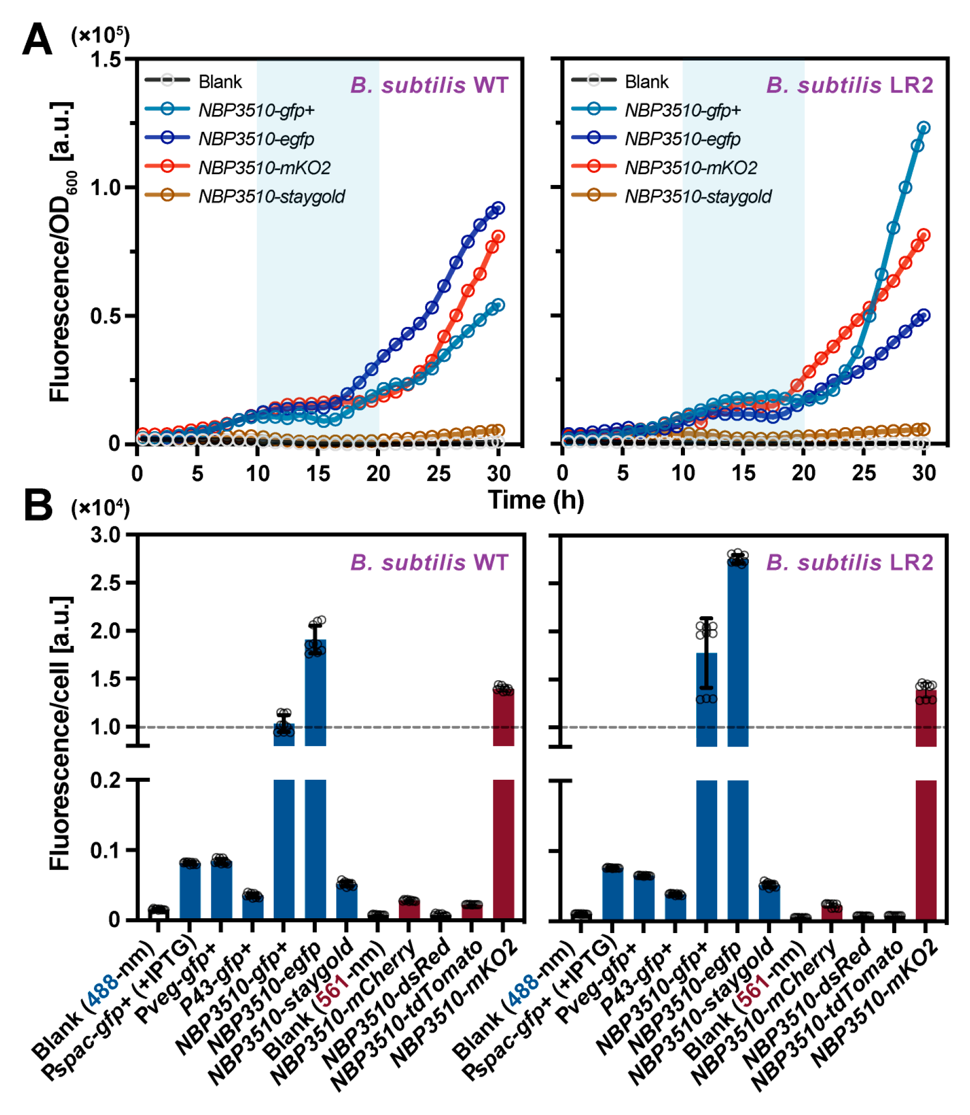
We then used fluorometry to quantitatively examine the dynamics of the expressed FPs during bacterial growth in the constructed cell lines. As summarized in Figures S4 and S5 and Figure 2A, the results suggested that the GFP+, EGFP and mKO2 induced by the promoter NBP3510 effectively indicate the bacterial growth in the B. subtilis WT and LR2 cultured in the minimal medium, while the low-expressed FPs poorly indicate growth in the other strains (Figure S5A). Moreover, the expression level per OD (fluorescence intensity/OD) dramatically increased after the exponential phase (10–20 h, blue area) in all the tested strains, which is consistent with the previous result that NBP3510 is a strong promoter during the stationary phase (Figure 2A) [36,37]. Meanwhile, the average fluorescence intensity at the single-cell level from the late exponential phase was also quantified using IFC. The bacterial cells expressing GFP+, EGFP or mKO2 exhibited an average intensity greater than 1 × 10⁴/cell (dashed line), which was significantly higher (>10-fold) than that of the other fluorescent strain cells (Figure 2B). Direct observation under a microscope and quantitative analysis at both the population and single-cell levels confirmed NBP3510 induces relatively high-level EGFP and mKO2 expression in single-copy status without additional inducers. This characteristic makes it an ideal indicator for the B. subtilis WT and L-form bacteria LR2.
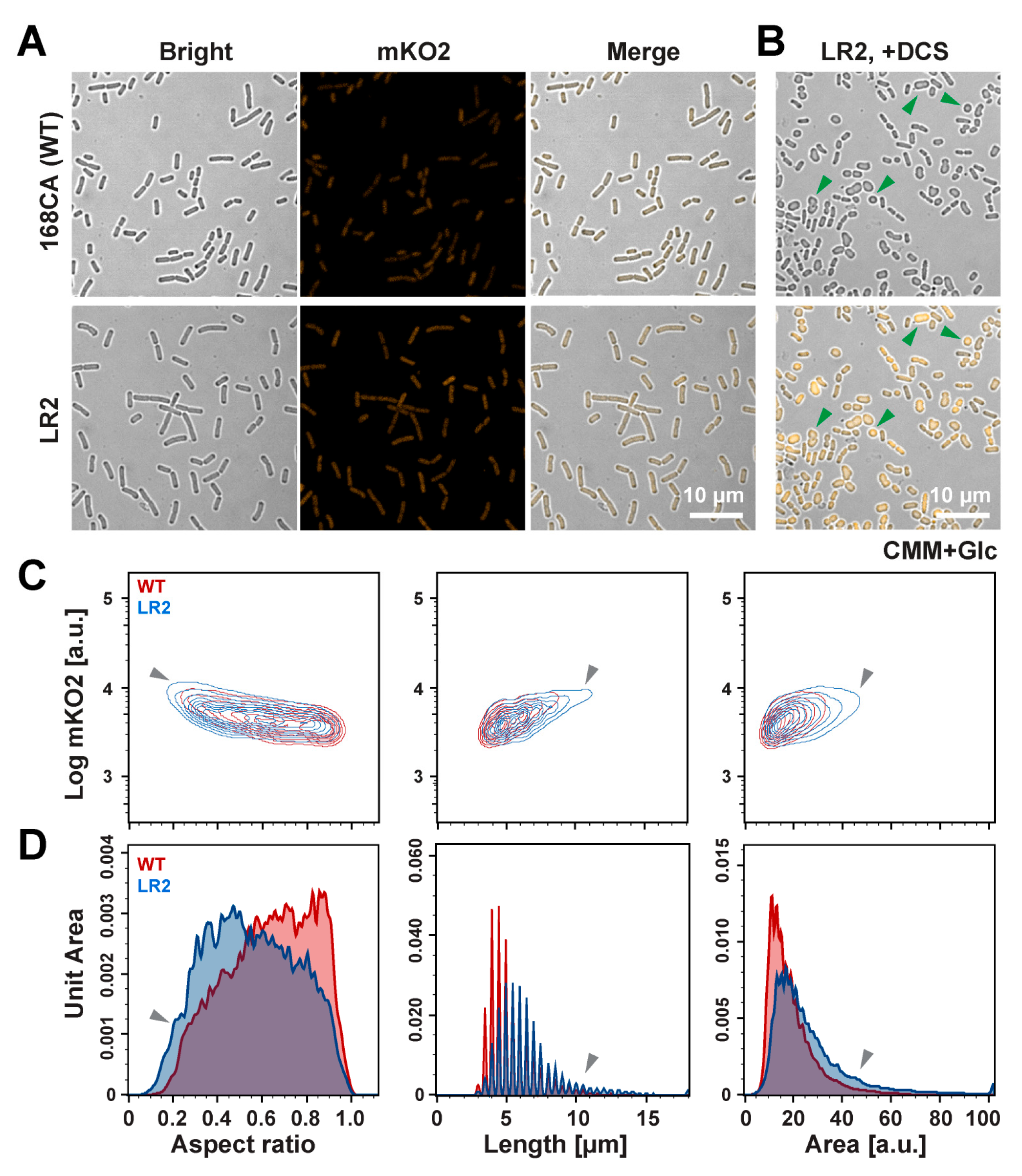
Both LR2 and its WT strain display a similar short rod-shaped morphology in the minimal medium (Figure 3A), which is inconsistent with the typical spherical morphology of most of the L-form bacteria in the previous reports [50]. We speculate this discrepancy may have been caused by the nutrient deprivation environment in the CMM or the residual cell walls remaining in the LR2. It is known that D-cycloserine (DCS) can efficiently induce the L-form transition in many bacteria by inhibiting cell wall lipid II precursor synthesis [50]. We then examined the morphological changes in the mKO2-labeled LR2 cells cultured in the CMM medium with DCS (Figure 3B). The direct observations under the confocal microscope reveal that LR2 can grow and exhibits more typical small spherical cells and irregular division (Figure 3B, green arrowhead). It is worth noting that the cells cultured in NB/MSM showed a remarkable diversity of shapes, regardless of DCS addition. Surprisingly, there appears to be a correlation between the higher mKO2 fluorescence level and the presence of DCS, as enhanced fluorescence was observed (Figure S6A), presumably implying that DCS may affect promoters or other unknown intracellular targets [51].
Taking advantage of the mKO2 fluorescence marker, we utilized the IFC platform to distinguish LR2 from other non-cell impurities. We then performed statistical analysis on key characteristics such as the fluorescence, cell aspect ratio, length and area. The results from the cells (n > 5000) showed significant differences in the aspect ratio between the DCS treatment and control groups of LR2, suggesting a shift toward a spherical shape in the bacterial morphology (Figure S6B,C). Although it is extremely difficult to differentiate between the two cell strains, the IFC analysis showed that more LR2 cells are longer, with a lower aspect ratio and larger area (gray arrowheads, Figure 3C,D), suggesting a change in bacterial elongation direction in the LR2 population. The IFC results confirm the distinct morphological changes in the LR2 cell populations in minimal medium, even though these changes were not discernible in the microscopic images.

3.3. NBP3510 Promoter Is Also Active in E. coli Strains

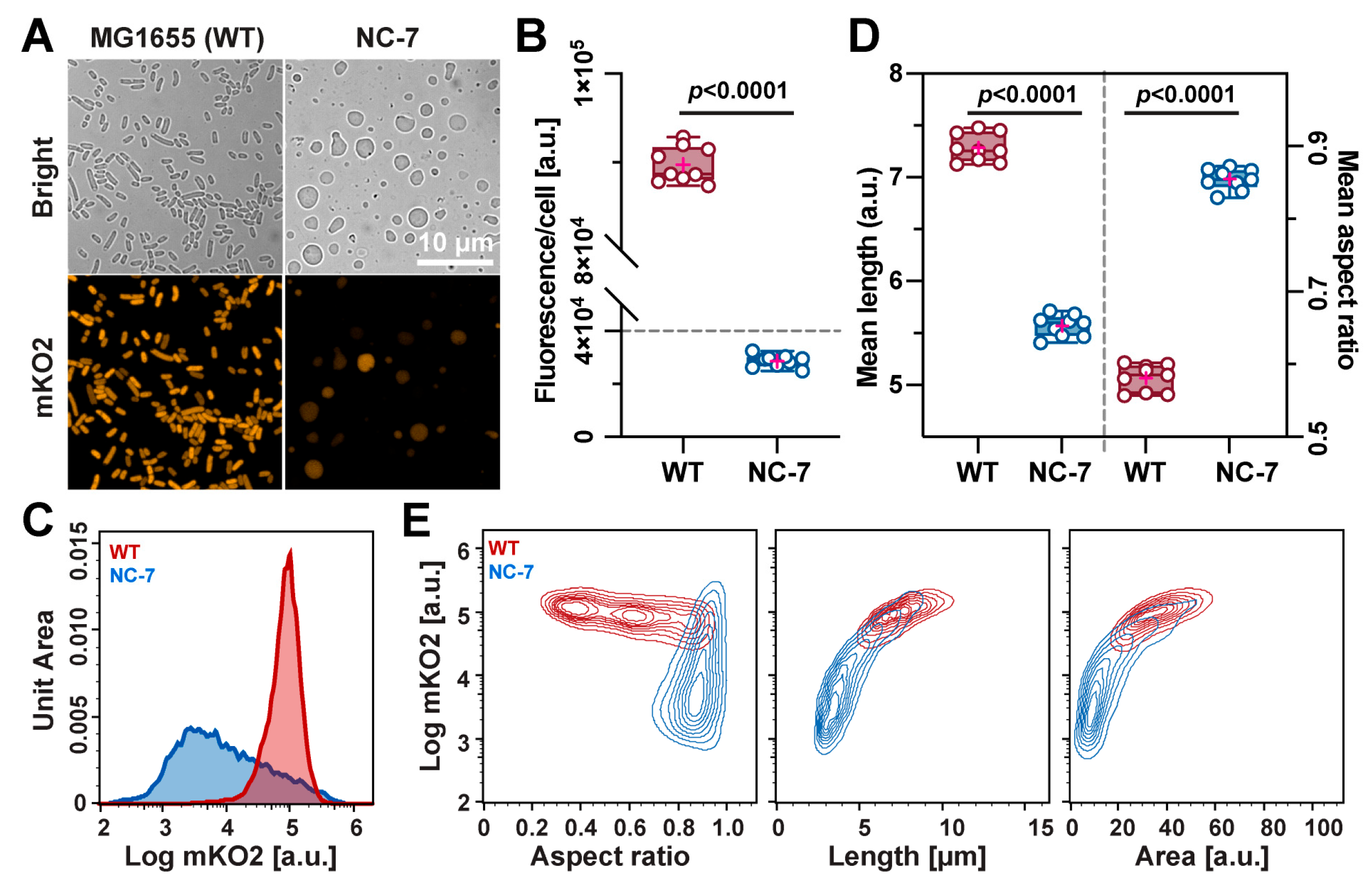
During the plasmid preparation of pNBP3510-fp for B. subtilis, we unexpectedly discovered that E. coli DH5α positive colonies showed recognizable colors (Figure S7A), indicating that NBP3510 is also recognizable by E. coli. To further explore this promoter’s potential in E. coli, we designed a new plasmid (pNBP3510-mKO2-Ecoli) assembled from three fragments containing Ori (pMB1) of pUC19, CmR of pETcoco1 and the NBP3510-mKO2 expression cassette (Figure S7B). The positive transformants in E. coli WT MG1655 and L-form NC-7 were then cultured in LB and MLB media, respectively. Compared to the control groups lacking the corresponding plasmid, the resulting bacterial cell pellets of each positive strain exhibited a detectable pink color, while NC-7 displayed a weak pink color, as shown in Figure S7C. To further visualize the cells, we gained fluorescent images using the confocal microscope (Figure 4A), which are consistent with the results in Figure S7C. The expression levels in NC-7 also varied notably among cells in the whole population. The fluorescence per OD in NC-7 showed a lower increase rate in mKO2 fluorescence during the exponential phase (Figure S5B), presumably due to low viability and significant differences in the plasmid copy numbers at the single-cell level in L-form cells.

3.4. Quantification Analysis in the E. coli WT and NC-7 L-Form Cells

The results of the nine replicates showed that the MG1655 cells had a much higher mKO2 fluorescence intensity than the NC-7 cells (Figure 4B,C). Further examination of the IFC data in Figure 4D unveiled that the MG1655 cells were longer (~6.84 μm) than the NC-7 cells (~4.86 μm), although many giant cells were present in the microscopic images (Figure 4A, right panel). The aspect ratio of the MG1655 cells (~0.57) was smaller than that of the NC-7 cells (~0.85), indicating NC-7 bacteria are more spherical. The detailed distributions of the aspect ratio, length and area versus the expressed fluorescence intensity are depicted in Figure 4E, demonstrating that NC-7 cells are spherical (aspect ratio) and contain more smaller (length and area) cells in the population. Collectively, the quantitative results from the IFC correlate well with the microscopy observations, confirming that constructed fluorescent NC-7 cells can be further applied in fluorescence-based quantitative analyses under various conditions.

4. Discussion

In this work, different promoters and FPs were chosen to achieve fluorescence labeling for the B. subtilis WT and L-form strains. While the integrated expression mode can address the possible plasmid loss during cell divisions, it requires stronger promoters to compensate for the lower expression levels. The NBP3510 promoter is a strong stationary phase promoter [37], showing significantly higher activity than the other three promoters (Pspac, Pveg and P43) tested in this study. The best performance was observed with GFP+, EGFP and mKO2 among the FPs chosen, validated using confocal microscopy, a plate reader and IFC (Figure 1, Figure 2 and Figures S1–S5). While other FPs have been shown to be highly photostable in previous studies, such as Staygold and mCherry [42,43,52], their expressions tested in our experiments were unsatisfactory, which could be attributed to the codon bias or protein stability, or other differences in the plasmid constructions [53,54,55,56,57]. Future investigation could improve their expressions using further codon optimization and reconsiderations in the plasmid reconstruction.
Moreover, we found that the B. subtilis promoter NBP3510 exhibits excellent activity in E. coli strains, suggesting a possible application as a universal promoter for large-scale protein productions in an inducer-free manner [37]. In general, most promoters show strain-specificity [58], with few functioning in heterologous hosts, such as PsrfA [59]. NBP3510 is an enhanced version of Pylb, created by replacing the −35, −10 core region and upstream sequence (UP), with consensus sequences [36,37]. These alterations may explain why this promoter is recognizable and drives strong gene expression in E. coli, though the detailed mechanism remains unknown.
Unexpectedly, we found in this study that B. subtilis LR2 can grow directly in CMM medium without additional osmotic stabilizers (Figure 1B). It is known that L-forms can proliferate better only when osmoprotective conditions are present [5,60]. Unlike commonly used nutrient broths like NB/MSM, CMM has a defined low-complexity composition ideal for synthetic biology applications [61]. Based on the fluorescent marker mKO2, a quantitative analysis was conducted to characterize the morphological changes in the LR2 cells in CMM in a quantitative manner. The results indicated that the LR2 cells exhibited a lower growth rate when cultured in the CMM medium (Figure 2A), potentially due to the inadequate nutritional supply in the culture environment. It is believed that cell elongation is driven by an FtsZ-independent mechanism, requiring excess membrane synthesis to generate an unbalanced surface-area-to-volume ratio, promoting cell division [45]. In environments with sufficient nutrition (NB/MSM), L-form bacteria are able to synthesize an adequate membrane from rich resources, allowing for the formation of extruded division morphologies. We hypothesize L-form growth is mostly hindered in nutrient-deficient environments like the CMM medium used in our experiments, and the typical L-form traits are less prominent than in rich medium.
Similarly, the mKO2 marker driven by NBP3510 was applied in E. coli to monitor the protein expression and cell morphology. The IFC analysis showed that the NC-7 L-form was statistically rounder and smaller than the wild-type (Figure 4D). NC-7 also showed significantly lower mKO2 expression than MG1655, indicating that the L-form bacteria have a higher tendency to lose plasmids and maintain only a low copy number. The heavy mutations in NC-7, including essential genes, make it a valuable resource for minimal genome research [5,62]. To study the gene function and essentiality in NC-7 and other L-forms, it is crucial to develop a plasmid DNA transformation method for gene knockout or overexpression. However, we encountered a poor transformation efficiency and failed to establish an efficient gene manipulation platform for NC-7, highlighting the need for future efforts on NC-7-based genetics and applications. Considering the cell-wall-less status of L-form cells, it might be interesting to test whether a liposome-based DNA transfection approach is usable to improve the transformation efficacy. L-form bacteria have been already found in plants, recurrent urinary tract infections and the human tumor microbiome, implying their clinical and environmental significance [63,64,65]. The methods and plasmids generated in our research are valuable for studying the L-form bacteria in clinical and environmental samples.

5. Conclusions

Based on small-scale screening of various promoters and fluorescent genes, we validated a strong promoter NBP3510, which induced satisfactory gene expressions in both B. subtilis and E. coli. Both the WT and cell-wall-less bacteria were successfully labeled with the fluorescent mKO2 protein and employed for quantitative analysis of the cell morphology in two L-form bacteria models. The findings provided insights into the L-form cell behavior in different environments, offering new opportunities for using L-forms as synthetic cell models.

Supplementary Materials

The following supporting information can be downloaded at https://www.mdpi.com/article/10.3390/bioengineering11010081/s1, Figure S1: Plasmid construction and direct observation of expressed FPs in B. subtilis WT and L-form LR2; Figure S2: The sequence of different promoters used in this study; Figure S3: Microscope images of all constructed fluorescent strains; Figure S4: Growth curve of all strains used in this study; Figure S5: The expression of fluorescent protein mKO2 in B. subtilis WT, L-form LR2, E. coli MG1655 and L-form NC-7; Figure S6: Quantitative analysis of morphology in B. subtilis L-form LR2 with DCS; Figure S7: Construction and analysis of fluorescent expression strains of E. coli MG1655 and L-form NC-7; Table S1: List of primers used in this study.

Author Contributions

Conceptualization, J.X. and T.Y.; methodology, D.T., Y.L. (Yiyuan Liu), Y.Z., Y.L. (Yunfei Liu), Y.X. and B.X.; validation, D.T., J.X. and T.Y.; formal analysis, J.X. and D.T.; investigation, J.X.; resources, D.T., Y.L. (Yiyuan Liu), Y.Z., Y.X. and B.X.; data curation, D.T., Y.L. (Yiyuan Liu), Y.Z. and J.X.; writing—original draft preparation, J.X. and D.T.; writing—review and editing, J.X. and T.Y.; visualization, J.X.; supervision, J.X. and T.Y.; project administration, J.X. and T.Y.; funding acquisition, T.Y. All authors have read and agreed to the published version of the manuscript.

Funding

This research was funded by the National Key R&D Program of China, Synthetic Biology Research (no. 2019YFA0904500).

Institutional Review Board Statement

Not applicable.

Informed Consent Statement

Not applicable.

Data Availability Statement

All supporting data are included within the main article and its Supplementary Files.

Acknowledgments

The authors thank Akinobu Oshima of Shimane University (Japan), Akiko Kashiwagi of Hirosaki University (Japan) and Jeff Errington of Newcastle University (UK) for providing the NC-7 strain and LR2 strain, respectively.

Conflicts of Interest

The authors declare no conflicts of interest.

References

  1. Allan, E.J.; Hoischen, C.; Gumpert, J. Bacterial L-forms. Adv. Appl. Microbiol. 2009, 68, 1–39. [Google Scholar] [CrossRef] [PubMed]
  2. Markova, N.; Slavchev, G.; Michailova, L.; Jourdanova, M. Survival of Escherichia coli under lethal heat stress by L-form conversion. Int. J. Biol. Sci. 2010, 6, 303–315. [Google Scholar] [CrossRef] [PubMed]
  3. Wohlfarth, J.C.; Feldmuller, M.; Schneller, A.; Kilcher, S.; Burkolter, M.; Meile, S.; Pilhofer, M.; Schuppler, M.; Loessner, M.J. L-form conversion in Gram-positive bacteria enables escape from phage infection. Nat. Microbiol. 2023, 8, 387–399. [Google Scholar] [CrossRef] [PubMed]
  4. Mattman, L.H. Cell Wall Deficient Forms: Stealth Pathogens; CRC Press: Boca Raton, FL, USA, 2000. [Google Scholar]
  5. Osawa, M.; Erickson, H.P. L form bacteria growth in low-osmolality medium. Microbiology 2019, 165, 842–851. [Google Scholar] [CrossRef]
  6. Schneider, C.A.; Rasband, W.S.; Eliceiri, K.W. NIH Image to ImageJ: 25 years of image analysis. Nat. Methods 2012, 9, 671–675. [Google Scholar] [CrossRef] [PubMed]
  7. Ducret, A.; Quardokus, E.M.; Brun, Y.V. MicrobeJ, a tool for high throughput bacterial cell detection and quantitative analysis. Nat. Microbiol. 2016, 1, 16077. [Google Scholar] [CrossRef]
  8. Sycuro, L.K.; Rule, C.S.; Petersen, T.W.; Wyckoff, T.J.; Sessler, T.; Nagarkar, D.B.; Khalid, F.; Pincus, Z.; Biboy, J.; Vollmer, W. Flow cytometry-based enrichment for cell shape mutants identifies multiple genes that influence Helicobacter pylori morphology. Mol. Microbiol. 2013, 90, 869–883. [Google Scholar] [CrossRef]
  9. Laubacher, M.E.; Melquist, A.L.; Chandramohan, L.; Young, K.D. Cell sorting enriches Escherichia coli mutants that rely on peptidoglycan endopeptidases to suppress highly aberrant morphologies. J. Bacteriol. 2013, 195, 855–866. [Google Scholar] [CrossRef]
  10. Tian, D.; Wang, C.; Liu, Y.; Zhang, Y.; Caliari, A.; Lu, H.; Xia, Y.; Xu, B.; Xu, J.; Yomo, T. Cell Sorting-Directed Selection of Bacterial Cells in Bigger Sizes Analyzed by Imaging Flow Cytometry during Experimental Evolution. Int. J. Mol. Sci. 2023, 24, 3243. [Google Scholar] [CrossRef]
  11. Felip, M.; Andreatta, S.; Sommaruga, R.; Straskrabova, V.; Catalan, J. Suitability of flow cytometry for estimating bacterial biovolume in natural plankton samples: Comparison with microscopy data. Appl. Environ. Microbiol. 2007, 73, 4508–4514. [Google Scholar] [CrossRef]
  12. Brown, M.R.; Hands, C.L.; Coello-Garcia, T.; Sani, B.S.; Ott, A.I.G.; Smith, S.J.; Davenport, R.J. A flow cytometry method for bacterial quantification and biomass estimates in activated sludge. J. Microbiol. Methods 2019, 160, 73–83. [Google Scholar] [CrossRef] [PubMed]
  13. Ou, F.; McGoverin, C.; Swift, S.; Vanholsbeeck, F. Absolute bacterial cell enumeration using flow cytometry. J. Appl. Microbiol. 2017, 123, 464–477. [Google Scholar] [CrossRef] [PubMed]
  14. Alba, C.; Marin, A.C.; McNicholl, A.G.; Montalban-Arques, A.; Mora-Gutierrez, I.; Sanchez-Arroyo, A.J.; Soler, T.; Garcia-Fresnadillo, D.; Gisbert, J.P.; Alarcon, T.; et al. A quick flow cytometry protocol to assess Helicobacter pylori viability. J. Microbiol. Methods 2020, 177, 106043. [Google Scholar] [CrossRef] [PubMed]
  15. Narayana, S.K.; Mallick, S.; Siegumfeldt, H.; van den Berg, F. Bacterial flow cytometry and imaging as potential process monitoring tools for industrial biotechnology. Fermentation 2020, 6, 10. [Google Scholar] [CrossRef]
  16. Power, A.L.; Barber, D.G.; Groenhof, S.R.M.; Wagley, S.; Liu, P.; Parker, D.A.; Love, J. The Application of Imaging Flow Cytometry for Characterisation and Quantification of Bacterial Phenotypes. Front. Cell Infect. Microbiol. 2021, 11, 716592. [Google Scholar] [CrossRef] [PubMed]
  17. Andreatta, S.; Wallinger, M.M.; Piera, J.; Catalan, J.; Psenner, R.; Hofer, J.S.; Sommaruga, R. Tools for discrimination and analysis of lake bacterioplankton subgroups measured by flow cytometry in a high-resolution depth profile. Aquat. Microb. Ecol. 2004, 36, 107–115. [Google Scholar] [CrossRef]
  18. Huang, Y.; Chen, W.; Chung, J.; Yin, J.; Yoon, J. Recent progress in fluorescent probes for bacteria. Chem. Soc. Rev. 2021, 50, 7725–7744. [Google Scholar] [CrossRef]
  19. Nienhaus, K.; Nienhaus, G.U. Fluorescent proteins of the EosFP clade: Intriguing marker tools with multiple photoactivation modes for advanced microscopy. RSC Chem. Biol. 2021, 2, 796–814. [Google Scholar] [CrossRef]
  20. Nienhaus, K.; Nienhaus, G.U. Genetically encodable fluorescent protein markers in advanced optical imaging. Methods Appl. Fluoresc. 2022, 10, 042002. [Google Scholar] [CrossRef]
  21. Li, X.; Bai, H.; Yang, Y.; Yoon, J.; Wang, S.; Zhang, X. Supramolecular Antibacterial Materials for Combatting Antibiotic Resistance. Adv. Mater. 2019, 31, e1805092. [Google Scholar] [CrossRef]
  22. Giepmans, B.N.; Adams, S.R.; Ellisman, M.H.; Tsien, R.Y. The fluorescent toolbox for assessing protein location and function. Science 2006, 312, 217–224. [Google Scholar] [CrossRef] [PubMed]
  23. Terpe, K. Overview of bacterial expression systems for heterologous protein production: From molecular and biochemical fundamentals to commercial systems. Appl. Microbiol. Biotechnol. 2006, 72, 211–222. [Google Scholar] [CrossRef] [PubMed]
  24. Calero, P.; Nikel, P.I. Chasing bacterial chassis for metabolic engineering: A perspective review from classical to non-traditional microorganisms. Microb. Biotechnol. 2019, 12, 98–124. [Google Scholar] [CrossRef]
  25. Yansura, D.G.; Henner, D.J. Use of the Escherichia coli lac repressor and operator to control gene expression in Bacillus subtilis. Proc. Natl. Acad. Sci. USA 1984, 81, 439–443. [Google Scholar] [CrossRef]
  26. Bhavsar, A.P.; Zhao, X.; Brown, E.D. Development and characterization of a xylose-dependent system for expression of cloned genes in Bacillus subtilis: Conditional complementation of a teichoic acid mutant. Appl. Environ. Microbiol. 2001, 67, 403–410. [Google Scholar] [CrossRef] [PubMed]
  27. Phan, T.T.; Nguyen, H.D.; Schumann, W. Novel plasmid-based expression vectors for intra- and extracellular production of recombinant proteins in Bacillus subtilis. Protein Expr. Purif. 2006, 46, 189–195. [Google Scholar] [CrossRef]
  28. Le, A.T.T.; Schumann, W. A novel cold-inducible expression system for Bacillus subtilis. Protein Expr. Purif. 2007, 53, 264–269. [Google Scholar] [CrossRef]
  29. Phan, T.T.; Schumann, W. Development of a glycine-inducible expression system for Bacillus subtilis. J. Biotechnol. 2007, 128, 486–499. [Google Scholar] [CrossRef]
  30. Fukushima, T.; Ishikawa, S.; Yamamoto, H.; Ogasawara, N.; Sekiguchi, J. Transcriptional, functional and cytochemical analyses of the veg gene in Bacillus subtilis. J. Biochem. 2003, 133, 475–483. [Google Scholar] [CrossRef]
  31. Zhang, A.L.; Liu, H.; Yang, M.M.; Gong, Y.S.; Chen, H. Assay and characterization of a strong promoter element from B. subtilis. Biochem. Biophys. Res. Commun. 2007, 354, 90–95. [Google Scholar] [CrossRef]
  32. Zhang, X.Z.; Cui, Z.L.; Hong, Q.; Li, S.P. High-level expression and secretion of methyl parathion hydrolase in Bacillus subtilis WB800. Appl. Environ. Microbiol. 2005, 71, 4101–4103. [Google Scholar] [CrossRef] [PubMed]
  33. Lee, S.J.; Pan, J.G.; Park, S.H.; Choi, S.K. Development of a stationary phase-specific autoinducible expression system in Bacillus subtilis. J. Biotechnol. 2010, 149, 16–20. [Google Scholar] [CrossRef] [PubMed]
  34. Guan, C.; Cui, W.; Cheng, J.; Zhou, L.; Guo, J.; Hu, X.; Xiao, G.; Zhou, Z. Construction and development of an auto-regulatory gene expression system in Bacillus subtilis. Microb. Cell Fact. 2015, 14, 150. [Google Scholar] [CrossRef] [PubMed]
  35. Guan, C.; Cui, W.; Cheng, J.; Zhou, L.; Liu, Z.; Zhou, Z. Development of an efficient autoinducible expression system by promoter engineering in Bacillus subtilis. Microb. Cell Fact. 2016, 15, 66. [Google Scholar] [CrossRef]
  36. Yu, X.; Xu, J.; Liu, X.; Chu, X.; Wang, P.; Tian, J.; Wu, N.; Fan, Y. Identification of a highly efficient stationary phase promoter in Bacillus subtilis. Sci. Rep. 2015, 5, 18405. [Google Scholar] [CrossRef]
  37. Zhou, C.; Ye, B.; Cheng, S.; Zhao, L.; Liu, Y.; Jiang, J.; Yan, X. Promoter engineering enables overproduction of foreign proteins from a single copy expression cassette in Bacillus subtilis. Microb. Cell Fact. 2019, 18, 111. [Google Scholar] [CrossRef]
  38. Jan, J.; Valle, F.; Bolivar, F.; Merino, E. Construction of protein overproducer strains in Bacillus subtilis by an integrative approach. Appl. Microbiol. Biotechnol. 2001, 55, 69–75. [Google Scholar] [CrossRef]
  39. Nijland, R.; Lindner, C.; van Hartskamp, M.; Hamoen, L.W.; Kuipers, O.P. Heterologous production and secretion of Clostridium perfringens beta-toxoid in closely related Gram-positive hosts. J. Biotechnol. 2007, 127, 361–372. [Google Scholar] [CrossRef]
  40. Panahi, R.; Vasheghani-Farahani, E.; Shojaosadati, S.A.; Bambai, B. Auto-inducible expression system based on the SigB-dependent ohrB promoter in Bacillus subtilis. Mol. Biol. 2014, 48, 970–976. [Google Scholar] [CrossRef]
  41. Leaver, M.; Dominguez-Cuevas, P.; Coxhead, J.; Daniel, R.; Errington, J. Life without a wall or division machine in Bacillus subtilis. Nature 2009, 457, 849–853. [Google Scholar] [CrossRef]
  42. Kawai, Y.; Mercier, R.; Wu, L.J.; Domínguez-Cuevas, P.; Oshima, T.; Errington, J. Cell growth of wall-free L-form bacteria is limited by oxidative damage. Curr. Biol. 2015, 25, 1613–1618. [Google Scholar] [CrossRef] [PubMed]
  43. Kawai, Y.; Mickiewicz, K.; Errington, J. Lysozyme Counteracts beta-Lactam Antibiotics by Promoting the Emergence of L-Form Bacteria. Cell 2018, 172, 1038–1049. [Google Scholar] [CrossRef] [PubMed]
  44. Errington, J. L-form bacteria, cell walls and the origins of life. Open Biol. 2013, 3, 120143. [Google Scholar] [CrossRef] [PubMed]
  45. Mercier, R.; Kawai, Y.; Errington, J. Excess membrane synthesis drives a primitive mode of cell proliferation. Cell 2013, 152, 997–1007. [Google Scholar] [CrossRef]
  46. Onoda, T.; Oshima, A.; Nakano, S.; Matsuno, A. Morphology, growth and reversion in a stable L-form of Escherichia coli K12. J. Gen. Microbiol. 1987, 133, 527–534. [Google Scholar] [CrossRef] [PubMed]
  47. Faires, N.; Tobisch, S.; Bachem, S.; Martin-Verstraete, I.; Hecker, M.; Stulke, J. The catabolite control protein CcpA controls ammonium assimilation in Bacillus subtilis. J. Mol. Microbiol. Biotechnol. 1999, 1, 141–148. [Google Scholar]
  48. Lewis, P.J.; Marston, A.L. GFP vectors for controlled expression and dual labelling of protein fusions in Bacillus subtilis. Gene 1999, 227, 101–110. [Google Scholar] [CrossRef]
  49. Kaltwasser, M.; Wiegert, T.; Schumann, W. Construction and application of epitope- and green fluorescent protein-tagging integration vectors for Bacillus subtilis. Appl. Environ. Microbiol. 2002, 68, 2624–2628. [Google Scholar] [CrossRef]
  50. Mercier, R.; Kawai, Y.; Errington, J. General principles for the formation and proliferation of a wall-free (L-form) state in bacteria. Elife 2014, 3, e04629. [Google Scholar] [CrossRef]
  51. Zhang, Q.; Cornilleau, C.; Muller, R.R.; Meier, D.; Flores, P.; Guerin, C.; Wolf, D.; Fromion, V.; Carballido-Lopez, R.; Mascher, T. Comprehensive and Comparative Transcriptional Profiling of the Cell Wall Stress Response in Bacillus subtilis. bioRxiv 2023. [Google Scholar] [CrossRef]
  52. Hirano, M.; Ando, R.; Shimozono, S.; Sugiyama, M.; Takeda, N.; Kurokawa, H.; Deguchi, R.; Endo, K.; Haga, K.; Takai-Todaka, R. A highly photostable and bright green fluorescent protein. Nat. Biotechnol. 2022, 40, 1132–1142. [Google Scholar] [CrossRef] [PubMed]
  53. Veening, J.W.; Smits, W.K.; Hamoen, L.W.; Jongbloed, J.D.; Kuipers, O.P. Visualization of differential gene expression by improved cyan fluorescent protein and yellow fluorescent protein production in Bacillus subtilis. Appl. Environ. Microbiol. 2004, 70, 6809–6815. [Google Scholar] [CrossRef] [PubMed]
  54. Sastalla, I.; Chim, K.; Cheung, G.Y.; Pomerantsev, A.P.; Leppla, S.H. Codon-optimized fluorescent proteins designed for expression in low-GC gram-positive bacteria. Appl. Environ. Microbiol. 2009, 75, 2099–2110. [Google Scholar] [CrossRef]
  55. Fortineau, N.; Trieu-Cuot, P.; Gaillot, O.; Pellegrini, E.; Berche, P.; Gaillard, J.-L. Optimization of green fluorescent protein expression vectors for in vitro and in vivo detection of Listeria monocytogenes. Res. Microbiol. 2000, 151, 353–360. [Google Scholar] [CrossRef] [PubMed]
  56. Makino, T.; Skretas, G.; Georgiou, G. Strain engineering for improved expression of recombinant proteins in bacteria. Microb. Cell Fact. 2011, 10, 32. [Google Scholar] [CrossRef]
  57. Shaner, N.C.; Patterson, G.H.; Davidson, M.W. Advances in fluorescent protein technology. J. Cell Sci. 2007, 120, 4247–4260. [Google Scholar] [CrossRef]
  58. Ulatowski, L.M.; Whitmore, K.L.; Romigh, T.; VanderWyden, A.S.; Satinover, S.M.; Drumm, M.L. Strain-specific variants of the mouse Cftr promoter region reveal transcriptional regulatory elements. Hum. Mol. Genet. 2004, 13, 1933–1941. [Google Scholar] [CrossRef]
  59. Guan, C.; Ma, Y.; Chen, X.; Zhao, R.; Huang, X.; Su, J.; Chen, D.; Lu, Z.; Li, Q.; Gu, R. Broad-host-range application of the srfA promoter from Bacillus subtilis in Escherichia coli. J. Microbiol. Methods 2020, 168, 105798. [Google Scholar] [CrossRef]
  60. Errington, J.; Mickiewicz, K.; Kawai, Y.; Wu, L.J. L-form bacteria, chronic diseases and the origins of life. Philos. Trans. R. Soc. Lond. B Biol. Sci. 2016, 371, 20150494. [Google Scholar] [CrossRef]
  61. Xavier, J.C.; Patil, K.R.; Rocha, I. Systems biology perspectives on minimal and simpler cells. Microbiol. Mol. Biol. Rev. 2014, 78, 487–509. [Google Scholar] [CrossRef]
  62. Liu, Y.; Zhang, Y.; Kang, C.; Tian, D.; Lu, H.; Xu, B.; Xia, Y.; Kashiwagi, A.; Westermann, M.; Hoischen, C.; et al. Comparative genomics hints at dispensability of multiple essential genes in two Escherichia coli L-form strains. Biosci. Rep. 2023, 43, BSR20231227. [Google Scholar] [CrossRef] [PubMed]
  63. Ferguson, C.M.; Booth, N.A.; Allan, E.J. An ELISA for the detection of Bacillus subtilis L-form bacteria confirms their symbiosis in strawberry. Lett. Appl. Microbiol. 2000, 31, 390–394. [Google Scholar] [CrossRef] [PubMed]
  64. Mickiewicz, K.M.; Kawai, Y.; Drage, L.; Gomes, M.C.; Davison, F.; Pickard, R.; Hall, J.; Mostowy, S.; Aldridge, P.D.; Errington, J. Possible role of L-form switching in recurrent urinary tract infection. Nat. Commun. 2019, 10, 4379. [Google Scholar] [CrossRef] [PubMed]
  65. Nejman, D.; Livyatan, I.; Fuks, G.; Gavert, N.; Zwang, Y.; Geller, L.T.; Rotter-Maskowitz, A.; Weiser, R.; Mallel, G.; Gigi, E.; et al. The human tumor microbiome is composed of tumor type-specific intracellular bacteria. Science 2020, 368, 973–980. [Google Scholar] [CrossRef]
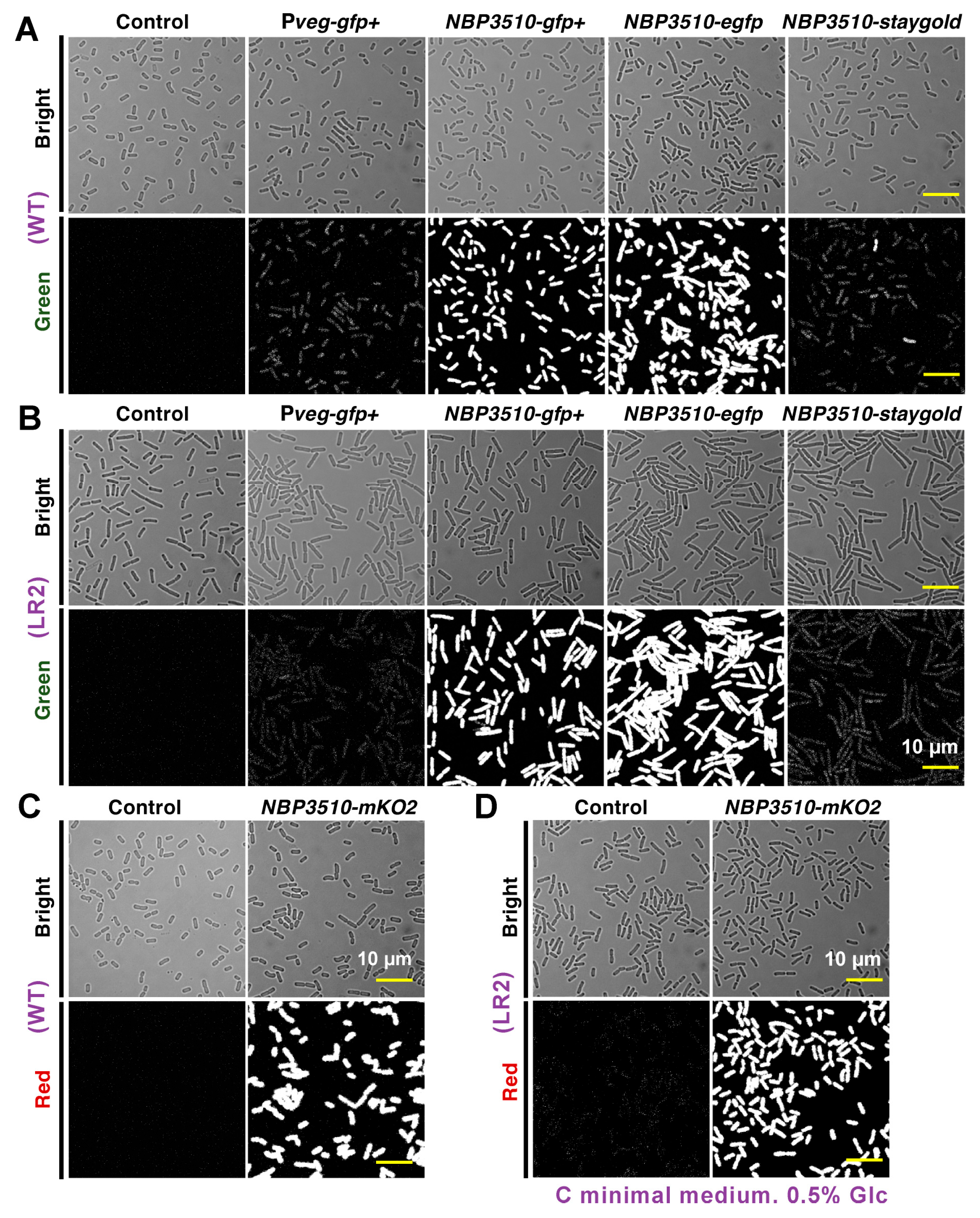
Figure 1. Comparison of expression levels of FPs in B. subtilis WT and L-form LR2. Microscopy images of strains with different promoters and FPs in B. subtilis WT (A,C) and L-form LR2 (B,D). The top panel for each strain shows bright field and the bottom panel shows green/red fluorescence field. All strains were cultured to late logarithmic phase in C minimal medium (CMM) and images were taken. Scale bar = 10 µm.
Bioengineering 11 00081 g001
Figure 2. Quantitative analysis of FPs in B. subtilis WT and L-form LR2. (A) The expression levels over time were measured using a microplate reader in B. subtilis WT and L-form LR2 at the cell population level. (B) Mean fluorescent intensity from 1 × 10⁴ cells was analyzed using IFC in B. subtilis WT and L-form LR2 at single-cell level. All cultures were grown in triplicate, and each experiment was performed at least three times (n = 9). Data are mean ± standard deviation.
Bioengineering 11 00081 g002
Figure 3. Quantitative analysis of morphology in B. subtilis WT and L-form LR2. Microscope images of B. subtilis 168CA WT ((A), upper panel) and L-form LR2 grown in C minimal medium ((A), lower panel) with or without 200 μg/mL D-cycloserine (DCS) (B) at late logarithmic phase. Scale bar = 10 µm. Green arrowheads indicate several typical cells with spherical shapes or undergoing irregular cell divisions. Detailed morphological changes and mKO2 expression in LR2 were analyzed using IFC and are shown in (C,D). The contour plot (contour levels: 10%) and distribution were performed for aspect ratio ((C)/(D): left panels), length ((C)/(D): middle panels) and area ((C)/(D): right panels). Gray arrowheads indicate the cell populations with a smaller aspect ratio, longer length and bigger area.
Bioengineering 11 00081 g003
Figure 4. Quantitative analysis of morphology in E. coli MG1655 and L-form NC-7. Microscope images of fluorescent cells with pNBP3510-mKO2 from E. coli MG1655 and NC-7 are shown in (A). Scale bar = 10 µm. IFC analysis of E. coli MG1655 and L-form NC-7 was performed to compare the fluorescent intensity (per cell) (B), mean length ((D), left panel) and mean aspect ratio ((C), right panel). All cultures were grown in triplicate and each experiment was performed at least three times (n = 9). Data are mean ± standard deviation. * p < 0.0001 (unpaired two-sided Student’s t-test). Red +: mean. The distributions of the mKO2 intensity in WT and NC-7 cells were compared (C). The contour plot (contour levels: 10%) was performed for aspect ratio ((E): left panel), length ((E): middle panel) and area ((E): right panel).
Bioengineering 11 00081 g004
Disclaimer/Publisher’s Note: The statements, opinions and data contained in all publications are solely those of the individual author(s) and contributor(s) and not of MDPI and/or the editor(s). MDPI and/or the editor(s) disclaim responsibility for any injury to people or property resulting from any ideas, methods, instructions or products referred to in the content.