Article

A Study on the Biofilm Removal Efficacy of a Bioelectric Toothbrush

1
PAIST (ProxiHealthcare Advanced Institute for Science and Technology), Seoul 04513, Republic of Korea
2
Graduate Program of Biomedical Engineering, Yonsei University, Seoul 03722, Republic of Korea
3
Department of Medical Engineering, Yonsei University College of Medicine, Seoul 03722, Republic of Korea
*
Authors to whom correspondence should be addressed.
Bioengineering 2023, 10(10), 1184; https://doi.org/10.3390/bioengineering10101184
Submission received: 12 September 2023 / Revised: 7 October 2023 / Accepted: 11 October 2023 / Published: 13 October 2023

Abstract

:
Effective oral care is a critical requirement to maintain a high quality of life. Most oral diseases are caused by plaque (oral biofilm), which is also correlated with systemic diseases. A common method to remove biofilm is brushing teeth with toothpaste. However, 3.5 billion people in the world have oral diseases, meaning that more efficient methods of removing biofilms are needed. We have developed a toothbrush that applies a bioelectric effect (BE) utilizing an electric force for biofilm removal. It demonstrated significantly higher biofilm removal efficiency than non-BE manual toothbrushes. Tests were performed in saline and toothpaste conditions using various pressures. Results showed that the BE toothbrush had a significantly higher biofilm removal efficiency in saline (0.5 N: 215.43 ± 89.92%, 2.5 N: 116.77 ± 47.02%) and in a toothpaste slurry (0.5 N: 104.96 ± 98.93%, 2.5 N: 96.23 ± 35.16%) than non-BE manual toothbrushes. Results also showed that BE toothbrushes were less dependent on toothpaste. This study suggests that the application of BE can be a new solution to plaque problems in oral care.

Graphical Abstract

1. Introduction

To enhance quality of life, maintaining optimal oral health is paramount. Regrettably, according to the World Health Organization (WHO), approximately 3.5 billion individuals globally are afflicted by oral diseases [1,2]. These oral diseases (including periodontitis, gingivitis, etc.) arise due to the inadequate management of biofilms, commonly referred to as plaque [3,4]. Plaque is characterized as a deposit forming a biofilm composed of adherent bacteria within the oral cavity [4]. This biofilm can trigger inflammation in periodontal tissues. It has been identified as a significant contributor to gingivitis and periodontitis [5]. Notably, deposits of plaque within the oral cavity can manifest clinical indications within 10 days [6,7]. Hence, to avert or manage periodontal diseases, it becomes imperative to either remove or impede the formation of plaque [8]. Efficient plaque removal proves pivotal in regulating periodontal diseases.
Notably, plaque has direct correlations with systemic illnesses such as stroke [9], diabetes [10], cardiovascular disease [11], and Alzheimer’s disease [12]. Consequently, the effective removal of plaque is important for both public and private healthcare. Plaque is typically removed through a mechanical method using a toothbrush or oral products [13,14]. In the 1960s, electric toothbrushes that increased plaque removal efficiency were developed [15].
Nevertheless, with 3.5 billion people worldwide suffering from oral diseases, typical plaque removal methods might not be effective. Therefore, a more efficient means of removing plaque is needed.
Toothpaste is commonly employed during tooth-brushing routines. While toothpaste could effectively aid in plaque removal, ongoing discussions regarding its safety arise due to the presence of abrasives, surfactants, and fluoride [16,17,18]. Overdosing with fluoride has been associated with adverse effects such as skeletal deformities, cancer, and gastric mucosa damage [18,19,20]. Among pregnant women, certain studies have indicated potential risks of birth defects [21]. Additionally, concerns are being raised, particularly concerning children, regarding potential serious issues such as teeth and skeletal underdevelopment [17,22,23].
Plaque is a type of biofilm that forms in the oral cavity [4]. The durability of a biofilm extends to the point where antibiotic resistance becomes 500 to 5000 times more pronounced than that exhibited by individual bacteria [8,24,25].
Studies on photodynamic therapy to remove biofilm have been reported [26,27,28]. Photodynamic therapy (PDT) is a proposed method to treat oral diseases and plaque by applying a combination of light, photosensitizers, and oxygen [26,27,28]. However, PDT has disadvantages in terms of time and pain [28].
Biofilms consist of polysaccharides and electrically polarized bacterial cells [29,30]. External electrical stimuli can disrupt biofilms’ structural integrity and impact the metabolic state of bacteria [28,29,30,31]. As demonstrated in a recent study, the combined application of an electrical signal known as the bioelectric effect (BE) along with both direct current (DC) and alternating current (AC) is remarkably effective for biofilm removal [31,32,33,34,35,36,37].
Recently, our group developed a toothbrush with a bioelectric effect and demonstrated its effectiveness in reducing plaque-induced dental gingival index in clinical trials [8,35]. This result means that applying a bioelectric effect to oral products has a clinically significant effect on oral care.
However, clinical trials have the disadvantage of being difficult to quantitatively evaluate due to variations among subjects [38]. In vitro tests are more convenient and more reproducible than in vivo tests. Therefore, they are widely used in research related to the human body [38,39,40]. Likewise, in relation to toothbrushes, studies using various in vitro brushing simulators have been reported [41,42,43,44].
In this study, we quantitatively compared the plaque removal efficiency of a bioelectric toothbrush and a regular toothbrush to observe the bioelectrical effect on plaque. We performed a quantitative plaque removal efficiency test using a developed brushing simulator for four types of toothbrushes (two types of regular toothbrushes and the proposed toothbrush (with or without BE)). The experiment was conducted under two conditions (with only saline and with toothpaste slurry) to investigate whether BE could replace toothpaste. Based on experimental results, we investigated the plaque removal efficiency of BE and toothpaste dependence for each.

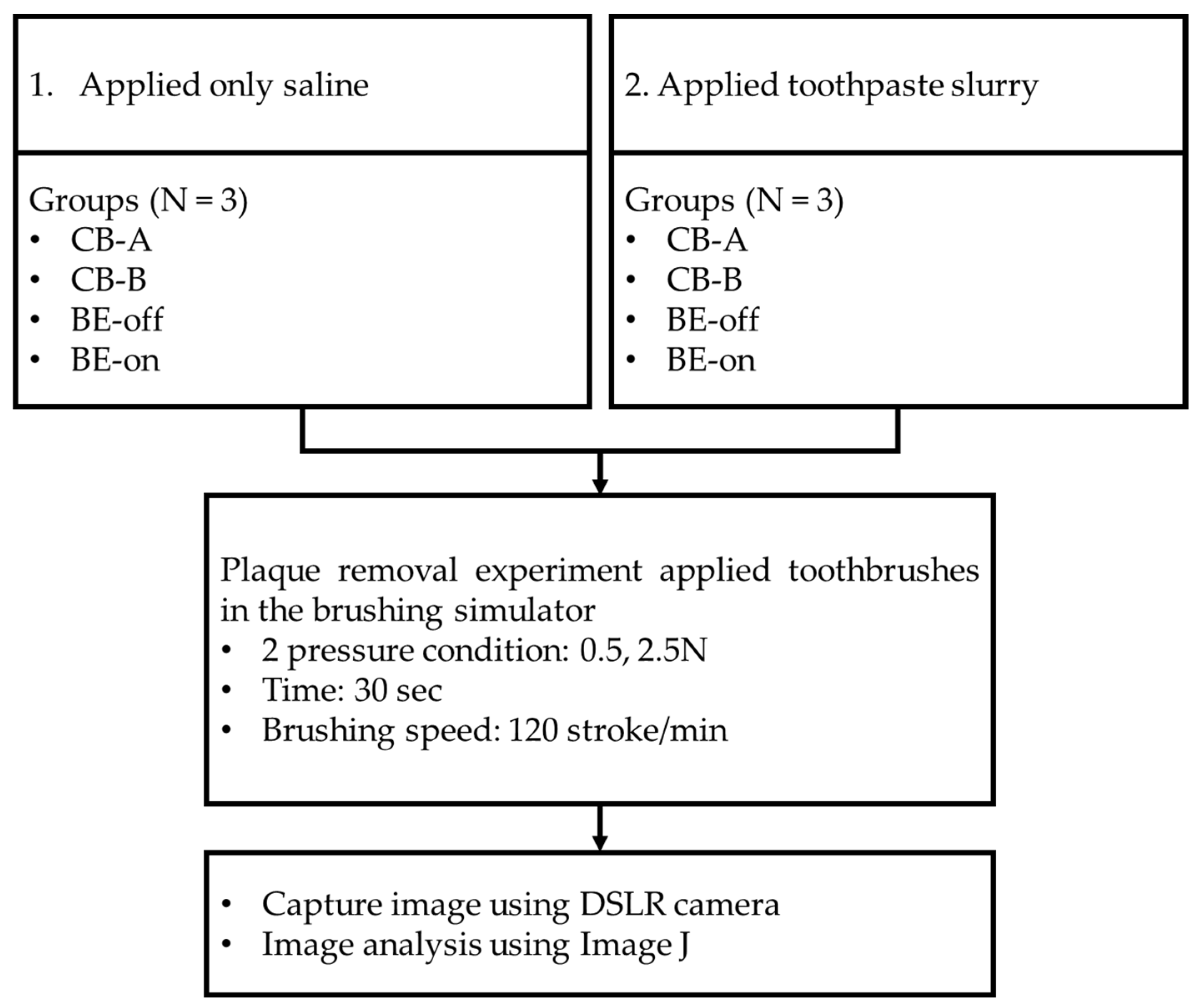
2. Materials and Methods

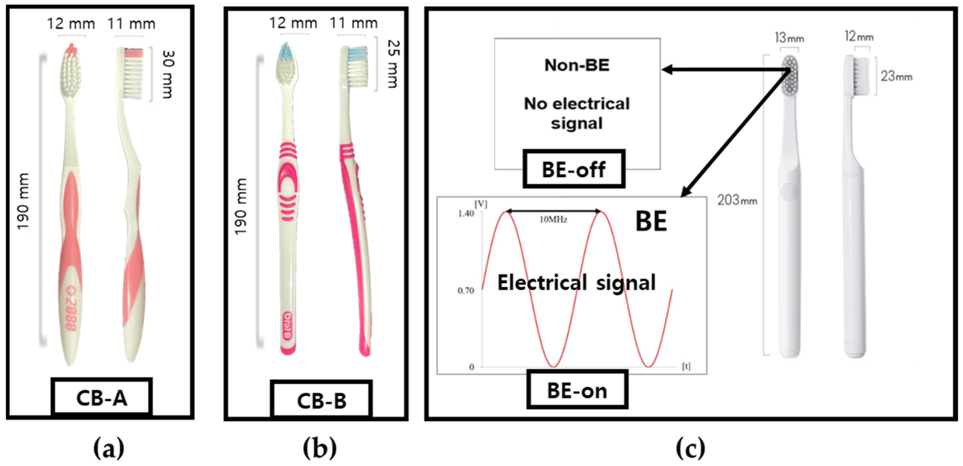
2.1. Tested Toothbrushes and Design of BE Toothbrush

The tested toothbrushes and their abbreviations are listed in Table 1 and Figure 1. We aimed to compare the plaque removal efficiency of a toothbrush with BE. Two commonly used toothbrushes and two developed toothbrushes were tested under two conditions: BE off and on. These conditions were specified to investigate the effect of the bristles (typical toothbrush versus proposed toothbrush) on plaque removal.
BE applies a combination of electrical signals to remove biofilms. The electrical signal was a combination of a 0.7 V sinusoidal signal at a frequency of 10 MHz with a DC offset of 0.7 V, as shown in Table 2 [8,45].
The frequency of the electrical signal was selected based on previous studies. The DC offset was set below the electrolysis threshold of 0.82 V [31]. Our previous study revealed that this signal does not induce electrolysis [45]. The electrical signal is shown in Figure 2a. An electronic circuit for generating the output signal was developed, as shown in Figure 2b. Electronic circuits were embedded in the proposed toothbrush. A stainless-steel electrode was chosen due to its corrosion resistance and conductivity essential for supplying an electric field. The signal for BE is output through these two electrodes.
The current from the signal output was measured using a multimeter (EDU34450A, Keysight Technologies, Santa Rosa, CA, USA) in a 0.9% saline condition. The measured current was about 40.7 ± 1.5 μA (n = 3). The current of BE was determined to be safe because it was within the biocompatible current range [46].

2.2. Development of Brushing Simulator

The brushing simulator consists of five linear actuators with a stepper motor (Scipia, Gwangju, Republic of Korea), five motor drivers (TB6600, DFRobot, Shanghai, China), four piezoelectric pressure sensors (QA3040P, Marveldex Inc., Bucheon, Republic of Korea), a switching-mode power supply (HU10748-13002E, Yueqing Hengwei Electric Co., Yueqing, China), and a construct of typodonts and toothbrushes. The construct was produced using a 3D printer (X1-carbon, Bambu lab, Shenzhen, China) and polylactic acid (PLA). A standard typodonts was used to construct the brushing simulator. For the brushing control and pressure exerted by the toothbrush data acquisition (DAQ), we used an Arduino Uno (Arduino.cc, Ivrea, Italy). The toothbrushing simulator was capable of simulating toothbrushing at up to 150 strokes/min in a 5 cm driving range. The brushing simulator is shown in Figure 3.
The pressure sensor of the simulator was calibrated by applying various masses from 20 to 300 g. The result showed a standard correlation of the piezoelectric material (Figure 4). A piezoelectric pressure sensor can be calibrated using a power function [47]. A standard weight was applied to the pressure sensor and the output voltage was recorded (n = 3). According to ISO/TR 14569-1, the pressure that the toothbrush presses on an artificial tooth is between 0.5 and 2.5 N [48]. The measured weight was converted to newtons (N).

2.3. Plaque Culture

Streptococcus mutans (KCTC 3065, Korean Collection for Type Cultures (KCTC), Jeongeup, Republic of Korea) used in this research were distributed from KCTC. They were cultured in a growth medium (LB Broth, Ambrothia Inc., Daejeon, Republic of Korea) at 37 °C for 48 h to provide sufficient time for maturation.

2.4. Experiment of Plaque Removal

Typodonts were evenly coated with the cultured S. mutans. Green marker spray (OccludeTM, Pascal, Bellevue, WA, USA) was used to visualize S. mutans. (plaques) [40]. The coated typodont was mounted on the brushing simulator. The force that the toothbrush pressed on the tooth was set to be 0.5 N and 2.5 N. According to ISO/TR 14569-1, the force that a toothbrush presses on teeth is between 0.5 N and 2.5 N [48]. The molar area was brushed [43]. The brushing frequency was 120 strokes/min [48] for 30 s. The recommended time for brushing is generally 2 min [49,50]. The target area was approximately 1/4 of the entire tooth. The driving range was set to 4 cm as an approximate range of the molar area. After brushing, tested typodonts were eluted in tap water for 30 s. This process was used to remove the plaque that was separated by brushing the teeth so that only the residual plaque would be quantified. According to our preliminary test, it was determined that this process did not cause unintended plaque elution. Plaques of typodonts before and after the experiment were captured under controlled lighting conditions using a DSLR camera (A350, Sony Co., Tokyo, Japan). Image analysis was performed using ImageJ (National Institutes of Health, Bethesda, MD, USA). Two experiments were conducted (n = 3). The experimental design is shown in Figure 5.
  • Only saline was applied.
  • A toothpaste slurry (toothpaste–saline at a ratio of 1:3) was applied.
The first experiment (only saline applied) was conducted to determine the biofilm removal effect of using only BE without toothpaste. Saline solution has been used in several studies to simulate saliva [51,52]. The second experiment (toothpaste slurry applied) was designed to represent typical brushing conditions. The toothpaste used was a toothpaste containing fluorine at 1450 ppm (Grate regular-flavor toothpaste, Colgate, Hamilton, NY, USA).
In our previous work, we found that a toothbrush with BE in a clinical trial could significantly reduce the gingivitis index [8,45]. Therefore, an additional comparison was made between a typical toothbrush using toothpaste and a BE toothbrush without toothpaste.

2.5. Statistical Analysis
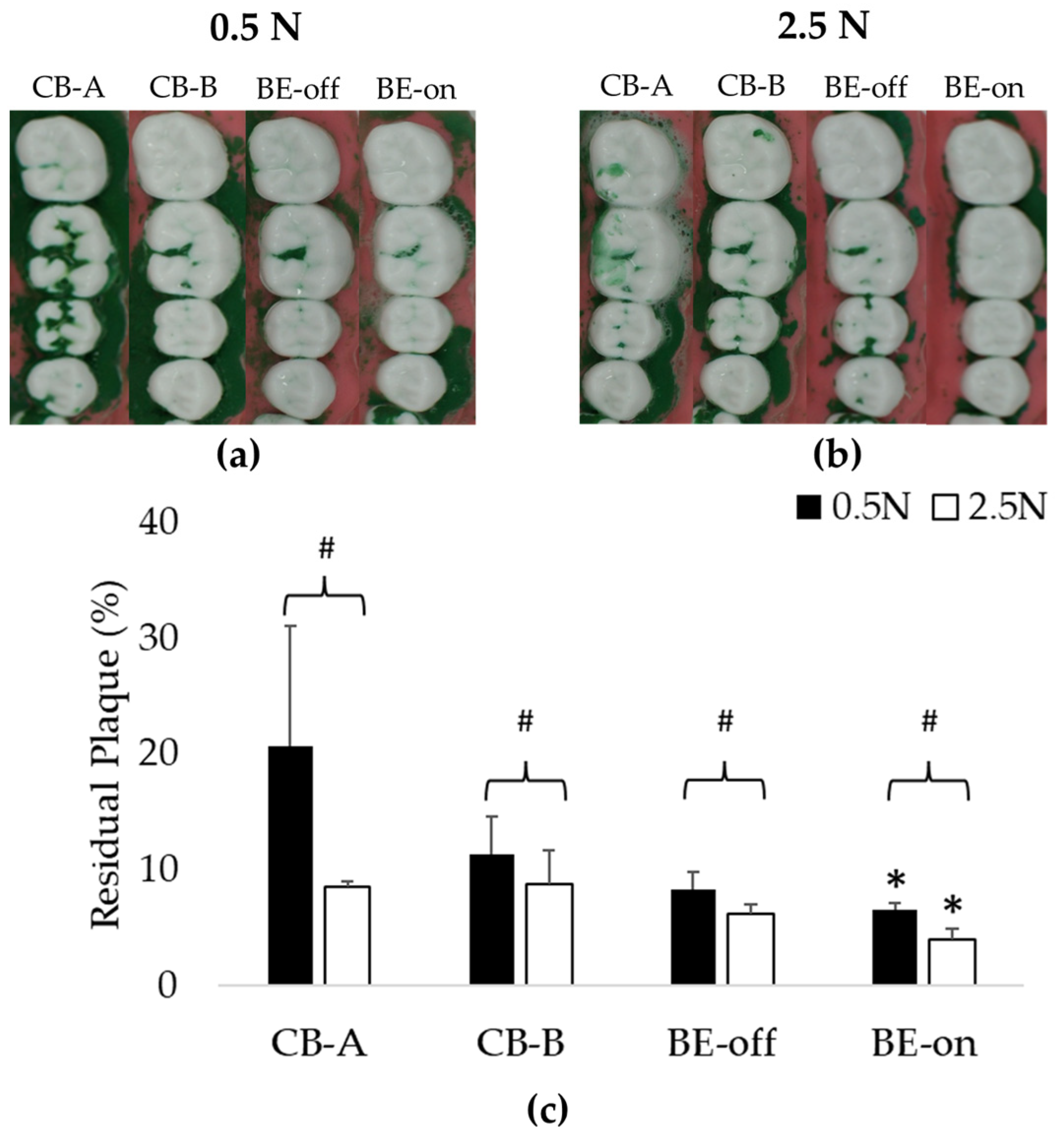
Residual plaques are presented as means and standard deviations (SDs). A two-way analysis of variance (ANOVA) followed by Dunnett’s post hoc test was used to determine the significance of differences between the experimental condition and the CB-A at 0.5 N as control. Paired t-tests were used to compare differences between BE-on saline and toothpaste slurry experiments. All statistical analyses were performed using R-studio version 4.3.1 (Posit, Boston, MA, USA). p-values of less than 0.05 were considered significant.

3. Results

3.1. Experiment Using Only Saline

This experiment was designed to evaluate plaque removal efficiencies to observe the effect of BE without using toothpaste. In all pressure conditions, the residual plaque using the BE-on toothbrush was significantly lower than that using other toothbrushes. Toothbrushes except for BE-on showed no significant difference in residual plaque, as shown in Figure 6. A previous study reported that there is no significant difference in plaque removal rate according to the shape of bristles [53]. This result suggests that the high plaque removal rate might be due to the application of BE. The order of residual plaque was 0.5 N > 2.5 N. The higher the pressure with which the toothbrush presses against the teeth, the higher the plaque removal rate. These results are consistent with those of previous reports [54].
At 0.5 N, BE-on showed 7.96 ± 1.67% of residual plaque, while CB-A, CB-B, and BE-off showed 27.42 ± 4.30%, 17.45 ± 0.59%, and 17.03 ± 5.99% of residual plaque, respectively. At 2.5 N, BE-on showed 5.24 ± 1.86% of residual plaque, while CB-A, CB-B, and BE-off showed 9.08 ± 3.18%, 10.20 ± 2.18%, and 6.55 ± 1.59% of residual plaque, respectively. The highest plaque removal efficiency was observed with BE-on at 2.5 N. On average, the plaque removal rate with BE-on was 215.43 ± 89.92% higher at 0.5 N and 116.77 ± 47.02% higher at 2.5 N than those using other toothbrushes.

3.2. Experiment Appling a Toothpaste Slurry

This experiment was designed to evaluate plaque removal efficiencies in typical situations. As observed in the experiment applying only saline, the residual plaque using BE-on was significantly lower than in those using other toothbrushes under all pressure conditions, as shown in Figure 7. Using toothpaste can increase plaque removal efficiency [16,17,18]. Therefore, the residual plaque in this experiment using a toothpaste slurry was lower than that when using only saline.
The order of residual plaque was 0.5 N > 2.5 N, as shown in Figure 7. At 0.5 N, BE-on showed 6.54 ± 0.59% of residual plaque, while CB-A, CB-B, and BE-off showed 20.66 ± 10.38%, 11.32 ± 3.24%, and 8.23 ± 1.54% of residual plaque, respectively. At 2.5 N, BE-on showed 3.97 ± 0.97% of residual plaque, while CB-A, CB-B, and BE-off showed 8.51 ± 0.53%, 8.69 ± 2.95%, and 6.19 ± 0.84% of residual plaque, respectively. BE-on was the most efficient in all pressure conditions. The highest plaque removal efficiency was observed for BE-on at 2.5 N. On average, the plaque removal rate using BE-on was 104.96 ± 98.93% higher at 0.5 N and 96.23% ± 35.16% higher at 2.5 N than when using other toothbrushes.

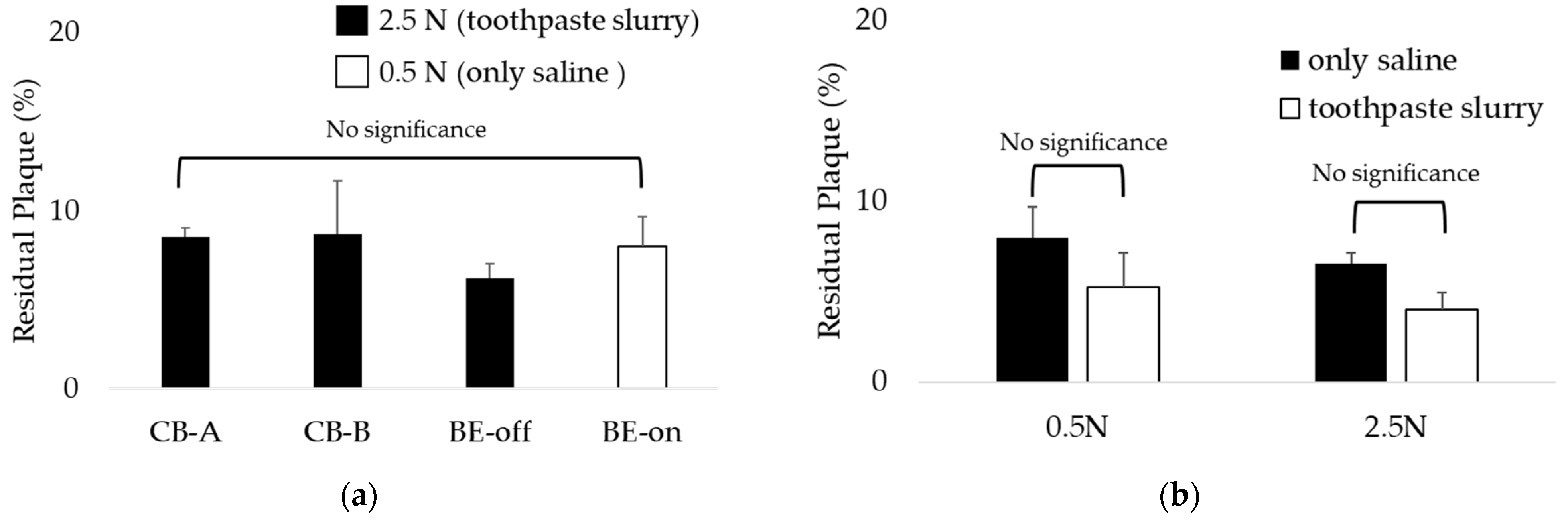
3.3. Comparison of Residual Plaque in Applied Saline with BE versus Toothpaste Slurry without BE

Through this comparison, we investigated whether BE could replace toothpaste. We investigated whether using BE-on with only saline resulted in significantly lower residual plaque than using other toothbrushes with toothpaste and saline slurry under all pressure conditions. As shown in Figure 8, the highest plaque removal efficiency was observed for BE-on without toothpaste at 2.5 N. These results suggest that applying BE to a toothbrush can replace toothpaste.
Interestingly, the BE-on under the 0.5 N condition using only saline showed residual plaque levels similar to those using other toothbrushes under the 2.5 N condition using toothpaste and saline slurry, as shown in Figure 9a. This result suggests that even if brushing is weak without toothpaste, plaque can be removed similarly to general brushing conditions if BE is applied. Also, as a result of comparing the residual plaque of BE-on using only saline or a toothpaste and saline slurry, there was no significant difference, as shown in Figure 9b. Therefore, if BE is applied, the dependence on toothpaste for plaque removal is expected to decrease.

4. Discussion

In both the only-saline and toothpaste-slurry experiments, there was no significant difference in residual plaque among toothbrushes except for BE-on. According to a previous report, there was no significant difference in plaque removal efficiency according to toothbrush bristles alone [53], similar to the results of the present study. There is a report showing that brushing under high-pressure conditions is more effective in removing plaque than under low-pressure conditions [54]. Similar results were observed in this study as residual plaque was lower under high-pressure (2.5 N) conditions. The use of toothpaste also affected the plaque removal efficiency [16,17,18]. The condition of using toothpaste was 34.67 ± 33.10% more effective for plaque removal on average than not using toothpaste. This result is similar to that of a previously reported study [55].
BE-on had a significantly lower residual plaque than other toothbrushes without BE under the same condition (Figure 6 and Figure 7). Plaque is a type of biofilm. Previous studies have reported that the treatment efficiency of biofilm is high when BE is applied [29,30,31]. BE is caused by the propagation of electromagnetic currents, waves, and voltages. Utilizing an AC at a specific frequency can enhance the porosity of the biofilm’s structure by inducing vibrations [35,36]. Conversely, a DC can induce changes in the electrolyte state [31,37]. The experimental condition is an electrically dielectric state due to only saline or toothpaste slurry being used. Saline has been used in several studies to simulate saliva [51,52]. It seems that effective propagation of the electric field can increase the biofilm removal effect. Therefore, a toothbrush with BE is expected to have a higher plaque removal efficiency.
Interestingly, it was observed that the residual plaque level using BE-on without toothpaste was significantly lower than that using toothbrushes with toothpaste under the same conditions, as shown in Figure 8. Because the BE weakens the structure of biofilm [31,32,33,34], it can increase biofilm removal efficiency. Based on these results, we propose BE as a possible substitute for toothpaste. Regardless of toothpaste application, no significant difference was observed in the residual plaque of BE-on (Figure 9b). This result indicates that if BE is applied, the dependence on toothpaste for tooth brushing can be reduced. These results mean that BE can increase the plaque removal rate regardless of the presence or absence of toothpaste. There is controversy regarding the safety of toothpaste due to abrasives, surfactants, and fluoride [16,17,18]. Generally, it includes potential carcinogenic risk, skeletal deformity, and gastric mucosal damage [18,19,20]. In children, infants and fetuses, this includes developmental disorders and bone un-development [17,22,23]. However, BE applies a microcurrent of 40.7 ± 1.5 μA, so it is harmless to the human body [46]. Thus, we anticipate that BE will be a good option for people who find it difficult to use toothpaste because of its controversial features.
Studies have reported that brushing teeth under high-pressure conditions can accelerate tooth wear [42,43]. In this study, we found that there was no significant difference in residual plaque between BE-on at a low pressure (0.5 N) without toothpaste and other toothbrushes at a high pressure (2.5 N) and with toothpaste (Figure 9a). Under the condition of the lowest pressure, brushing with only BE applied without toothpaste showed a plaque removal efficiency similar to that using the highest pressure and toothpaste. This means that a toothbrush using BE can show good plaque removal efficiency without excessive brushing or toothpaste. Therefore, it can be proposed that the application of BE can effectively remove plaque even without toothpaste or aggressive brushing. Hard brushing can increase tooth wear [42,43]. Thus, this suggests that the application of BE may reduce the intensity of tooth brushing. Therefore, it is expected that the wear of permanent teeth can be reduced if BE is applied. We believe that the application of BE will reduce not only the potential risk of toothpaste, but also the potential risk of tooth wear caused by strong brushing.
The use of an in vitro simulator not only has high experimental reproducibility compared to clinical trials, but it also enables quantitative testing [38]. In this study, we quantitatively compared the plaque removal efficiency of BE toothbrushes and other toothbrushes using an in vitro simulator. In our previous clinical study, a BE-applied toothbrush significantly reduced the gingival index, but not the plaque index [8]. This might be because there was variation among the subjects in the clinical study. It is possible that a significant plaque index was not observed due to human bias. However, since in this study we conducted an in vitro experiment using a brushing simulator capable of quantitative evaluation, we were able to observe significant plaque removal efficiency.
Based on these research results, we intend to additionally develop various oral care products including toothbrushes. Plaque can cause various problems in the oral cavity; not only on the teeth, but also on parts such as the tongue [56]. This applies not only to people; pets also have various problems due to plaque [57]. Therefore, we plan to develop oral care products through mechanical improvement with BE, including tongue cleaners and pet toothbrushes, to solve various problems related to plaque.
In this study, we evaluated the plaque removal efficiency of a BE toothbrush in a simulated brushing environment. However, our study has some limitations. First, we used consistent brushing speed and pressure, which might not reflect the variability in brushing patterns that occurs in real life [58]. Second, we used the same typodont for our tests, which does not account for differences in oral cavity structure that exist from person to person [59]. Third, we only evaluated plaque removal in one area of the teeth, although the structure of teeth varies from region to region. There is a possibility that the plaque removal rate may vary depending on the brushing pattern, tooth structure, and area. Therefore, additional studies are needed to determine the plaque removal efficiency of BE in more realistic conditions. These studies should investigate the effects of different brushing patterns, pressure conditions, and oral cavity structures.

5. Conclusions

In this study, we developed a BE toothbrush and investigated the plaque removal effect of BE through quantitative plaque removal experiments using a brushing simulator. A BE toothbrush was observed to be effective in removing plaque. We believe that the application of BE will contribute to effective oral care. In addition, the application of BE not only shows the possibility of replacing toothpaste, but also shows the ability to reduce the intensity of brushing. Thus, it is expected that the wear of permanent teeth can also be reduced. In the future, we will develop various oral care products applying BE, including tongue scrapers, toothbrushes for pets, and so on.

Author Contributions

Conceptualization, H.M.P. and Y.W.K.; methodology, H.M.P.; software, H.M.P.; validation, H.M.P.; formal analysis, E.J.; investigation, Y.W.K.; data curation, S.R.; writing—original draft preparation, H.M.P., E.J., S.R. and Y.W.K.; writing—review and editing, H.M.P., E.J., S.R., S.K.Y. and Y.W.K.; visualization, H.M.P. All authors have read and agreed to the published version of the manuscript.

Funding

This work was supported by the Technology development Program (No. S3301078) and funded by the Ministry of SMEs and Startups (MSS, Korea).

Institutional Review Board Statement

Not applicable.

Informed Consent Statement

Not applicable.

Data Availability Statement

Not applicable.

Conflicts of Interest

The authors declare no conflict of interest.

References

  1. Lorenzo-Pouso, A.I.; Pérez-Sayáns, M.; Bravo, S.B.; López-Jornet, P.; García-Vence, M.; Alonso-Sampedro, M.; Carballo, J.; García-García, A. Protein-Based Salivary Profiles as Novel Biomarkers for Oral Diseases. Dis. Mrk. 2018, 2018, 6141845. [Google Scholar] [CrossRef]
  2. World Health Organization. Available online: https://www.who.int/health-topics/oral-health#tab=tab_1 (accessed on 8 September 2023).
  3. Allaker, R.P.; Douglas, C.I. Novel anti-microbial therapies for dental plaque-related diseases. Int. J. Antimicrob. Agents 2009, 33, 8–13. [Google Scholar] [CrossRef]
  4. Marsh, P.D. Dental plaque: Biological significance of a biofilm and community life-style. J. Clin Periodontol. 2005, 32, 7–15. [Google Scholar] [CrossRef] [PubMed]
  5. Abebe, G.M. Oral Biofilm and Its Impact on Oral Health, Psychological and Social Interaction. Int. J. Oral Dent. Health 2021, 7, 127. [Google Scholar]
  6. Löe, H.; Theilade, E.; Jensen, S.B. Experimental Gingivitis in Man. J. Periodontol. 1965, 36, 177–187. [Google Scholar] [CrossRef] [PubMed]
  7. Lovdal, A.; Arno, A.; Waerhaug, J. Incidence of clinical manifestations of periodontal disease in light of oral hygiene and calculus formation. J. Amer. Dent. Assoc. 1958, 56, 21–33. [Google Scholar] [CrossRef] [PubMed]
  8. Kim, Y.W.; Lee, J.; Lee, T.H. Bioelectric effect utilized a healthcare device for effective management of dental biofilms and gingivitis. Med. Eng. Phys. 2022, 104, 103804. [Google Scholar] [CrossRef]
  9. Alhadainy, H.A.; Keefe, T.; Abdel-Karim, A.H.; Abdulrab, S.; Halboub, E. Association between dental diseases and history of stroke in the United States. Clin. Exp. Dent. Res. 2021, 7, 845–851. [Google Scholar] [CrossRef]
  10. Albert, D.A.; Ward, A.; Allweiss, P. Diabetes and oral disease: Implications for health professionals. Ann. N. Y. Acad. Sci. 2012, 1255, 1–15. [Google Scholar] [CrossRef]
  11. Kotronia, E.; Brown, H.; Papacosta, A.O.; Lennon, L.T.; Weyant, R.J.; Whincup, P.H.; Wannamethee, S.G.; Ramsay, S.E. Oral health and all-cause, cardiovascular disease, and respiratory mortality in older people in the UK and USA. Sci. Rep. 2021, 11, 16452. [Google Scholar] [CrossRef]
  12. Ming, Y.; Hsu, S.W.; Yen, Y.; Lan, S.J. Association of oral health–related quality of life and Alzheimer disease: A systematic review. J. Prosthet. Dent. 2020, 124, 168–175. [Google Scholar] [CrossRef] [PubMed]
  13. Sälzer, S.; Slot, D.E.; Van der Weijden, F.A.; Dörfer, C.E. Efficacy of inter-dental mechanical plaque control in managing gingivitis—A meta-review. J. Clin. Periodontol. 2015, 42, 92–105. [Google Scholar] [CrossRef] [PubMed]
  14. Westfelt, E. Rationale of mechanical plaque control. J. Clin. Periodontol. 1996, 23, 263–267. [Google Scholar] [CrossRef] [PubMed]
  15. Jepsen, S. Role of m toothbrushes in effective plaque control: Advantages and limitations. In Proceedings of European Workship on Mechanical Plaque Control, 1st ed.; Lang, N.P., Attstrom, R., Love, H., Eds.; Quintessence Publishing: Batavia, IL, USA; pp. 121–137.
  16. Hara, A.T.; Turssi, C.P. Baking soda as an abrasive in toothpastes: Mechanism of action and safety and effectiveness considerations. J. Am. Dent. Assoc. 2017, 148, 27–33. [Google Scholar] [CrossRef]
  17. Stovell, A.G.; Newton, B.M.; Lynch, R.J.M. Important considerations in the development of toothpaste formulations for children. Int. Dent. J. 2013, 63, 57–63. [Google Scholar] [CrossRef]
  18. Ullah, R.; Zafar, M.S.; Shahani, N. Potential fluoride toxicity from oral medicaments: A review. Iran. J. Basic Med. Sci. 2017, 20, 841–848. [Google Scholar]
  19. Mohideen, H.; Dahiya, D.S.; Parsons, D.; Hussain, H.; Ahmed, R.S. Skeletal Fluorosis: A Case of Inhalant Abuse Leading to a Diagnosis of Colon Cancer. J. Investig. Med. High Impact Case Rep. 2022, 10, 23247096221084919. [Google Scholar] [CrossRef]
  20. Pashley, D.H.; Allison, N.B.; Easmann, R.P.; McKinney, R.V.; Horner, J.A.; Whitford, G.M. The effects of fluoride on the gastric mucosa of the rat. J. Oral Path. Med. 1984, 13, 535–545. [Google Scholar] [CrossRef]
  21. Wang, Y.; Jiang, L.; Zhao, Y. Awareness of the Benefits and Risks Related to Using Fluoridated Toothpaste Among Doctors: A Population-Based Study. Med. Sci. Monit 2019, 25, 6397–6404. [Google Scholar] [CrossRef]
  22. Basch, C.H.; Rajan, S. Marketing strategies and warning labels on children’s toothpaste. J. Am. Dent. Hyg. Assoc. 2014, 88, 316–319. [Google Scholar]
  23. Buzalaf, M.A.; Levy, S.M. Fluoride intake of children:considerations for dental caries and dental fluorosis. Monogr. Oral. Sci. 2011, 22, 1–19. [Google Scholar] [PubMed]
  24. Hathroubi, S.; Mekni, M.A.; Domenico, P.; Nguyen, D.; Jacques, M. Biofilms: Microbial shelters against antibiotics. Microb. Drug Resist. 2017, 23, 147–156. [Google Scholar] [CrossRef] [PubMed]
  25. Costerton, J.W.; Ellis, B.; Lam, K.; Johnson, F.; Khoury, A.E. Mechanism of electrical enhancement of efficacy of antibiotics in killing biofilm bacteria. Antimicrob. Agents Chemother. 1994, 38, 2803–2809. [Google Scholar] [CrossRef] [PubMed]
  26. Park, J.H.; Kim, Y.-G.; Um, H.-S.; Lee, S.Y.; Lee, J.-K.; Chang, B.-S. Antimicrobial effect of toothbrush with light emitting diode on dental biofilm attached to zirconia surface: An in vitro study. J. Dent. Rehabil. Appl. Sci. 2019, 35, 160–169. [Google Scholar] [CrossRef]
  27. Bagchi, D.; Rathnam, V.S.; Lemmens, P.; Banerjee, I.; Pal, S.K. NIR-light-active ZnO-based nanohybrids for bacterial biofilm treatment. ACS Omega 2018, 3, 10877–10885. [Google Scholar] [CrossRef]
  28. Pérez-Pérez, L.; García-Gavín, J.; Gilaberte, Y. Daylight-mediated photodynamic therapy in Spain: Advantages and disadvantages. Actas Dermosifiliogr. 2014, 105, 663–674. [Google Scholar] [CrossRef]
  29. Costerton, J.W.; Stewart, P.S.; Greenberg, E.P. Bacterial biofilms: A common cause of persistent infections. Science 1999, 284, 1318–1322. [Google Scholar] [CrossRef]
  30. Daubert, D.M.; Weinstein, B.F. Biofilm as a risk factor in implant treatment. Periodontology 2000 2019, 81, 29–40. [Google Scholar] [CrossRef]
  31. Kim, Y.W.; Subramanian, S.; Gerasopoulos, K.; Ben-Yoav, H.; Wu, H.C.; Quan, D.; Carter, K.; Meyer, M.T.; Bentley, W.E.; Ghodssi, R. Effect of Electrical Energy on the Efficacy of Biofilm Treatment Using the Bioelectric Effect. NPJ Biofilms Microbiomes 2015, 1, 15016. [Google Scholar] [CrossRef]
  32. Kim, Y.W.; Meyer, M.T.; Berkovich, A.; Subramanian, S.; Iliadis, A.A.; Bentley, W.E.; Ghodssi, R. A surface acoustic wave biofilm sensor integrated with a treatment method based on the bioelectric effect. Sens. Actuators A Phys. 2016, 238, 140–149. [Google Scholar] [CrossRef]
  33. Pareilleux, A.; Sicard, N. Lethal effects of electric current on Escherichia coli. Appl. Microbiol. 1970, 19, 421–424. [Google Scholar] [CrossRef] [PubMed]
  34. Blenkinsopp, S.A.; Khoury, A.; Costerton, J. Electrical enhancement of biocide efficacy against Pseudomonas aeruginosa biofilms. Appl. Environ. Microbiol. 1992, 58, 3770–3773. [Google Scholar] [CrossRef] [PubMed]
  35. Caubet, R.; Pedarros-Caubet, F.; Chu, M.; Freye, E.; de Belém Rodrigues, M.; Moreau, J.M.; Ellison, W.J. A radio frequency electric current enhances antibiotic efficacy against bacterial biofilms. Antimicrob. Agents Chemother. 2004, 48, 4662–4664. [Google Scholar] [CrossRef] [PubMed]
  36. del Pozo, J.L.; Rouse, M.S.; Mandrekar, J.N.; Steckelberg, J.M.; Patel, R. The electricidal effect: Reduction of staphylococcus and pseudomonas biofilms by prolonged exposure to low-intensity electrical current. Antimicrob. Agents Chemother. 2009, 53, 41–45. [Google Scholar] [CrossRef]
  37. Stoodley, P.; DeBeer, D.; Lappin-Scott, H.M. Influence of Electric Fields and pH on Biofilm Structure as Related to the Bioelectric Effect. Antimicrob. Agents Chemother. 1997, 41, 1876–1879. [Google Scholar] [CrossRef]
  38. Park, H.M.; Chang, K.H.; Moon, S.H.; Park, B.J.; Yoo, S.K.; Nam, K.C. In vitro delivery efficiencies of nebulizers for different breathing patterns. Biomed. Eng. OnLine 2021, 20, 59. [Google Scholar] [CrossRef]
  39. Wisotzki, M.; Mair, A.; Schlett, P.; Lindner, B.; Oberhardt, M.; Bernhard, S. In Vitro Major Arterial Cardiovascular Simulator to Generate Benchmark Data Sets for In Silico Model Validation. Data 2022, 7, 145. [Google Scholar] [CrossRef]
  40. Szalma, J.; Lovasz, B.V.; Vajta, L.; Soos, B.; Lempel, E.; Mohlhenrich, S.C. The influence of the chosen in vitro bone simulation model on intraosseous temperatures and drilling times. Sci. Rep. 2019, 9, 11817. [Google Scholar] [CrossRef]
  41. Lee, J.-H.; Kim, S.-H.; Han, J.-S.; Yeo, I.-S.L.; Yoon, H.-I. Optical and Surface Properties of Monolithic Zirconia after Simulated Toothbrushing. Materials 2019, 12, 1158. [Google Scholar] [CrossRef]
  42. Bizhang, M.; Schmidt, I.; Chun, Y.P.; Arnold, W.H.; Zimmer, S. Toothbrush abrasivity in a long-term simulation on human dentin depends on brushing mode and bristle arrangement. PLoS ONE 2017, 12, e0172060. [Google Scholar] [CrossRef]
  43. Ledder, R.G.; Latimer, J.; Forbes, S.; Penney, J.L.; Sreenivasan, P.K.; Mcbain, A.J. Visualization and quantification of the oral hygiene effects of brushing, dentifrice use, and brush wear using a tooth brushing stimulator. Front. Public Health 2019, 7, 91. [Google Scholar] [CrossRef] [PubMed]
  44. Lang, T.; Staufer, S.; Jennes, B.; Gaengler, P. Clinical validation of robot simulation of toothbrushing-comparative plaque removal efficacy. BMC Oral Health 2014, 14, 82. [Google Scholar] [CrossRef] [PubMed]
  45. Kim, Y.W.; Lee, J.; Han, S.K.; Koo, B.-S.; Park, T.; Park, H.M.; Lee, B. A Non-Electrolysis Bioelectric Effect for Gingivitis and Hygiene Contamination Biofilm Removal. Appl. Microbiol. 2023, 3, 675–686. [Google Scholar] [CrossRef]
  46. Kim, Y.-J.; Lee, S.G.; Go, S.J.; An, S.; Kim, Y.E.; Kim, Y.I.; Hyun, K.-Y.; Cho, D.S.; Choi, G.E. Anti-inflammatory Effects of Low-frequency Stimulator using Superposition of Alternating Microcurrent Wave in the Animal Models. Biomed. Sci. Lett. 2021, 27, 99–104. [Google Scholar] [CrossRef]
  47. Hao, L.; Li, C.; Wang, L.; Bai, B.; Zhao, Y.; Luo, C. Design and Fabrication of a High-Temperature SOI Pressure Sensor with Optimized Crossbeam Membrane. Micromachines 2023, 14, 1045. [Google Scholar] [CrossRef]
  48. ISO TR 14569-1; Dental Materials-Guidance on Testing of Wear Resistance. Part 1: Wear by Tooth Brushing; International Organization for Standardization: Geneva, Switzerland, 2007.
  49. van Leeuwen, M.P.C.; van der Weijden, F.A.; Slot, D.E.; Rosema, M.A.M. Toothbrush wear in relation to toothbrushing effectiveness. Int. J. Dent. Hyg. 2019, 17, 77–84. [Google Scholar] [CrossRef]
  50. García-Godoy, F.; Villalta, P.; Bartizek, R.D.; Barker, M.L.; Biesbrock, A.R. Tooth whitening effects of an experimental power whitening toothbrush relative to an 8.7% hydrogen peroxide paint-on gel control. Am. J. Dent. 2004, 17, 25A–30A. [Google Scholar]
  51. Addy, M.; Jenkins, S.; Newcombe, R. The effect of triclosan, stannous fluoride and chlorhexidine products on: (I) Plaque regrowth over a 4-day period. J. Clin. Periodontol. 1990, 17, 693–697. [Google Scholar] [CrossRef]
  52. Binney, A.; Addy, M.; McKeown, S.; Everatt, L. The effect of a commercially available triclosan-containing toothpaste compared to a sodium-fluoride-containing toothpaste and a chlorhexidine rinse on 4-day plaque regrowth. J. Clin. Periodontol. 1995, 22, 693–697. [Google Scholar] [CrossRef]
  53. Sripriya, N.; and Shaik Hyder Ali, K.H. A comparative study of the efficacy of four different bristle designs of tooth brushes in plaque removal. J. Indian Soc. Pedod. Prev. Dent. 2007, 25, 76–81. [Google Scholar] [CrossRef]
  54. Pujarern, P.; Rodanant, P.; Warinsiriruk, E.; Rattanasuwan, K. Evaluation of the optimum brushing force on dental plaque removal: An in vitro study. M. Dent. J. 2021, 41, 75–82. [Google Scholar]
  55. Mankodi, S.; Berkowitz, H.; Durbin, K.; Nelson, B. Evaluation of the effects of brushing on the removal of dental plaque. J. Clin. Dent. 1998, 9, 57–60. [Google Scholar] [PubMed]
  56. Ozdemir, A.; Mas, M.R.; Sahin, S.; Sağlamkaya, U.; Ateşkan, U. Detection of Helicobacter pylori colonization in dental plaques and tongue scrapings of patients with chronic gastritis. Quintessence Int. 2001, 32, 131–134. [Google Scholar] [PubMed]
  57. Pérez-Serrano, R.M.; Domínguez-Pérez, R.A.; Ayala-Herrera, J.L.; Luna-Jaramillo, A.E.; De Larrea, G.Z.-L.; Solís-Sainz, J.C.; García-Solís, P.; Loyola-Rodríguez, J.P. Dental plaque microbiota of pet owners and their dogs as a shared source and reservoir of antimicrobial resistance genes. J. Glob. Antimicrob. Resist. 2020, 21, 285–290. [Google Scholar] [CrossRef] [PubMed]
  58. Essalat, M.; Morrison, D.; Kak, S.; Chang, E.J.; Penso, I.R.; Kulchar, R.J.; Padilla, O.H.M.; Shetty, V. A Naturalistic Study of Brushing Patterns Using Powered Toothbrushes. PLoS ONE 2022, 17, e0263638. [Google Scholar] [CrossRef]
  59. Collins, L.M.; Dawes, C. The surface area of the adult human mouth and thickness of the salivary film covering the teeth and oral mucosa. J. Dent. Res. 1987, 66, 1300–1302. [Google Scholar] [CrossRef]
Figure 1. Actual figure of tested toothbrushes: (a) CB-A, (b) CB-B. (c) Proposed toothbrush (applied non-BE: BE-off, applied BE: BE-on).
Bioengineering 10 01184 g001
Figure 2. (a) Example of bioelectric effect signal; (b) schematic of electronic system for generating the bioelectric effect signal [8,45].
Bioengineering 10 01184 g002
Figure 3. The development of the brushing simulator: (a) Schematic of the brushing simulator; (b) actual figure of the brushing simulator.
Bioengineering 10 01184 g003
Figure 4. Calibration of the piezoelectric pressure sensor corresponding to a previous report [47].
Bioengineering 10 01184 g004
Figure 5. Graphic description of the plaque removal procedure.
Bioengineering 10 01184 g005
Figure 6. Residual plaques in only-saline experiment: (a) Representative image of 0.5 N condition showing significant plaque reduction under BE-on condition; (b) representative image of 2.5 N condition showing significant plaque reduction under BE-on condition; (c) results of residual plaque using only saline. Results are presented as means ± SDs. * p < 0.05 versus CB-A, # p < 0.05 versus 0.5 N.
Bioengineering 10 01184 g006
Figure 7. Residual plaques in toothpaste slurry experiment: (a) Representative image of 0.5 N condition showing significant plaque reduction using BE-on; (b) representative image of 2.5 N condition showing significant plaque reduction using BE-on; (c) results of residual plaque after applying a toothpaste slurry. Results are presented as means ± SDs. * p < 0.05 versus CB-A, # p < 0.05 versus 0.5 N.
Bioengineering 10 01184 g007
Figure 8. Comparison of residual plaque in applied toothpaste slurry with a typical toothbrush versus applying only saline with BE-on. Results are presented as means ± SDs. * p < 0.05 versus CB-A, # p < 0.05 versus 0.5 N.
Bioengineering 10 01184 g008
Figure 9. The dependence of BE on toothpaste: (a) Comparison of BE-on at 0.5 N in only saline versus other toothbrushes at 2.5 N in a toothpaste slurry; (b) comparison of BE-on using only saline versus a toothpaste and saline slurry. Results are presented as means ± SDs. There was no significant difference in residual plaque.
Bioengineering 10 01184 g009
Table 1. Description of condition for tested toothbrushes.
AbbreviationsToothbrushesBE
Applied
CB-A2080 Original Toothbrush, Aekyung Co., Ltd., Seoul, Republic of KoreaX
CB-BOral-B Ultra-fine, Oral-B Laboratories, Boston, MA, USAX
BE-OffNon-bioelectric effectX
BE-On0.7 V amplitude of 10 MHz with 0.7 V offsetO
Table 2. Details of bioelectric signals [8,45].
ContentsDetailsComments
Intensity0.7 VBelow-water electrolysis 0.82 V
Frequency10 MHzEffective frequency
Composition (AC:DC)1:1Effective biofilm treatment
Disclaimer/Publisher’s Note: The statements, opinions and data contained in all publications are solely those of the individual author(s) and contributor(s) and not of MDPI and/or the editor(s). MDPI and/or the editor(s) disclaim responsibility for any injury to people or property resulting from any ideas, methods, instructions or products referred to in the content.

