Article

Multigenerational Consequences of Prenatal Exposure to Benzophenone-3 Demonstrate Sex- and Region-Dependent Neurotoxic and Pro-Apoptotic Effects in Mouse Brain

by
Karolina Przepiórska-Drońska
1,†,
Andrzej Łach
1,†,
Bernadeta Angelika Pietrzak-Wawrzyńska
1,
Joanna Rzemieniec
2,
Małgorzata Kajta
1,
Agnieszka Wawrzczak-Bargieła
3,
Wiktor Bilecki
3,
Karolina Noworyta
4 and
Agnieszka Wnuk
1,*
1
Laboratory of Neuropharmacology and Epigenetics, Department of Drug Addiction Pharmacology, Maj Institute of Pharmacology, Polish Academy of Sciences, Smętna 12, 31-343 Krakow, Poland
2
Department of Pharmaceutical Sciences, University of Milan, 20133 Milan, Italy
3
Department of Pharmacology and Brain Biostructure, Maj Institute of Pharmacology, Polish Academy of Sciences, Smętna 12, 31-343 Kraków, Poland
4
Affective Cognitive Neuroscience Laboratory, Department of Pharmacology, Maj Institute of Pharmacology, Polish Academy of Sciences, Smętna 12, 31-343 Kraków, Poland
*
Author to whom correspondence should be addressed.
These authors contributed equally to this work.
Toxics 2024, 12(12), 906; https://doi.org/10.3390/toxics12120906
Submission received: 6 November 2024 / Revised: 6 December 2024 / Accepted: 9 December 2024 / Published: 13 December 2024

Abstract

:
Benzophenone-3 (BP-3), commonly used as a UV filter in personal care products and as a stabilizer, is an alleged endocrine disruptor with potential neurodevelopmental impacts. Despite its abundance in the environment, the studies on its effect on brain development are scarce, especially in terms of multigenerational impact. In this work, for the first time, we examined neurotoxic and pro-apoptotic effects of BP-3 on mouse brain regions (cerebral cortex and hippocampus) in both the first (F1) and second (F2) generations after maternal exposure to environmentally relevant BP-3 levels. We found disregulated markers of cell damage (LDH, H2O2, caspase-3 and -8) and observed increased expression of pro-apoptotic Fas/FAS or Fasl/FASL. BP-3 exposure disrupted the BAX/BCL2 pathway, showing stronger effects in the F1 than in the F2 generation, with a dominance of extrinsic pathway (FAS, FASL, caspase-8) over intrinsic one (BAX, BCL2), suggesting that BP-3-induced apoptosis primarily operates via the extrinsic pathway and could impair brain homeostasis across generations. This study underscores the potential of BP-3 to increase multigenerational risks associated with disrupted neurodevelopment and highlights the importance of understanding its long-term neurotoxic effects.

Graphical Abstract

1. Introduction

The knowledge of dangers associated with overexposure to sun radiation has become increasingly common [1,2,3,4,5,6], followed by increased demand and consumption of products protecting us from such rays. Photodegradation can also damage a wide variety of products, e.g., food packaging and building materials. Substances that answer this demand are chemical filters of UV, among which one of the most commonly used is benzophenone-3 (also known as BP-3, 2-hydroxy-4-methoxybenzophenone, oxybenzone, or 2OH-4MeO-BP). BP-3 is one of twelve members of benzophenones, a family of aromatic ketones, which are chemical absorbents of UV light. It is extensively used in the cosmetic industry, most notably as a component of sunscreen creams, as a scent stabilizer in perfumes, or as protection from photodegradation in a wide variety of products, e.g., plastic packaging, paints, and detergents [7,8,9]. Individual products differ in BP-3 concentrations; until 2017, the legally accepted concentration was 10% and in 2017, the ordinance of the EU reduced that threshold to 6% or 0.5% if the only use of BP-3 in a given product is to protect it from photodegradation [10]. The thresholds introduced in 2017 were maintained in the 2022 amendment to the EU Cosmetics Regulation [11]. In 2018 and 2019, Hawaii and Key West (Florida) banned the sale of sunscreens containing BP-3 due to its toxic and harmful impact on marine ecosystems [12]. This compound can be washed off into water from bodies covered in sunscreen products, contributing to environmental pollution. In particular, BP-3 was proven to exert negative effects on the reproduction of aquatic life and to evoke toxicity, contributing to the emergence of developmental abnormalities and enhanced mortality of marine fauna [8,13,14,15]. Importantly, BP-3 was also found to be accumulating in tissues of aquatic organisms, especially in adipose tissue, indicating that it can further accumulate through the food chain [16]. Such wide application of BP-3 translates to high levels of human exposure to this compound; solely in Europe, up to 1000 tons of BP-3 are being produced/imported annually [17].
According to the US Center for Disease Control and Prevention (CDC) and the EU Scientific Committee on Consumer Safety (SCCS), almost the entire world’s population is exposed to BP-3 [18,19,20,21,22]. Within 48 h of a single topical application of sunscreen containing BP-3, only 0.5% of the substance is removed from the body, while it still can be found in the urine after 96 h [23,24]. Recent reports indicate a link between urinary BP-3 levels and the prevalence of autoimmunity, as measured by antinuclear antibodies in adolescents and young adults [25]. Furthermore, BP-3 can enter the digestive system with potable water and other food products as a contamination originating from plastic packaging [26,27,28]. The respiratory system enables BP-3 entry with inhaled dust [29]. Additionally, BP-3 is lipophilic, which allows for its accumulation in adipose tissue [8]. This was demonstrated in a populational study on New York habitants, where a maximum concentration of up to 5 mg of BP-3 per kilogram of fat tissue was observed [30]. It was shown that BP-3 can cross the blood–brain barrier (BBB), since its presence was detected in post mortem adult brains [31]. This observation was confirmed by previous research, including our team using a new in vitro model of the BBB that consists of the primary culture of Wistar rat brain capillary endothelial cells, pericytes, and astrocytes [32,33,34]. It is also important to note the threats that BP-3 poses for prenatally developing organisms, since it easily crosses the placenta and its presence has been confirmed in amniotic fluid, placental tissue, the umbilical cord, and fetal blood [35,36,37]. Exposure to a high concentration of BP-3 follows a human into the postnatal period as it has been detected in human breast milk [8,35,36,37,38,39,40]. In addition, it is believed that BP-3 reaches significantly higher concentrations in newborns and young organisms, due to the low activity of their metabolic enzymes in comparison to adolescent and adult organisms [41,42]. Finally, human studies on prenatal exposure to BP-3 showed the association between maternal serum concentrations of BP-3 in pregnancy and reduced Leydig cell function in adult sons, also indicating an impact of BP-3 on the reproductive system [43].
To understand the meaning of studying the influence of a given substance on the cells, it is crucial to firstly become acquainted with its terminal effects, which can be potential outcomes of exposure to said substance. Apoptosis is a form of programmed cell death resulting in cell elimination that is mediated via DNA fragmentation and apoptotic body formation. It plays a crucial role in the proper functioning of an organism, and in terms of this work, particularly in the formation of a functional nervous system and maintaining its neuroplasticity [44]. Importantly, during the neurodevelopmental phase, neurons utilize apoptosis-related pathways for non-detrimental processes, such as axon pruning and axonal outgrowth [45]. Our previous research described molecular mechanisms induced by BP-3 in neurons, with a particular emphasis on the developing mammalian brain. Our recent in vitro setups involved both (1) primary cultures of mice neuronal cells treated with BP-3 and (2) primary cultures of mice neuronal cells settled from embryos prenatally exposed to BP-3. These studies showed the increase in neurotoxicity and apoptosis induced by BP-3, along with the downregulation of autophagy [32,46,47,48]. In the context of the nervous system, apoptosis is particularly important during neurogenesis, when 50% of neuronal cells are eliminated in the course of this process; apoptosis is thus crucial for the emergence of a correctly formed nervous system in a developed organism [49]. Therefore, determining the role of cerebral apoptosis in multigenerational BP-3-induced cell death may be the basis for predicting the prevalence of various types of neurodegenerative and neurodevelopmental disorders in adult life. In order to distinguish different kinds of multigenerational effects, we differentiate between intergenerational effects, describing the impact of the exposure of the paternal (P) generation on their immediate offspring (P→F1), and transgenerational effects, the impact exerted on subsequent generations (P→F2).
Prenatal exposure to a substance such as BP-3 may be the basis for the development of disorders like autism and schizophrenia, that will manifest themselves only in the adult nervous system [50,51,52]. Although studies on effects of BP-3 in this regard remain scarce, this speculation is heavily supported by research on other environmental contaminants, mainly other endocrine disruptors [53,54,55,56,57,58,59,60]. Despite evidence of the presence of BP-3 in human tissues, little is known about the effect of this substance on the nervous system, especially in the early stages of brain development.
Therefore, the aim of the current study was to investigate effects of prenatal exposure to BP-3 in the cerebral cortex and hippocampus of brains derived from F1 (in the offspring of mothers exposed to environmentally relevant concentrations of BP-3) and F2 mice (second-generation mice). In the present study, we focused on two brain structures crucially involved in neurodevelopmental and neurodegenerative disorders, i.e., the cerebral cortex and the hippocampus. Developmental disturbances in these regions can result in lasting neurological and behavioral impairments [61,62]. The existing literature highlights how specific chemicals can disrupt the brain’s normal development, leading to long-term cognitive and behavioral issues [63]. The brain cortex is involved in higher-order functions, such as perception, reasoning, and decision-making, whereas the hippocampus is integral to learning and memory, playing a vital role in episodic memory formation. Both structures undergo significant development during the prenatal and early postnatal periods, resulting in the particular vulnerability of individuals at those stages to exposure to environmental chemicals. Such exposure, particularly to endocrine-disrupting chemicals (EDCs), may result in neuropathologies such as autism spectrum disorder (ASD) or attention deficit hyperactivity disorder (ADHD) [64]. Analysis of intergenerational and transgenerational consequences of BP-3 exposure furthers our understanding of the mechanisms of environmental chemicals with neurotoxic effects and their impacts on neurodevelopmental disorders’ onsets in later stages of life.

2. Materials and Methods

2.1. Materials

A Lactate Dehydrogenase Activity (LDH) Assay Kit, Peroxide Assay Kit, Caspase 3 and 8 Assay Kit, BP-3 (C14H12O3, molecular weight: 228.2433, CAS no. 131-57-7), peanut oil and Tween 20 were provided by Sigma-Aldrich (St. Louis, MO, USA). Mini-PROTEAN TGX precast gels (7.5%), Sodium dodecyl sulfate (SDS), a Bradford reagent and Laemmli sample buffer were purchased from Bio-Rad Laboratories (Hercules, CA, USA). The 2-mercaptoethanol was obtained from Carl Roth GmbH + Co. KG (Karlsruhe, Germany). The Fast Probe qPCR Master Mix and Protein Markers were purchased from EURx (Gdańsk, Poland). The High-Capacity cDNA reverse transcription kit, radioimmunoprecipitation assay (RIPA) lysis buffer and TaqMan probes for the apoptosis-related genes; Fas (Mm00433237_m1), Fasl (Mm00438864_m1), Bax (Mm00432051_m1), Bcl2 (Mm00477631_m1), Gsk3b (Mm00444911_m1), and the reference genes; Hprt (Mm01318741_m1), Actb (Mm00607939_s1), and Gapdh (Mm05724508_g1) were obtained from Thermo Fisher Scientific (Waltham, MA, USA). Polyvinylidene difluoride (PVDF) membranes were obtained from Merck Millipore (Billerica, MA, USA). BM chemiluminescence blotting substrate was obtained from Roche Diagnostics GmbH (Mannheim, Germany). The RNeasy Mini Kit and QIAzol Lysis Reagent were obtained from Qiagen (Hilden, Germany). Anti-mouse antibody conjugated to horseradish peroxidase (sc-516102), donkey anti-goat antibody conjugated to horseradish peroxidase (sc-2020), anti-ACTB mouse monoclonal antibody (sc-47778), anti-GSK3β rabbit polyclonal antibody (sc-9166), anti-BCL2 mouse monoclonal antibody (sc-7382), anti-BAX mouse monoclonal antibody (sc-7480), anti-FAS mouse monoclonal antibody (sc-74540), and anti-FASL mouse monoclonal antibody (sc-19681) were purchased from Santa Cruz Biotechnology Inc. (Santa Cruz, CA, USA). All other chemicals were of analytical or laboratory grade and were purchased from standard suppliers.

2.2. Animals and Treatment

In our study, all pregnant Albino Swiss mice (Charles River Laboratories, Sulzfeld, Germany) were housed individually in cages with free access to water and food, and under environmentally controlled conditions (21 ± 1 °C; 40–50% humidity; 12:12 light:dark cycle). All animals were handled daily before the beginning of the experiments. The pregnant mice were treated with peanut oil (control group, 6 mice) or BP-3 (experimental group, 9 mice) by subcutaneous injections, administered from the 7th day of gestation until the 21st day (Scheme 1). BP-3 was dissolved in peanut oil and the animals were injected in a volume of 10 mL per kg of body weight, receiving a dose of 50 mg/kg daily. The selected dose of BP-3 was environmentally relevant, since it corresponded with the exposure determined for an average female (weighing from 50.3 kg to 99.2 kg, with a mean value of 75 kg according to the NCD [65] in the course of a full-body topical application of sunscreen—40 g per person, which translates to 2 mg per cm2 of skin). A singular application of sunscreen consists 4 g of BP-3, which translates to a dose of 53.33 mg/kg of substance per kg of body mass of an average female [66]. According to Wnuk et al. [48], exposure to BP-3 in the aforementioned form did not induce visible side effects in pregnant mice and did not affect the course of pregnancy. After the animals from the F1 generation reached sexual maturity, one group of the animals (3-month-old–20 males and 32 females, and 5-month-old–30 males and 43 females) were sacrificed to collect tissues, and the other group was used for crossbreeding to obtain the F2 generation. In order to avoid inbreeding, animals from different litters were used for obtaining the F2 generation. Mice from the F2 generation (1-month-old animals, 50 males and 49 females) were sacrificed to obtain brain tissues, as previously described [67]. The cortices and hippocampi were isolated from both the males and females. All efforts were made to minimize the suffering and the number of animals in accordance with the National Institutes of Health Guidelines for the Care and Use of Laboratory Animals and the European Communities Council Directive for the Care and Use of Laboratory Animals (86/609/EEC). Experimental protocols were approved by the Committee for Laboratory Animal Welfare and the Ethics Committee of the Maj Institute of Pharmacology PAS in Krakow, Poland, resolution no 137/2019.

2.3. Assessment of Lactate Dehydrogenase (LDH) Level

LDH is an oxidoreductase enzyme catalyzing a bidirectional conversion of lactate and pyruvate, and the assessment of its activity is a widely used marker of tissue damage. In this study, the levels of LDH were assessed with an LDH Assay Kit on a 96-well plate, according to protocol provided by manufacturer. The isolated structures were first homogenized in RIPA buffer and centrifuged (10,000× g for 15 min in 4 °C) in order to separate the insoluble elements. The final reaction volume consisted of 50 µL of a hippocampus or cerebral cortex sample and 50 µL of a master reaction mix, and the protein concentration was set to be the same in each sample. The absorbance measurement was conducted at a wavelength of 450 nm using an Infinite M200PRO microplate reader (Tecan, Männedorf, Switzerland). The data were analyzed with i-control 1.11 software and are presented as a percentage of the control value ± SEM.

2.4. Assessment of Hydrogen Peroxide (H2O2) Concentration

H2O2 arises in the course of molecular oxygen metabolism and serves as a messenger taking part in intracellular signaling, as well as a source of oxidative stress. The quantitative determination of the hydrogen peroxide in a hippocampus or cerebral cortex sample is possible due to the formation of the colorimetric purple complex in response to the oxidation of Fe2+ to Fe3+ by peroxides. In our study, the concentration of H2O2 was measured with a Peroxide Assay Kit on a standard 96-well plate, according to a protocol provided by the manufacturer. The tissue lysates originating from the hippocampus or cerebral cortex were incubated with 200 µL of detection reagent for 30 min at room temperature. Next, the absorbance was measured with an Infinite M200PRO microplate reader and a wavelength of 585 nm. The intensity of the reaction color was proportional to the peroxide concentration in the sample. The obtained results are presented as a percentage of the control value ± SEM.

2.5. Assessment of Caspase-8 Activity

The role of caspase-8 is the activation of caspases-3, -6, and -7, making it an important factor in apoptotic processes. Caspase-8 activity was measured using the Caspase-8 Assay Kit (Sigma Aldrich, St. Louis, MO, USA) according to the manufacturer’s instruction. The colorimetric assessment is based on measuring the hydrolysis of the peptide substrate Ac-IETD-pNA (Acetyl-Ile-Glu-Thr-Asp p-Nitroanilide) and the release of p-nitroanilide moieties (pNA) that exhibit high levels of absorption at a wavelength of 405 nm. The assessment was conducted on a standard 96-well plate, with a positive and negative control. Subsequent wells were filled with 10 µL of a hippocampus or cerebral cortex sample and a corresponding amount of 1× Assay Buffer. The reaction was initialized by adding caspase-8 colorimetric substrate using a multichannel pipette. The absorbance was measured at a wavelength of 405 nm. The data were analyzed with i-control 1.11 software and the results are presented as a percentage of the control value ± SEM.

2.6. Assessment of Caspase-3 Activity

In our study, the activity of caspase-3, an effector caspase during apoptotic processes, was assessed with the use of the Caspase 3 Assay Kit (Sigma Aldrich, USA) in accordance with the manufacturer’s protocol. This colorimetric assay was based on measuring the hydrolysis of the chromogenic substrate of caspase-3 (Ac-DEVD-pNA; Acetyl-ASP-Glu-Val-Asp p-nitroanilide) and the release of p-nitroanilide moieties (pNA) that were detected as the change in the absorbance value at a wavelength of 405 nm. The assessment was performed in a 100 µL volume consisting of a hippocampus or cerebral cortex sample, the 1× Assay Buffer, and the caspase-3 substrate. After incubation, the absorbance was measured at a wavelength of 405 nm using an Infinite M200PRO microplate reader and the results are presented as a percentage of the control value  ±  SEM.

2.7. qPCR Analysis of the Apoptosis-Related Factors

Mouse cerebral cortices and hippocampi were lysed using the QIAzol Lysis Reagent and the total RNA was isolated with the RNeasy Mini Kit, as previously described [68]. The amount of extracted RNA was quantified spectrophotometrically at 260 nm and 260/280 nm, with the accepted purity of RNA determined at an A260/A280 ratio value of ~2.0. To generate the cDNA, the RNA template was reverse transcribed with a High-Capacity cDNA Reverse Transcription Kit using the CFX96 Real-Time system (Bio-Rad, USA), in accordance with manufacturer’s protocol. The cDNA obtained from the reaction was amplified during the qPCR procedure consisting of the following steps: 2 min at 50 °C and 10 min at 95 °C, followed by 40 cycles of 15 s at 95 °C and 1 min at 60 °C. In addition to cDNA, the reaction mixture was composed of Fast Probe qPCR Master Mix, the TaqMan Gene Expression Assay (specific for Gapdh, Hprt, Actb, Fas, Fasl, Bax, Bcl2, and Gsk3b) and RNase-free water. The qPCR was performed with the CFX96 Real-Time system and the data were analyzed using the delta delta Ct method. The reference genes were selected from Gapdh, Hprt, and Actb with the use of the following algorithms: geNorm, NormFinder, and BestKeeper. A full analysis of candidates for reference genes is available in the Supplementary Information (SI 1). The results are presented as fold change ± SEM. TaqMan probes targeting apoptosis-related genes and reference genes used in this study are presented in Table 1.

2.8. Western Blot Analysis of the Apoptosis-Related Factors

After the isolation of the cerebral cortex and hippocampus, the structures underwent mechanical homogenization and sonication in the RIPA lysing buffer. Subsequently, the homogenate was centrifuged at 15,000× g and 4 °C for 20 min. The protein concentration was quantified using the Bradford reagent and bovine serum albumin as a standard. Next, the Laemmli buffer was added along with β-mercaptoethanol, and the samples were subjected to denaturation on a block heater for 5 min at 95 °C. Samples were then separated using 7.5% polyacrylamide gels and TriColor or Rainbow markers. After electrophoresis, the separated proteins were electrotransferred onto a PVDF membrane with the Bio-Rad Mini Trans-Blot apparatus. To reduce non-specific signals, the membranes were washed with 5% dried milk and 0.2% Tween-20 in 0.02M Tris-buffered saline (TBS) for two hours. Subsequently, the membranes were incubated overnight at 4 °C with one of the primary antibodies targeting the reference protein ACTB or the studied proteins: FAS, FASL, BAX, BCL2, and GSK3β. Antibodies used in this research and their appropriate dilutions are as follows: anti-ACTB mouse monoclonal antibody (1:3500), anti-GSK3β rabbit polyclonal antibody (1:150), anti-BCL2 mouse monoclonal antibody (1:100), anti-BAX mouse monoclonal antibody (1:100), anti-FAS mouse monoclonal antibody (1:100), and anti-FASL mouse monoclonal antibody (1:100). The next day, the membranes were washed and incubated with secondary antibodies (diluted 1:1000) conjugated with horseradish peroxidase (HRP) to detect the target to which the primary antibody was bound. The chemiluminescence band signals were developed using the BM Chemiluminescence Blotting Substrate and visualized with Luminescent Image Analyzer Fuji-Las 4000 (Fuji, Japan). Luminescence intensity was quantified with a MultiGauge V3.0 image analyser and normalized to ACTB, followed by presenting the results as a percentage of the control value ± SEM. Antibodies targeting the apoptosis-related protein and reference protein used in this study are presented in Table 2.

2.9. Data Analysis

The statistical analysis was accomplished with the use of raw data expressed in the following ways: mean absorbance intensity (in arbitrary units) per sample for analyses of LDH, H2O2, caspase-3 and -8 activities; fluorescence units for qPCR; and mean optical density for Western blot. Each control or experimental group consisted of animals from different litters and the number of animals per group was 5–6. The analyses were preceded by a Shapiro–Wilk normality test. Student’s t-test was applied to assess the differences between the control (peanut oil injections) and experimental groups (BP-3 injections). The differences were considered significant when p values reached less than 0.05 and were indicated as follows: * p < 0.05; ** p < 0.01; and *** p < 0.001 (versus the control mice).

3. Results

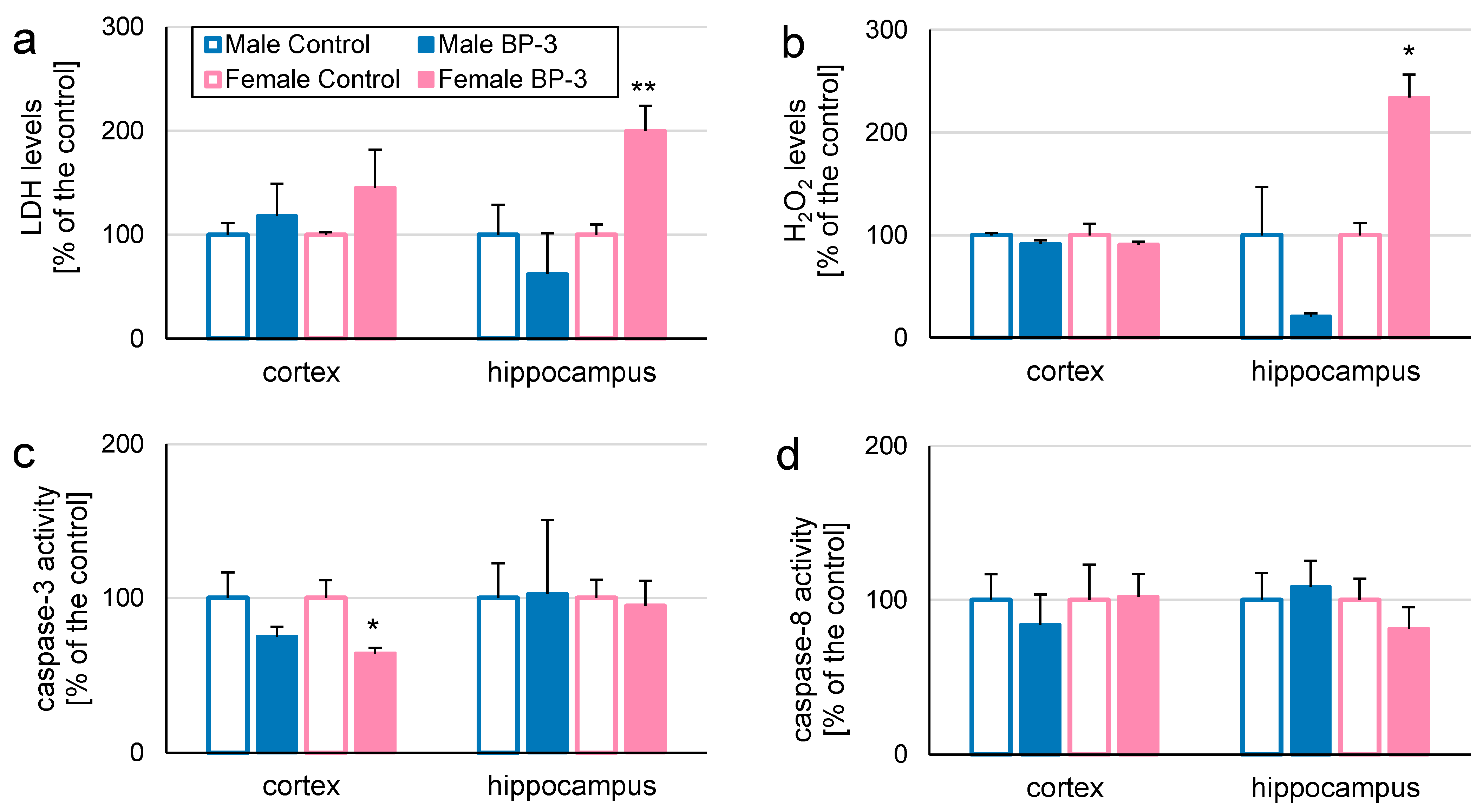
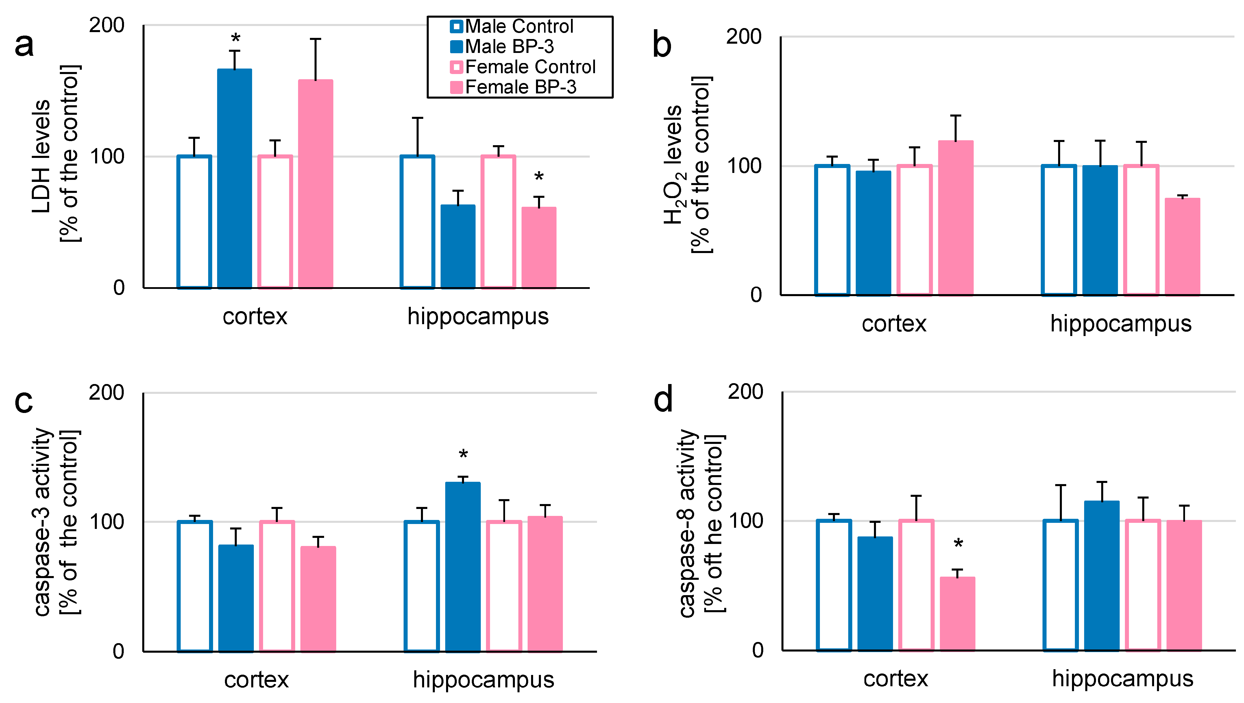
3.1. In 3-Month-Old Offspring, Prenatal Exposure to BP-3 Elevates LDH and H2O2 Levels in Female Hippocampi, but Decreases Caspase-3 Activity in Female Cerebral Cortices

In our experiment, pregnant female mice were exposed to BP-3 in concentrations of 50 mg/kg between the 7th and 21st day of pregnancy. We aimed to assess the effects of prenatal exposure to BP-3 in 3-month-old (adolescent) and 5-month-old (adult) offspring (F1) and transgenerational changes in 1-month-old individuals (F2). We chose to study intergenerational/transgenerational effects of BP-3 on two brain structures, i.e., the cerebral cortex and the hippocampus, since they are known for their crucial involvement in developmental and degenerative brain disorders [61,62,63,64].
Prenatal exposure to BP-3 caused pronounced changes in levels of LDH and H2O2 in the hippocampi of adolescent (3-month-old) female offspring (200 and 234% of the control, respectively; Figure 1a,b). Furthermore, in the cerebral cortices of 3-month-old female offspring, we observed decreased caspase-3 activity (64% of the control; Figure 1c). We noticed no biochemical changes in the cerebral cortex, nor in the hippocampus, in 3-month-old male offspring (Figure 1a–d). In females, cortical LDH, H2O2 and caspase-8 levels as well as hippocampal caspase-3 and caspase-8 levels were not affected (Figure 1a–d).

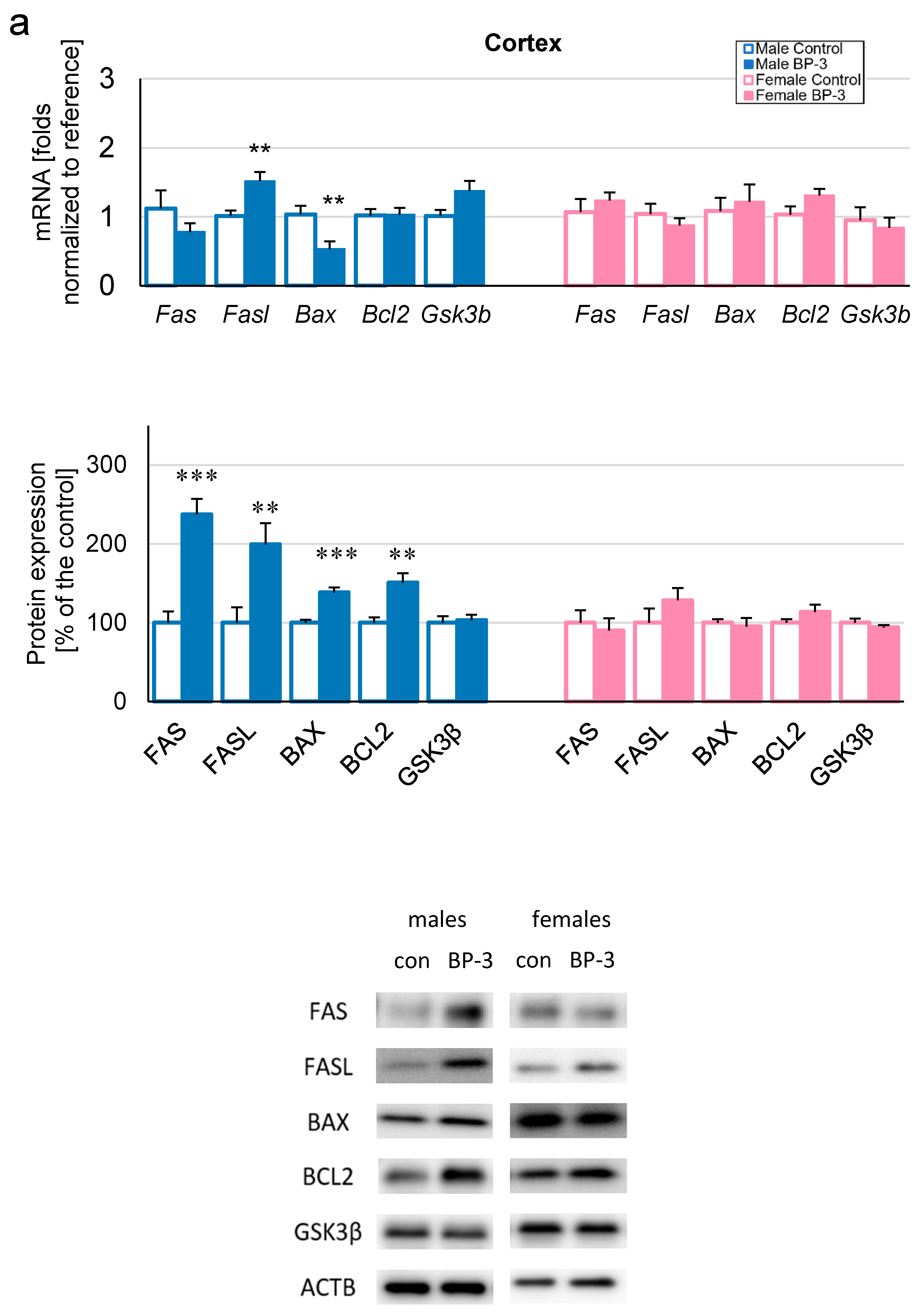
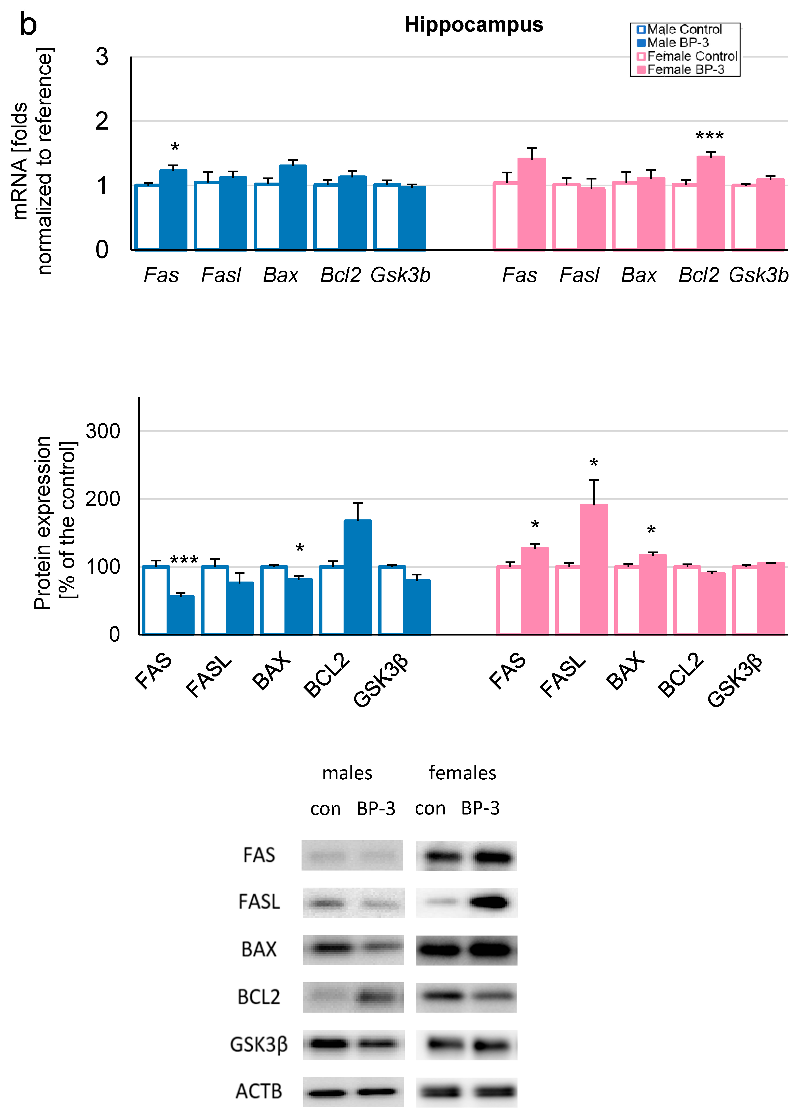
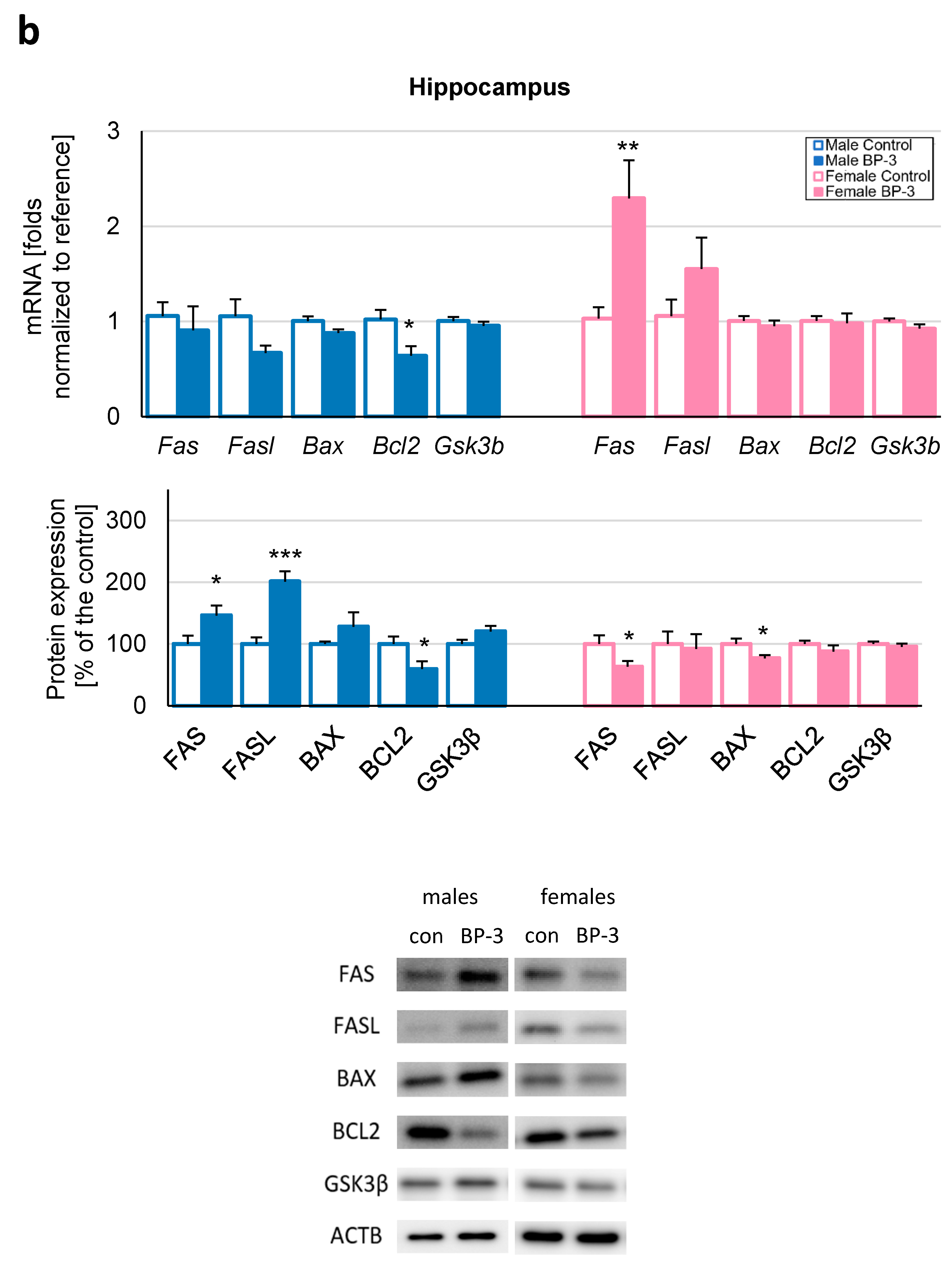
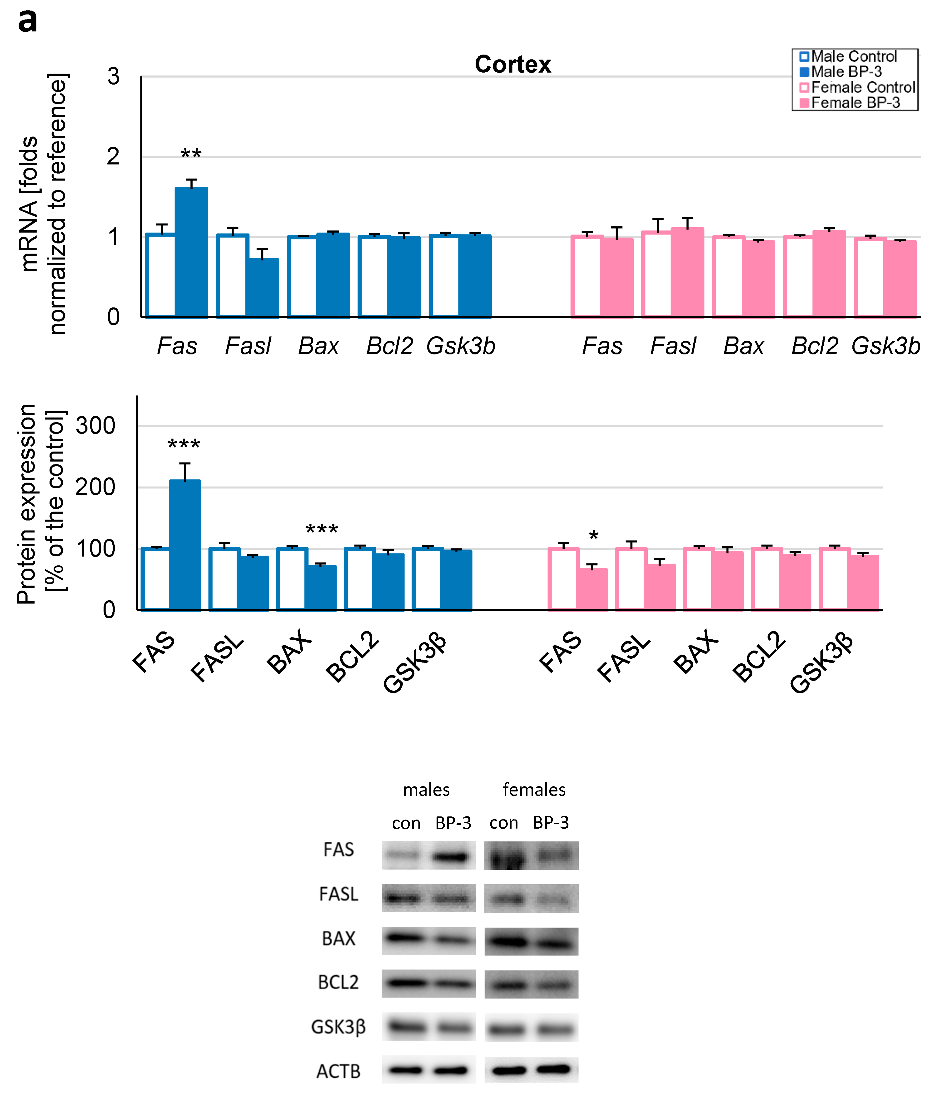
3.2. Prenatal Exposure to BP-3 Causes Pronounced Changes in the Expression of Apoptosis-Related Factors in Male Cerebral Cortices and Hippocampi, and Female Hippocampi of 3-Month-Old Offspring

Apoptosis plays a crucial role in neurodevelopment and neurodegeneration. This fact prompted us to investigate the intergenerational/transgenerational effects of BP-3 on apoptosis-related factors in the chosen brain regions (cerebral cortex and hippocampus). In 3-month-old offspring, dysregulation of the apoptosis-related mRNAs and protein was most prominent in the cerebral cortices of males (Figure 2a), where prenatal exposure to BP-3 caused the upregulation of FAS (237% of the control), Fasl (to 1.52 fold)/FASL (199% of the control), BAX (138% of the control) and BCL2 (151% of the control), but the downregulation of Bax (to 0.53 fold). In contrast, no changes were observed in the cerebral cortices of females (Figure 2a).
In the hippocampi of 3-month-old males, prenatal exposure to BP-3 increased the mRNA of Fas (1.23 fold) but decreased the FAS protein level (55% of the control), while also causing BAX downregulation (81% of the control) (Figure 2b). The levels of remaining factors (Fasl/FASL, Bax, Bcl2/BCL2, and Gsk3b/GSK3β) were not affected. In the hippocampi of 3-month-old females, Bcl2 (1.44 fold), FAS (127% of the control), FASL (191% of the control), and BAX (117% of the control) were upregulated, while the rest of the investigated factors (Fas, Fasl, Bax, BCL2, and Gsk3b/GSK3β) remained unchanged (Figure 2b).

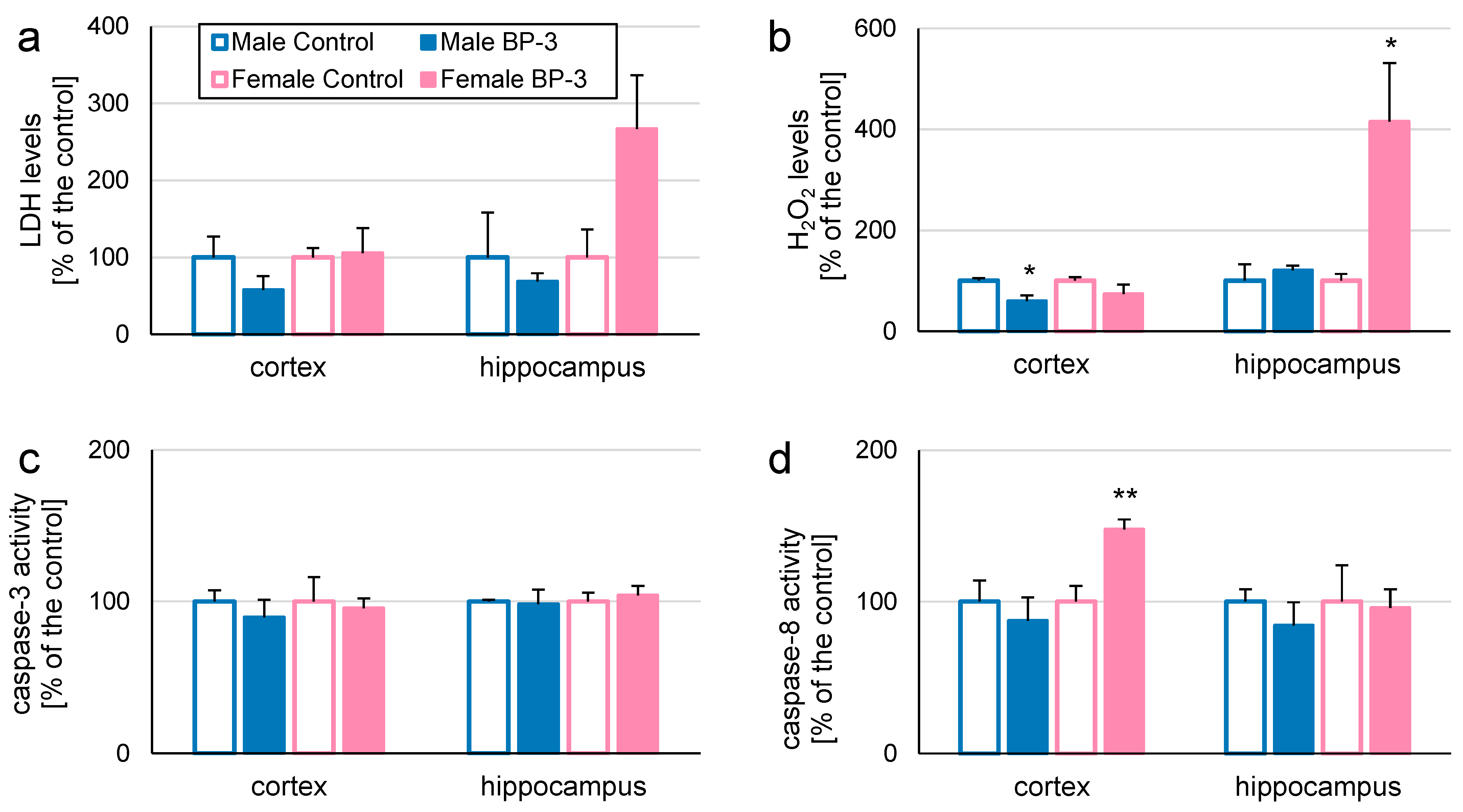
3.3. Prenatal Exposure to BP-3 Alters H2O2 Levels and Caspase-8 Activity in Cerebral Cortices and/or Hippocampi of 5-Month-Old Offspring

The most pronounced effects of prenatal exposure to BP-3 in 5-month-old offspring were noticed in H2O2 levels in the female hippocampus (416% of the control) but not in the cerebral cortices of females (Figure 3b). In contrast, no changes in this parameter were present in the hippocampi of males, while decreased H2O2 levels were observed in cerebral cortices (60% of the control; Figure 3b). In 5-month-old offspring, prenatal exposure to BP-3 altered caspase-8 only in the cerebral cortices of females, increasing its activity to 148% of the control (Figure 3d). No changes in LDH levels and caspase-3 activity were observed in these groups in either the cerebral cortex or the hippocampus (Figure 3a,c).

3.4. Prenatal Exposure to BP-3 Changes Expression of Apoptosis-Related Factors in Cerebral Cortices and/or Hippocampi of 5-Month-Old Offspring

In the cerebral cortices of 5-month-old male offspring prenatally exposed to BP-3, increased expressions of Fas (2.31-fold increase), FAS (302% of the control), and FASL (196% of the control) were observed. In females, prenatal exposure to BP-3 caused a decrease in Fas (to 0.66-fold) at an mRNA level, but increases at the protein level of FAS (221% of the control) and FASL (219% of the control). In the cerebral cortices of 5-month-old offspring, prenatal exposure to BP-3 did not alter Bax/BAX, Bcl2/BCL2, and Gsk3b/GSK3β, except for the mRNA of Bax (decrease to 0.87-fold) and BCL2 in females (increased to 141% of the control; Figure 4a).
In the hippocampi of 5-month-old male offspring prenatally exposed to BP-3, we observed increased FAS (160% of the control) and FASL (201% of the control) levels and a decrease in Bcl2/BCL2 (to 0.64 fold and 59% of the control, respectively). In females, there is a discrepancy in effects of prenatal exposure to BP-3 in Fas/FAS mRNA and protein levels, i.e., Fas increased 2.29-fold and FAS decreased to 63% of the control. In female offspring, BP-3 also caused the BAX level decrease (71% of the control; Figure 4b). Levels of remaining the apoptosis-related mRNAs and protein did not change (Figure 4a,b).

3.5. Transgenerational Effects of Exposure to BP-3 Alters LDH, Caspase-3, and Caspase-8 Activity in Cerebral Cortices and/or Hippocampi of 1-Month-Old Mice

In 1-month-old males from the F2 generation, transgenerational effects of exposure to BP-3 increased LDH levels in the cerebral cortex (166% of the control), but not in the hippocampus and elevated caspase-3 activity in the hippocampus (130% of the control), but not in the cerebral cortex (Figure 5a,c). On the other hand, in females, transgenerational effects of exposure to BP-3 decreased LDH levels in the hippocampus (61% of the control), but not in the cerebral cortex, and weakened caspase-8 activity in the cerebral cortex (56% of the control), but not in the hippocampus (Figure 5a,d). F2 individuals were not affected by transgenerational effects of exposure to BP-3 in terms of H2O2 levels (Figure 5b).

3.6. Transgenerational Effects of Exposure to BP-3 Changes Expression of Apoptosis-Related Factors in Cerebral Cortices and/or Hippocampi of 1-Month-Old Individuals

In the cerebral cortices of 1-month-old individuals from the F2 generation, transgenerational effects of exposure to BP-3 increased Fas and FAS (1.60 fold and 210% of the control, respectively), but decreased BAX (71% of the control) in males, while in females, FAS decreased to 66% of the control (Figure 6a).
In the hippocampi of 1-month-old individuals, FAS and FASL were elevated in males (189 and 177% of the control, respectively), while in females, an increase in Bax (1.10 fold) and BCL2 was observed (210% of the control; Figure 6b). Levels of the remaining apoptosis-related mRNAs and protein did not change (Figure 6a,b).

4. Discussion

BP-3 is widely applied as a UV filter in personal care products and as a stabilizer for commercial use that prevents photodegradation. Emerging evidence suggests that BP-3 may act as an endocrine disruptor, interfering with the body’s endocrine system. Although studies showed that BP-3 crosses the BBB and its presence was detected in post mortem adult brains, the mechanisms of BP-3 action in the central nervous system are still an important subject of interest for researchers [31]. In our recent in vitro studies, we conducted experiments on primary cultures of mice neuronal cells treated with BP-3, and primary neuronal cells settled from embryos prenatally exposed to BP-3. We demonstrated that BP-3 induced neurotoxicity and apoptosis, and downregulated autophagy [32,46,47,48], suggesting its contribution to neurodevelopmental abnormalities and the onset of neurological disorders. However, little is known about its action on neurodevelopment, especially in the contexts of intergenerational and transgenerational consequences. Therefore, the aim of our study was to determine the toxic and apoptotic effects of prenatal exposure to BP-3 in mice from the F1 and F2 generations, both in males and females. The administered dose of BP-3 to the parental generation was relevant to the amount that an average woman is exposed to as a result of applying sunscreen to the entire body (40 g of cream/person − 2 mg/cm2 of skin) [65,66,69].
Here, we provide evidence for the neurotoxic action of BP-3 on the mouse cerebral cortex and hippocampus in terms of LDH, caspase-3, caspase-8 and/or H2O2 levels in the offspring of mothers exposed to environmentally relevant concentrations of BP-3, as well as in the second-generation mice. Sensitivity to BP-3, as regards the levels of LDH and caspase-3 activity, was observed in both generations (F1 and F2), whereas sensitivity to BP-3 regarding H2O2 levels was observed only in the F1 generation. Most vulnerable to LDH- and H2O2-related effects was the female hippocampus, whereas vulnerability to caspase-3-related effects was only observed in the female cerebral cortex (F1 generation) and the male hippocampus (F2 generation). These data show that BP-3 evokes a variety of effects which may either initiate apoptosis (caspase-3), possible via the induction of oxidative stress (H2O2), and finally evoke brain cell death (LDH). Increases in LDH and H2O2 release are related to the onset of neurodegenerative disorders [70,71], autism [72], schizophrenia, or social adaptation disorders [73] and similar results to ours were obtained in in vitro studies on primary neurons [46,47,48] or the SH-SY5Y cell line [74] exposed to BP-3. On the other hand, our data indicating decreases in LDH or H2O2 release may be associated with a cellular defense response aimed at delaying neurodegeneration [75]. The obtained results indicating increasing levels of caspase-3 activity are consistent with the results previously obtained in vitro [46,47,48]. Therefore, our present study shows that BP-3 may predispose brain tissue to caspase-3-dependent apoptosis and cause multigenerational neurotoxic effects.
The pro-apoptotic effect of BP-3 on the brain was confirmed by increases in Fas/FAS and/or Fasl/FASL expression levels that were independent from generation, gender, or cerebral structure, except for in 3-month-old F1 females and 1-month-old F2 females. In addition, BP-3 affected caspase-8 activity (known to participate in the FAS/FASL-dependent extrinsic apoptosis pathway) in both generations, but the only vulnerable structure was the female cerebral cortex. Enhanced FAS and FASL expression levels in brain cells accompany neurodegenerative and neurological disorders’ onsets, i.e., during experimental autoimmune encephalomyelitis (EAE model), exposure to heavy metals (e.g., cadmium), as well as in Alzheimer’s disease [76,77,78]. Our data indicated that BP-3 also caused the dysregulation of the BAX/BCL2 pathway which was much more prominent in the F1 than in the F2 generation. In addition, we observed the prevalence of extrinsic apoptosis (FAS, FASL, and caspase-8) over intrinsic apoptosis (BAX, BCL2) in both generations, except for in the 3-month-old F1 generation. Similarly to our studies, simultaneous increase in anti-apoptotic BCL2 and pro-apoptotic BAX was observed during prenatal exposure to cocaine [79] and cellular models of stroke and Alzheimer’s disease [80,81,82,83,84]. The anti-apoptotic properties of BCL2 are also prominent in the mechanism of cell protection against the effects of hypoxia and reperfusion, as evidenced by an enhanced BCL2 expression level and the increased survival of cerebral cortex cells in rats subjected to hypoxia [71]. Therefore, apoptotic processes induced by BP-3 were mostly related to the extrinsic pathway and these changes affected F1 and F2 males to a greater extent.
Taking into account the main goal of the research to demonstrate the transgenerational consequences of BP-3 exposure, it can be said that BP-3 has an ability to induce brain malfunction (due to neurotoxicity and apoptosis) and in this way, it may contribute to neurodevelopmental diseases in F2 generation mice. Considering gender differences, males appear to be more vulnerable than females to the impact of BP-3 exposure, as evidenced by the increase in LDH and Fas/FAS levels in the cerebral cortex or the enhancement in caspase-3 activity as well as FAS and FASL expression levels in the hippocampus. The fewer effects observed in females may be associated with the protective role of estrogens in the brain [85]. Males’ sensitivity to transgenerational action was also observed after exposure to vinclozolin, an agricultural fungicide, known to be an androgenic endocrine disruptor, that induced epigenetic changes and the transgenerational inheritance of neurodevelopmental disorders and abnormalities [86,87]. Therefore, our studies indicated that ancestral exposure of gestating mice to BP-3 induces toxic and pro-apoptotic effects in the brains of second-generation mice; however, mechanisms of inheritance or such long-term influences of this substance still must be specified. While considering the results achieved in the current study, it is also important to note its limitations; we have conducted ex vivo experiments including biochemical and molecular assessments, and we observed and studied effects evoked in this regard. The results obtained in the course of our work encourage further efforts; the next stage of our work is to include neurobehavioral assessment in order to determine if the effects observed ex vivo correspond with changes in animals’ behavior. Additionally, our study did not assess the effects of BP-3 exposure on the P generation, including factors such as maternal health, food consumption, and weight gain during pregnancy. These factors could influence intrauterine conditions and, consequently, developmental outcomes in the offspring. While our research focused on the indirect effects of BP-3 on the nervous system and its transgenerational impacts, future studies should incorporate these aspects to provide a more comprehensive understanding of BP-3′s effects on both parental and subsequent generations.
In summary, we proved for the first time the toxic and pro-apoptotic effects of prenatal exposure to BP-3 that are both intergenerational, i.e., in the offspring of mothers exposed to environmentally relevant concentrations of BP-3, as well as transgenerational, i.e., in second-generation mice. Our studies indicate that apoptotic processes induced by BP-3 in the central nervous system are more closely related to the extrinsic pathway than to the intrinsic mitochondrial pathway. Interestingly, taking into account expression levels of apoptotic factors, we observe much stronger effects of prenatal exposure to BP-3 in males and in the F1 generation. In our studies, we identified transgenerational mechanisms, which confirm the capacity of BP-3 to impair brain homeostasis and in this way increase the risk of multigenerational disorders associated with the disruption of neurodevelopment.

Supplementary Materials

The following supporting information can be downloaded at: https://www.mdpi.com/article/10.3390/toxics12120906/s1.

Author Contributions

K.P.-D.—data curation, formal analysis, investigation, validation, writing—original draft; A.Ł.—data curation, formal analysis, investigation, validation, writing—original draft; B.A.P.-W.—data curation, formal analysis, investigation, validation, writing—original draft; J.R.—investigation; M.K.—writing—review and editing; A.W.-B.—validation; W.B.—validation; K.N.—investigation; A.W.—conceptualization, methodology, data curation, formal analysis, investigation, supervision, writing—original draft. All authors have read and agreed to the published version of the manuscript.

Funding

This research was funded by the statutory fund of the Maj Institute of Pharmacology, Polish Academy of Sciences in Krakow, Poland. Karolina Przepiórska-Drońska was supported by Twinning TrePsy, funded by the European Union’s Horizon Europe research and innovation programme under agreement no 101079256.

Institutional Review Board Statement

The animal study protocol was approved by the 2nd Local Institutional Animal Care and Use Committee (resolution no 137/2019, 27 June 2019).

Informed Consent Statement

Not applicable.

Data Availability Statement

The datasets generated during and/or analyzed during the current study are available from the corresponding author on reasonable request.

Acknowledgments

Schemes were created with BioRender.com.

Conflicts of Interest

The authors have no relevant financial or non-financial interests to disclose.

Abbreviations

Actb—β-actin; Bax/BAX—BCL2-associated X protein; BBB—blood–brain barrier; Bcl2/BCL2—B-cell lymphoma 2; BP-3—benzophenone-3; Ct—threshold cycle; F1—first-generation mice; F2—second-generation mice; Fas/FAS—cell surface death receptor; Fasl/FASL—FAS ligand; Gapdh—glyceraldehyde-3-phosphate dehydrogenase; Gsk3b/GSK3β—glycogen synthase kinase 3 beta; Hprt1—Hypoxanthine phosphoribosyltransferase 1; H2O2—hydrogen peroxide; LDH—lactate dehydrogenase; P—parental generation; UV—ultraviolet light; qPCR—quantitative polymerase chain reaction.

References

  1. Watson, M.; Holman, D.M.; Maguire-Eisen, M. Ultraviolet Radiation Exposure and Its Impact on Skin Cancer Risk. Seminars Oncol. Nurs. 2016, 32, 241–254. [Google Scholar] [CrossRef]
  2. Hasbani, D.J.; Ghaoui, N.; Haddad, F.; Shahla, W.A.; Saade, S.; Bandali, T.; Menhem, Z.; Saade, D. Sun Protection Knowledge, Attitudes and Practices: A Lebanese-Based Survey. JEADV Clin. Pract. 2024, 3, 565–574. [Google Scholar] [CrossRef]
  3. Afiouni, R.; Helou, J.; Bou-Orm, I. Knowledge of the Risks of Ultraviolet Radiation, Sun Exposure Attitudes and Practices among Lebanese University Students. Prev. Med. Rep. 2024, 47, 102900. [Google Scholar] [CrossRef]
  4. Tang, X.; Yang, T.; Yu, D.; Xiong, H.; Zhang, S. Current Insights and Future Perspectives of Ultraviolet Radiation (UV) Exposure: Friends and Foes to the Skin and Beyond the Skin. Environ. Int. 2024, 185, 108535. [Google Scholar] [CrossRef] [PubMed]
  5. Sani, M.A.; Khezerlou, A.; Tavassoli, M.; Abedini, A.H.; McClements, D.J. Development of Sustainable UV-Screening Food Packaging Materials: A Review of Recent Advances. Trends Food Sci. Technol. 2024, 145, 104366. [Google Scholar] [CrossRef]
  6. Yousif, E.; Haddad, R. Photodegradation and Photostabilization of Polymers, Especially Polystyrene: Review. SpringerPlus 2013, 2, 398. [Google Scholar] [CrossRef] [PubMed]
  7. Wnuk, A.; Kajta, M. Is the Commonly Used UV Filter Benzophenone-3 a Risk Factor for the Nervous System? Acta Biochim. Pol. 2021, 68, 557–563. [Google Scholar] [CrossRef] [PubMed]
  8. Kim, S.; Choi, K. Occurrences, Toxicities, and Ecological Risks of Benzophenone-3: A Common Component of Organic Sunscreen Products: A Mini-Review. Environ. Int. 2014, 70, 143–157. [Google Scholar] [CrossRef]
  9. Mustieles, V.; Balogh, R.K.; Axelstad, M.; Montazeri, P.; Márquez, S.; Vrijheid, M.; Draskau, M.K.; Taxvig, C.; Peinado, F.M.; Berman, T.; et al. Benzophenone-3: Comprehensive Review of the Toxicological and Human Evidence with Meta-Analysis of Human Biomonitoring Studies. Environ. Int. 2023, 173, 107739. [Google Scholar] [CrossRef]
  10. Commission Regulation (EU) 2017/238 of 10 February 2017 Amending Annex VI to Regulation (EC) No 1223/2009 of the European Parliament and of the Council on Cosmetic Products. Available online: https://eur-lex.europa.eu/legal-content/EN/TXT/?uri=CELEX%3A32017R0238 (accessed on 8 December 2024).
  11. Commission Regulation (EU) 2022/1176 of 7 July 2022 Amending Regulation (EC) No 1223/2009 of the European Parliament and of the Council as Regards the Use of Certain UV Filters in Cosmetic Products. Available online: https://eur-lex.europa.eu/eli/reg/2022/1176 (accessed on 8 December 2024).
  12. Suh, S.; Pham, C.; Smith, J.; Mesinkovska, N.A. The Banned Sunscreen Ingredients and Their Impact on Human Health: A Systematic Review. Int. J. Dermatol. 2020, 59, 1033–1042. [Google Scholar] [CrossRef]
  13. Du, Y.; Wang, W.-Q.; Pei, Z.-T.; Ahmad, F.; Xu, R.-R.; Zhang, Y.-M.; Sun, L.-W. Acute Toxicity and Ecological Risk Assessment of Benzophenone-3 (BP-3) and Benzophenone-4 (BP-4) in Ultraviolet (UV)-Filters. Int. J. Environ. Res. Public Health 2017, 14, 1414. [Google Scholar] [CrossRef] [PubMed]
  14. Ortiz-Román, M.I.; Casiano-Muñiz, I.M.; Román-Velázquez, F.R. Toxicity of UV Filter Benzophenone-3 in Brine Shrimp Nauplii (Artemia salina) and Zebrafish (Danio rerio) Embryos. J. Xenobiotics 2024, 14, 537–553. [Google Scholar] [CrossRef]
  15. Wnuk, W.; Michalska, K.; Krupa, A.; Pawlak, K. Benzophenone-3, a Chemical UV-Filter in Cosmetics: Is It Really Safe for Children and Pregnant Women? Postepy Dermatol. Alergol. 2022, 39, 26–33. [Google Scholar] [CrossRef] [PubMed]
  16. Sun, Y.; Lu, G.; Zhang, P.; Ling, X.; Zhou, R.; Yan, Z.; Liu, J. Influence of Organic Colloids on the Uptake, Accumulation and Effects of Benzophenone-3 in Aquatic Animals. Environ. Sci. Nano 2021, 8, 1366–1378. [Google Scholar] [CrossRef]
  17. European Chemicals Agency. 2020 Substance Information: Oxybenzone; Annankatu 18; European Chemicals Agency: Helsinki, Finland, 2020. [Google Scholar]
  18. CDC (Centers for Disease Control and Prevention). 2017 Fourth National Report on Human Exposure to Environmental Chemicals; Centers for Disease Control and Prevention: Atlanta, GA, USA, 2017. Available online: https://stacks.cdc.gov/view/cdc/21813 (accessed on 8 December 2024).
  19. SCCS (Scientific Committee on Consumer Safety). Opinion on Benzophenone-3 (CAS No 131-57-7, EC No 205-031-5), Preliminary Version of 15 December 2020, Final Version of 30–31 March 2021, SCCS/1625/20. Available online: https://health.ec.europa.eu/system/files/2022-08/sccs_o_247.pdf (accessed on 8 December 2024).
  20. Ruszkiewicz, J.A.; Pinkas, A.; Ferrer, B.; Peres, T.V.; Tsatsakis, A.; Aschner, M. Neurotoxic Effect of Active Ingredients in Sunscreen Products: A Contemporary Review. Toxicol. Rep. 2017, 4, 245–259. [Google Scholar] [CrossRef]
  21. Mao, J.F.; Li, W.; Ong, C.N.; He, Y.; Jong, M.C.; Gin, K.Y. Assessment of Human Exposure to Benzophenone-Type UV Filters: A Review. Environ. Int. 2022, 167, 107405. [Google Scholar] [CrossRef]
  22. Tibbetts, J. Shining a Light on BP-3 Exposure. Environ. Health Perspect. 2008, 116, A306. [Google Scholar] [CrossRef]
  23. Gustavsson Gonzalez, H.; Farbrot, A.; Larkö, O. Percutaneous Absorption of Benzophenone-3: A Common Component of Topical Sunscreens. Clin. Exp. Dermatol. 2002, 27, 691–694. [Google Scholar] [CrossRef]
  24. Janjua, N.R.; Kongshoj, B.; Andersson, A.M.; Wulf, H.C. Sunscreens in Human Plasma and Urine after Repeated Whole-body Topical Application. J. Eur. Acad. Dermatol. Venereol. 2008, 22, 456–461. [Google Scholar] [CrossRef] [PubMed]
  25. Parks, C.G.; Meier, H.C.S.; Jusko, T.A.; Wilkerson, J.; Miller, F.W.; Sandler, D.P. Benzophenone-3 and Antinuclear Antibodies in U.S. Adolescents and Adults Ages 12–39 Years. Front. Immunol. 2022, 13, 958527. [Google Scholar] [CrossRef] [PubMed]
  26. IARC Working Group on the Evaluation of Carcinogenic Risks to Humans. Some Chemicals Present in Industrial and Consumer Products, Food and Drinking-Water. IARC Monogr. Eval. Carcinog. Risks Hum. 2013, 101, 9–549. [Google Scholar]
  27. Yao, Y.N.; Wang, Y.; Zhang, H.; Gao, Y.; Zhang, T.; Kannan, K. A Review of Sources, Pathways, and Toxic Effects of Human Exposure to Benzophenone Ultraviolet Light Filters. Eco-Environ. Health 2024, 3, 30–44. [Google Scholar] [CrossRef]
  28. Gavrila, A.A.; Dasteridis, I.S.; Tzimas, A.A.; Chatzimitakos, T.G.; Stalikas, C.D. Benzophenones in the Environment: Occurrence, Fate and Sample Preparation in the Analysis. Molecules 2023, 28, 1229. [Google Scholar] [CrossRef]
  29. Wan, Y.; Xue, J.; Kannan, K. Occurrence of Benzophenone-3 in Indoor Air from Albany, New York, USA, and Its Implications for Inhalation Exposure. Sci. Total Environ. 2015, 537, 304–308. [Google Scholar] [CrossRef] [PubMed]
  30. Wang, L.; Asimakopoulos, A.G.; Kannan, K. Accumulation of 19 Environmental Phenolic and Xenobiotic Heterocyclic Aromatic Compounds in Human Adipose Tissue. Environ. Int. 2015, 78, 45–50. [Google Scholar] [CrossRef]
  31. Van der Meer, T.P.; Artacho-Cordón, F.; Swaab, D.F.; Struik, D.; Makris, K.C.; Wolffenbuttel, B.H.R.; Frederiksen, H.; Van Vliet-Ostaptchouk, J.V. Distribution of Non-Persistent Endocrine Disruptors in Two Different Regions of the Human Brain. Int. J. Environ. Res. Public Health 2017, 14, 1059. [Google Scholar] [CrossRef] [PubMed]
  32. Wnuk, A.; Rzemieniec, J.; Staroń, J.; Litwa, E.; Lasoń, W.; Bojarski, A.; Kajta, M. Prenatal Exposure to Benzophenone-3 Impairs Autophagy, Disrupts RXRs/PPARγ Signaling, and Alters Epigenetic and Post-Translational Statuses in Brain Neurons. Mol. Neurobiol. 2019, 56, 4820–4837. [Google Scholar] [CrossRef]
  33. Pomierny, B.; Krzyżanowska, W.; Broniowska, Ż.; Strach, B.; Bystrowska, B.; Starek-Świechowicz, B.; Maciejska, A.; Skórkowska, A.; Wesołowska, J.; Walczak, M.; et al. Benzophenone-3 Passes Through the Blood-Brain Barrier, Increases the Level of Extracellular Glutamate, and Induces Apoptotic Processes in the Hippocampus and Frontal Cortex of Rats. Toxicol. Sci. 2019, 171, 485–500. [Google Scholar] [CrossRef] [PubMed]
  34. Maciejska, A.; Pomierny, B.; Krzyżanowska, W.; Starek-Świechowicz, B.; Skórkowska, A.; Budziszewska, B. Mechanism of Microglial Cell Activation in the Benzophenone-3 Exposure Model. Neuroscience 2023, 533, 63–76. [Google Scholar] [CrossRef]
  35. Vela-Soria, F.; Jiménez-Díaz, I.; Rodríguez-Gómez, R.; Zafra-Gómez, A.; Ballesteros, O.; Navalón, A.; Vílchez, J.; Fernández, M.; Olea, N. Determination of Benzophenones in Human Placental Tissue Samples by Liquid Chromatography-Tandem Mass Spectrometry. Talanta 2011, 85, 1848–1855. [Google Scholar] [CrossRef] [PubMed]
  36. Philippat, C.; Wolff, M.S.; Calafat, A.M.; Ye, X.; Bausell, R.; Meadows, M.; Stone, J.; Slama, R.; Engel, S.M. Prenatal Exposure to Environmental Phenols: Concentrations in Amniotic Fluidand Variability in Urinary Concentrations During Pregnancy. Environ. Health Perspect. 2013, 121, 1225–1231. [Google Scholar] [CrossRef] [PubMed]
  37. Krause, M.; Frederiksen, H.; Sundberg, K.; Jørgensen, F.; Jensen, L.; Nørgaard, P.; Jørgensen, C.; Ertberg, P.; Juul, A.; Drzewiecki, K.; et al. Presence of Benzophenones Commonly Used as UV Filtersand Absorbers in Paired Maternal and Fetal Samples. Environ. Int. 2018, 110, 51–60. [Google Scholar] [CrossRef] [PubMed]
  38. Schlumpf, M.; Kypke, K.; Wittassek, M.; Angerer, J.; Mascher, H.; Mascher, D.; Vökt, C.; Birchler, M.; Lichtensteiger, W. Exposure Patterns of UV Filters, Fragrances, Parabens, Phthalates, Organochlor Pesticides, PBDEs, and PCBs in Human Milk: Correlation of UV Filters with Use of Cosmetics. Chemosphere 2010, 81, 1171–1183. [Google Scholar] [CrossRef] [PubMed]
  39. Molins-Delgado, D.; Olmo-Campos, M.D.M.; Valeta-Juan, G.; Pleguezuelos-Hernández, V.; Barceló, D.; Díaz-Cruz, M.S. Determination of UV Filters in Human Breast Milk Using Turbulent Flow Chromatography and Babies’ Daily Intake Estimation. Environ. Res. 2018, 161, 532–539. [Google Scholar] [CrossRef]
  40. Iribarne-Durán, L.M.; Serrano, L.; Peinado, F.M.; Peña-Caballero, M.; Hurtado, J.A.; Vela-Soria, F.; Fernández, M.F.; Freire, C.; Artacho-Cordón, F.; Olea, N. Biomonitoring Bisphenols, Parabens, and Benzophenones in Breast Milk from a Human Milk Bank in Southern Spain. Sci. Tot. Environ. 2022, 830, 154737. [Google Scholar] [CrossRef]
  41. Wang, L.; Kannan, K. Characteristic Profiles of Benzophenone-3 and Its Derivatives in Urine of Children and Adults from the United States and China. Environ. Sci. Technol. 2013, 47, 12532–12538. [Google Scholar] [CrossRef] [PubMed]
  42. Zhang, T.; Sun, H.; Qin, X.; Wu, Q.; Zhang, Y.; Ma, J.; Kannan, K. Benzophenone-Type UV Filters in Urine and Blood from Children, Adults, and Pregnant Women in China: Partitioning Between Blood and Urine as Well as Maternal and Fetal Cord Blood. Sci. Total Environ. 2013, 461–462, 49–55. [Google Scholar] [CrossRef] [PubMed]
  43. Holmboe, S.A.; Henriksen, L.S.; Frederiksen, H.; Andersson, A.-M.; Priskorn, L.; Jørgensen, N.; Juul, A.; Toppari, J.; Skakkebæk, N.E.; Main, K.M. Prenatal Exposure to Phenols and Benzophenones in Relation to Markers of Male Reproductive Function in Adulthood. Front. Endocrinol. 2022, 13, 1071761. [Google Scholar] [CrossRef] [PubMed]
  44. Wnuk, A.; Kajta, M. Steroid and Xenobiotic Receptor Signalling in Apoptosis and Autophagy of the Nervous System. Int. J. Mol. Sci. 2017, 18, 2394. [Google Scholar] [CrossRef]
  45. Hollville, E.; Romero, S.E.; Deshmukh, M. Apoptotic Cell Death Regulation in Neurons. FEBS J. 2019, 286, 3276–3298. [Google Scholar] [CrossRef]
  46. Wnuk, A.; Rzemieniec, J.; Lasoń, W.; Krzeptowski, W.; Kajta, M. Apoptosis Induced by the UV Filter Benzophenone-3 in Mouse Neuronal Cells Is Mediated via Attenuation of Erα/Pparγ and Stimulation of Erβ/Gpr30 Signaling. Mol. Neurobiol. 2018, 55, 2362–2383. [Google Scholar] [CrossRef] [PubMed]
  47. Wnuk, A.; Rzemieniec, J.; Lasoń, W.; Krzeptowski, W.; Kajta, M. Benzophenone-3 Impairs Autophagy, Alters Epigenetic Status, and Disrupts Retinoid X Receptor Signaling in Apoptotic Neuronal Cells. Mol. Neurobiol. 2018, 55, 5059–5074. [Google Scholar] [CrossRef]
  48. Wnuk, A.; Rzemieniec, J.; Litwa, E.; Lasoń, W.; Kajta, M. Prenatal Exposure to Benzophenone-3 (BP-3) Induces Apoptosis, Disrupts Estrogen Receptor Expression and Alters the Epigenetic Status of Mouse Neurons. J. Steroid Biochem. Mol. Biol. 2018, 182, 106–118. [Google Scholar] [CrossRef] [PubMed]
  49. Rudin, C.M.; Thompson, C.B. Apoptosis and Disease: Regulation and Clinical Relevance of Programmed Cell Death. Annu. Rev. Med. 1997, 48, 267–281. [Google Scholar] [CrossRef] [PubMed]
  50. Margolis, R.L.; Chuang, D.M.; Post, R.M. Programmed Cell Death: Implications for Neuropsychiatric Disorders. Biol. Psychiatry 1994, 35, 946–956. [Google Scholar] [CrossRef]
  51. Jarskog, L.F.; Glantz, L.A.; Gilmore, J.H.; Lieberman, J.A. Apoptotic Mechanisms in the Pathophysiology of Schizophrenia. Prog. Neuropsychopharmacol. Biol. Psychiatry 2005, 29, 846–858. [Google Scholar] [CrossRef]
  52. Dong, D.; Zielke, H.R.; Yeh, D.; Yang, P. Cellular Stress and Apoptosis Contribute to the Pathogenesis of Autism Spectrum Disorder. Autism Res. 2018, 11, 1076–1090. [Google Scholar] [CrossRef] [PubMed]
  53. Rossignol, D.A.; Genuis, S.J.; Frye, R.E. Environmental Toxicants and Autism Spectrum Disorders: A Systematic Review. Transl. Psychiatry 2014, 4, e360. [Google Scholar] [CrossRef] [PubMed]
  54. Ye, B.S.; Leung, A.O.W.; Wong, M.H. The Association of Environmental Toxicants and Autism Spectrum Disorders in Children. Environ. Pollut. 2017, 227, 234–242. [Google Scholar] [CrossRef] [PubMed]
  55. Khan, A.; Plana-Ripoll, O.; Antonsen, S.; Brandt, J.; Geels, C.; Landecker, H.; Sullivan, P.F.; Pedersen, C.B.; Rzhetsky, A. Environmental Pollution is Associated with Increased Risk of Psychiatric Disorders in the US and Denmark. PLoS Biol. 2019, 17, e3000353. [Google Scholar] [CrossRef] [PubMed]
  56. Robinson, N.; Bergen, S.E. Environmental Risk Factors for Schizophrenia and Bipolar Disorder and Their Relationship to Genetic Risk: Current Knowledge and Future Directions. Front. Genet. 2021, 12, 686666. [Google Scholar] [CrossRef] [PubMed]
  57. Attademo, L.; Bernardini, F.; Garinella, R.; Compton, M.T. Environmental Pollution and Risk of Psychotic Disorders: A Review of the Science to Date. Schizophr. Res. 2017, 181, 55–59. [Google Scholar] [CrossRef] [PubMed]
  58. Jiang, Y.; Zhao, H.; Xia, W.; Li, Y.; Liu, H.; Hao, K.; Chen, J.; Sun, X.; Liu, W.; Li, J.; et al. Prenatal Exposure to Benzophenones, Parabens and Triclosan and Neurocognitive Development at 2 Years. Environ. Int. 2019, 126, 413–421. [Google Scholar] [CrossRef]
  59. Lopez-Torres, B.; Ares, I.; Martínez, M.; Maximiliano, J.E.; Martínez-Larrañaga, M.R.; Anadón, A.; Martínez, M.A. Neurotoxicity Induced by the Pyrethroid Lambda-Cyhalothrin: Alterations in Monoaminergic Systems and Dopaminergic and Serotoninergic Pathways in the Rat Brain. Food Chem. Toxicol. 2022, 169, 113434. [Google Scholar] [CrossRef] [PubMed]
  60. Sotelo-Orozco, J.; Calafat, A.M.; Cook Botelho, J.; Schmidt, R.J.; Hertz-Picciotto, I.; Bennett, D.H. Exposure to Endocrine Disrupting Chemicals Including Phthalates, Phenols, and Parabens in Infancy: Associations with Neurodevelopmental Outcomes in the MARBLES Study. Int. J. Hyg. Environ. Health 2024, 261, 114425. [Google Scholar] [CrossRef] [PubMed]
  61. Rubenstein, J.L. Annual Research Review: Development of the Cerebral Cortex: Implications for Neurodevelopmental Disorders. J. Child. Psychol. Psychiatry 2011, 52, 339–355. [Google Scholar] [CrossRef]
  62. Li, Y.; Shen, M.; Stockton, M.E.; Zhao, X. Hippocampal Deficits in Neurodevelopmental Disorders. Neurobiol. Learn. Mem. 2019, 165, 106945. [Google Scholar] [CrossRef] [PubMed]
  63. Schug, T.T.; Blawas, A.M.; Gray, K.; Heindel, J.J.; Lawler, C.P. Elucidating the Links Between Endocrine Disruptors and Neurodevelopment. Endocrinology 2015, 156, 1941–1951. [Google Scholar] [CrossRef] [PubMed]
  64. Doi, M.; Usui, N.; Shimada, S. Prenatal Environment and Neurodevelopmental Disorders. Front. Endocrinol. 2022, 13, 860110. [Google Scholar] [CrossRef]
  65. NCD Risk Factor Collaboration (NCD-RisC). Worldwide trends in underweight and obesity from 1990 to 2022: A pooled analysis of 3663 population-representative studies with 222 million children, adolescents, and adults. Lancet 2024, 403, 1027–1050. [Google Scholar] [CrossRef]
  66. Janjua, N.R.; Mogensen, B.; Andersson, A.-M.; Petersen, J.H.; Henriksen, M.; Skakkebæk, N.E.; Wulf, H.C. Systemic Absorption of the Sunscreens Benzophenone-3, Octyl-Methoxycinnamate, and 3-(4-Methyl-Benzylidene) Camphor After Whole-Body Topical Application and Reproductive Hormone Levels in Humans. J. Investig. Dermatol. 2004, 123, 57–61. [Google Scholar] [CrossRef]
  67. Kajta, M.; Wnuk, A.; Rzemieniec, J.; Litwa, E.; Lason, W.; Zelek-Molik, A.; Nalepa, I.; Rogóż, Z.; Grochowalski, A.; Wojtowicz, A.K. Depressive-Like Effect of Prenatal Exposure to DDT Involves Global DNA Hypomethylation and Impairment of GPER1/ESR1 Protein Levels but Not ESR2 and AHR/ARNT Signaling. J. Steroid Biochem. Mol. Biol. 2017, 171, 94–109. [Google Scholar] [CrossRef] [PubMed]
  68. Wnuk, A.; Rzemieniec, J.; Przepiórska, K.; Pietrzak, B.A.; Maćkowiak, M.; Kajta, M. Prenatal Exposure to Triclocarban Impairs ESR1 Signaling and Disrupts Epigenetic Status in Sex-Specific Ways as Well as Dysregulates Expression of Neurogenesis-and Neurotransmitter-Related Genes in the Postnatal Mouse Brain. Int. J. Mol. Sci. 2021, 22, 13121. [Google Scholar] [CrossRef] [PubMed]
  69. Wang, J.; Ganley, C.J. Safety Threshold Considerations for Sunscreen Systemic Exposure: A Simulation Study. Clin. Pharmacol. Ther. 2019, 105, 161–167. [Google Scholar] [CrossRef]
  70. Halliwell, B. Biochemistry of Oxidative Stress. Biochem. Soc. Trans. 2007, 35(Pt5), 1147–1150. [Google Scholar] [CrossRef]
  71. Xing, B.; Chen, H.; Zhang, M.; Zhao, D.; Jiang, R.; Liu, X.; Zhang, S. Ischemic Postconditioning Inhibits Apoptosis After Focal Cerebral Ischemia/Reperfusion Injury in the Rat. Stroke 2008, 39, 2362–2369. [Google Scholar] [CrossRef] [PubMed]
  72. Khemakhem, A.M.; Frye, R.E.; El-Ansary, A.; Al-Ayadhi, L.; Bacha, A.B. Novel Biomarkers of Metabolic Dysfunctionis Autism Spectrum Disorder: Potential for Biological Diagnostic Markers. Metab. Brain Dis. 2017, 32, 1983–1997. [Google Scholar] [CrossRef]
  73. Giné-Servén, E.; Martinez-Ramirez, M.; Boix-Quintana, E.; Davi-Loscos, E.; Guanyabens, N.; Casado, V.; Muriana, D.; Torres-Rivas, C.; Crespo-Facorro, B.; Labad, J. Routine Cerebrospinal Fluid Parameters as Biomarkers in First-Episode Psychosis: A Prospective Observational Study. Prog. Neuropsychopharmacol. Biol. Psychiatry 2022, 112, 110424. [Google Scholar] [CrossRef]
  74. Broniowska, Ż.; Pomierny, B.; Smaga, I.; Filip, M.; Budziszewska, B. The Effect of UV-Filters on the Viability of Neuroblastoma (SH-SY5Y) Cell Line. Neurotoxicology 2016, 54, 44–52. [Google Scholar] [CrossRef]
  75. Long, D.M.; Frame, A.K.; Reardon, P.N.; Cumming, R.C.; Hendrix, D.A.; Kretzschmar, D.; Giebultowicz, J.M. Lactate Dehydrogenase Expression Modulates Longevity and Neurodegeneration in Drosophila melanogaster. Aging 2020, 12, 10041–10058. [Google Scholar] [CrossRef]
  76. de la Monte, S.M.; Sohn, Y.K.; Wands, J.R. Correlates of p53- and Fas (CD95)-Mediated Apoptosis in Alzheimer’s Disease. J. Neurol. Sci. 1997, 152, 73–83. [Google Scholar] [CrossRef] [PubMed]
  77. Ethell, D.W.; Buhler, L.A. Fas Ligand-Mediated Apoptosis in Degenerative Disorders of the Brain. J. Clin. Immunol. 2003, 23, 439–446. [Google Scholar] [CrossRef] [PubMed]
  78. Yuan, Y.; Zhang, Y.; Zhao, S.; Chen, J.; Yang, J.; Wang, T.; Zou, H.; Wang, Y.; Gu, J.; Liu, X.; et al. Cadmium-Induced Apoptosis in Neuronal Cells is mediated by Fas/FasL-Mediated Mitochondrial Apoptotic Signaling Pathway. Sci. Rep. 2018, 8, 8837. [Google Scholar] [CrossRef] [PubMed]
  79. Xiao, D.; Zhang, L. Upregulation of Bax and Bcl-2 following Prenatal Cocaine Exposure Induces Apoptosis in Fetal Rat Brain. Int. J. Med. Sci. 2008, 5, 295–302. [Google Scholar] [CrossRef] [PubMed]
  80. Satou, T.; Cummings, B.J.; Cotman, C.W. Immunoreactivity for Bcl-2 Protein Within Neurons in the Alzheimer’s Disease Brain Increases with Disease Severity. Brain Res. 1995, 697, 35–43. [Google Scholar] [CrossRef]
  81. O’Barr, S.; Schultz, J.; Rogers, J. Expression of the Protooncogene Bcl-2 in Alzheimer’s Disease Brain. Neurobiol. Aging 1996, 17, 131–136. [Google Scholar] [CrossRef]
  82. Kitamura, Y.; Shimohama, S.; Kamoshima, W.; Ota, T.; Matsuoka, Y.; Nomura, Y.; A Smith, M.; Perry, G.; Whitehouse, P.J.; Taniguchi, T. Alteration of Proteins Regulating Apoptosis, Bcl-2, Bcl-x, Bax, Bak, Bad, ICH-1 and CPP32, in Alzheimer’s Disease. Brain Res. 1998, 780, 260–269. [Google Scholar] [CrossRef] [PubMed]
  83. Wnuk, A.; Przepiórska, K.; Rzemieniec, J.; Pietrzak, B.; Kajta, M. Selective Targeting of Non-Nuclear Estrogen Receptors with PaPE-1 as a New Treatment Strategy for Alzheimer’s Disease. Neurotox. Res. 2020, 38, 957–966. [Google Scholar] [CrossRef] [PubMed]
  84. Przepiórska, K.; Wnuk, A.; Beyer, C.; Kajta, M. Amorfrutin B Protects Mouse Brain Neurons from Hypoxia/Ischemia By Inhibiting Apoptosis and Autophagy Processes Through Gene Methylation- and miRNA-Dependent Regulation. Mol. Neurobiol. 2023, 60, 576–595. [Google Scholar] [CrossRef] [PubMed]
  85. Zárate, S.; Stevnsner, T.; Gredilla, R. Role of Estrogen and Other Sex Hormones in Brain Aging. Neuroprotection and DNA Repair. Front. Aging Neurosci. 2017, 9, 430. [Google Scholar] [CrossRef]
  86. Guerrero-Bosagna, C.; Settles, M.; Lucker, B.; Skinner, M.K. Epigenetic Transgenerational Actions of Vinclozolin on Promoter Regions of the Sperm Epigenome. PLoS ONE 2010, 5, e13100. [Google Scholar] [CrossRef] [PubMed]
  87. Nilsson, E.; King, S.E.; McBirney, M.; Kubsad, D.; Pappalardo, M.; Beck, D.; Sadler-Riggleman, I.; Skinner, M.K. Vinclozolin Induced Epigenetic Transgenerational Inheritance of Pathologies and Sperm Epimutation Biomarkers for Specific Diseases. PLoS ONE 2018, 13, e0202662. [Google Scholar] [CrossRef] [PubMed]
Scheme 1. Illustration of experimental paradigm–prenatal exposure to BP-3 and analyses performed on the F1 and F2 generations to determine the multigenerational neurotoxicity and apoptosis induced by BP-3.
Toxics 12 00906 sch001
Figure 1. The effects of prenatal exposure to BP-3 in 3-month-old mice from the F1 generation on levels of LDH, H2O2, and activity of caspase-3 and -8. Prenatal exposure to BP-3 increased LDH (a) and H2O2 levels (b) in female hippocampi and decreased caspase-3 activity in female cerebral cortices (c) in 3-month-old F1 mice. In the case of caspase-8, there were no significant changes in its activity in female and male cerebral cortices or hippocampi (d). Each control or experimental group consisted of animals from different litters, and the samples were collected from 5 to 6 animals per group. All results are expressed as percentages of the control ± SEM. * p < 0.05 and ** p < 0.01 versus the control animals.
Toxics 12 00906 g001
Figure 2. The effects of prenatal exposure to BP-3 in 3-month-old mice from the F1 generation on the gene and protein expression of apoptosis-related factors. Prenatal exposure to BP-3 diminished the mRNA of Bax, but enhanced levels of FAS, Fasl/FASL, BAX, and BCL2 in 3-month-old F1 males cortices; however, there were no expression changes in apoptosis-related factors in the cerebral cortices of 3-month-old F1 females (a). For the hippocampi, prenatal exposure to BP-3 increased the mRNA expression of Fas, but decreased FAS and BAX in 3-month-old F1 males. Prenatal exposure of 3-month-old F1 females to BP-3 resulted in an increase in Bcl2 mRNA, as well as FAS, FASL, BAX protein levels in hippocampi (b). Each control or experimental group consisted of animals from different litters, and the samples were collected from 5 to 6 animals per group. All results are expressed as a fold change in the case of qPCR and a percentage of the control ± SEM in the case of Western blotting. * p < 0.05, ** p < 0.01, and *** p < 0.001 versus the control animals.
Toxics 12 00906 g002aToxics 12 00906 g002b
Figure 3. The effects of prenatal exposure to BP-3 in 5-month-old mice from the F1 generation on levels of LDH, H2O2, and the activity of caspase-3 and -8. Prenatal exposure of 5-month-old F1 males reduced H2O2 levels in the cerebral cortex (b); while for females, significant changes concerned only an increase in caspase-8 activity in the cerebral cortex (d) and increases in H2O2 levels in the hippocampus (b). No changes were noticed in parameters determining LDH levels (a) and caspase-3 activity (c) in 5-month-old F1 males and females. Each control or experimental group consisted of animals from different litters, and the samples were collected from 5 to 6 animals per group. All results are expressed as a percentage of the control ± SEM. * p < 0.05 and ** p < 0.01 versus the control animals.
Toxics 12 00906 g003
Figure 4. The effects of prenatal exposure to BP-3 in 5-month-old mice from the F1 generation on the gene and protein expression of apoptosis-related factors. Prenatal exposure to BP-3 increased Fas/FAS and FASL mRNA or protein levels in the cerebral cortices of 5-month-old males, while in females, there were decreases in mRNA Fas and Bax expression and enhancements in FAS, FASL, and BCL2 protein levels (a). Within the hippocampus, significant changes included a Bcl2/BCL2 decrease and FAS, FASL increases in 5-month-old males; in females, significant changes included an enhancement in Fas mRNA expression and diminishment in FAS and BAX protein levels (b). Each control or experimental group consisted of animals from different litters, and the samples were collected from 5 to 6 animals per group. All results are expressed as a fold change in the case of qPCR and a percentage of the control ± SEM in the case of Western blotting. * p < 0.05, ** p < 0.01, and *** p < 0.001 versus the control animals.
Toxics 12 00906 g004aToxics 12 00906 g004b
Figure 5. The effects of BP-3 in 1-month-old mice from the F2 generation on levels of LDH, H2O2, and the activity of caspase-3 and -8. Ancestral exposure of gestating mice to BP-3 resulted in increases in LDH levels in the cerebral cortices (a) and caspase-3 activity in the hippocampi (c) of 1-month-old F2 males. In females, the significant changes included decreases in caspase-8 activity in the cerebral cortex (d) and LDH levels in the hippocampus (a). The levels of H2O2 were not influenced (b). Each control or experimental group consisted of animals from different litters, and the samples were collected from 5 to 6 animals per group. All results are expressed as a percentage of the control ± SEM. * p < 0.05 versus the control animals.
Toxics 12 00906 g005
Figure 6. The effects of BP-3 in 1-month-old mice from the F2 generation on the gene and protein expression of apoptosis-related factors. Ancestral exposure of gestating mice to BP-3 resulted in the enhancement of Fas/FAS and the diminishment of BAX expression in cerebral cortices of 1-month-old F2 males, while in females, only a decrease in the FAS level (a) was observed. Within the hippocampus, significant changes included an increase in FAS and FASL protein levels in 1-month-old F2 males; in females, the significant changes included the increase in the Bax mRNA expression level and in the BCL2 protein level (b). Each control or experimental group consisted of animals from different litters, and the samples were collected from 5 to 6 animals per group. All results are expressed as a fold change in the case of qPCR and as a percentage of the control ± SEM in the case of Western blotting. * p < 0.05, ** p < 0.01, and *** p < 0.001 versus the control animals.
Toxics 12 00906 g006aToxics 12 00906 g006b
Table 1. TaqMan probes targeting apoptosis-related genes and reference genes used in this study.
Gene NameCatalog Number of TaqMan Probe
FasMm00433237_m1
FaslMm00438864_m1
BaxMm00432051_m1
Bcl2Mm00477631_m1
Gsk3bMm00444911_m1
HprtMm01318741_m1
ActbMm00607939_s1
GapdhMm05724508_g1
Table 2. Antibodies targeting the apoptosis-related protein and reference protein used in this study.
Protein NameCatalog Number of an Anti-Body
FASsc-74540
FASLsc-19681
BAXsc-7480
BCL2sc-7382
GSK3βsc-9166
ACTBsc-47778
Disclaimer/Publisher’s Note: The statements, opinions and data contained in all publications are solely those of the individual author(s) and contributor(s) and not of MDPI and/or the editor(s). MDPI and/or the editor(s) disclaim responsibility for any injury to people or property resulting from any ideas, methods, instructions or products referred to in the content.

