

New Surface Modification of Hydrophilic Polyvinyl Alcohol via Predrying and Electrospinning of Hydrophobic Polycaprolactone Nanofibers


Abstract
1. Introduction
2. Materials and Methods
2.1. Materials
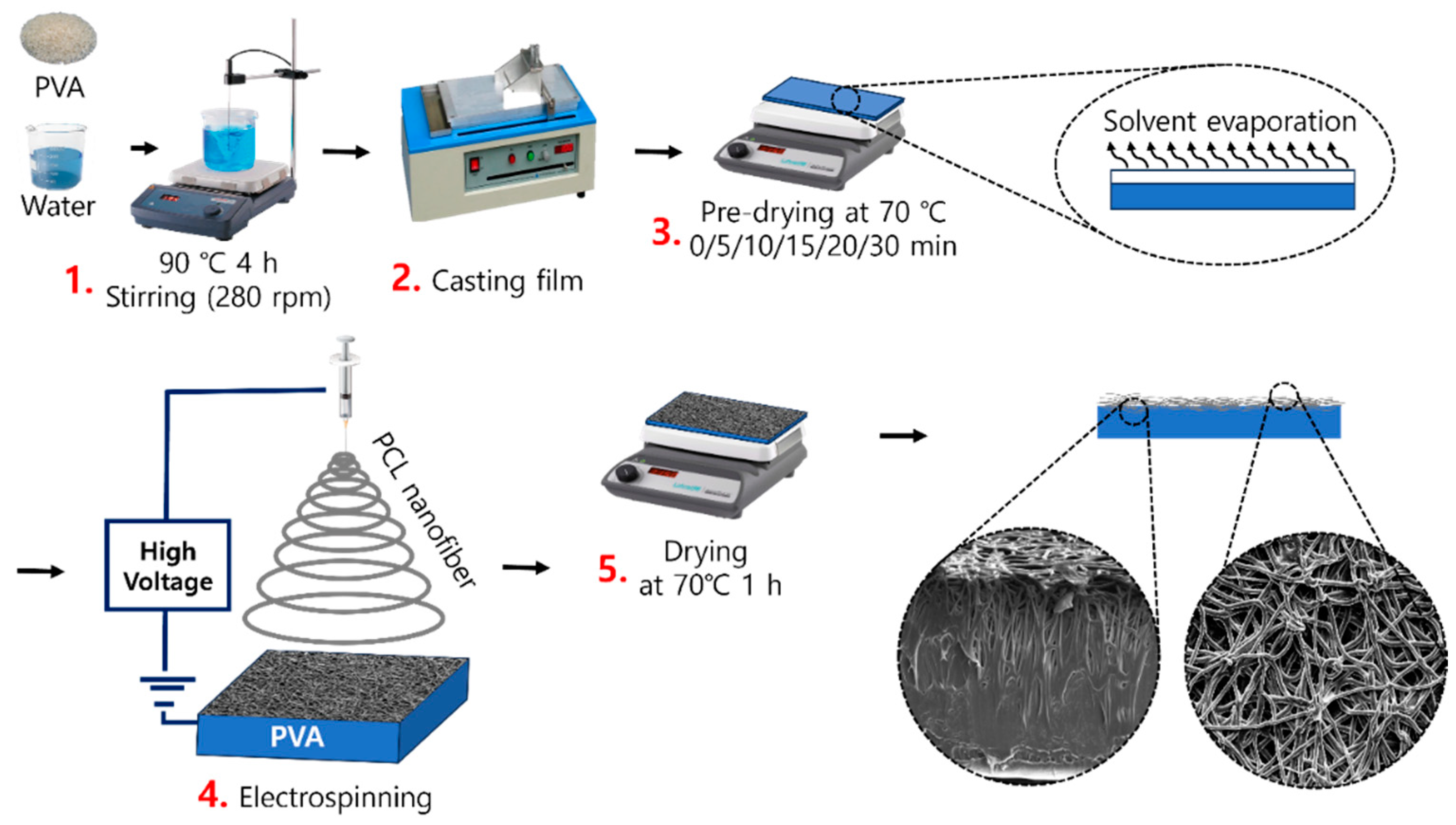
2.2. Sample Preparation
2.3. Characterization
2.3.1. FTIR Analysis
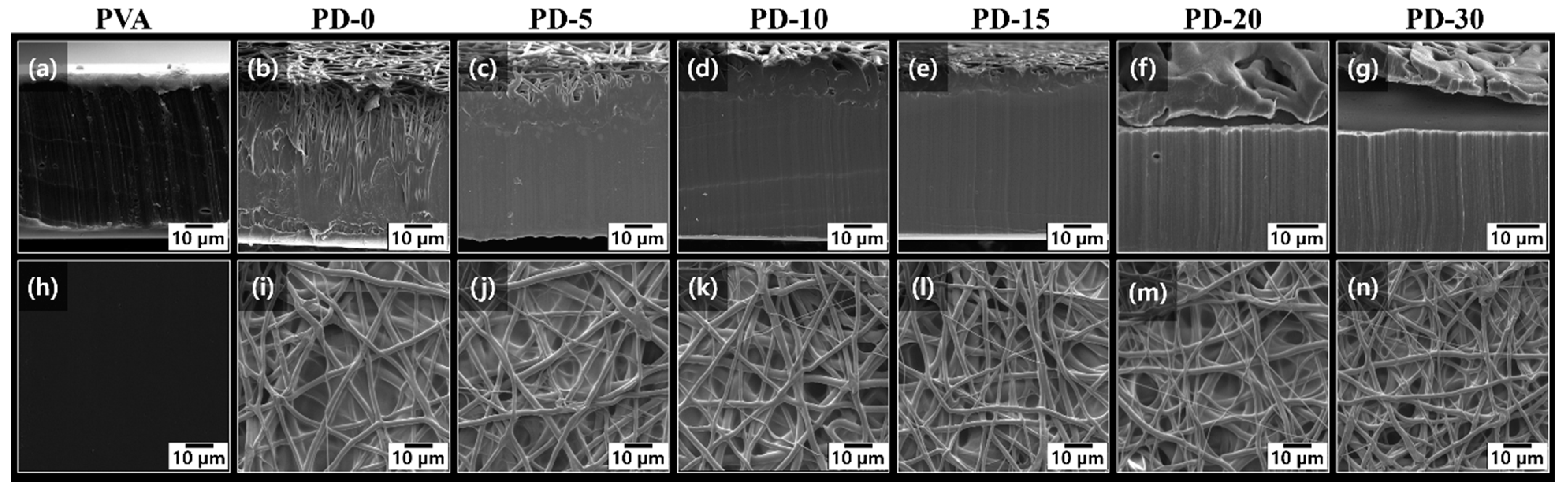
2.3.2. Morphology
2.3.3. Water Contact Angle (WCA)
2.3.4. Mechanical Properties
2.3.5. Thermogravimetric Analysis (TGA)
2.3.6. Oxygen Transmission Rate (OTR)
3. Discussion
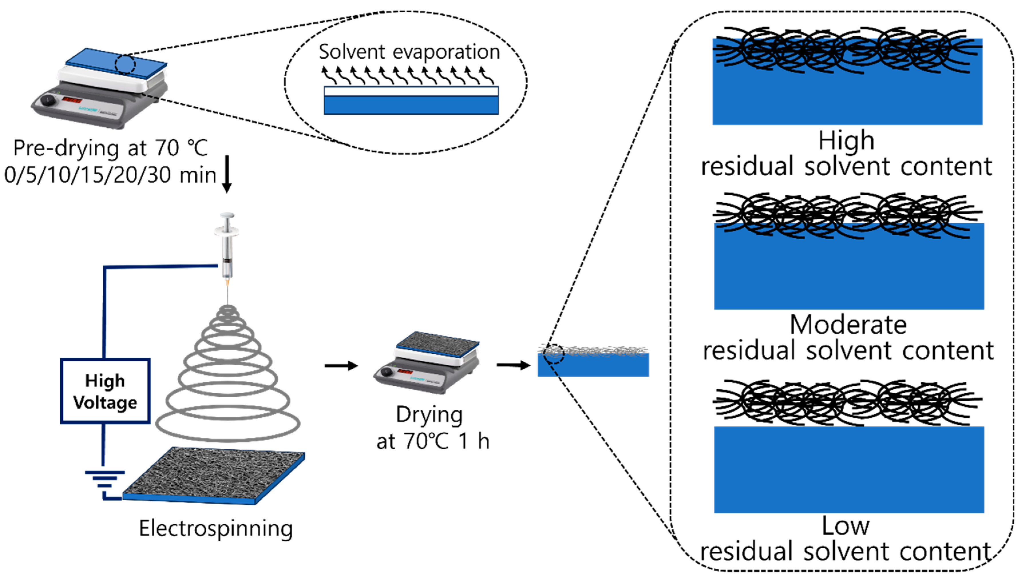
3.1. Effect of Predrying Time on the Residual Solvent and Penetration Depth of PCL Nanofibers onto the PVA Film
3.2. ATR-FTIR
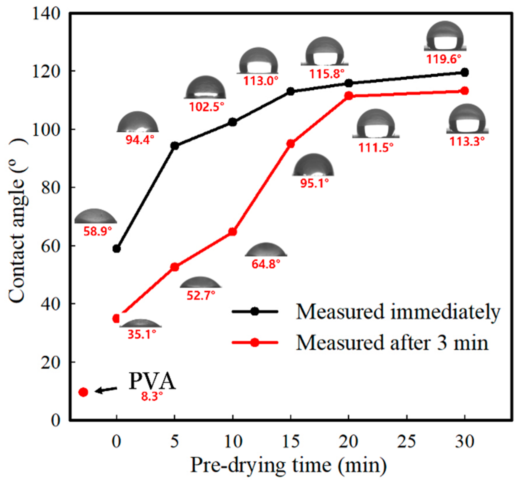
3.3. WCA
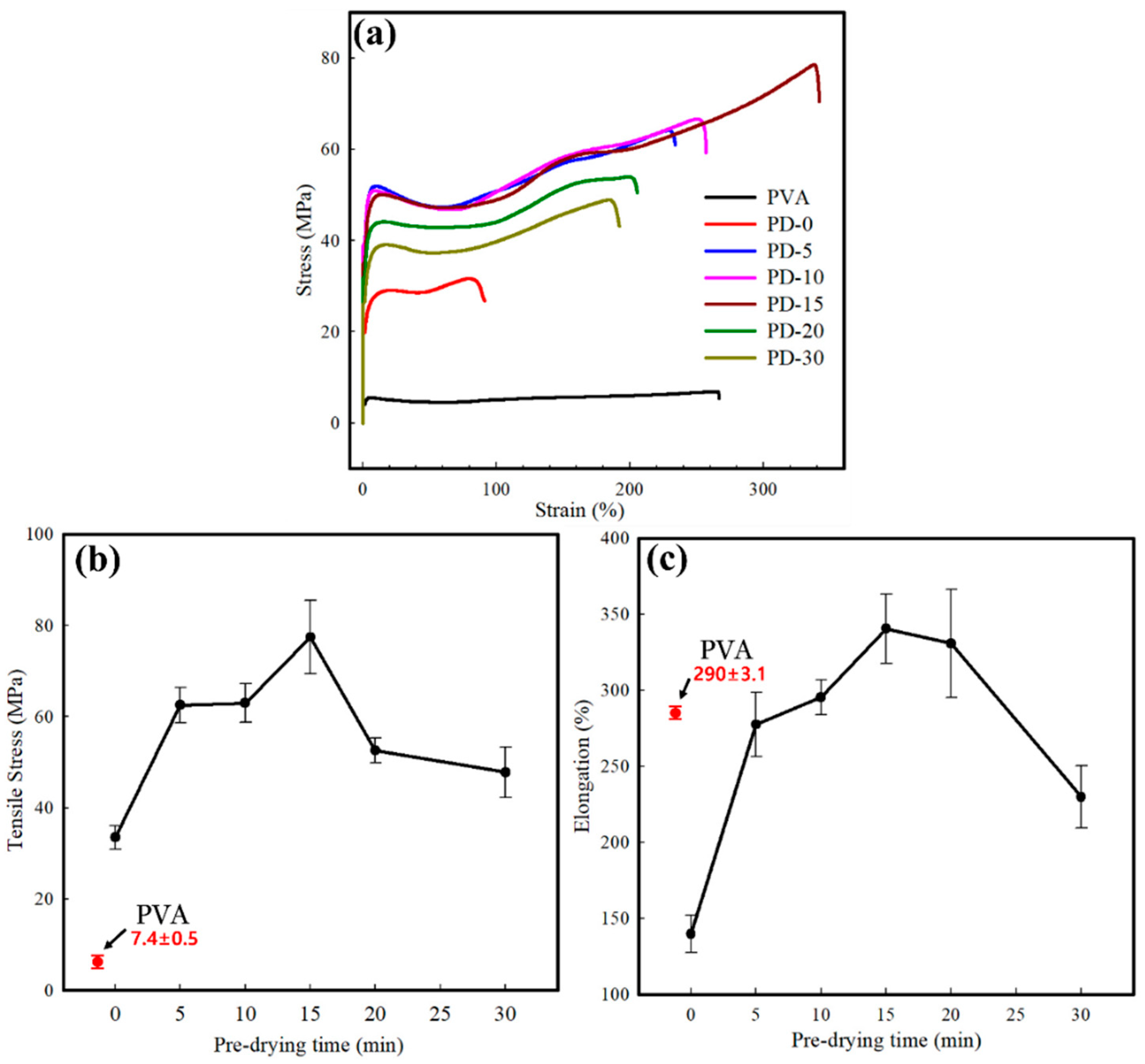
3.4. Mechanical Properties
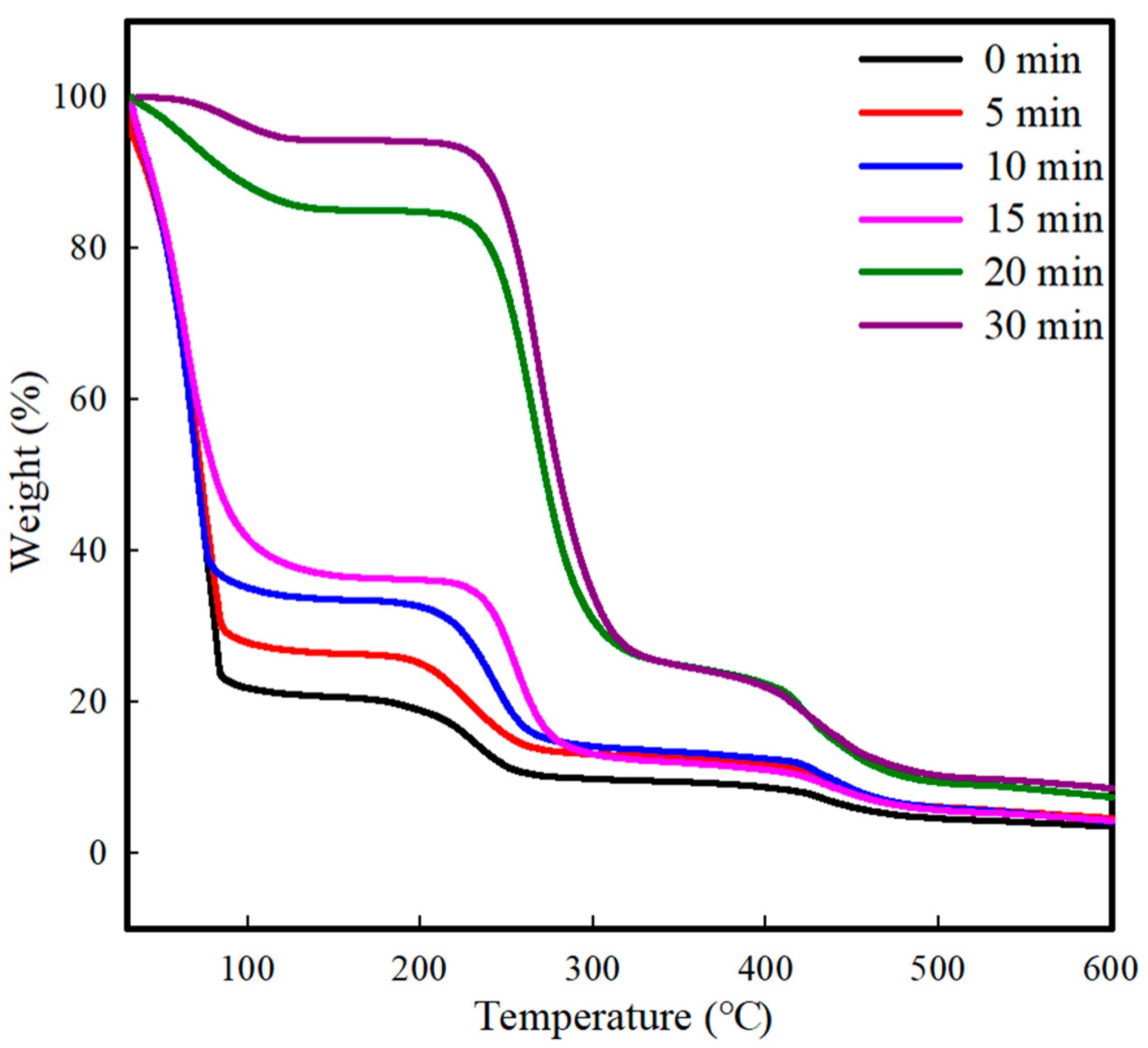
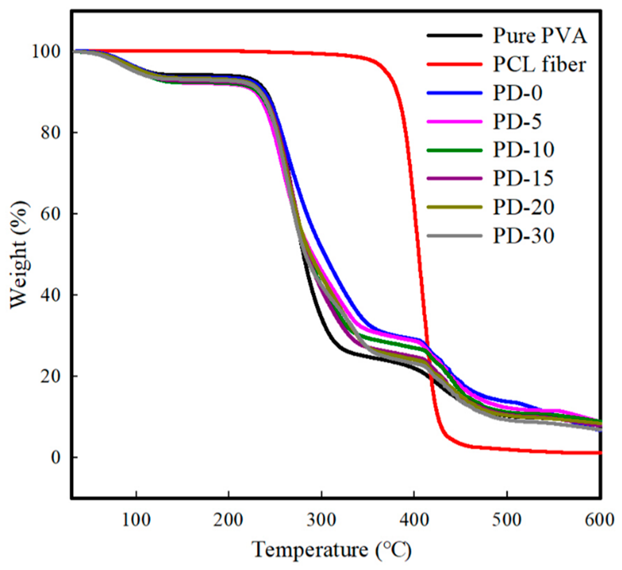
3.5. Thermal Properties
3.6. Oxygen Barrier Properties
4. Conclusions
Author Contributions
Funding
Institutional Review Board Statement
Informed Consent Statement
Data Availability Statement
Conflicts of Interest
References
- Liu, X. The impact of China’s high-quality development of energy on carbon neutrality. Energy Rep. 2023, 9, 2665–2675. [Google Scholar] [CrossRef]
- Teng, X.; Zhuang, W.; Liu, F.-p.; Chang, T.-h.; Chiu, Y.-h. China’s path of carbon neutralization to develop green energy and improve energy efficiency. Renew. Energy 2023, 206, 397–408. [Google Scholar] [CrossRef]
- Raza, S.; Ghasali, E.; Raza, M.; Chen, C.; Li, B.; Orooji, Y.; Lin, H.; Karaman, C.; Maleh, H.K.; Erk, N. Advances in technology and utilization of natural resources for achieving carbon neutrality and a sustainable solution to neutral environment. Environ. Res. 2022, 220, 115135. [Google Scholar] [CrossRef]
- Zhou, X.; Wang, G.; Li, D.; Wang, Q.; Zhu, K.; Hao, Y.; Xu, Y.; Li, N. Shape-memory polyurethane elastomer originated from waste PET plastic and their composites with carbon nanotube for sensitive and stretchable strain sensor. Compos. Part A Appl. Sci. Manuf. 2023, 177, 107920. [Google Scholar] [CrossRef]
- Jafarzadeh, S.; Forough, M.; Amjadi, S.; Javan Kouzegaran, V.; Almasi, H.; Garavand, F.; Zargar, M. Plant protein-based nanocomposite films: A review on the used nanomaterials, characteristics, and food packaging applications. Crit. Rev. Food Sci. Nutr. 2022, 63, 9667–9693. [Google Scholar] [CrossRef] [PubMed]
- Arunan, I.; Crawford, R.H. Greenhouse gas emissions associated with food packaging for online food delivery services in Australia. Resour. Conserv. Recycl. 2021, 168, 105299. [Google Scholar] [CrossRef]
- Leslie, H.A.; Van Velzen, M.J.; Brandsma, S.H.; Vethaak, A.D.; Garcia-Vallejo, J.J.; Lamoree, M.H. Discovery and quantification of plastic particle pollution in human blood. Environ. Int. 2022, 163, 107199. [Google Scholar] [CrossRef] [PubMed]
- Zhang, W.; Cao, J.; Jiang, W. Effect of different cation in situ cross-linking on the properties of pectin-thymol active film. Food Hydrocoll. 2022, 128, 107594. [Google Scholar] [CrossRef]
- Tan, C.J.; Tong, Y.W. The effect of protein structural conformation on nanoparticle molecular imprinting of ribonuclease a using miniemulsion polymerization. Langmuir 2007, 23, 2722–2730. [Google Scholar] [CrossRef]
- Gaaz, T.S.; Sulong, A.B.; Akhtar, M.N.; Kadhum, A.A.H.; Mohamad, A.B.; Al-Amiery, A.A. Properties and applications of polyvinyl alcohol, halloysite nanotubes and their nanocomposites. Molecules 2015, 20, 22833–22847. [Google Scholar] [CrossRef]
- Siracusa, V.; Rocculi, P.; Romani, S.; Dalla Rosa, M. Biodegradable polymers for food packaging: A review. Trends Food Sci. Technol. 2008, 19, 634–643. [Google Scholar] [CrossRef]
- Gulino, E.F.; Citarrella, M.C.; Maio, A.; Scaffaro, R. An innovative route to prepare in situ graded crosslinked PVA graphene electrospun mats for drug release. Compos. Part A Appl. Sci. Manuf. 2022, 155, 106827. [Google Scholar] [CrossRef]
- Panda, P.K.; Park, K.; Seo, J. Development of poly (vinyl alcohol)/regenerated chitosan blend film with superior barrier, antioxidant, and antibacterial properties. Prog. Org. Coat. 2023, 183, 107749. [Google Scholar] [CrossRef]
- Hong, X.; He, J.; Zou, L.; Wang, Y.; Li, Y.V. Preparation and characterization of high strength and high modulus PVA fiber via dry-wet spinning with cross-linking of boric acid. J. Appl. Polym. Sci. 2021, 138, 51394. [Google Scholar] [CrossRef]
- Sapalidis, A.A. Porous Polyvinyl alcohol membranes: Preparation methods and applications. Symmetry 2020, 12, 960. [Google Scholar] [CrossRef]
- Yang, J.M.; Panda, P.K.; Jie, C.J.; Dash, P.; Chang, Y.H. Poly (vinyl alcohol)/chitosan/sodium alginate composite blended membrane: Preparation, characterization, and water-induced shape memory phenomenon. Polym. Eng. Sci. 2022, 62, 1526–1537. [Google Scholar] [CrossRef]
- Gautam, L.; Warkar, S.G.; Ahmad, S.I.; Kant, R.; Jain, M. A review on carboxylic acid cross-linked polyvinyl alcohol: Properties and applications. Polym. Eng. Sci. 2022, 62, 225–246. [Google Scholar] [CrossRef]
- Panda, P.K.; Yang, J.-M.; Chang, Y.-H. Water-induced shape memory behavior of poly (vinyl alcohol) and p-coumaric acid-modified water-soluble chitosan blended membrane. Carbohydr. Polym. 2021, 257, 117633. [Google Scholar] [CrossRef] [PubMed]
- Wang, L.; Wei, R.; Liu, C.; Liu, X.; Li, D. Construction of alternating multilayer films with stable absorption-dominated electromagnetic shielding performance and reinforced mechanical properties via interface engineering. Compos. Part A Appl. Sci. Manuf. 2024, 176, 107862. [Google Scholar] [CrossRef]
- Frias, C.F.; Fonseca, A.C.; Coelho, J.F.; Serra, A.C. Crosslinked poly (hydroxyurethane) films from biobased carbonates: Structure-properties relationships and the influence of moisture in the mechanical properties. Prog. Org. Coat. 2024, 187, 108100. [Google Scholar] [CrossRef]
- Islam, M.S.; Ang, B.C.; Andriyana, A.; Afifi, A.M. A review on fabrication of nanofibers via electrospinning and their applications. SN Appl. Sci. 2019, 1, 1248. [Google Scholar] [CrossRef]
- Zhang, W.; Shi, Y.; Wang, B.; Han, Y.; Zhang, R. High-strength electrospun polydimethylsiloxane/polytetrafluoroethylene hybrid membranes with stable and controllable coral-like structures. Compos. Part A Appl. Sci. Manuf. 2023, 164, 107316. [Google Scholar] [CrossRef]
- Bili, O.; Elkalaaoui, K.; Boukhriss, A.; Chaoui, M.A.; Majid, S.; El Kouali, M.; Gmouh, S. Novel lightweight and flexible functional textile based on PVDF and [Im, PF6] developed via the electrospinning technique. Prog. Org. Coat. 2024, 186, 108019. [Google Scholar] [CrossRef]
- Abdul Hameed, M.M.; Mohamed Khan, S.A.P.; Thamer, B.M.; Rajkumar, N.; El-Hamshary, H.; El-Newehy, M. Electrospun nanofibers for drug delivery applications: Methods and mechanism. Polym. Adv. Technol. 2023, 34, 6–23. [Google Scholar] [CrossRef]
- Biswas, R.; Alam, M.; Sarkar, A.; Haque, M.I.; Hasan, M.M.; Hoque, M. Application of nanotechnology in food: Processing, preservation, packaging and safety assessment. Heliyon 2022, 8, e11795. [Google Scholar] [CrossRef] [PubMed]
- Zaghloul, M.M.Y.; Steel, K.; Veidt, M.; Heitzmann, M.T. Mechanical and Tribological Performances of Thermoplastic Polymers Reinforced with Glass Fibres at Variable Fibre Volume Fractions. Polymers 2023, 15, 694. [Google Scholar] [CrossRef]
- Zhao, K.; Tian, X.; Huang, N.; Zhang, K.; Wang, Y.; Zhang, Y.; Wang, W. Tunable mechanical performances of collagen-based film: Effect of collagens in different hierarchies and cellulose nanofiber. Prog. Org. Coat. 2023, 176, 107404. [Google Scholar] [CrossRef]
- Chen, G.; Liu, H. Electrospun cellulose nanofiber reinforced soybean protein isolate composite film. J. Appl. Polym. Sci. 2008, 110, 641–646. [Google Scholar] [CrossRef]
- Zhang, Y.; Deng, L.; Zhong, H.; Pan, J.; Li, Y.; Zhang, H. Superior water stability and antimicrobial activity of electrospun gluten nanofibrous films incorporated with glycerol monolaurate. Food Hydrocoll. 2020, 109, 106116. [Google Scholar] [CrossRef]
- Kang, S.; Zhao, K.; Yu, D.-G.; Zheng, X.; Huang, C. Advances in biosensing and environmental monitoring based on electrospun nanofibers. Adv. Fiber Mater. 2022, 4, 404–435. [Google Scholar] [CrossRef]
- Scaffaro, R.; Lopresti, F.; Maio, A.; Botta, L.; Rigogliuso, S.; Ghersi, G. Electrospun PCL/GO-g-PEG structures: Processing-morphology-properties relationships. Compos. Part A Appl. Sci. Manuf. 2017, 92, 97–107. [Google Scholar] [CrossRef]
- El Mouat, A.; El Assimi, T.; Raihane, M.; Ternel, J.; Bricout, H.; Monflier, E.; Tilloy, S.; Lahcini, M. Exploiting poly (ε-caprolactone) grafted from hydrohydroxymethylated sunflower oil as biodegradable coating material of water-soluble fertilizers. Prog. Org. Coat. 2023, 179, 107513. [Google Scholar] [CrossRef]
- Zou, Y.; Sun, Y.; Shi, W.; Wan, B.; Zhang, H. Dual-functional shikonin-loaded quaternized chitosan/polycaprolactone nanofibrous film with pH-sensing for active and intelligent food packaging. Food Chem. 2023, 399, 133962. [Google Scholar] [CrossRef] [PubMed]
- Shi, C.; Zhou, A.; Fang, D.; Lu, T.; Wang, J.; Song, Y.; Lyu, L.; Wu, W.; Huang, C.; Li, W. Oregano essential oil/β-cyclodextrin inclusion compound polylactic acid/polycaprolactone electrospun nanofibers for active food packaging. Chem. Eng. J. 2022, 445, 136746. [Google Scholar] [CrossRef]
- Ullah, A.; Haider, M.K.; Wang, F.-f.; Morita, S.; Kharaghani, D.; Ge, Y.; Yoshiko, Y.; Lee, J.S.; Kim, I.S. “Clay-corn-caprolactone” a novel bioactive clay polymer nanofibrous scaffold for bone tissue engineering. Appl. Clay Sci. 2022, 220, 106455. [Google Scholar] [CrossRef]
- Reis, P.; Ferreira, J.; Antunes, F.; Richardson, M. Effect of interlayer delamination on mechanical behavior of carbon/epoxy laminates. J. Compos. Mater. 2009, 43, 2609–2621. [Google Scholar] [CrossRef]
- Pommet, M.; Juntaro, J.; Heng, J.Y.; Mantalaris, A.; Lee, A.F.; Wilson, K.; Kalinka, G.; Shaffer, M.S.; Bismarck, A. Surface modification of natural fibers using bacteria: Depositing bacterial cellulose onto natural fibers to create hierarchical fiber reinforced nanocomposites. Biomacromolecules 2008, 9, 1643–1651. [Google Scholar] [CrossRef]
- Li, Y.; Yao, S.; Shi, H.; Zhang, Y.; Han, C.; Yu, Y. Enhancing the crystallization of biodegradable poly (ε-caprolactone) using a polyvinyl alcohol fiber favoring nucleation. Thermochim. Acta 2021, 706, 179065. [Google Scholar] [CrossRef]
- Shi, J.; Xu, L.; Qiu, D. Effective antifogging coating from hydrophilic/hydrophobic polymer heteronetwork. Adv. Sci. 2022, 9, 2200072. [Google Scholar] [CrossRef]
- ASTM Standard D618; Standard Practice for Conditioning Plastics for Testing. ASTM International: West Conshohocken, PA, USA, 2021. Available online: https://www.astm.org/d0618-21.html (accessed on 26 April 2024).
- Zhou, G.; Mo, L.; Zhou, C.; Wu, Y.; Lai, F.; Lv, Y.; Ma, J.; Miao, Y.-E.; Liu, T. Ultra-strong capillarity of bioinspired micro/nanotunnels in organic cathodes enabled high-performance all-organic sodium-ion full batteries. Chem. Eng. J. 2021, 420, 127597. [Google Scholar] [CrossRef]
- ASTM D638-14; Standard Test Method for Tensile Properties of Plastics. ASTM International: West Conshohocken, PA, USA, 2014. Available online: https://www.astm.org/d0638-14.html (accessed on 26 April 2024).
- ASTM D3985-1; Standard Test Method for Oxygen Gas Transmission Rate through Plastic Film and Sheeting Using a Coulometric Sensor. ASTM International: West Conshohocken, PA, USA, 2017. Available online: https://www.astm.org/d3985-17.html (accessed on 26 April 2024).
- Espíndola, S.P.; Norder, B.; Koper, G.J.; Picken, S.J. The Glass Transition Temperature of Heterogeneous Biopolymer Systems. Biomacromolecules 2023, 24, 1627–1637. [Google Scholar] [CrossRef] [PubMed]
- Hellert, C.; Wortmann, M.; Frese, N.; Grötsch, G.; Cornelißen, C.; Ehrmann, A. Adhesion of electrospun poly (Acrylonitrile) nanofibers on conductive and isolating foil substrates. Coatings 2021, 11, 249. [Google Scholar] [CrossRef]
- Prasad, N.S.; Babarao, R.; Madapusi, S.; Sridhar, S.; Choudhury, N.R.; Bhargava, S.K. Residual solvent induced physical morphology and gas permeation in polyamide-imide membrane: Experimental investigation and molecular simulations. Eur. Polym. J. 2022, 165, 111012. [Google Scholar] [CrossRef]
- Dai, Y.; Tang, Q.; Zhang, Z.; Yu, C.; Li, H.; Xu, L.; Zhang, S.; Zou, Z. Enhanced mechanical, thermal, and UV-shielding properties of poly (vinyl alcohol)/metal–organic framework nanocomposites. RSC Adv. 2018, 8, 38681–38688. [Google Scholar] [CrossRef] [PubMed]
- Yang, N.; Ying, L.; Li, K.; Chen, F.; Zhao, F.; Sun, Z.; Feng, L.; Liu, J. Biodegradable Mulching Films Based on Polycaprolactone and Its Porous Structure Construction. Polymers 2022, 14, 5340. [Google Scholar] [CrossRef]
- Hoang, B.N.; Nguyen, T.T.; Bui, Q.P.T.; Bach, L.G.; Vo, D.V.N.; Trinh, C.D.; Bui, X.T.; Nguyen, T.D. Enhanced selective adsorption of cation organic dyes on polyvinyl alcohol/agar/maltodextrin water-resistance biomembrane. J. Appl. Polym. Sci. 2020, 137, 48904. [Google Scholar] [CrossRef]
- Alazzawi, M.; Kadim Abid Alsahib, N.; Turkoglu Sasmazel, H. Core/Shell glycine-polyvinyl alcohol/polycaprolactone nanofibrous membrane intended for guided bone regeneration: Development and characterization. Coatings 2021, 11, 1130. [Google Scholar] [CrossRef]
- Mouro, C.; Simões, M.; Gouveia, I.C. Emulsion electrospun fiber mats of PCL/PVA/chitosan and eugenol for wound dressing applications. Adv. Polym. Technol. 2019, 2019, 9859506. [Google Scholar] [CrossRef]
- Quan, B.; Wang, J.; Li, Y.; Sui, M.; Xie, H.; Liu, Z.; Wu, H.; Lu, X.; Tong, Y. Cellulose nanofibrous/MXene aerogel encapsulated phase change composites with excellent thermal energy conversion and storage capacity. Energy 2023, 262, 125505. [Google Scholar] [CrossRef]
- Zhao, D.; Liu, X.; Shen, Z. Effect of oxygen-containing functional groups on the wettability of coal through DFT and MD simulation. Arab. J. Chem. 2023, 16, 104606. [Google Scholar] [CrossRef]
- Yudianti, R.; Karina, M. Development of nanocomposites from bacterial cellulose and poly (vinyl alcohol) using casting-drying method. Procedia Chem. 2012, 4, 73–79. [Google Scholar]
- Mugica, G.W.; Tovio, D.O.; Cuyas, J.C.; González, A.C. Effect of porosity on the tensile properties of low ductility aluminum alloys. Mater. Res. 2004, 7, 221–229. [Google Scholar] [CrossRef]
- Chen, Z.; Zhu, D.; Tong, F.; Lu, X.; Lu, Q. Low dielectric constant polyimide hybrid films prepared by in situ blow-balloon method. ACS Appl. Polym. Mater. 2019, 1, 2189–2196. [Google Scholar] [CrossRef]
- Xu, J.; Xia, R.; Zheng, L.; Yuan, T.; Sun, R. Plasticized hemicelluloses/chitosan-based edible films reinforced by cellulose nanofiber with enhanced mechanical properties. Carbohydr. Polym. 2019, 224, 115164. [Google Scholar] [CrossRef]
- Bacha, E.G.; Demsash, H.D.; Shumi, L.D.; Debesa, B.E. Investigation on Reinforcement Effects of Nanocellulose on the Mechanical Properties, Water Absorption Capacity, Biodegradability, Optical Properties, and Thermal Stability of a Polyvinyl Alcohol Nanocomposite Film. Adv. Polym. Technol. 2022, 2022, 6947591. [Google Scholar] [CrossRef]
- Jung, J.; Sodano, H.A. High strength epoxy nanocomposites reinforced by epoxy functionalized aramid nanofibers. Polymer 2020, 195, 122438. [Google Scholar] [CrossRef]
- Zhang, Z.; Qu, Z.; Xu, L.; Liu, R.; Zhang, P.; Zhang, Z.; Langdon, T.G. Relationship between strength and uniform elongation of metals based on an exponential hardening law. Acta Mater. 2022, 231, 117866. [Google Scholar] [CrossRef]
- Soliman, S.; Pagliari, S.; Rinaldi, A.; Forte, G.; Fiaccavento, R.; Pagliari, F.; Franzese, O.; Minieri, M.; Di Nardo, P.; Licoccia, S. Multiscale three-dimensional scaffolds for soft tissue engineering via multimodal electrospinning. Acta Biomater. 2010, 6, 1227–1237. [Google Scholar] [CrossRef] [PubMed]
- Bryaskova, R.; Georgieva, N.; Andreeva, T.; Tzoneva, R. Cell adhesive behavior of PVA-based hybrid materials with silver nanoparticles. Surf. Coat. Technol. 2013, 235, 186–191. [Google Scholar] [CrossRef]
- Zhao, M.; Pan, W.; Wan, C.; Qu, Z.; Li, Z.; Yang, J. Defect engineering in development of low thermal conductivity materials: A review. J. Eur. Ceram. Soc. 2017, 37, 1–13. [Google Scholar] [CrossRef]
- Lim, M.; Kwon, H.; Kim, D.; Seo, J.; Han, H.; Khan, S.B. Highly-enhanced water resistant and oxygen barrier properties of cross-linked poly (vinyl alcohol) hybrid films for packaging applications. Prog. Org. Coat. 2015, 85, 68–75. [Google Scholar] [CrossRef]
- Lim, M.; Kim, D.; Seo, J.; Han, H. Preparation and properties of poly (vinyl alcohol)/vinyltrimethoxysilane (PVA/VTMS) hybrid films with enhanced thermal stability and oxygen barrier properties. Macromol. Res. 2014, 22, 1096–1103. [Google Scholar] [CrossRef]
- Syverud, K.; Stenius, P. Strength and barrier properties of MFC films. Cellulose 2009, 16, 75–85. [Google Scholar] [CrossRef]
- Möller, M.W.; Kunz, D.A.; Lunkenbein, T.; Sommer, S.; Nennemann, A.; Breu, J. UV-cured, flexible, and transparent nanocomposite coating with remarkable oxygen barrier. Adv. Mater. 2012, 24, 2142–2147. [Google Scholar] [CrossRef] [PubMed]
- Wongphan, P.; Panrong, T.; Harnkarnsujarit, N. Effect of different modified starches on physical, morphological, thermomechanical, barrier and biodegradation properties of cassava starch and polybutylene adipate terephthalate blend film. Food Packag. Shelf Life 2022, 32, 100844. [Google Scholar] [CrossRef]
- Mittal, V. Mechanical and gas permeation properties of compatibilized polypropylene–layered silicate nanocomposites. J. Appl. Polym. Sci. 2008, 107, 1350–1361. [Google Scholar] [CrossRef]


| Sample | Predrying Time (min) | Electrospinning Time (s) | Residual Solvent Content Measured by TGA (%) |
|---|---|---|---|
| PD-0 | 0 | 30 | 79.4 ± 5.6 a |
| PD-5 | 5 | 30 | 73.6 ± 5.8 ab |
| PD-10 | 10 | 30 | 66.5 ± 4.0 bc |
| PD-15 | 15 | 30 | 63.5 ± 3.5 c |
| PD-20 | 20 | 30 | 15.0 ± 0.2 d |
| PD-30 | 30 | 30 | 5.8 ± 0.3 e |
| Sample Code | Tensile Strength (MPa) | Elongation at Break (%) | OTR (cc/m2·day) |
|---|---|---|---|
| Pure PVA | 7.4 ± 0.5 e | 290.0 ± 3.1 c | 5.5 ± 0.1 a |
| PD-0 | 33.5 ± 2.5 d | 139.8 ± 12.2 e | 3.6 ± 0.3 b |
| PD-5 | 62.5 ± 3.8 b | 277.4 ± 21.1 c | 1.8 ± 0.6 c |
| PD-10 | 62.9 ± 4.2 b | 295.4 ± 11.4 bc | 1.6 ± 0.6 c |
| PD-15 | 77.4 ± 7.9 a | 340.5 ± 22.9 a | 1.9 ± 0.5 c |
| PD-20 | 52.5 ± 2.7 c | 330.8 ± 35.5 ab | 1.9 ± 0.3 c |
| PD-30 | 47.8 ± 5.5 c | 230.0 ± 20.4 d | 3.4 ± 0.9 b |
Disclaimer/Publisher’s Note: The statements, opinions and data contained in all publications are solely those of the individual author(s) and contributor(s) and not of MDPI and/or the editor(s). MDPI and/or the editor(s) disclaim responsibility for any injury to people or property resulting from any ideas, methods, instructions or products referred to in the content. |
© 2024 by the authors. Licensee MDPI, Basel, Switzerland. This article is an open access article distributed under the terms and conditions of the Creative Commons Attribution (CC BY) license (https://creativecommons.org/licenses/by/4.0/).
Share and Cite
Ahn, K.; Park, K.; Sadeghi, K.; Seo, J. New Surface Modification of Hydrophilic Polyvinyl Alcohol via Predrying and Electrospinning of Hydrophobic Polycaprolactone Nanofibers. Foods 2024, 13, 1385. https://doi.org/10.3390/foods13091385
Ahn K, Park K, Sadeghi K, Seo J. New Surface Modification of Hydrophilic Polyvinyl Alcohol via Predrying and Electrospinning of Hydrophobic Polycaprolactone Nanofibers. Foods. 2024; 13(9):1385. https://doi.org/10.3390/foods13091385
Chicago/Turabian StyleAhn, Kihyeon, Kitae Park, Kambiz Sadeghi, and Jongchul Seo. 2024. "New Surface Modification of Hydrophilic Polyvinyl Alcohol via Predrying and Electrospinning of Hydrophobic Polycaprolactone Nanofibers" Foods 13, no. 9: 1385. https://doi.org/10.3390/foods13091385
APA StyleAhn, K., Park, K., Sadeghi, K., & Seo, J. (2024). New Surface Modification of Hydrophilic Polyvinyl Alcohol via Predrying and Electrospinning of Hydrophobic Polycaprolactone Nanofibers. Foods, 13(9), 1385. https://doi.org/10.3390/foods13091385