Article

Antibacterial Mechanism of Linalool against Pseudomonas fragi: A Transcriptomic Study

1
School of Precision Instrument and Opto-Electronics Engineering, Tianjin University, 92 Weijin Road, Tianjin 300072, China
2
Hainan University-HSF/LWL Collaborative Innovation Laboratory, School of Food Sciences & Engineering, Hainan University, 58 People Road, Haikou 570228, China
3
Tianjin Engineering Research Center of Civil Aviation Energy Environment and Green Development, Civil Aviation University of China, 2898 Jinbei Road, Tianjin 300300, China
4
Innovation Department, Hainan Province Hochung Corporation Areca-Nut Deep Processing Technology Research Institute, 45 Yuesong Road, Dingan 571299, China
*
Author to whom correspondence should be addressed.
These authors contributed equally to this work.
Foods 2022, 11(14), 2058; https://doi.org/10.3390/foods11142058
Submission received: 9 June 2022 / Revised: 2 July 2022 / Accepted: 6 July 2022 / Published: 12 July 2022

Abstract

:
Pseudomonas fragi is the dominant spoilage bacterium that causes the deterioration of chilled meat. Our previous study showed that linalool has potent antibacterial activity against P. fragi, but its antibacterial mechanism is unclear. To explore the antibacterial mechanism of linalool against P. fragi, this study used RNA-seq technology to perform transcriptome analysis of P. fragi samples with or without linalool treatment (1.5 mL/L) for 2 h. The results showed that linalool treatment disrupted the extracellular lipopolysaccharide synthesis pathway in P. fragi and activated fatty acid metabolism and ribosomal function to compensate for cell membrane damage. The energy metabolism of P. fragi was severely disturbed by linalool, and multiple ATP synthases and ATP transportases were overexpressed in the cells but could not guarantee the consumption of ATP. The simultaneous overexpression of multiple ribosomal functional proteins and transporters may also place an additional burden on cells and cause them to collapse.

1. Introduction

“Zero Hunger” is one of the United Nations Sustainable Development Goals in 2015-2030 [1], and 2022 is an intermediate time-point in achieving this goal. Unfortunately, according to the report on the official website of the United Nations, this goal may not be achieved by 2030, which means that the issue of food security still requires great attention from all parties. Approximately 1.3 billion tons of food are destroyed or wasted for various reasons every year [2]. The loss of meat and meat products accounts for approximately 20% of this loss, and microorganisms are the main factor causing meat spoilage. Pseudomonas fragi has been reported to be the dominant spoilage bacterium in chilled beef, pork and seafood under aerobic storage conditions [3]. The presence of P. fragi can cause adverse changes in the colour, odour, viscosity and nutritional value of meat/meat products, and the most significant spoilage performance is the production of various volatile compounds [4]. Multiple reports indicated that the production of aldehydes, ketones and alcohol was associated with P. fragi. Papadopoulou et al. (2020) found abnormally elevated levels of ethyl caproate, ethyl caprylate and ethyl decanoate in beef contaminated with P. fragi, and these compounds gave beef an undesired fruity off-taste [5]. Therefore, taking effective measures to prevent P. fragi contamination can reduce the loss of meat.
Preservatives made from natural products showed good antibacterial properties. Linalool is a ring-free monoterpene that exists in the essential oils of many plants and demonstrates broad-spectrum antibacterial activity. According to the reports of Chen et al., He et al. and Varia et al., linalool can inhibit the physiological activities of yeast, Staphylococcus and Aspergillus niger [6,7,8]. The disruption of bacterial cell membranes by linalool resulting in the leakage of cellular contents is one of the well-established antibacterial mechanisms of linalool. In our previous studies, we found that linalool can exert a bacteriostatic effect by inhibiting the respiratory metabolism of Listeria monocytogenes, Pseudomonas aeruginosa, Shewanella putrefaciens, and Pseudomonas fluorescens, which explains the damage of linalool to bacteria from the perspective of energy metabolism [9,10,11]. While the effect of linalool on the genetic material in cells cannot be ignored, Ghosh et al. (2019) found that the combination of linalool with vitamin C and copper (LVC) promoted cell oxidation, and the free radicals generated by oxidation led to bacterial DNA damage, which achieved a bacteriostatic effect [12]. Noh et al. (2019) reported that the anticancer effect of linalool in prostate cancer cells is also related to the damage of DNA structure [13]. Slight DNA damage does not prevent microbes from multiplying but alters the expression of genes in the cell [14]. In other words, if the inhibitory concentration is not enough to kill bacterial cells, transcriptomic research is an effective means for determining the effect of antibacterial substances on bacterial cells.
The research object of transcriptome sequencing is all RNA transcripts contained in a specific cell or tissue in a certain state [15]. The sequence information of almost all transcripts in cells or tissues is comprehensively obtained by high-throughput sequencing. Transcriptome sequencing is a supportive technology (RNA-seq technology) for studying gene function and structure [16]. In recent years, the rapid development of RNA-seq technology has provided many valuable insights into the influence of natural products on bacteria-related genes. Yu et al. (2021) found that Cinnamomum camphora essential oil hindered the normal growth of E. coli by inhibiting metabolism and adhesion through RNA-seq analysis [17]. Park et al. (2019) found that the intervention of erythorbyl laurate (EL) upregulated Staphylococcus aureus cell wall gene expression and demonstrated that EL is a good material to act as a cell wall activator or a cell membrane targeting agent [18]. In addition, transcriptomics was used as a tool to discover new antimicrobial targets. Bacteria exposed to multiple bacteriostatic substances or antibiotics were subjected to global gene expression profiling using RNA-seq, and substances with broad targets were chosen as potential bacteriostatic agents [19,20].
In our previous work, it has been found that linalool has a good antibacterial effect on P. fragi [21], but the specific antibacterial mechanism is still unclear. In this study, the antibacterial mechanism of linalool against P. fragi was obtained by performing transcriptome analysis of P. fragi samples with or without linalool treatment (1.5 mL/L) for 2 h. This provides a theoretical basis for the application of linalool in food preservation.

2. Materials and Methods

2.1. Cells and Culture Conditions

P. fragi (ATCC 4973) was obtained from the China General Microorganism Culture Collection and Management Center. P. fragi was inoculated into a nutrient broth agar culture medium (NA) for passage activation. Then, P. fragi was inoculated in a liquid nutrient broth culture medium (NB) and cultured at 37 °C for 18 h to reach the logarithmic growth phase. The bacterial solution was centrifuged at 6000× g for 10 min, and the bacterial pellet was collected. The cells were washed twice with sterile normal saline and resuspended. Then, the bacterial concentration was adjusted to 106~107 CFU/mL by McFarland turbidimetry.
The activated P. fragi solution was added to NB and cultured at 37 °C for 18 h. Then, linalool was added to the treated group so that the concentration of linalool in the system was 1.5 mL/L (Minimum inhibitory concentration, MIC), which was confirmed in our previous study. An equal amount of sterile water was added to the control group. After treatment for 2 h at 37 °C, the cells were collected by centrifugation (6000 r/min, 4 °C, 10 min) and then washed 3 times with sterile phosphate-buffered saline (PBS) (0.1 mol/L, pH 7.4). Then, the clean cell pellets were quickly frozen in liquid nitrogen and stored at −80 °C until use.

2.2. RNA Isolation and cDNA Library Construction

Total mRNA was isolated from harvested cells using an RNA isolation kit (DP430, Qiagen, Hilden, Germany) and a Ribo-Zero rRNA Removal Kit (Bacteria, Epicentre, Madison, WI, USA), and then randomly fragmented into short fragments by adding a fragmentation buffer. The first strand of cDNA was synthesized in the Moloney Murine Leukaemia Virus (M-MuLV) reverse transcriptase system using the fragmented mRNA as a template and random oligonucleotides as primers. Subsequently, the RNA strand was degraded by RNaseH, and the second strand of cDNA was synthesized from dNTPs under the DNA polymerase I system. The second-strand cDNA was purified with a PCR purification kit (QIAQuick PCR Purification Kit, Qiagen, Hilden, Germany). The purified cDNA was end-repaired, A-tailed, and ligated with sequencing adapters. Then, AMPure XP beads (Beckman Coulter, Brea, CA, USA) were used to screen cDNAs of approximately 370–420 bp. The USER enzyme was used to degrade the second strand of cDNA containing U; then, PCR amplification was performed, and, finally, the library was obtained.

2.3. Transcriptome Sequencing and Quantification

The constructed library was sequenced using an Illumina NovaSeq 6000 (Illumina, San Diego, CA, USA). All data were stripped of sequences containing linkers, unknown sequences, and low-quality sequences. The clean reads were subjected to genomic mapping analysis using Bowtie2 software (Version 2.4.4, Langmead B, Washington, DC, USA). Quantitative analysis of gene expression in each sample was performed using subread software.

2.4. Differentially Expressed Gene (DEG) Screening and Analysis

Fragments per kilobase million (FPKM) were used as normalised values for gene expression across all samples. Then, the DEGs between the treated group and the control group were screened out according to the absolute value of log2 (fold change) > 0 along with Padj < 0.05. The DEGs were subjected to Gene Ontology (GO) functional enrichment analysis (http://geneontology.org/, 1 September 2021) and Kyoto Encyclopedia of Genes and Genomes (KEGG) pathway enrichment analysis (http://www.genome.jp/kegg/, 15 September 2021).

3. Results

3.1. Data Processing and Analysis

A total of 134,788,572 raw reads were collected from the Illumina sequencing platform, and 133,584,990 reads (99.11%) were considered clean reads after data filtering, sequencing error rate checks, and GC content distribution checks. The Q30 mass fraction values of samples in the two groups were both above 90%, and the GC content was above 56%. After comparing the reads of each sample with the reference genome, the proportion of all samples successfully aligned to the genome was found to be higher than 94.8%, and the proportion of multiple mapped reads did not exceed 1.8%. These results indicated that the quality of this sequencing was sufficiently high for subsequent analysis.

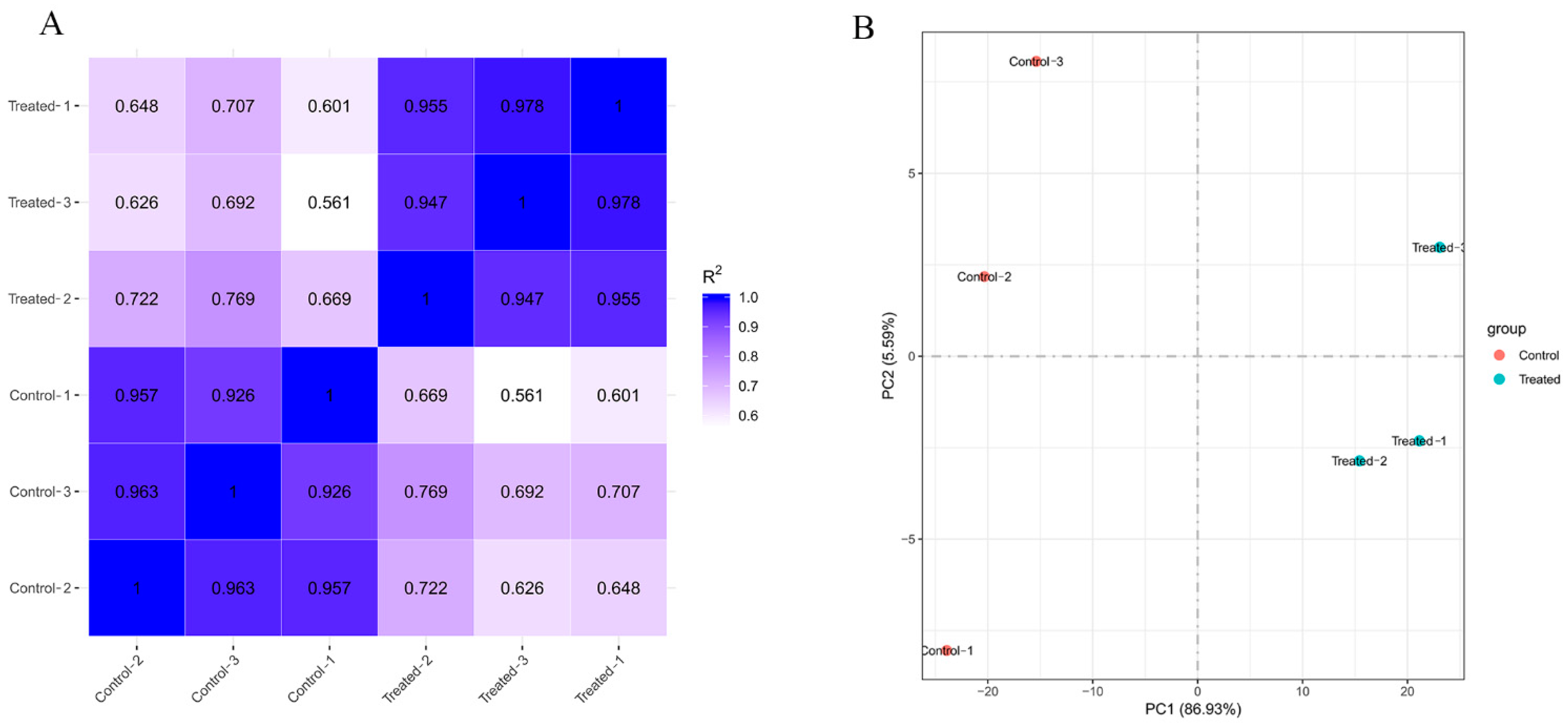
3.2. Diversity Analysis
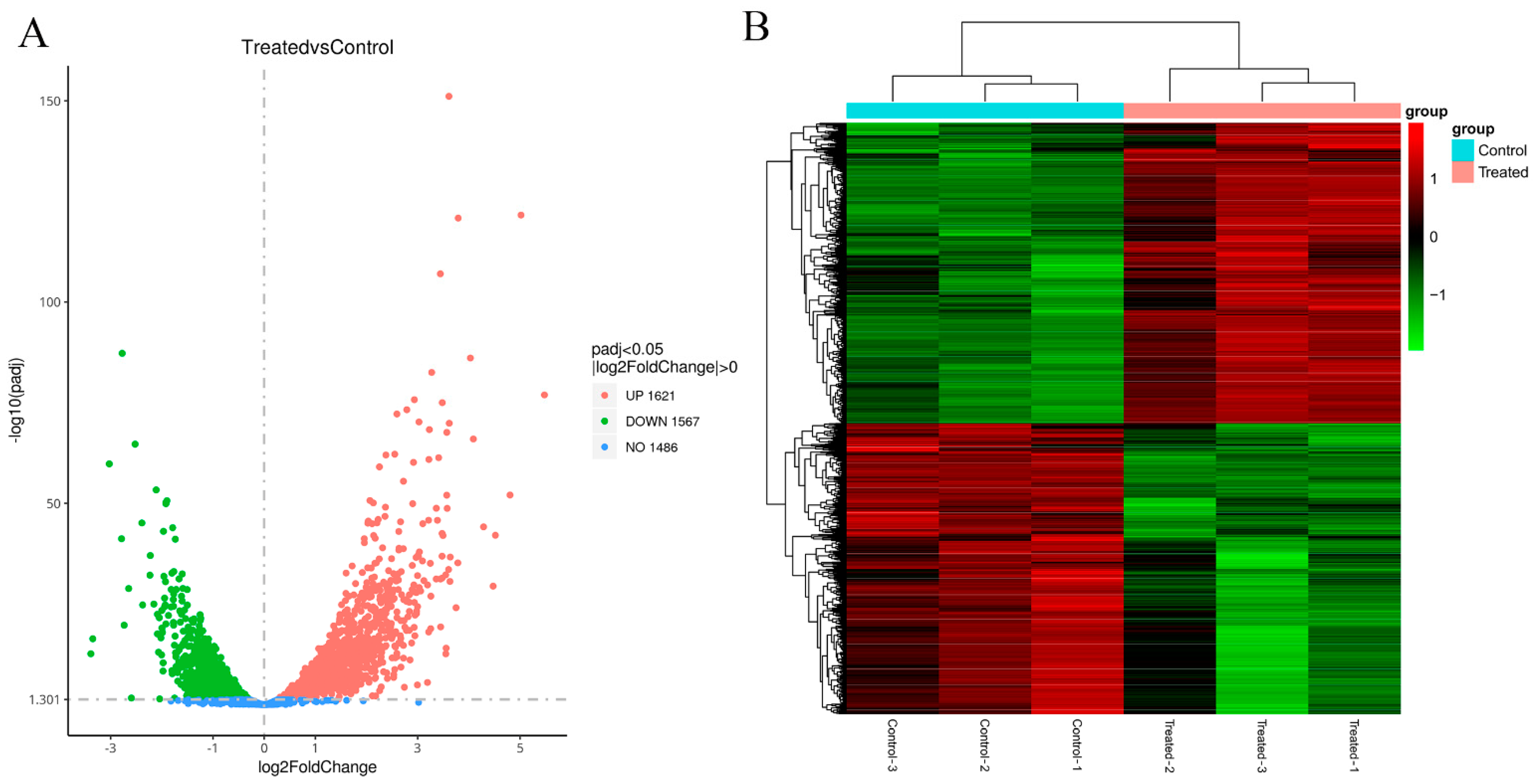
The correlation of gene expression levels among samples is an important indicator for testing the reliability of experiments. The similarity of expression patterns between samples is proportional to the degree of correlation coefficient distance 1. As shown in Figure 1A, the correlation between the samples was greater than 0.94 in the treated group and the control group, which indicated that the parallelism of the samples was good and that the experimental data obtained were reliable. However, the correlation of the treated group vs. the control group was 0.561–0.769, indicating that linalool treatment had a significant effect on the gene expression of P. fragi. The PCA score plot (Figure 1B) also showed the same results: the samples in the treated group and the control group were separated, which is a manifestation of the difference in gene expression levels between the two groups.

3.3. DEGs

DESeq2 software was used to analyse gene expression differences, and |log2 (fold change)| > 0 and Padj < 0.05 were selected as the criteria for screening DEGs. Volcano plots (Figure 2A) were created to show the differential gene distribution in the treated group vs. the control group, with upregulated genes in red and downregulated genes in green. A total of 3188 DEGs were identified between the treated group and the control group, of which 1567 DEGs were downregulated and 1621 DEGs were upregulated in the treated group.
However, when the top 1% of DEGs were screened in Table 1, 4 DEGs were downregulated, and 28 DEGs were upregulated. Table 1 shows that linalool regulated 13 enzyme-related genes, including N-acetyltransferase, sulphate adenyltransferase subunit CysD, ATP synthase subunit, sulphate adenylyltransferase subunit CysN, malate dehydrogenase, thioredoxin-disulphide reductase, ferredoxin-NADP reductase, ADP-forming succinate-CoA ligase subunit beta, quinone reductase, transglycosylase, methylcrotonoyl-CoA carboxylase, serine protease, and dienelactone hydrolase. Some functional protein genes that assemble ribosomes were also regulated, such as rpsT and groL (30S ribosomal protein and chaperonin GroEL). Ribosomal proteins can affect the synthesis of various enzymes and the formation of cell membranes. In addition, the expression levels of six transporters were regulated by linalool, including the biopolymer transporter ExbD, extracellular solute-binding protein, phosphate ABC transporter substrate-binding protein PstS, ABC transporter substrate-binding protein, MotA/TolQ/ExbB proton channel family proteins, and putative porins.
The DEGs were clustered using hierarchical clustering, and the genes or samples with similar expression patterns in the heatmap (Figure 2B) were clustered together. The results showed that the responses of the samples after linalool intervention were consistent. The effect of linalool on the activity of these enzyme-related genes should be discussed in further studies.

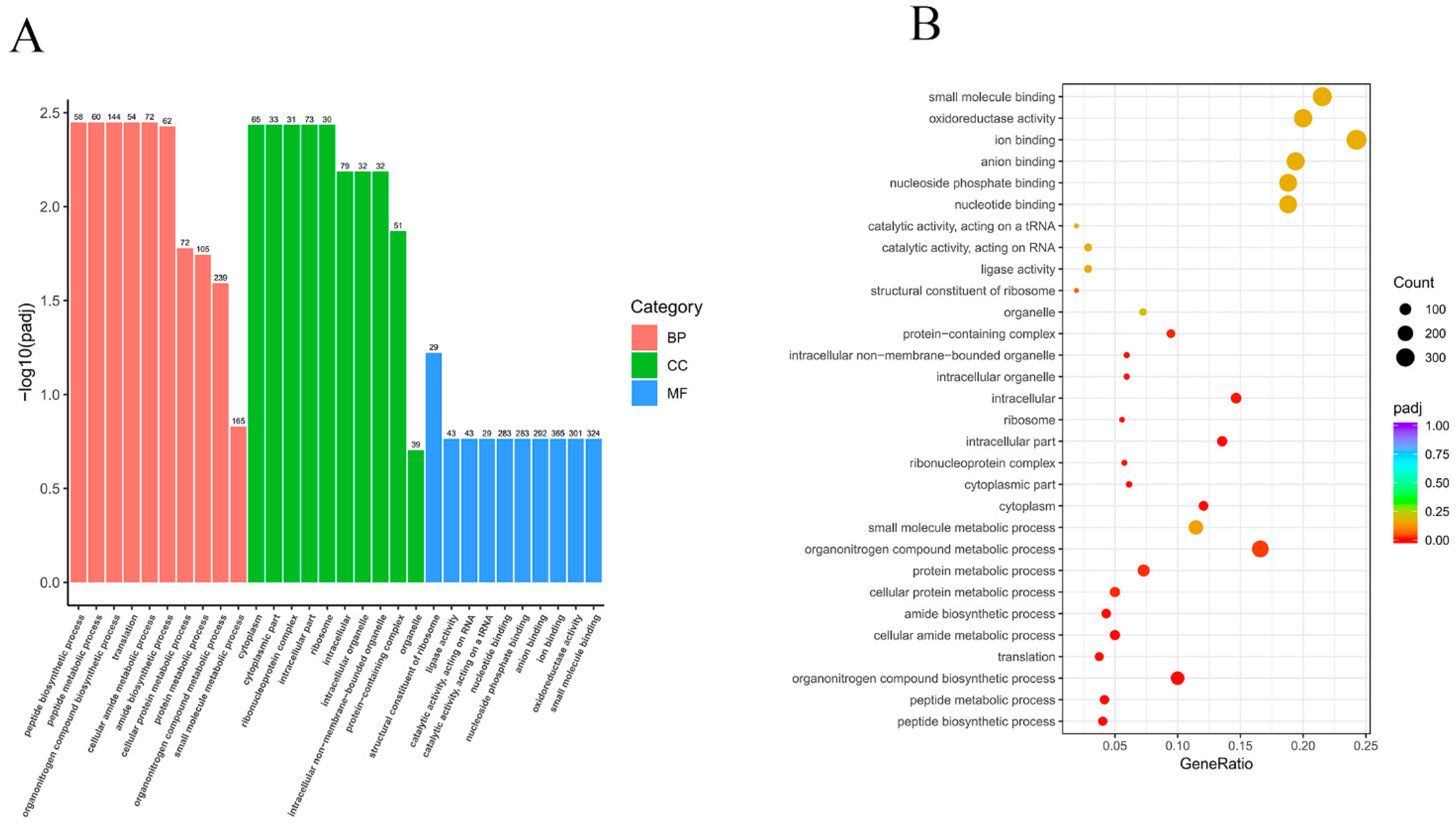
3.4. GO Enrichment Analysis

GO enrichment analysis utilises a comprehensive database describing gene function, which is divided into three parts: biological process (BP), cellular component (CC), and molecular function (MF). For GO functional enrichment, Padj < 0.05 was used as the threshold for significant enrichment. Here, a total of 232 BP terms, 27 CC terms, and 156 MF terms were confirmed. The top 10 significant terms in each section (30 in total) were selected to form a GO bar graph (Figure 3A). The biological processes enriched for the identified DEGs included peptide biosynthesis and metabolism (GO:0043043 and GO:0006518), amide biosynthesis and metabolism (GO:0043603 and GO:0043604), and organic nitrogen synthesis and metabolism (GO: 1901566 and GO:1901564). The major cellular components enriched for the identified DEGs were the cytoplasmic fraction (GO:0044444), ribosomes (GO:0005840), intracellular organelles (GO:0043229), and protein-containing complexes (GO:0032991). The molecular functions enriched for the identified DEGs were ligase activity (GO:0016874), nucleotide-binding (GO:0000166), and oxidoreductase activity (GO:0016491). The histograms and scatterplots of significantly enriched GO terms are shown in Figure 3A,B.

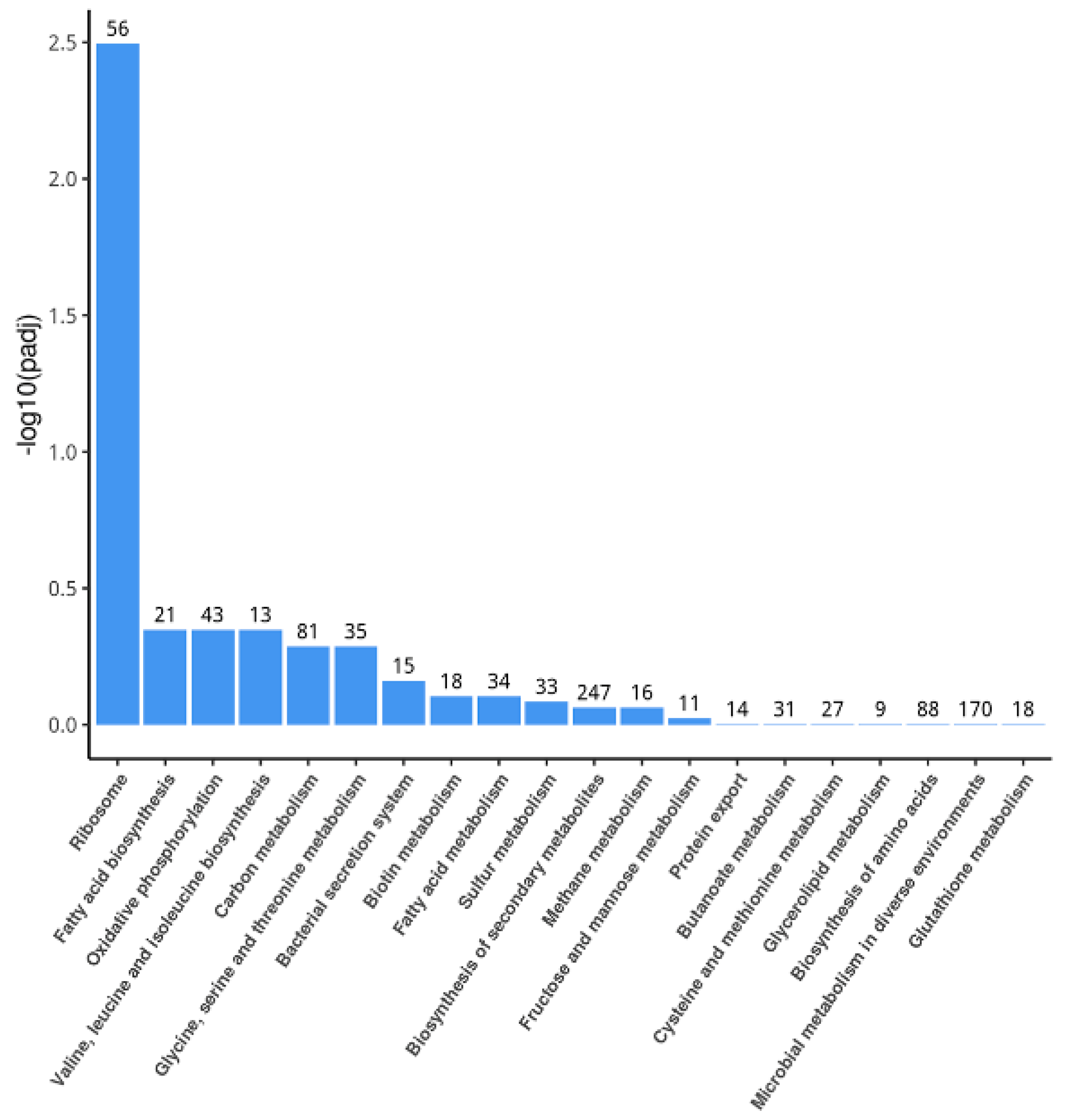
3.5. KEGG Pathway Analysis

In the KEGG pathway analysis, a total of 80 enrichment items were identified. The top 20 significant KEGG pathways were selected to draw a histogram for display (Figure 4). Among them, a total of six significant pathways were screened under the condition of P < 0.05, including ribosomal pathways; fatty acid biosynthesis; oxidative phosphorylation; valine, leucine and isoleucine biosynthesis; carbon metabolism; and glycine, serine and threonine metabolism. The number of upregulated genes in these pathways was much greater than the number of downregulated genes (Table 2), which showed the trend that these pathways were promoted by linalool.

4. Discussion

Screening DEGs and determining gene function by transcriptome sequencing technology has the characteristics of short time consumption, low research cost, high data yield, and high data accuracy. In fact, studies of Escherichia coli, L. monocytogenes, and S. species based on transcriptome sequencing have been reported [22,23,24]. P. fragi can easily lead to the spoilage of chilled meat in aerobic storage. Linalool is an excellent natural antibacterial substance that can effectively inhibit the growth of P. fragi and delay the spoilage of chilled meat. Our previous research showed that cell membrane rupture and intracellular metabolic disorder appeared in P. fragi with linalool treatment at the minimum inhibitory concentration for 8 h [21]. In this study, we investigated the effects of linalool on cell membrane formation and the energy metabolism pathways of P. fragi from the perspective of molecular biology. P. fragi with linalool treatment was found to have a different expression of several genes from the normal state, which means that linalool can regulate the transcription of P. fragi.
The disruption of cell walls or cell membranes is considered to be one of the antibacterial mechanisms of linalool. Linalool can prevent the formation of the cell walls of Listeria monocytogenes [25] and can destroy the biofilm of various bacteria, such as Salmonella typhimurium [26] and Candida albicans [27]. Lipopolysaccharide (LPS) attached to the cell wall is a typical feature of Gram-negative bacteria such as P. fragi [28]. LPS is considered to be the first barrier to protect Gram-negative bacteria [29]. The destruction and inhibition of LPS synthesis is the premise for bacteriostatic substances and antibiotics to function. As shown in Table 1, the expression of gene RS05125 (regulating transglucosylase domain-containing protein) was significantly inhibited in the linalool-treated group, which hindered the transfer of core polysaccharide in LPS and further affected LPS synthesis. Phospholipids and proteins are the main components that maintain the stability of cell membranes [30]. The production of phospholipids in bacterial cell membranes relies on fatty acid metabolism pathways, and the synthesis of membrane proteins requires the expression of the encoded genes and the successful construction of ribosomes. According to the KEGG pathway analysis, the ribosomal pathways and fatty acid biosynthesis of P. fragi were significantly increased under the stimulation of linalool, which may be the positive feedback regulation of P. fragi to compensate for cell membrane damage. Specifically, RS20760 is a gene encoding an outer membrane protein that is related to the biofilm formation of P. fragi, and the expression level of RS20760 was significantly increased after linalool treatment, which mean that P. fragi supplemented the outer membrane protein. The 30S ribosomal protein S20 encoded by the rpsT gene is a key component of the ribosome, responsible for translation initiation and the association of the 30S and 50S subunits [31]. The expression of the rpsT gene is directly related to the synthesis of membrane proteins, transporters, and various enzymes in cells. Among the GO functional enrichment of DEGs, linalool significantly upregulated the expression of the S20 protein in ribosomes, which indicated that multiple proteins of bacterial cells were overexpressed.
The regulation of energy metabolism is important for maintaining cell stability. Oxidative phosphorylation is the main source of energy for P. fragi. The enzymes and substrates involved in the oxidative phosphorylation pathway directly affect cell function and viability [32]. In our previous research, we found that linalool leaked the respiratory chain dehydrogenase and ATP of P. fragi, which led to the disturbance of respiratory metabolism. The transcriptome results in this study also provide evidence in support of the above conclusion. In this study, oxidative phosphorylation was an important pathway enriched for the identified DEGs in KEGG pathway analysis. As shown in Table 1, the gene expression levels of ATP synthase, malate dehydrogenase, ferredoxin-NADP reductase, and ADP-forming succinate-CoA ligase subunit beta were significantly upregulated under the influence of linalool, which may explain how P. fragi cells replenish key enzymes and ATP. Sulphate adenylyltransferase is one of the key enzymes in transferring ATP from mitochondria to the cytoplasm [33], and the upregulation of genes cysD and cysN in the linalool-treated group indicated that the cytoplasm needs a sufficient ATP supply to compensate for the loss caused by ATP leakage [34]. However, this strategy did not appear to work, and there was no evidence that ATP was consumed in the cell. The hydrogen ions generated by ATP consumption were the power source of flagellar movement. Table 1 showed that the expression level of the gene RS13000 regulating flagellar basal body rod protein FlgF is decreased, which was a symptom of hydrogen ion deficiency. Together, this evidence suggested that linalool is a heavy blow to the energy metabolism of P. fragi.
At the same time, P. fragi upregulates multiple transporters, including the biopolymer transporter ExbD, the phosphate ABC transporter substrate-binding protein PstS, ABC transporter substrate-binding protein, and putative porin, to satisfy substrates required for oxidative phosphorylation supply. However, Wang et al. (2003) reported that the overexpression of transporters causes an increased burden on the cell membrane [35], which may accelerate cell collapse.

5. Conclusions

Overall, linalool significantly inhibited the growth of P. fragi. By comparing the transcript levels between the treated and control groups, it was found that linalool treatment disrupted the extracellular lipopolysaccharide synthesis pathway in P. fragi and activated the fatty acid metabolism and ribosomal function to compensate for cell membrane damage. The energy metabolism of P. fragi was severely disturbed by linalool, and multiple ATP synthases and ATP transportases were overexpressed in the cells but could not guarantee the consumption of ATP. The simultaneous overexpression of multiple ribosomal functional proteins and transporters may also place an additional burden on cells and cause them to collapse.

Author Contributions

Writing—original draft preparation, Y.L. and F.R.; funding acquisition, H.C. and W.C.; software, project administration and investigation, H.C. and D.C. All authors have read and agreed to the published version of the manuscript.

Funding

This study was funded by the Natural Science Foundation of China (32160591) and Hainan Provincial Natural Science Foundation of China (320RC515).

Institutional Review Board Statement

This study did not involve any human or animal testing.

Informed Consent Statement

Written informed consent was obtained from all study participants.

Data Availability Statement

Data are contained within the article.

Conflicts of Interest

The authors declare no competing financial interests.

References

  1. Fritz, S.; See, L.; Carlson, T.; Haklay, M.M.; West, S.E. Citizen science and the United Nations Sustainable Development Goals. Nat. Sustain. 2019, 2, 922–930. [Google Scholar] [CrossRef]
  2. Kang, S.; Shi, C.; Chang, J.; Kong, F.; Li, M.; Guan, B.; Zhang, Z.; Shi, X.; Zhao, H.; Peng, Y. Label free-based proteomic analysis of the food spoiler Pseudomonas fluorescens response to lactobionic acid by SWATH-MS. Food Control. 2021, 123, 107834. [Google Scholar] [CrossRef]
  3. Bahmid, N.A.; Heising, J.; Fogliano, V.; Dekker, M. Packaging design using mustard seeds as a natural antimicrobial: A study on inhibition of Pseudomonas fragi in liquid medium. Foods 2020, 9, 789. [Google Scholar] [CrossRef] [PubMed]
  4. Agrimonti, C.; White, J.C.; Tonetti, S.; Marmiroli, N. Antimicrobial activity of cellulosic pads amended with emulsions of essential oils of oregano, thyme and cinnamon against microorganisms in minced beef meat. Int. J. Food Microbiol. 2019, 305, 108246. [Google Scholar] [CrossRef]
  5. Papadopoulou, O.S.; Iliopoulos, V.; Mallouchos, A.; Panagou, E.Z.; Chorianopoulos, N.; Tassou, C.C.; Nychas, G.E. Spoilage potential of Pseudomonas (P. fragi, P. putida) and LAB (Leuconostoc mesenteroides, Lactobacillus sakei) strains and their volatilome profile during storage of sterile pork meat using GC/MS and data analytics. Foods 2020, 9, 633. [Google Scholar] [CrossRef]
  6. Chen, Q.; Zhang, M.; Chen, M.; Li, M.; Zhang, H.; Song, P.; An, T.; Yue, P.; Gao, X. Influence of Eurotium cristatum and Aspergillus niger individual and collaborative inoculation on volatile profile in liquid-state fermentation of instant dark teas. Food Chem. 2021, 350, 129234. [Google Scholar] [CrossRef]
  7. He, F.; Li, K.; Zhang, X.; Yang, Y.; Fang, Y.; Xiang, F. Components and antibacterial activity of a novel essential oil from the nutrient broth of Eremothecium ashbyii H4565. LWT 2019, 101, 389–394. [Google Scholar] [CrossRef]
  8. Varia, R.D.; Patel, J.H.; Modi, F.D.; Vihol, P.D.; Bhavsar, S.K. In vitro and in vivo antibacterial and anti-inflammatory properties of linalool. Int. J. Curr. Microbiol. Appl. Sci. 2020, 9, 1481–1489. [Google Scholar] [CrossRef]
  9. Guo, F.; Chen, Q.; Liang, Q.; Zhang, M.; Chen, W.; Chen, H.; Yun, Y.; Zhong, Q.; Chen, W. Antimicrobial activity and proposed action mechanism of linalool against Pseudomonas fluorescens. Front. Microbiol. 2021, 49, 562094. [Google Scholar] [CrossRef]
  10. He, R.; Chen, W.; Chen, H.; Zhong, Q.; Zhang, H.; Zhang, M.; Chen, W. Antibacterial mechanism of linalool against L. monocytogenes, a metabolomic study. Food Control 2022, 132, 108533. [Google Scholar] [CrossRef]
  11. Liu, X.; Cai, J.; Chen, H.; Zhong, Q.; Hou, Y.; Chen, W.; Chen, W. Antibacterial activity and mechanism of linalool against Pseudomonas aeruginosa. Microb. Pathog. 2020, 141, 103980. [Google Scholar] [CrossRef] [PubMed]
  12. Ghosh, T.; Srivastava, S.K.; Gaurav, A.; Kumar, A.; Kumar, P.; Yadav, A.S.; Pathania, R.; Navani, N.K. A combination of linalool, vitamin C, and copper synergistically triggers reactive oxygen species and DNA damage and inhibits Salmonella enterica subsp. enterica Serovar Typhi and Vibrio fluvialis. Appl. Environ. Microb. 2019, 85, e2418–e2487. [Google Scholar]
  13. Noh, S.; Choi, E.; Hwang, C.; Jung, J.H.; Kim, S.; Kim, B. Dietary compounds for targeting prostate cancer. Nutrients 2019, 11, 2401. [Google Scholar] [CrossRef] [PubMed] [Green Version]
  14. Xu, L.; Zhang, C.; Xu, P.; Wang, X.C. Mechanisms of ultraviolet disinfection and chlorination of Escherichia coli: Culturability, membrane permeability, metabolism, and genetic damage. J. Environ. Sci. China 2018, 65, 356–366. [Google Scholar] [CrossRef] [PubMed]
  15. Hansen, K.D.; Brenner, S.E.; Sandrine, D. Biases in Illumina transcriptome sequencing caused by random hexamer priming. Nucleic Acids Res. 2010, 38, e131. [Google Scholar] [CrossRef] [Green Version]
  16. Lee, C.S.; Ungewickell, A.; Bhaduri, A.; Qu, K.; Khavari, P.A. Transcriptome sequencing in Sezary syndrome identifies Sezary cell and mycosis fungoides-associated lncRNAs and novel transcripts. Blood 2012, 120, 3288. [Google Scholar] [CrossRef] [Green Version]
  17. Yu, Y.; Dong, J.; Wang, Y.; Gong, X. RNA-seq analysis of antibacterial mechanism of Cinnamomum camphora essential oil against Escherichia coli. Peerj 2021, 9, e11081. [Google Scholar] [CrossRef]
  18. Jun-Young, P.; Su-Kyung, J.; Kyung-Min, P.; Yu, H.; Bai, J.; Sangryeol, R.; Chang, P.S. Transcriptomic analysis of Staphylococcus aureus under the stress condition of antibacterial erythorbyl laurate by RNA sequencing. Food Control 2018, 96, S963185694. [Google Scholar]
  19. Lathen, D.R.; Merrill, C.B.; Rothenfluh, A. Flying Together: Drosophila as a Tool to Understand the Genetics of Human Alcoholism. Int. J. Mol. Sci. 2020, 21, 6649. [Google Scholar] [CrossRef]
  20. Mcdevitt, D.; Rosenberg, M. Exploiting genomics to discover new antibiotics. Trends Microbiol 9:611–617. Trends Microbiol. 2002, 9, 611–617. [Google Scholar] [CrossRef]
  21. Li, Y.; He, R.; Cai, J.; Chang, J.; Chen, W. Antibacterial Activity and Mechanism of Linalool against Pseudomonas fragi. J. Food Sci. Technol. 2020, 141, 103980. [Google Scholar]
  22. Liu, Y.; Gao, W.; Wang, Y.; Wu, L.; Liu, X.; Yan, T.; Alm, E.; Arkin, A.; Thompson, D.K.; Fields, M.W. Transcriptome analysis of Shewanella oneidensis MR-1 in response to elevated salt conditions. J. Bacteriol. 2005, 187, 2501–2507. [Google Scholar] [CrossRef] [PubMed] [Green Version]
  23. Milohanic, E.; Glaser, P.; Coppée, J.Y.; Frangeul, L.; Vega, Y.; Vázquez Boland, J.A.; Kunst, F.; Cossart, P.; Buchrieser, C. Transcriptome analysis of Listeria monocytogenes identifies three groups of genes differently regulated by PrfA. Mol. Microbiol. 2003, 47, 1613–1625. [Google Scholar] [CrossRef] [PubMed] [Green Version]
  24. Polissi, A.; De Laurentis, W.; Zangrossi, S.; Briani, F.; Longhi, V.; Pesole, G.; Dehò, G. Changes in Escherichia coli transcriptome during acclimatization at low temperature. Res. Microbiol. 2003, 154, 573–580. [Google Scholar] [CrossRef]
  25. Gao, Z.; Zhong, W.; Liu, T.; Zhao, T.; Guo, J. Global Proteomic Analysis of Listeria monocytogenes’ Response to Linalool. Foods 2021, 10, 2449. [Google Scholar] [CrossRef]
  26. Prakash, A.; Vadivel, V.; Rubini, D.; Nithyanand, P. Antibacterial and antibiofilm activities of linalool nanoemulsions against Salmonella Typhimurium. Food Biosci. 2019, 28, 57–65. [Google Scholar] [CrossRef]
  27. Blaskó, Á.; Gazdag, Z.; Gróf, P.; Máté, G.; Sárosi, S.; Krisch, J.; Vágvölgyi, C.; Makszin, L.; Pesti, M. Effects of clary sage oil and its main components, linalool and linalyl acetate, on the plasma membrane of Candida albicans: An in vivo EPR study. Apoptosis 2017, 22, 175–187. [Google Scholar] [CrossRef]
  28. Nichols, W.K.; Prosser, F.H. Induction of ornithine decarboxylase in macrophages by bacterial lipopolysaccharides (LPS) and mycobacterial cell wall material. Life Sci. 1980, 27, 913–920. [Google Scholar] [CrossRef]
  29. Murphy, K.; Park, A.J.; Hao, Y.; Brewer, D.; Lam, J.S.; Khursigara, C.M. Influence of O Polysaccharides on Biofilm Development and Outer Membrane Vesicle Biogenesis in Pseudomonas aeruginosa PAO1. J. Bacteriol. 2014, 196, 1306–1317. [Google Scholar]
  30. Sharma, P.; Varma, R.; Sarasij, R.C.; Ira; Gousset, K.; Krishnamoorthy, G.; Rao, M.; Mayor, S. Nanoscale Organization of Multiple GPI-Anchored Proteins in Living Cell Membranes. Cell 2004, 116, 589. [Google Scholar] [CrossRef] [Green Version]
  31. Miryala, S.K.; Anbarasu, A.; Ramaiah, S. Gene interaction network to unravel the role of gut bacterial species in cardiovascular diseases: E. coli O157: H7 host-bacterial interaction study. Comput. Biol. Med. 2021, 133, 104417. [Google Scholar] [CrossRef] [PubMed]
  32. Li, L.; Hu, K.; Hong, B.; Lu, X.; Qian, D. The inhibitory effect of Bacillus amyloliquefaciens L1 on Aeromonas hydrophila and its mechanism. Aquaculture 2021, 539, 736590. [Google Scholar] [CrossRef]
  33. Shaw, G.W.S.A. Electrophoretic charaterization of ATP-sulfate adenylyltransferase (ATP-sulfurylase) using acrylamide gels. Anal. Biochem. 1972, 48, 259–265. [Google Scholar]
  34. Navarro-Suarez, A.M.; Hidalgo-Acosta, J.C.; Fadini, L.; Feliu, J.M.; Sua Rez-Herrera, M.F. Electrochemical Oxidation of Hydrogen on Basal Plane Platinum Electrodes in Imidazolium Ionic Liquids. J. Phys. Chem. C 2011, 115, 7. [Google Scholar] [CrossRef]
  35. Wang, D.; Safferling, M.; Lemieux, M.J.; Griffith, H.; Chen, Y.; Li, X. Practical aspects of overexpressing bacterial secondary membrane transporters for structural studies. Biochim. Biophys. Acta Biomembr. 2003, 1610, 23–36. [Google Scholar] [CrossRef] [Green Version]
Figure 1. Correlation analysis of samples in the treated group and the control group. (A) Pearson correlation between samples. (B) Principal component analysis (PCA) based on FPKM.
Foods 11 02058 g001
Figure 2. Differentially expressed gene analysis. (A) Volcano plot for all DEGs. (B) Cluster heatmap of DEGs. The columns represent samples, and the rows represent genes.
Foods 11 02058 g002
Figure 3. (A) Histogram of GO functional enrichment analysis based on DEGs. (B) Scatter plot of GO functional enrichment analysis based on DEGs.
Foods 11 02058 g003
Figure 4. Histogram of KEGG enrichment analysis.
Foods 11 02058 g004
Table 1. The DEGs of P. fragi’s responses to linalool (top 1%).
Gene NameDescriptionLog2 (Fold Change)PP adj
RS12095N-acetyltransferase3.611.56 × 10−1557.30 × 10−152
RS19715biopolymer transporter ExbD5.021.06 × 10−1252.49 × 10−122
cysDsulphate adenylyltransferase subunit CysD3.798.97 × 10−1251.40 × 10−121
RS20760extracellular solute-binding protein3.448.44 × 10−1119.87 × 10−108
RS05965Lrp/AsnC family transcriptional regulator−2.776.23 × 10−915.83 × 10−88
rpsT30S ribosomal protein S204.03 1.10 × 10−898.56 × 10−87
RS19705TonB-dependent receptor3.27 4.91 × 10−863.28 × 10−83
RS08005phosphate ABC transporter substrate-binding protein5.48 2.21 × 10−801.29 × 10−77
RS120901-acyl-sn-glycerol-3-phosphate acyltransferase2.94 3.87 × 10−792.01 × 10−76
RS10370SCP2 sterol-binding domain-containing protein3.48 2.29 × 10−781.07 × 10−75
RS08405F0F1 ATP synthase subunit delta2.79 1.48 × 10−766.29 × 10−74
cysNsulphate adenylyltransferase subunit CysN2.59 1.80 × 10−757.01 × 10−73
RS00275ABC transporter substrate-binding protein3.03 1.85 × 10−736.65 × 10−71
groLchaperonin GroEL3.62 4.20 × 10−731.40 × 10−70
RS00755malate dehydrogenase3.23 1.70 × 10−715.30 × 10−69
RS04250hypothetical protein3.57 8.84 × 10−712.58 × 10−68
RS08110hypothetical protein4.09 4.11 × 10−691.13 × 10−66
trxBthioredoxin-disulphide reductase2.55 2.70 × 10−656.65 × 10−63
RS19700MotA/TolQ/ExbB proton channel family protein2.38 4.53 × 10−651.06 × 10−62
RS18445YggL family protein3.41 2.21 × 10−644.91 × 10−62
RS06165hypothetical protein3.22 6.39 × 10−641.36 × 10−61
RS08105LysR family transcriptional regulator2.92 3.53 × 10−637.18 × 10−61
fprferredoxin-NADP reductase2.25 4.97 × 10−629.29 × 10−60
sucCADP-forming succinate--CoA ligase subunit beta2.72 1.91 × 10−583.43 × 10−56
RS13000flagellar basal body rod protein FlgF−2.112.79 × 10−564.82 × 10−54
RS12740putative porin4.805.39 × 10−559.00 × 10−53
RS22490co-chaperone GroES3.576.21 × 10−551.00 × 10−52
RS08580NADPH:quinone reductase2.071.34 × 10−532.09 × 10−51
RS05125transglycosylase domain-containing protein−1.901.58 × 10−532.38 × 10−51
RS06800methylcrotonoyl-CoA carboxylase−1.924.51 × 10−536.59 × 10−51
ecoserine protease inhibitor ecotin2.135.84 × 10−538.27 × 10−51
RS20605dienelactone hydrolase family protein2.909.24 × 10−531.27 × 10−50
Table 2. Partial results of KEGG enrichment analysis (top 10).
KEGGIDDescriptionPPadjCountUpDown
pfz03010Ribosome4.01 × 10−50.00320756551
pfz00061Fatty acid biosynthesis0.0136080.45022121147
pfz00190Oxidative phosphorylation0.0197010.45022143376
pfz00290Valine, leucine and isoleucine biosynthesis0.0225110.4502211394
pfz01200Carbon metabolism0.0376210.515821815922
pfz00260Glycine, serine and threonine metabolism0.0386870.515821351916
pfz03070Bacterial secretion system0.0603240.68941715141
pfz00780Biotin metabolism0.0865150.78489218108
pfz01212Fatty acid metabolism0.08830.784892341816
pfz00920Sulfur metabolism0.1027240.821793331914
Publisher’s Note: MDPI stays neutral with regard to jurisdictional claims in published maps and institutional affiliations.

