Development of a Radiographic Index for Periodontitis
Abstract
1. Introduction
2. Materials and Methods
- -
- Participant Information Sheet;
- -
- Consent Form;
- -
- Instruction Sheet;
- -
- Fifty printed DPTs (two equal batches numbered from 1–25 and 26–50, respectively);
- -
- A Scoring Grid to fill out their recordings (to be returned to investigator ZS).
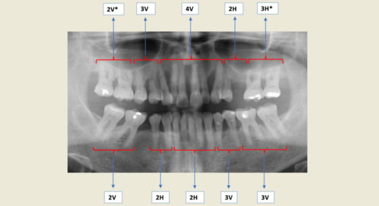
- The dentition is divided into five maxillary quintets and five mandibular quintets (Table 2A);
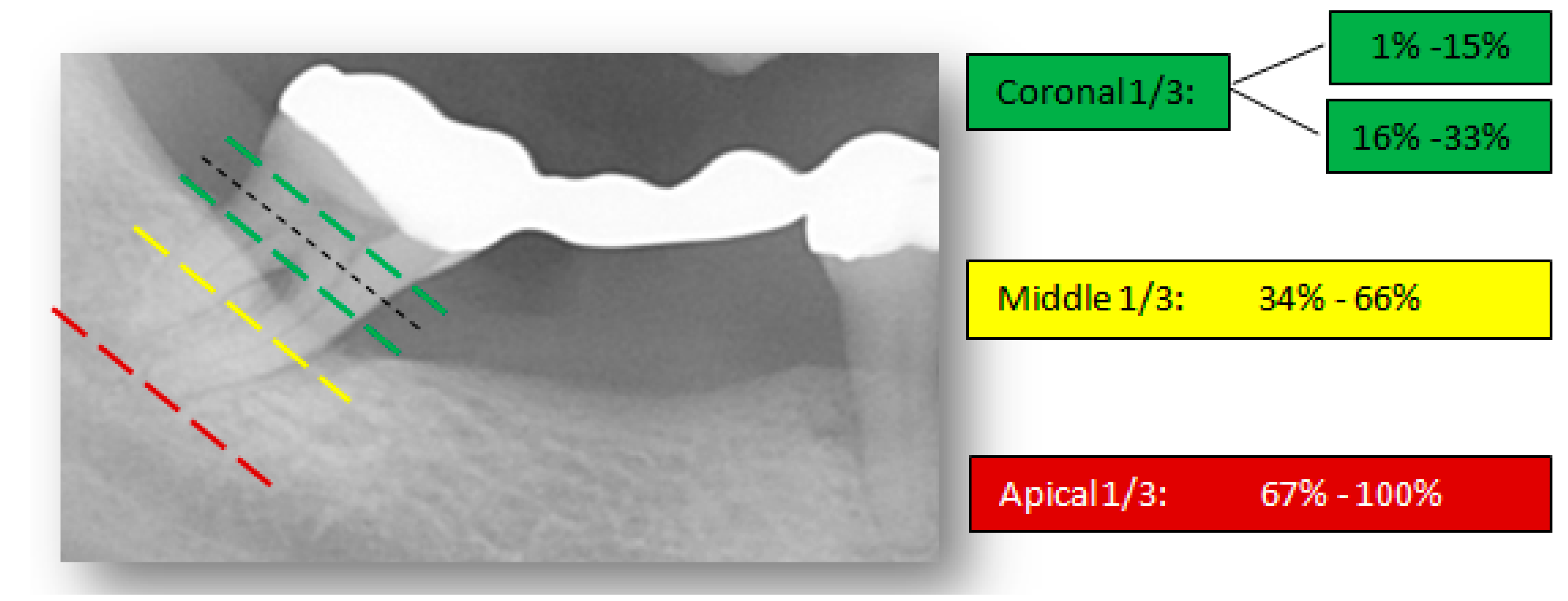
- The highest (worst) bone loss score for each quintet is recorded. The proposed scoring codes with additional descriptions are shown in Table 2B,C respectively;
- All teeth in each quintet were examined (with the exception of third molars—unless first and/or second molars are missing);
- For a quintet to qualify for recording, it must contain at least one tooth.
- -
- Participants’ visual interpretations via the proposed scoring codes;
- -
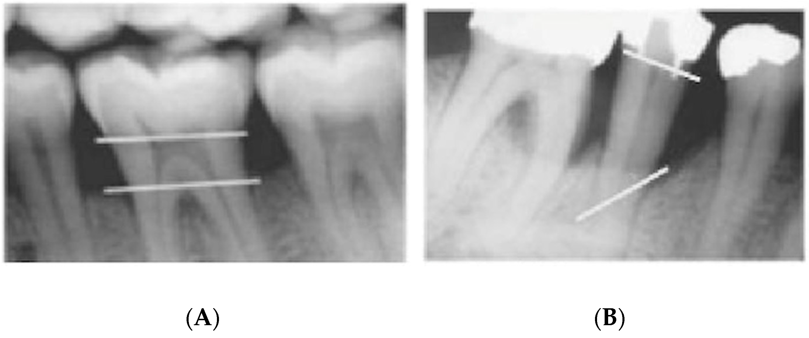
- Gold standard measurements via the Schei technique.
3. Results
3.1. Intra-Examiner Reliability
3.2. Inter-Examiner Reliability
3.3. Validity
4. Discussion
5. Conclusions
Author Contributions
Funding
Institutional Review Board Statement
Informed Consent Statement
Data Availability Statement
Acknowledgments
Conflicts of Interest
References
- Tonetti, M.S.; Jepsen, S.; Jin, L.; Otomo-Corgel, J. Impact of the global burden of periodontal diseases on health, nutrition and wellbeing of mankind: A call for global action. J. Clin. Periodontol. 2017, 44, 456–462. [Google Scholar] [CrossRef]
- Russell, A.L. A system of classification and scoring for prevalence surveys of periodontal disease. J. Dent. Res. 1956, 35, 350–359. [Google Scholar] [CrossRef] [PubMed]
- British Society of Periodontology—BSP. Basic Periodontal Examination—BPE; British Society of Periodontology: London, UK, 2011; pp. 10–14. [Google Scholar]
- Listgarten, M.A. Periodontal probing: What does it mean? J. Clin. Periodontol. 1980, 7, 165–176. [Google Scholar] [CrossRef] [PubMed]
- Hunter, F. Periodontal probes and probing. IDJ 1994, 44 (Suppl. 1), 577–583. [Google Scholar]
- Heft, M.W.; Perelmuter, S.H.; Cooper, B.Y.; Magnusson, I.; Clark, W.B. Relationship between gingival inflammation and painfulness of periodontal probing. J. Clin. Periodontol. 1991, 18, 213–215. [Google Scholar] [CrossRef]
- Bujang, M.A.; Baharum, N. A simplified guide to determination of sample size requirements for estimating the value of intraclass correlation coefficient: A review. AOS 2017, 1, 12. [Google Scholar]
- Schei, O.; Waerhaug, J.; Lovdal, A.; Arno, A. Alveolar bone loss as related to oral hygiene and age. J. Periodontol. 1959, 30, 7–16. [Google Scholar] [CrossRef]
- Björn, H.I.; Halling, A.R.; Thyberg, H.Å. Radiographic assessment of marginal bone loss. Odontol. Revy 1969, 20, 165–179. [Google Scholar]
- American Association of Endodontics—AAE. Glossary of Endodontic Terms, 9th ed.; American Association of Endodontics: Chicago, IL, USA, 2016. [Google Scholar]
- McHugh, M.L. Interrater reliability: The kappa statistic. Biochem. Med.(Zagreb) 2012, 15, 276–282. [Google Scholar] [CrossRef]
- Koo, T.K.; Li, M.Y. A guideline of selecting and reporting intraclass correlation coefficients for reliability research. J. Chiropr. Med. 2016, 1, 155–163. [Google Scholar] [CrossRef]
- Akoglu, H. User’s guide to correlation coefficients. Turk. J. Emerg. Med. 2018, 1, 91–93. [Google Scholar] [CrossRef]
- Hausmann, E. Radiographic and digital imaging in periodontal practice. J. Periodontol. 2000, 71, 497–503. [Google Scholar] [CrossRef] [PubMed]
- Afrashtehfar, K.I.; Brägger, U.; Hicklin, S.P. Reliability of interproximal bone height measurements in bone- and tissue-level implants: A methodological study for improved calibration purposes. Int. J. Oral Maxillofac. Implants 2020, 35, 289–296. [Google Scholar] [CrossRef]
- Mol, A. Imaging methods in periodontology. Periodontol. 2000 2004, 34, 34–48. [Google Scholar] [CrossRef] [PubMed]
- Takeshita, W.M.; Iwaki, L.C.; Da Silva, M.C.; Tonin, R.H. Evaluation of diagnostic accuracy of conventional and digital periapical radiography, panoramic radiography, and cone-beam computed tomography in the assessment of alveolar bone loss. Contemp. Clin. Dent. 2014, 5, 318. [Google Scholar] [CrossRef] [PubMed]
- Faculty of General Dental Practice—FGDP. Radiographs in Periodontal Assessment. In Selection Criteria for Dental Radiography; Faculty of General Dental Practice: London, UK, 2018. [Google Scholar]
- Pepelassi, E.A.; Diamanti-Kipioti, A. Selection of the most accurate method of conventional radiography for the assessment of periodontal osseous destruction. J. Clin. Periodontol. 1997, 24, 557–567. [Google Scholar] [CrossRef] [PubMed]
- Hausmann, E.; Allen, K.; Christersson, L.; Genco, R.J. Effect of X-ray beam vertical angulation on radiographic alveolar crest level measurement. J. Periodontol. Res. 1989, 24, 8–19. [Google Scholar] [CrossRef]
- Albandar, J.M.; Abbas, D.K. Radiographic quantification of alveolar bone level changes: Comparison of 3 currently used methods. J. Clin. Periodontol. 1986, 13, 810–813. [Google Scholar] [CrossRef] [PubMed]
- Bassiouny, M.A.; Grant, A.A. The accuracy of the Schei ruler: A laboratory investigation. J. Periodontol. 1975, 46, 748–752. [Google Scholar] [CrossRef]
- Teeuw, W.J.; Coelho, L.; Silva, A.; Van Der Palen, C.J.; Lessmann, F.G.; Van der Velden, U.; Loos, B.G. Validation of a dental image analyzer tool to measure alveolar bone loss in periodontitis patients. J. Periodontal Res. 2009, 44, 94–102. [Google Scholar] [CrossRef]
- Farook, F.F.; Alodwene, H.; Alharbi, R.; Alyami, M.; Alshahrani, A.; Almohammadi, D.; Alnasyan, B.; Aboelmaaty, W. Reliability assessment between clinical attachment loss and alveolar bone level in dental radiographs. Clin. Exp. Dent. Res. 2020, 6, 596–601. [Google Scholar] [CrossRef] [PubMed]
- Tonetti, M.S.; Greenwell, H.; Kornman, K.S. Staging and grading of periodontitis: Framework and proposal of a new classification and case definition. J. Periodontol. 2018, 89, S159–S172. [Google Scholar] [CrossRef]


| [R0 = 0.5] vs. [R1 = 0.7] | ||
|---|---|---|
| Observation per Subject | Number of Subjects (Power = 80%) | Number of Subjects (Power = 90%) |
| 2 | 63 | 87 |
| 3 | 39 | 55 |
| 4 | 32 | 45 |
| 5 | 28 | 40 |
| 6 | 26 | 37 |
| 7 | 25 | 35 |
| 8 | 24 | 33 |
| 9 | 23 | 32 |
| 10 | 22 | 32 |
| 20 | 20 | 28 |
| 30 | 19 | 27 |
| 40 | 19 | 27 |
 50 50 | 19 | 26 |
| 60 | 19 | 26 |
| 70 | 18 | 26 |
| 80 | 18 | 26 |
| 90 | 18 | 26 |
| 100 | 18 | 26 |
| (A) | |||||||
| UPPER RIGHT MOLARS (#17–#16) | UPPER RIGHT PREMOLARS (#15–#14) | UPPER ANTERIORS (#13–#23) | UPPER LEFT PREMOLARS (#24–#25) | UPPER LEFT MOLARS (#26–#27) | |||
| LOWER RIGHT MOLARS (#47–#46) | LOWER RIGHT PREMOLARS (#45–#44) | LOWER ANTERIORS (#43–#33) | LOWER LEFT PREMOLARS (#34–#35) | LOWER LEFT MOLARS (#36–#37) | |||
| (B) | |||||||
| CODE | DEFINITION | PERCENTAGES (%) Interproximal Alveolar Bone Loss | |||||
| 0 | No Bone Loss | 0 | |||||
| 1 | Mild Bone Loss | 1–15 | |||||
| 2 | Moderate Bone Loss | 16–33 | |||||
| 3 | Severe Bone Loss | 34–66 | |||||
| 4 | Very Severe Bone Loss | 67–100 | |||||
| (C) | |||||||
| CODE | DEFINITION | ||||||
| * | Furcation Involvement | ||||||
| H | Horizontal Pattern of Bone Loss | ||||||
| V | Vertical Pattern of Bone Loss | ||||||
| - | Teeth Absent in Quintet | ||||||
| Site (Quintet) | iABL Severity (K) | iABL Pattern/Furcation (K) | p-Value |
|---|---|---|---|
| UR Molars (1) | 0.847 | 0.718 | 0.000 |
| UR Premolars (2) | 0.762 | 0.849 | 0.000 |
| U Anteriors (3) | 0.839 | 0.642 | 0.000 |
| UL Premolars (4) | 0.735 | 0.849 | 0.000 |
| UL Molars (5) | 0.844 | 0.527 | 0.000 |
| LL Molars (6) | 0.824 | 0.788 | 0.000 |
| LL Premolars (7) | 0.809 | 1.000 | 0.000 |
| L Anteriors (8) | 0.877 | 0.645 | 0.000 |
| LR Premolars (9) | 0.679 | 1.000 | 0.000 |
| LR Molars (10) | 0.862 | 1.000 | 0.000 |
| Mean Agreement (K) | 0.808 | 0.802 | 0.000 |
| (A) | |||
| Site (Quintet) | iABL Severity (ICC) | iABL Pattern/Furcation (ICC) | p-Value |
| UR Molars (1) | 0.932 | 0.847 | 0.000 |
| UR Premolars (2) | 0.848 | 0.779 | 0.000 |
| U Anteriors (3) | 0.830 | 0.480 | 0.000 |
| UL Premolars (4) | 0.874 | 0.744 | 0.000 |
| UL Molars (5) | 0.933 | 0.875 | 0.000 |
| LL Molars (6) | 0.970 | 0.906 | 0.000 |
| LL Premolars (7) | 0.951 | 0.764 | 0.000 |
| L Anteriors (8) | 0.907 | 0.731 | 0.000 |
| LR Premolars (9) | 0.937 | 0.793 | 0.000 |
| LR Molars (10) | 0.968 | 0.863 | 0.000 |
| Mean Agreement ICC | 0.915 | 0.778 | 0.000 |
| (B) | |||
| Site (Quintet) | iABL Severity (ICC) | iABL Pattern/Furcation (ICC) | p-Value |
| UR Molars (1) | 0.866 | 0.852 | 0.000 |
| UR Premolars (2) | 0.814 | 0.646 | 0.000 |
| U Anteriors (3) | 0.743 | 0.534 | 0.000 |
| UL Premolars (4) | 0.856 | 0.674 | 0.000 |
| UL Molars (5) | 0.906 | 0.862 | 0.000 |
| LL Molars (6) | 0.956 | 0.875 | 0.000 |
| LL Premolars (7) | 0.861 | 0.655 | 0.000 |
| L Anteriors (8) | 0.821 | 0.419 | 0.000 |
| LR Premolars (9) | 0.913 | 0.682 | 0.000 |
| LR Molars (10) | 0.945 | 0.791 | 0.000 |
| Mean Agreement ICC | 0.868 | 0.699 | 0.000 |
| Participants (n = 20) | iABL Severity (ICC) | iABL Pattern/Furcation (ICC) | p-Value |
|---|---|---|---|
| PGT & Consultants (n = 14) | 0.915 | 0.778 | 0.000 |
| UGs (n = 6) | 0.868 | 0.699 | 0.000 |
| Total Mean Agreement ICC | 0.892 | 0.739 | 0.000 |
Publisher’s Note: MDPI stays neutral with regard to jurisdictional claims in published maps and institutional affiliations. |
© 2021 by the authors. Licensee MDPI, Basel, Switzerland. This article is an open access article distributed under the terms and conditions of the Creative Commons Attribution (CC BY) license (http://creativecommons.org/licenses/by/4.0/).
Share and Cite
Shaker, Z.M.H.; Parsa, A.; Moharamzadeh, K. Development of a Radiographic Index for Periodontitis. Dent. J. 2021, 9, 19. https://doi.org/10.3390/dj9020019
Shaker ZMH, Parsa A, Moharamzadeh K. Development of a Radiographic Index for Periodontitis. Dentistry Journal. 2021; 9(2):19. https://doi.org/10.3390/dj9020019
Chicago/Turabian StyleShaker, Zeyad M. H., Azin Parsa, and Keyvan Moharamzadeh. 2021. "Development of a Radiographic Index for Periodontitis" Dentistry Journal 9, no. 2: 19. https://doi.org/10.3390/dj9020019
APA StyleShaker, Z. M. H., Parsa, A., & Moharamzadeh, K. (2021). Development of a Radiographic Index for Periodontitis. Dentistry Journal, 9(2), 19. https://doi.org/10.3390/dj9020019


