Maxillary First Premolars’ Internal Morphology: A Systematic Review and Meta-Analysis
Abstract
1. Introduction
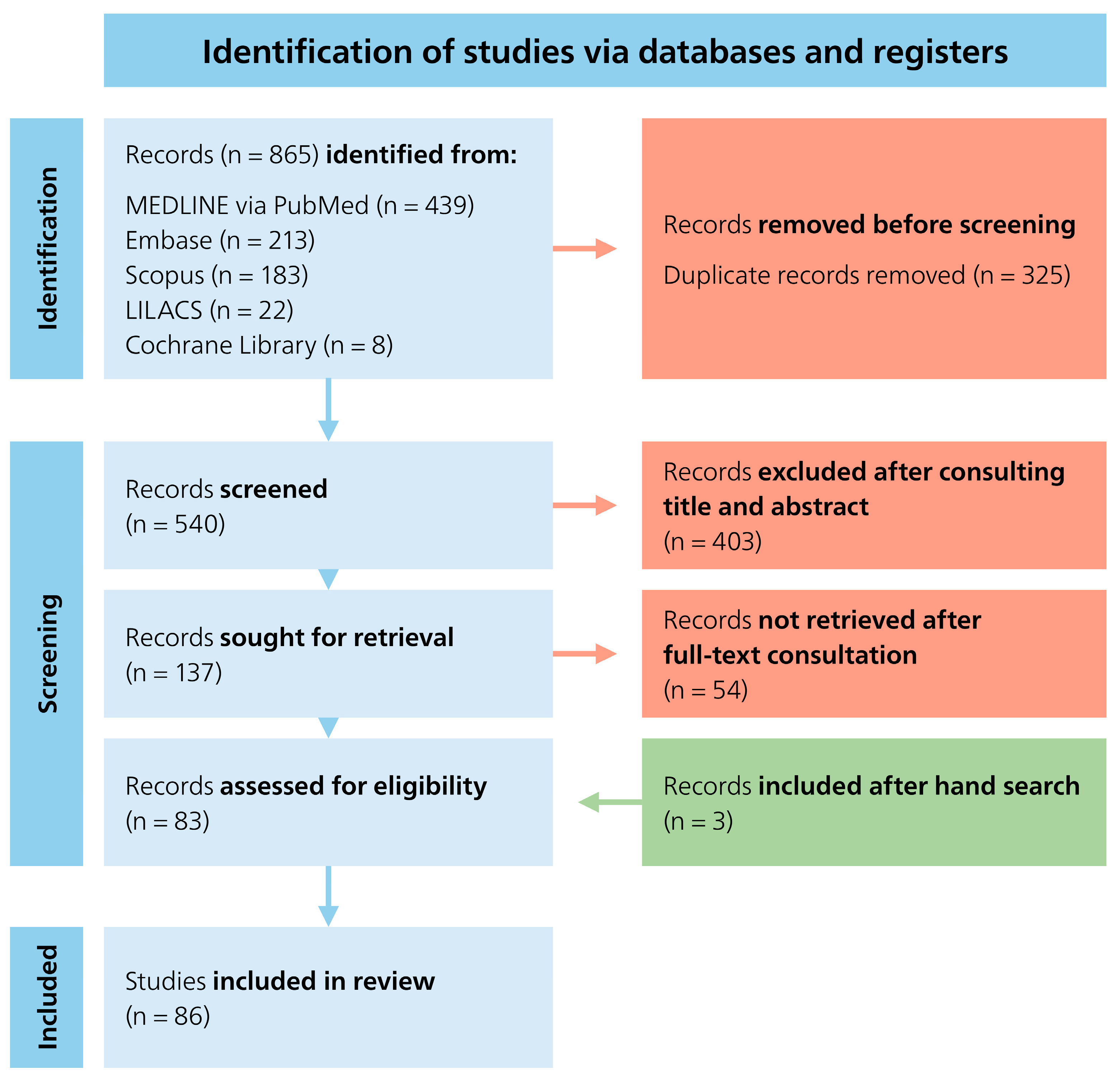
2. Materials and Methods
2.1. Inclusion and Exclusion Criteria
2.2. Data Collection and Synthesis
2.3. Quality Assessment and Risk of Bias
3. Results
3.1. Number of Roots
3.2. Root Canal Configuration (RCC)
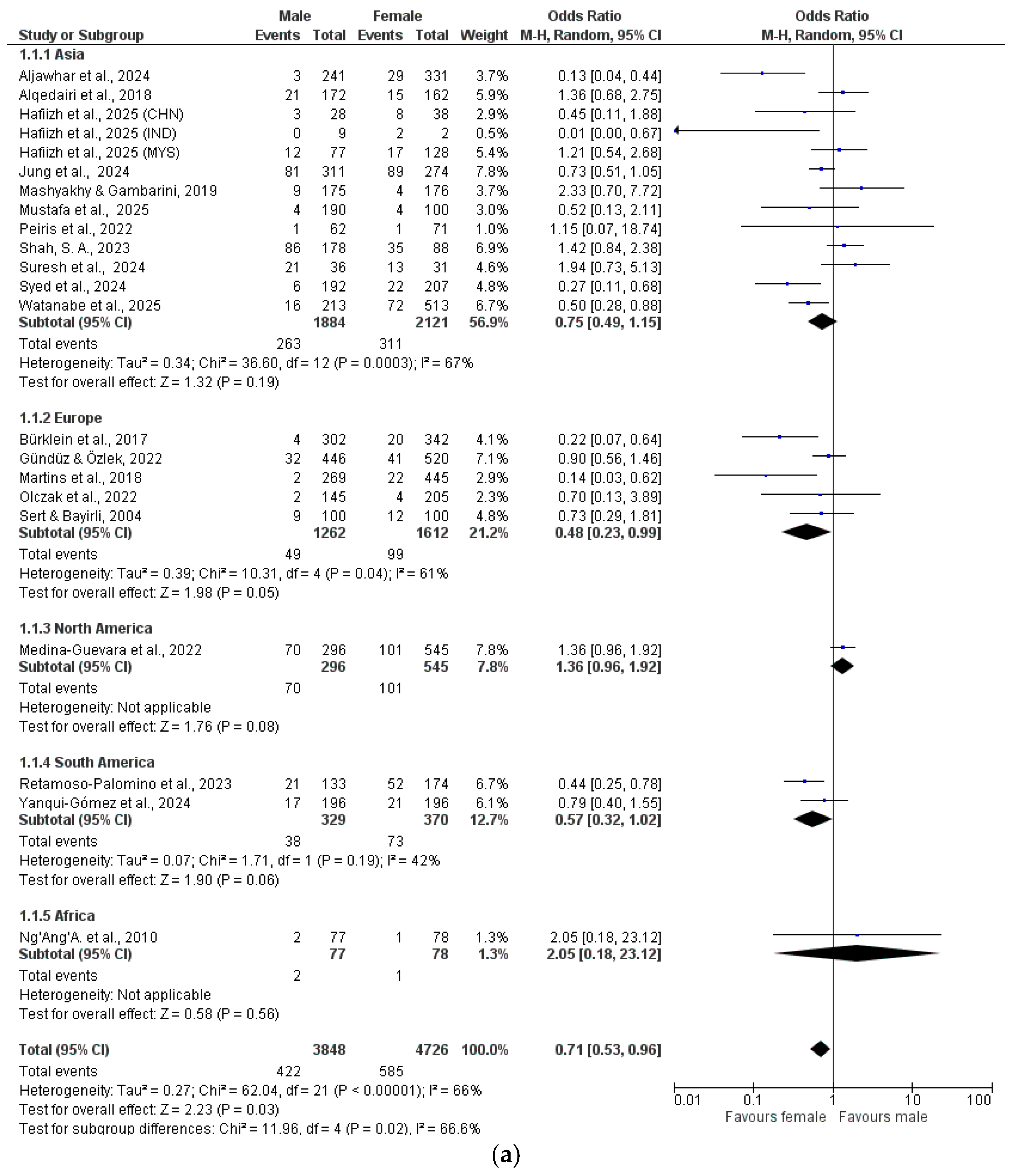
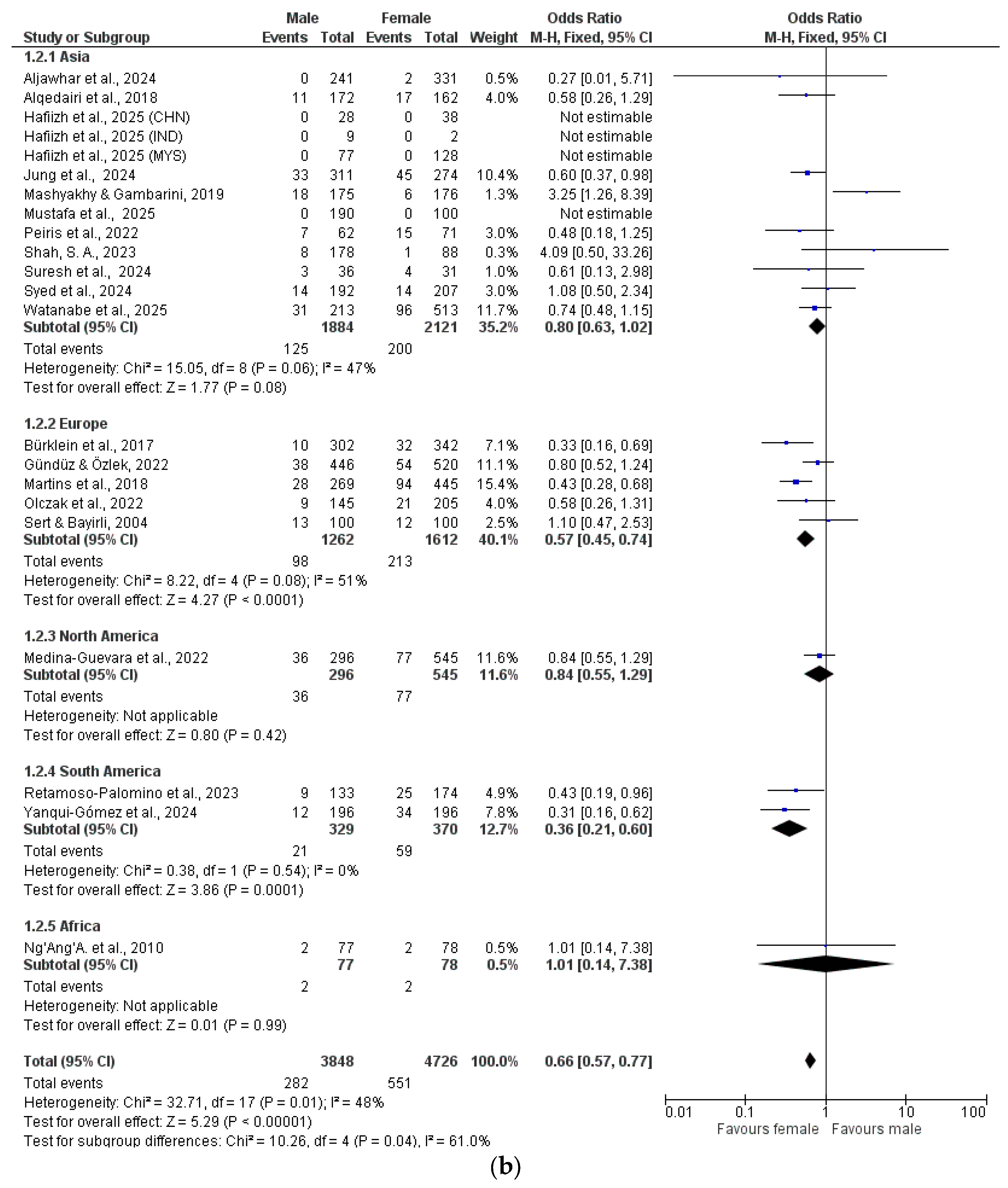
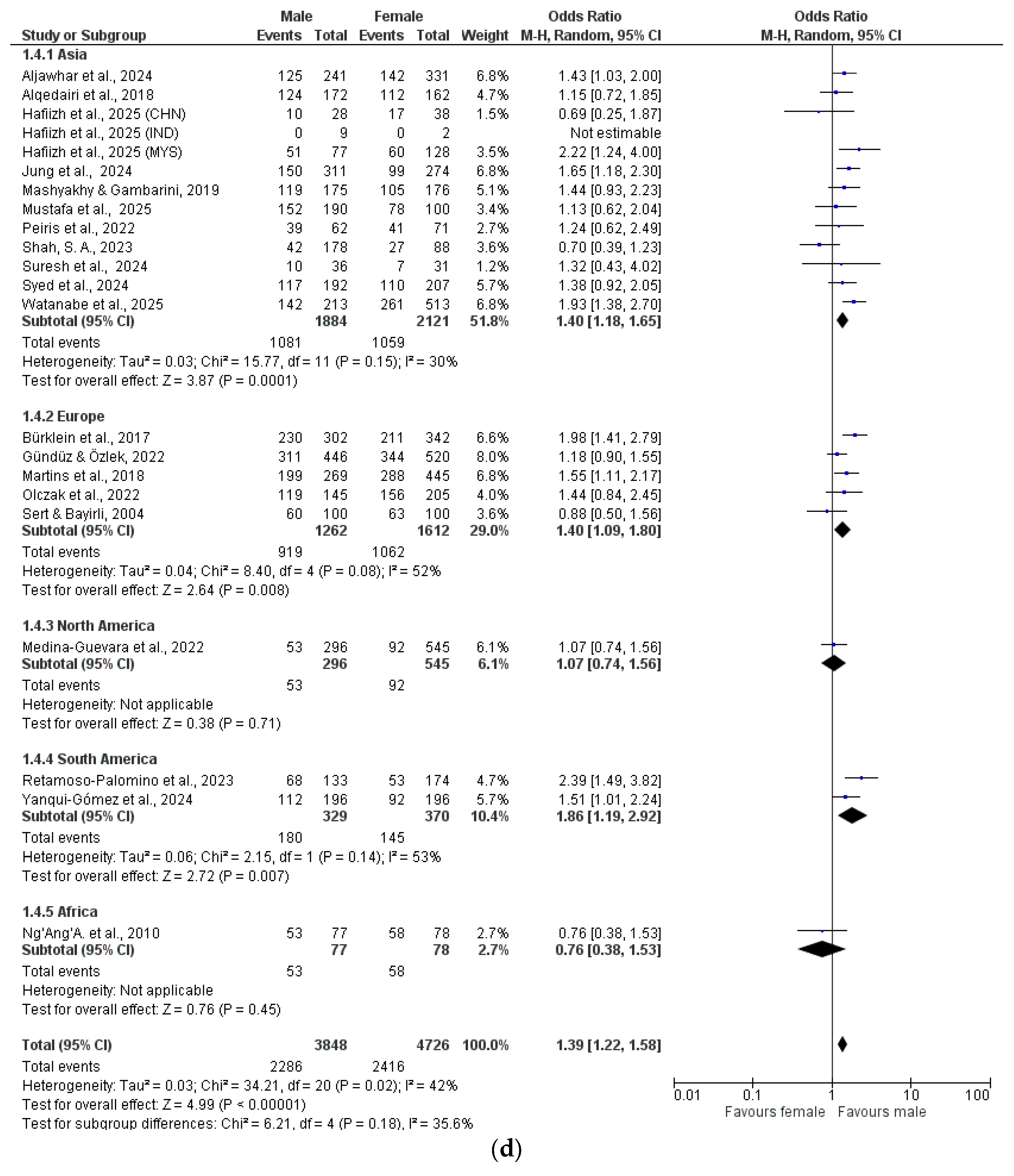
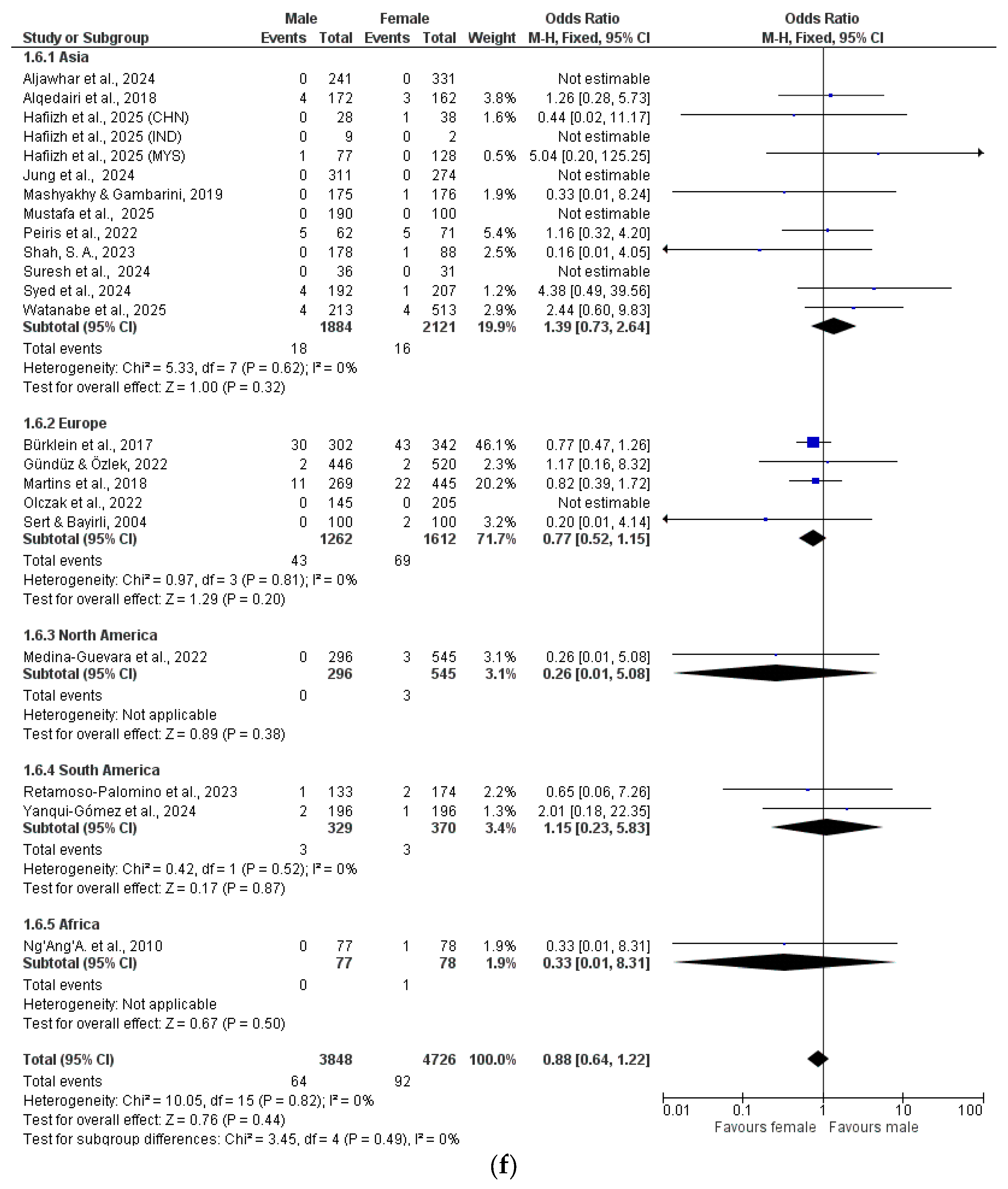
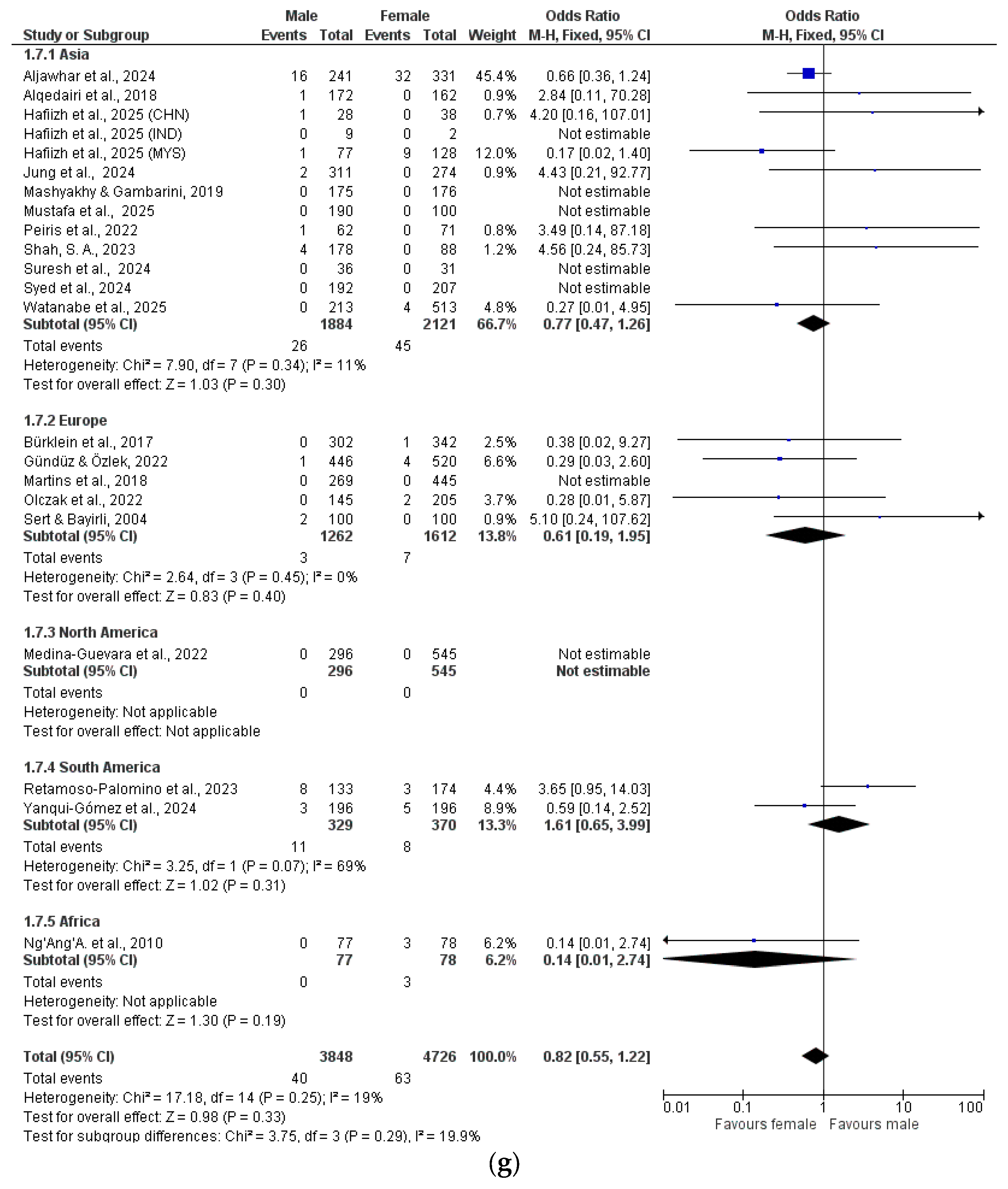
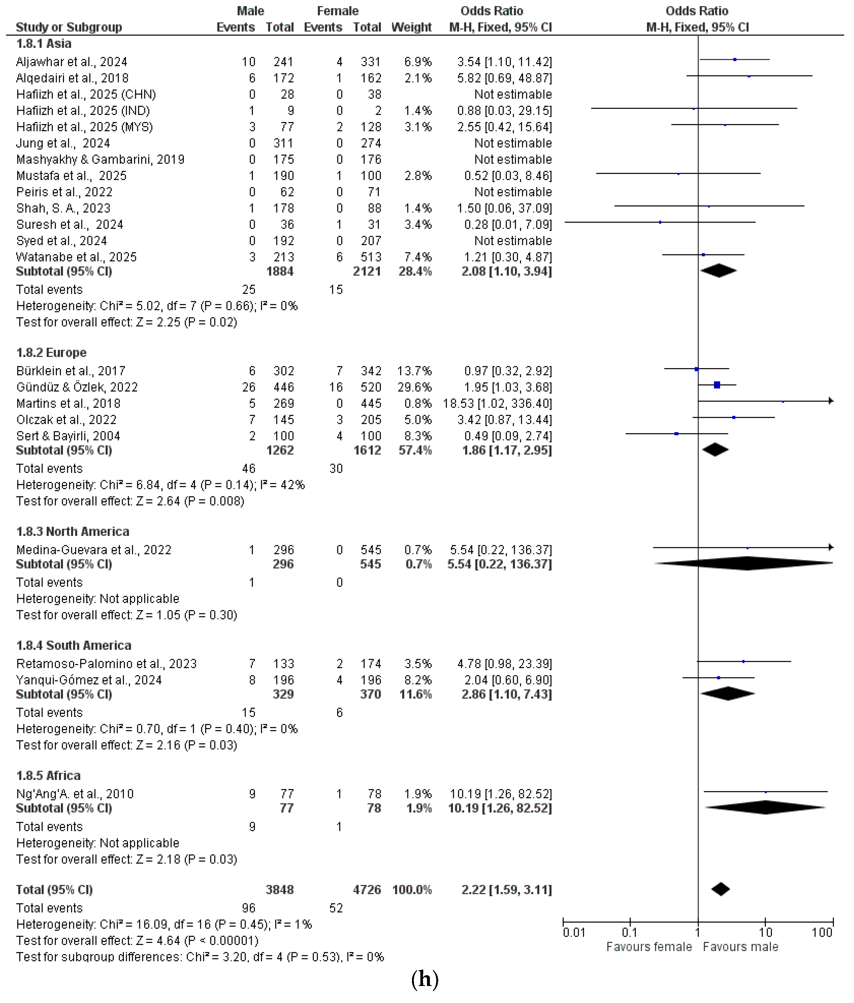
3.3. Comparative Studies
4. Discussion
4.1. Number of Roots and Root Canal Configuration (RCC)
4.2. Differences Between Women and Men
4.3. Sample Size and Research Methods
4.4. Strengths and Limitations
5. Conclusions
Supplementary Materials
Author Contributions
Funding
Data Availability Statement
Acknowledgments
Conflicts of Interest
References
- Vertucci, F.J. Root canal anatomy of the human permanent teeth. Oral Surg. Oral Med. Oral Pathol. 1984, 58, 589–599. [Google Scholar] [CrossRef]
- Özcan, E.; Çolak, H.; Hamidi, M.M. Root and canal morphology of maxillary first premolars in a Turkish population. J. Dent. Sci. 2012, 7, 390–394. [Google Scholar] [CrossRef]
- Briseño-Marroquín, B.; Paqué, F.; Maier, K.; Willershausen, B.; Wolf, T.G. Root canal morphology and configuration of 179 maxillary first molars by means of micro-computed tomography: An ex vivo study. J. Endod. 2015, 41, 2008–2013. [Google Scholar] [CrossRef]
- Pineda, F.; Kuttler, Y. Mesiodistal and buccolingual roentgenographic investigation of 7,275 root canals. Oral Surg. Oral Med. Oral Pathol. 1972, 33, 101–110. [Google Scholar] [CrossRef]
- Atieh, M.A. Root and canal morphology of maxillary first premolars in a Saudi population. J. Contemp. Dent. Pract. 2008, 9, 46–53. [Google Scholar] [CrossRef]
- Agholor, C.N.; Sede, M.A. An evaluation of root and canal morphology of maxillary premolars in a Nigerian tertiary hospital: An in-vivo study. Ghana Dent. J. 2021, 18, 30–36. [Google Scholar]
- Qiao, X.; Xu, T.; Chen, L.; Yang, D. Analysis of root canal curvature and root canal morphology of maxillary posterior teeth in Guizhou, China. Med. Sci. Monit. 2021, 27, e928758. [Google Scholar] [CrossRef]
- Faraj, B.M.; Abdulrahman, M.S.; Faris, T.M. Visual inspection of root patterns and radiographic estimation of its canal configurations by confirmation using sectioning method: An ex vivo study on maxillary first premolar teeth. BMC Oral Health 2022, 22, 166. [Google Scholar] [CrossRef]
- Green, D. Double canals in single roots. Oral Surg. Oral Med. Oral Pathol. 1973, 35, 689–696. [Google Scholar] [CrossRef]
- Vertucci, F.J.; Gegauff, A. Root canal morphology of the maxillary first premolar. J. Am. Dent. Assoc. 1979, 99, 194–198. [Google Scholar] [CrossRef]
- Calişkan, M.K.; Pehlivan, Y.; Sepetçioglu, F.; Türkün, M.; Tuncer, S.S. Root canal morphology of human permanent teeth in a Turkish population. J. Endod. 1995, 21, 200–204. [Google Scholar] [CrossRef]
- Kartal, N.; Ozçelik, B.; Cimilli, H. Root canal morphology of maxillary premolars. J. Endod. 1998, 24, 417–419. [Google Scholar] [CrossRef]
- Sert, S.; Bayirli, G.S. Evaluation of the root canal configurations of the mandibular and maxillary permanent teeth by gender in the Turkish population. J. Endod. 2004, 30, 391–398. [Google Scholar] [CrossRef]
- Awawdeh, L.; Abdullah, H.; Al-Qudah, A. Root form and canal morphology of Jordanian maxillary first premolars. J. Endod. 2008, 34, 956–961. [Google Scholar] [CrossRef]
- Peiris, R. Root and canal morphology of human permanent teeth in a Sri Lankan and Japanese population. Anthropol. Sci. 2008, 116, 123–133. [Google Scholar] [CrossRef]
- Weng, X.-L.; Yu, S.-B.; Zhao, S.-L.; Wang, H.-G.; Mu, T.; Tang, R.-Y.; Zhou, X.-D. Root canal morphology of permanent maxillary teeth in the Han nationality in Chinese Guanzhong area: A new modified root canal staining technique. J. Endod. 2009, 35, 651–656. [Google Scholar] [CrossRef]
- Ng’ang’a, R.N.; Masiga, M.A.; Maina, S.W. Internal root morphology of the maxillary first premolars in Kenyans of African descent. East Afr. Med. J. 2010, 87, 20–24. [Google Scholar] [CrossRef]
- Neelakantan, P.; Subbarao, C.; Ahuja, R.; Subbarao, C.V. Root and canal morphology of Indian maxillary premolars by a modified root canal staining technique. Odontology 2011, 99, 18–21. [Google Scholar] [CrossRef]
- Gupta, S.; Sinha, D.J.; Gowhar, O.; Tyagi, S.P.; Singh, N.N.; Gupta, S. Root and canal morphology of maxillary first premolar teeth in north Indian population using clearing technique: An in vitro study. J. Conserv. Dent. 2015, 18, 232–236. [Google Scholar] [CrossRef]
- Dinakar, C.; Shetty, U.A.; Salian, V.V.; Shetty, P. Root canal morphology of maxillary first premolars using the clearing technique in a South Indian population: An in vitro study. Int. J. Appl. Basic Med. Res. 2018, 8, 143–147. [Google Scholar] [CrossRef]
- Senan, E.M.; Alhadainy, H.A.; Genaid, T.M.; Madfa, A.A. Root form and canal morphology of maxillary first premolars of a Yemeni population. BMC Oral Health 2018, 18, 94. [Google Scholar] [CrossRef]
- Khattak, M.A.; Ijaz, F.; Ahmad, N.; Arbab, S.; Khattak, I.; Khattak, Y.J. Root canal configuration of maxillary first premolar teeth in a subpopulation of Peshawar using tooth cross-sectioning method: An in vitro study. J. Ayub Med. Coll. Abbottabad 2022, 34, S627–S631. [Google Scholar] [CrossRef]
- Peiris, R.; Arambawatta, K.; Pitakotuwage, N. Root and canal morphology of maxillary premolars and their relationship with the crown morphology. J. Oral Biosci. 2022, 64, 148–154. [Google Scholar] [CrossRef]
- Carns, E.J.; Skidmore, A.E. Configurations and deviations of root canals of maxillary first premolars. Oral Surg. Oral Med. Oral Pathol. 1973, 36, 880–886. [Google Scholar] [CrossRef]
- Tian, Y.Y.; Guo, B.; Zhang, R.; Yu, X.; Wang, H.; Hu, T.; Dummer, P.M.H. Root and canal morphology of maxillary first premolars in a Chinese subpopulation evaluated using cone-beam computed tomography. Int. Endod. J. 2012, 45, 996–1003. [Google Scholar] [CrossRef]
- Ok, E.; Altunsoy, M.; Nur, B.G.; Aglarci, O.S.; Çolak, M.; Güngör, E. A cone-beam computed tomography study of root canal morphology of maxillary and mandibular premolars in a Turkish population. Acta Odontol. Scand. 2014, 72, 701–706. [Google Scholar] [CrossRef]
- Abella, F.; Teixidó, L.M.; Patel, S.; Sosa, F.; Duran-Sindreu, F.; Roig, M. Cone-beam computed tomography analysis of the root canal morphology of maxillary first and second premolars in a Spanish population. J. Endod. 2015, 41, 1241–1247. [Google Scholar] [CrossRef]
- Bulut, D.G.; Kose, E.; Ozcan, G.; Sekerci, A.E.; Canger, E.M.; Sisman, Y. Evaluation of root morphology and root canal configuration of premolars in Turkish individuals using cone-beam computed tomography. Eur. J. Dent. 2015, 9, 551–557. [Google Scholar] [CrossRef]
- Felsypremila, G.; Vinothkumar, T.S.; Kandaswamy, D. Anatomic symmetry of root and root canal morphology of posterior teeth in an Indian subpopulation using cone-beam computed tomography: A retrospective study. Eur. J. Dent. 2015, 9, 500–507. [Google Scholar] [CrossRef]
- Celikten, B.; Orhan, K.; Aksoy, U.; Tufenkci, P.; Kalender, A.; Basmaci, F.; Dabaj, P. Cone-beam CT evaluation of root canal morphology of maxillary and mandibular premolars in a Turkish Cypriot population. BDJ Open 2016, 2, 15006. [Google Scholar] [CrossRef]
- Bürklein, S.; Heck, R.; Schäfer, E. Evaluation of the root canal anatomy of maxillary and mandibular premolars in a selected German population using cone-beam computed tomographic data. J. Endod. 2017, 43, 1448–1452. [Google Scholar] [CrossRef]
- Martins, J.N.R.; Marques, D.; Mata, A.; Caramês, J. Root and root canal morphology of the permanent dentition in a Caucasian population: A cone-beam computed tomography study. Int. Endod. J. 2017, 50, 1013–1026. [Google Scholar] [CrossRef]
- Shi, Z.Y.; Hu, N.; Shi, X.W.; Dong, X.X.; Ou, L.; Cao, J.K. Root canal morphology of maxillary premolars among the elderly. Chin. Med. J. 2017, 130, 2999–3000. [Google Scholar] [CrossRef]
- Alqedairi, A.; Alfawaz, H.; Al-Dahman, Y.; Alnassar, F.; Al-Jebaly, A.; Alsubait, S. Cone-beam computed tomographic evaluation of root canal morphology of maxillary premolars in a Saudi population. Biomed. Res. Int. 2018, 2018, 8170620. [Google Scholar] [CrossRef]
- Li, Y.H.; Bao, S.J.; Yang, X.W.; Tian, X.M.; Wei, B.; Zheng, Y.L. Symmetry of root anatomy and root canal morphology in maxillary premolars analyzed using cone-beam computed tomography. Arch. Oral Biol. 2018, 94, 84–92. [Google Scholar] [CrossRef]
- Martins, J.N.R.; Ordinola-Zapata, R.; Marques, D.; Francisco, H.; Caramês, J. Differences in root canal system configuration in human permanent teeth within different age groups. Int. Endod. J. 2018, 51, 931–941. [Google Scholar] [CrossRef]
- Martins, J.N.R.; Marques, D.; Francisco, H.; Caramês, J. Gender influence on the number of roots and root canal system configuration in human permanent teeth of a Portuguese subpopulation. Quintessence Int. 2018, 49, 103–111. [Google Scholar]
- Martins, J.N.R.; Gu, Y.; Marques, D.; Francisco, H.; Caramês, J. Differences on the root and root canal morphologies between Asian and White ethnic groups analyzed by cone-beam computed tomography. J. Endod. 2018, 44, 1096–1104. [Google Scholar] [CrossRef]
- Nazeer, M.R.; Khan, F.R.; Ghafoor, R. Evaluation of root morphology and canal configuration of maxillary premolars in a sample of Pakistani population by using cone beam computed tomography. J. Pak. Med. Assoc. 2018, 68, 423–427. [Google Scholar]
- de Lima, C.O.; de Souza, L.C.; Devito, K.L.; do Prado, M.; Campos, C.N. Evaluation of root canal morphology of maxillary premolars: A cone-beam computed tomography study. Aust. Endod. J. 2019, 45, 196–201. [Google Scholar] [CrossRef]
- Liu, X.; Gao, M.; Ruan, J.; Lu, Q. Root canal anatomy of maxillary first premolar by microscopic computed tomography in a Chinese adolescent subpopulation. Biomed. Res. Int. 2019, 2019, 4327046. [Google Scholar] [CrossRef]
- Maghfuri, S.; Keylani, H.; Chohan, H.; Dakkam, S.; Atiah, A.; Mashyakhy, M. Evaluation of root canal morphology of maxillary first premolars by cone beam computed tomography in Saudi Arabian southern region subpopulation: An in vitro study. Int. J. Dent. 2019, 2019, 2063943. [Google Scholar] [CrossRef]
- Mashyakhy, M.; Gambarini, G. Root and root canal morphology differences between genders: A comprehensive in-vivo CBCT study in a Saudi population. Acta Stomatol. Croat. 2019, 53, 213–246. [Google Scholar] [CrossRef]
- Pan, J.Y.Y.; Parolia, A.; Chuah, S.R.; Bhatia, S.; Mutalik, S.; Pau, A. Root canal morphology of permanent teeth in a Malaysian subpopulation using cone-beam computed tomography. BMC Oral Health 2019, 19, 14. [Google Scholar] [CrossRef]
- Rajakeerthi, R.; Nivedhitha, M.S.B. Use of cone beam computed tomography to identify the morphology of maxillary and mandibular premolars in Chennai population. Braz. Dent. Sci. 2019, 22, 55–62. [Google Scholar] [CrossRef]
- Saber, S.E.D.M.; Ahmed, M.H.M.; Obeid, M.; Ahmed, H.M.A. Root and canal morphology of maxillary premolar teeth in an Egyptian subpopulation using two classification systems: A cone beam computed tomography study. Int. Endod. J. 2019, 52, 267–278. [Google Scholar] [CrossRef]
- Asheghi, B.; Momtahan, N.; Sahebi, S.; Zangoie Booshehri, M. Morphological evaluation of maxillary premolar canals in Iranian population: A cone-beam computed tomography study. J. Dent. 2020, 21, 215–224. [Google Scholar]
- Buchanan, G.D.; Gamieldien, M.Y.; Tredoux, S.; Vally, Z.I. Root and canal configurations of maxillary premolars in a South African subpopulation using cone beam computed tomography and two classification systems. J. Oral Sci. 2020, 62, 93–97. [Google Scholar] [CrossRef]
- Kfir, A.; Mostinsky, O.; Elyzur, O.; Hertzeanu, M.; Metzger, Z.; Pawar, A.M. Root canal configuration and root wall thickness of first maxillary premolars in an Israeli population: A cone-beam computed tomography study. Sci. Rep. 2020, 10, 434. [Google Scholar] [CrossRef]
- Nikkerdar, N.; Asnaashari, M.; Karimi, A.; Araghi, S.; Seifitabar, S.; Golshah, A. Root and canal morphology of maxillary teeth in an Iranian subpopulation residing in western Iran using cone-beam computed tomography. Iran. Endod. J. 2020, 15, 31–37. [Google Scholar]
- Wu, D.; Hu, D.Q.; Xin, B.C.; Sun, D.G.; Ge, Z.P.; Su, J.Y. Root canal morphology of maxillary and mandibular first premolars analyzed using cone-beam computed tomography in a Shandong Chinese population. Medicine 2020, 99, e20116. [Google Scholar] [CrossRef] [PubMed]
- Al-Zubaidi, S.M.; Almansour, M.I.; Al Manour, N.N.; Alshammari, A.S.; Alshammari, A.F.; Altamimi, Y.S.; Madfa, A.A. Assessment of root morphology and canal configuration of maxillary premolars in a Saudi subpopulation: A cone-beam computed tomographic study. BMC Oral Health 2021, 21, 26. [Google Scholar] [CrossRef] [PubMed]
- Dhaimy, S.; Dhoum, S.; Diouri, M.; Bedida, L.; Elmerini, H.; Benkiran, I. Cone-beam computed tomography evaluation of the root morphology of the maxillary and mandibular premolars in a Moroccan subpopulation: Canal configurations and root curvatures (Part 2). Saudi Endod. J. 2021, 11, 162–167. [Google Scholar] [CrossRef]
- Haider, I.; Sddique, S.N.; Tirmazi, S.M.; Moazzam, M.; Sana, U.; Rahseed, M.; Batool, I. Evaluation of root canal morphology of maxillary first premolars by cone-beam computed tomography. Pak. J. Med. Health Sci. 2021, 15, 3663–3665. [Google Scholar] [CrossRef]
- Malik, S.; Singla, R.; Gill, G.; Jain, N.; Kumar, T.; Arora, S. Evaluation of root canal anatomy of maxillary premolars in a North Indian subpopulation using cone-beam computed tomography. Endodontology 2021, 33, 158–164. [Google Scholar] [CrossRef]
- Mashyakhy, M. Anatomical evaluation of maxillary premolars in a Saudi population: An in vivo cone-beam computed tomography study. J. Contemp. Dent. Pract. 2021, 22, 284–289. [Google Scholar] [CrossRef]
- Monardes, H.; Herrera, K.; Vargas, J.; Steinfort, K.; Zaror, C.; Abarca, J. Root anatomy and canal configuration of maxillary premolars: A cone-beam computed tomography study. Int. J. Morphol. 2021, 39, 463–468. [Google Scholar] [CrossRef]
- Yoza, T.; Serikawa, M.; Sugita, T.; Harada, T.; Usami, A. Cone-beam computed tomography observation of maxillary first premolar canal shapes. Anat. Cell Biol. 2021, 54, 424–430. [Google Scholar] [CrossRef]
- Aguilera, J.; Vallette, M.; Navarro, P.; Betancourt, P. Root and root canal system morphology of maxillary first premolars in a Chilean subpopulation: A cone-beam computed tomography study. Int. J. Morphol. 2022, 40, 449–454. [Google Scholar] [CrossRef]
- Alnaqbi, H.S.Y.; Gorduysus, M.O.; Al Shehadat, S.; Al Bayatti, S.W.; Mahmoud, I. Evaluation of variations in root canal anatomy and morphology of permanent maxillary premolars among the Emirate population using CBCT. Open Dent. J. 2022, 16, 1. [Google Scholar] [CrossRef]
- Gündüz, H.; Özlek, E. Evaluation of root morphology and root canal configuration of mandibular and maxillary premolar teeth in Turkish subpopulation by using cone-beam computed tomography. East J. Med. 2022, 27, 465–471. [Google Scholar] [CrossRef]
- Hanif, F.; Ahmed, A.; Javed, M.Q.; Khan, Z.J.; Ulfat, H. Frequency of root canal configurations of maxillary premolars as assessed by cone-beam computed tomography scans in the Pakistani subpopulation. Saudi Endod. J. 2022, 12, 100–105. [Google Scholar]
- Iqbal, A.; Khattak, O.; Issrani, R.; Alonazi, M.A.; Ali, A.H. Cone-beam computed tomography evaluation of root morphology of the premolars in Saudi Arabian subpopulation. Pesqui. Bras. Odontopediatria Clín. Integr. 2022, 22, e210140. [Google Scholar] [CrossRef]
- Medina-Guevara, C.A.; Oliva-Rodríguez, R.; Calvillo-Martínez, D.H.; Mariel-Cárdenas, J.; Muñoz-Ruiz, A.I.; Gutiérrez-Cantú, F.J. Anatomical and morphological findings of the root canal of maxillary premolars and their prevalence: CBCT study in a Mexican population. Int. J. Morphol. 2022, 40, 573–578. [Google Scholar] [CrossRef]
- Olczak, K.; Pawlicka, H.; Szymański, W. Root form and canal anatomy of maxillary first premolars: A cone-beam computed tomography study. Odontology 2022, 110, 365–375. [Google Scholar] [CrossRef] [PubMed]
- Allawi, S.; Ayoubi, H.; Al-Tayyan, M.; Toutangy, E.; Tolibah, Y.A. Evaluation of roots, root canal morphology, and bilateral symmetry of maxillary first molars in a Syrian subpopulation using cone-beam computed tomography. Clin. Exp. Dent. Res. 2023, 9, 1149–1155. [Google Scholar] [CrossRef]
- Erkan, E.; Olcay, K.; Eyüboğlu, T.F.; Şener, E.; Gündoğar, M. Assessment of the canal anatomy of the premolar teeth in a selected Turkish population: A cone-beam computed tomography study. BMC Oral Health 2023, 23, 403. [Google Scholar] [CrossRef]
- Khanna, S.; Jobanputra, L.; Mehta, J.; Parmar, A.; Panchal, A.; Mehta, F. Revisiting premolars using cone-beam computed tomography analysis and classifying their roots and root canal morphology using newer classification. Cureus 2023, 15, e38623. [Google Scholar] [CrossRef]
- Merhej, M.J.; El Hachem, R.; Sacre, H.; Salameh, P.; Ghosn, N.; Naaman, A. The root canal morphology of premolars in a sample of the Lebanese population: Clinical considerations. Int. Arab J. Dent. 2023, 14, 37–46. [Google Scholar] [CrossRef]
- Mirah, M.A.; Bafail, A.; Baik, A.; Abu Zaid, B.; Hakeem, M.; Ghabbani, H. Root canal morphology of premolars in Saudis. Cureus 2023, 15, e45888. [Google Scholar] [CrossRef]
- Retamoso-Palomino, M.; Mayta-Tovalino, F.; García-Rupaya, C. A cone-beam computed tomography study of the root and root canal morphology of maxillary first premolars in young Peruvians. Saudi Endod. J. 2023, 13, 231–235. [Google Scholar] [CrossRef]
- Shah, S.A. Cone beam computed tomography evaluation of root canal morphology of maxillary premolars in North-West sub-population of Pakistan. Khyber Med. Univ. J. 2023, 15, 116–121. [Google Scholar]
- Akotiya, B.R.; Kassa, S.C.; Saha, M.K.; Saha, S.G.; Agarwal, R.S.; Balakrishnan, S.P. Assessment of root morphology and canal configuration of maxillary first and second premolars in Central Indian population using CBCT and two classification systems—An observational study. Afr. J. Biol. Sci. 2024, 6, 6321–6332. [Google Scholar]
- Aljawhar, A.M.; Ibrahim, N.; Abdul Aziz, A.; Ahmed, H.M.A.; Azami, N.H. Characterization of the root and canal anatomy of maxillary premolar teeth in an Iraqi subpopulation: A cone beam computed tomography study. Odontology 2024, 112, 570–587. [Google Scholar] [CrossRef] [PubMed]
- Mirza, M.B. Evaluating the internal anatomy of maxillary premolars in an adult Saudi subpopulation using two classifications: A CBCT-based retrospective study. Med. Sci. Monit. 2024, 30, e943455. [Google Scholar] [CrossRef]
- Syed, G.A.; Pullishery, F.; Alhazmi, K.A.; Nazer, M.I.; Alkhamis, A.; Meer, F.M.; Halteet, F.A.; Sendiyoni, A.M.I.; Taher, A.O.H. CBCT evaluation of root canal morphology of maxillary first premolar in Saudi subpopulation. J. Pharm. Bioallied Sci. 2024, 16 (Suppl. S2), S1619–S1622. [Google Scholar] [CrossRef]
- Aljawhar, A.M.; Ibrahim, N.; Abdul Aziz, A.; Ahmed, H.M.A.; Azami, N.H. Micro-computed tomographic evaluation of root and canal anatomy of maxillary first premolars in Iraqi sub-population. Sci. Rep. 2025, 15, 10821. [Google Scholar] [CrossRef]
- Almehrzi, H.; Khawajah, S.; Alharbi, N.S.; El Abed, R.; Jamal, M. Evaluation of the root and canal morphology of maxillary and mandibular premolars in an Emirati sub-population. Int. Dent. Journal. 2025, 75, 1864–1873. [Google Scholar] [CrossRef]
- Mustafa, M.; Karobari, M.I.; Al-Maqtari, A.A.A.; Abdulwahed, A.; Almokhatieb, A.A.; Almufleh, L.S.; Hashem, Q.; Alsakaker, A.; Alam, M.K.; Ahmed, H.M.A. Investigating root and canal morphology of anterior and premolar teeth using CBCT with a novel coding classification system in Saudi subpopulation. Sci. Rep. 2025, 15, 4392. [Google Scholar] [CrossRef]
- Suresh, S.; Kalhoro, F.A.; Rani, P.; Memon, M. Root canal morphology of premolars in population of Hyderabad, Pakistan: A cone beam computerised tomographic analysis. J. Coll. Physicians Surg. Pak. 2024, 34, 1238–1244. [Google Scholar] [CrossRef]
- Yanqui-Gómez, J.S.; Dulanto-Vargas, J.A.; Carranza-Samanez, K.M. Morphology of roots and canals of maxillary first premolars: A CBCT study in a Peruvian sample. Int. J. Dent. 2024, 2024, 2341041. [Google Scholar] [CrossRef] [PubMed]
- Jung, Y.-H.; Hwang, J.-J.; Lee, J.-S.; Cho, B.-H. Analysis of root number and canal morphology of maxillary premolars using cone-beam computed tomography. Imaging Sci. Dent. 2024, 54, 370–380. [Google Scholar] [CrossRef] [PubMed]
- Martins, J.N.R.; Tummala, S.; Nallapati, S.; Marques, D.N.D.S.; Silva, E.S.; Caramês, J.M.M.; Versiani, M. Population-specific anatomical variations in premolar root canal systems: A cross-sectional cone-beam computed tomography study of Jamaican and Portuguese subpopulations. Dent. J. 2025, 13, 50. [Google Scholar] [CrossRef]
- Acevedo-Tavie, J.; Méndez-Vera, P.; Ríos, P.; Rosas-Méndez, C. Descripción de la morfología del sistema de canales radiculares en premolares maxilares mediante tomografía computarizada cone beam en una población chilena. Int. J. Odontostomatol. 2024, 18, 51–55. [Google Scholar] [CrossRef]
- Hafiizh, A.M.M.; Yusuf, S.N.M.; Omar, S.H.; Mohammad, N. Cone-beam computed tomography evaluation of canal morphology of maxillary premolars in Malaysian subpopulation using two canals classification. Endodontology 2025, 37, 118–127. [Google Scholar] [CrossRef]
- Watanabe, S.; Yabumoto, S.; Okiji, T. Evaluation of root and root canal morphology in maxillary premolar teeth: A cone-beam computed tomography study using two classification systems in a Japanese population. J. Dent. Sci. 2024, 20, 927–935. [Google Scholar] [CrossRef]
- Wolf, T.G.; Kozaczek, C.; Siegrist, M.; Betthäuser, M.; Paqué, F.; Briseño-Marroquín, B. An ex vivo study of root canal system configuration and morphology of 115 maxillary first premolars. J. Endod. 2020, 46, 794–800. [Google Scholar] [CrossRef]
- Alenezi, M.A.; Al-Nazhan, S.A.; Al-Omari, M.A. Three-dimensional evaluation of root canal morphology of maxillary first premolars: Micro–computed tomographic study. Saudi Dent. J. 2022, 34, 611–616. [Google Scholar] [CrossRef]
- Weine, F.S.; Healey, H.J.; Gerstein, H.; Evanson, L. Canal configuration in the mesiobuccal root of the maxillary first molar and its endodontic significance. Oral Surg. Oral Med. Oral Pathol. 1969, 28, 419–425. [Google Scholar] [CrossRef]
- Ahmed, H.M.A.; Versiani, M.A.; De-Deus, G.; Dummer, P.M.H. A new system for classifying root and root canal morphology. Int. Endod. J. 2017, 50, 761–770. [Google Scholar] [CrossRef]
- Liberati, A.; Altman, D.G.; Tetzlaff, J.; Mulrow, C.; Gøtzsche, P.C.; Ioannidis, J.P.; Clarke, M.; Devereaux, P.J.; Kleijnen, J.; Moher, D. The PRISMA statement for reporting systematic reviews and meta-analyses of studies that evaluate health care interventions: Explanation and elaboration. PLoS Med. 2009, 6, e1000100. [Google Scholar] [CrossRef]
- Higgins, J.P.; Thompson, S.G. Quantifying heterogeneity in a meta-analysis. Stat. Med. 2002, 21, 1539–1558. [Google Scholar] [CrossRef] [PubMed]
- Henry, B.M.; Tomaszewski, K.A.; Ramakrishnan, P.K.; Roy, J.; Vikse, J.; Loukas, M.; Tubbs, R.S.; Walocha, J.A. Development of the anatomical quality assessment (AQUA) tool for the quality assessment of anatomical studies included in meta-analyses and systematic reviews. Clin. Anat. 2017, 30, 6–13. [Google Scholar] [CrossRef] [PubMed]
- Page, M.J.; McKenzie, J.E.; Bossuyt, P.M.; Boutron, I.; Hoffmann, T.C.; Mulrow, C.D.; Shamseer, L.; Tetzlaff, M.J.; Akl, A.E.; Brennan, E.S.; et al. The PRISMA 2020 statement: An updated guideline for reporting systematic reviews. BMJ 2021, 372, n71. [Google Scholar] [CrossRef] [PubMed]
- Plotino, G.; Grande, N.M.; Pecci, R.; Bedini, R.; Pameijer, C.H.; Somma, F. Three-dimensional imaging using microcomputed tomography for studying tooth macromorphology. J. Am. Dent. Assoc. 2006, 137, 1555–1561. [Google Scholar] [CrossRef]
- Limoeiro, A.G.; Braitt, A.H.; Machado, A.S.; Bueno, C.E.; Fontana, C.E.; Freire, L.G.; Lopes, R.T.; De Martin, A.S. Micro-computed Tomography Evaluation of Filling Material Removal by Three Reciprocating Systems with Different Thermal Treatments. Giorn. Ital. Endod. 2021, 35, 13–18. [Google Scholar]
- Ahmed, H.M.A.; Keleş, A.; Wolf, T.G.; Nagendrababu, V.; Duncan, H.F.; Peters, O.A.; Dummer, P.M.H. Controversial Terminology in Root and Canal Anatomy: A Comprehensive Review. Eur. Endod. J. 2024, 9, 308–334. [Google Scholar] [CrossRef]
- Spagnuolo, G.; Ametrano, G.; D’Antò, V.; Formisano, A.; Simeone, M.; Riccitiello, F.; Amato, M.; Rengo, S. Microcomputed Tomography Analysis of Mesiobuccal Orifices and Major Apical Foramen in First Maxillary Molars. Open Dent. J. 2012, 6, 118–125. [Google Scholar] [CrossRef]


Disclaimer/Publisher’s Note: The statements, opinions and data contained in all publications are solely those of the individual author(s) and contributor(s) and not of MDPI and/or the editor(s). MDPI and/or the editor(s) disclaim responsibility for any injury to people or property resulting from any ideas, methods, instructions or products referred to in the content. |
© 2025 by the authors. Licensee MDPI, Basel, Switzerland. This article is an open access article distributed under the terms and conditions of the Creative Commons Attribution (CC BY) license (https://creativecommons.org/licenses/by/4.0/).
Share and Cite
Wolf, T.G.; Ulugöl, D.S.; Wierichs, R.J.; Holtkamp, A.K.M.; Spagnuolo, G.; Donnermeyer, D.; Waber, A.L. Maxillary First Premolars’ Internal Morphology: A Systematic Review and Meta-Analysis. Dent. J. 2025, 13, 510. https://doi.org/10.3390/dj13110510
Wolf TG, Ulugöl DS, Wierichs RJ, Holtkamp AKM, Spagnuolo G, Donnermeyer D, Waber AL. Maxillary First Premolars’ Internal Morphology: A Systematic Review and Meta-Analysis. Dentistry Journal. 2025; 13(11):510. https://doi.org/10.3390/dj13110510
Chicago/Turabian StyleWolf, Thomas Gerhard, Dilara Sare Ulugöl, Richard Johannes Wierichs, Agnes Klara Maria Holtkamp, Gianrico Spagnuolo, David Donnermeyer, and Andrea Lisa Waber. 2025. "Maxillary First Premolars’ Internal Morphology: A Systematic Review and Meta-Analysis" Dentistry Journal 13, no. 11: 510. https://doi.org/10.3390/dj13110510
APA StyleWolf, T. G., Ulugöl, D. S., Wierichs, R. J., Holtkamp, A. K. M., Spagnuolo, G., Donnermeyer, D., & Waber, A. L. (2025). Maxillary First Premolars’ Internal Morphology: A Systematic Review and Meta-Analysis. Dentistry Journal, 13(11), 510. https://doi.org/10.3390/dj13110510


