

Synchrotron X-ray Studies of the Structural and Functional Hierarchies in Mineralised Human Dental Enamel: A State-of-the-Art Review

 and
and 
Abstract
1. Introduction

2. Demineralisation and Remineralisation of Teeth
2.1. Tooth Structure
2.1.1. Enamel
2.1.2. Dentine

2.2. The Role and Structure of Biofilm in Caries Disease
2.3. Effect of Dental Caries on Enamel Structure
2.4. Dental Remineralisation Strategies
3. Synchrotron (Circular Particle Accelerator)
3.1. X-ray Scattering—Diffraction


3.2. X-ray Imaging Techniques—Tomography, Ptychography and ‘Rich’ Tomography

3.3. Spectroscopy—XRF, XANES, PIC Mapping, and FTIR
4. Conclusions and Perspectives
Supplementary Materials
Author Contributions
Funding
Data Availability Statement
Acknowledgments
Conflicts of Interest
References
- James, S.L.; Abate, D.; Abate, K.H.; Abay, S.M.; Abbafati, C.; Abbasi, N.; Abbastabar, H.; Abd-Allah, F.; Abdela, J.; Abdelalim, A.; et al. Global, regional, and national incidence, prevalence, and years lived with disability for 354 diseases and injuries for 195 countries and territories, 1990–2017: A systematic analysis for the Global Burden of Disease Study 2017. Lancet 2018, 392, 1789–1858. [Google Scholar] [CrossRef] [PubMed]
- Listl, S.; Galloway, J.; Mossey, P.A.; Marcenes, W. Global Economic Impact of Dental Diseases. J. Dent. Res. 2015, 94, 1355–1361. [Google Scholar] [CrossRef] [PubMed]
- Moussa, D.G.; Ahmad, P.; Mansour, T.A.; Siqueira, W.L. Current state and challenges of the global outcomes of dental caries research in the meta-omics era. Front. Cell. Infect. Microbiol. 2022, 12, 887907. [Google Scholar] [CrossRef] [PubMed]
- Barnhart, R.K. The Barnhart Dictionary of Etymology; H.W. Wilson Company: New York, NY, USA, 1988. [Google Scholar]
- Gerabek, W.E. The tooth-worm: Historical aspects of a popular medical belief. Clin. Oral Investig. 1999, 3, 1–6. [Google Scholar] [CrossRef] [PubMed]
- Ruel-Kellermann, M. Les vers dentaires du XVIe au XVIIIe siècle: Mythe ou réalité? In Actes. Société Française D’histoire de L’art dentaire, XXVIe Congrès; Actes SFHAD: Madrid, Spain, 2016; Volume 21, pp. 15–19. [Google Scholar]
- Suddick, R.P.; Harris, N.O. Historical perspectives of oral biology: A series. Crit. Rev. Oral Biol. Med. 1990, 1, 135–151. [Google Scholar] [CrossRef]
- Fauchard, P. Le Chirurgien Dentiste, ou Traité des Dents, 2nd ed.; Tome Premier: Paris, France, 1746. [Google Scholar]
- Brès, E.F.; Reyes-Gasga, J.; Hemmerlé, J. Human Tooth Enamel, a Sophisticated Material. In Extracellular Matrix Biomineralization of Dental Tissue Structures; Goldberg, M., Den Besten, P., Eds.; Springer Nature Switzerland AG: Cham, Switzerland, 2021; Chapter 9; Volume 10, pp. 243–259. [Google Scholar] [CrossRef]
- Freitas, R.A. Nanodentistry. J. Am. Dent. Assoc. 2000, 131, 1559–1565. [Google Scholar] [CrossRef]
- Hieber, S.E.; Müller, B. Nanodentistry. In Nanomedicine and Nanobiotechnology; Logothetidis, S., Ed.; Springer: Berlin/Heidelberg, Germany, 2012; pp. 95–107. [Google Scholar] [CrossRef]
- Neves, A.A.; Coutinho, E.; Alves HD, L.; de Assis, J.T. Stress and strain distribution in demineralized enamel: A micro-CT based finite element study. Microsc. Res. Tech. 2015, 78, 865–872. [Google Scholar] [CrossRef]
- Forssten, S.D.; Björklund, M.; Ouwehand, A.C. Streptococcus mutans, caries and simulation models. Nutrients 2010, 2, 290–298. [Google Scholar] [CrossRef]
- Hajishengallis, E.; Parsaei, Y.; Klein, M.I.; Koo, H. Advances in the microbial etiology and pathogenesis of early childhood caries. Mol. Oral Microbiol. 2017, 32, 24–34. [Google Scholar] [CrossRef]
- Mark Welch, J.L.; Rossetti, B.J.; Rieken, C.W.; Dewhirst, F.E.; Borisy, G.G. Biogeography of a human oral microbiome at the micron scale. Proc. Natl. Acad. Sci. USA 2016, 113, E791–E800. [Google Scholar] [CrossRef]
- Hwang, G.; Liu, Y.; Kim, D.; Sun, V.; Aviles-Reyes, A.; Kajfasz, J.K.; Lemos, J.A.; Koo, H. Simultaneous spatiotemporal mapping of in situ pH and bacterial activity within an intact 3D microcolony structure. Sci. Rep. 2016, 6, 32841. [Google Scholar] [CrossRef]
- Xiao, J.; Hara, A.; Kim, D.; Zero, D.T.; Koo, H.; Hwang, G. Biofilm three-dimensional architecture influences in situ pH distribution pattern on the human enamel surface. Int. J. Oral Sci. 2017, 9, 74–79. [Google Scholar] [CrossRef]
- Saavedra-Abril, J.A.; Balhen-Martin, C.; Zaragoza-Velasco, K.; Kimura-Hayama, E.T.; Saavedra, S.; Stoopen, M.E. Dental multisection CT for the placement of oral implants: Technique and applications. Radiographics 2010, 30, 1975–1991. [Google Scholar] [CrossRef]
- Baumann, T.; Carvalho, T.S.; Lussi, A. The effect of enamel proteins on erosion. Sci. Rep. 2015, 5, 15194. [Google Scholar] [CrossRef]
- Nanci, A. Ten Cate’s Oral Histology, 9th ed.; Elsevier: St. Louis, MO, USA, 2018. [Google Scholar]
- Trenouth, M.J. The origin of the terms enamel, dentine and cementum. Fac. Dent. J. 2014, 5, 26–31. [Google Scholar] [CrossRef]
- Hillson, S. Dental Tissues. In Teeth Cambridge Manuals in Archaeology; Hillson, S., Ed.; Cambridge University Press: Cambridge, UK, 2005; pp. 146–206. [Google Scholar] [CrossRef]
- Lacruz, R.S.; Habelitz, S.; Wright, J.T.; Paine, M.L. Dental enamel formation and implications for oral health and disease. Physiol. Rev. 2017, 97, 939–993. [Google Scholar] [CrossRef]
- Thompson, V.P. The tooth: An analogue for biomimetic materials design and processing. Dent. Mater. 2020, 36, 25–42. [Google Scholar] [CrossRef]
- Noemi, K.B.A.; Santiago, C. Histological comparison of amelodentinal junction and striae of retzius in donated secondary and primary teeth. Acta Sci. Dent. Sci. 2020, 4, 26–31. [Google Scholar] [CrossRef]
- Yu, C.; Abbott, P. An overview of the dental pulp: Its functions and responses to injury. Aust. Dent. J. 2007, 52, S4–S6. [Google Scholar] [CrossRef]
- Zhang, W.; Yelick, P.C. Vital pulp therapy-current progress of dental pulp regeneration and revascularization. Int. J. Dent. 2010, 2010, 856087. [Google Scholar] [CrossRef]
- Arana-Chavez, V.E.; Massa, L.F. Odontoblasts: The cells forming and maintaining dentine. Int. J. Biochem. Cell Biol. 2004, 36, 1367–1373. [Google Scholar] [CrossRef] [PubMed]
- Li, J.; Parada, C.; Chai, Y. Cellular and molecular mechanisms of tooth root development. Development 2017, 144, 374–384. [Google Scholar] [CrossRef] [PubMed]
- Yamamoto, T.; Hasegawa, T.; Yamamoto, T.; Hongo, H.; Amizuka, N. Histology of human cementum: Its structure, function, and development. Jpn. Dent. Sci. Rev. 2016, 52, 63–74. [Google Scholar] [CrossRef] [PubMed]
- Naito, K.; Kuwahara, Y.; Yamamoto, H.; Matsuda, Y.; Okuyama, K.; Ishimoto, T.; Nakano, T.; Yamashita, H.; Hayashi, M. Improvement of acid resistance of Zn-doped dentin by newly generated chemical bonds. Mater. Des. 2022, 215, 110412. [Google Scholar] [CrossRef]
- Foster, B.L. On the discovery of cementum. J. Periodontal Res. 2017, 52, 666–685. [Google Scholar] [CrossRef]
- Sudhakar, R.; Pratebha, B. Fibrous architecture of cementodentinal junction in disease: A scanning electron microscopic study. J. Oral Maxillofac. Pathol. 2015, 19, 325–329. [Google Scholar] [CrossRef]
- Lepora, N.F.; Verschure, P.; Prescott, T.J. The state of the art in biomimetics. Bioinspir. Biomim. 2013, 8, 013001. [Google Scholar] [CrossRef]
- Koenigswald, W.V.; Clemens, W.A. Levels of complexity in the microstructure of mammalian enamel and their application in studies of systematics. Scanning Microsc. 1992, 6, 195–217, discussion 217-218. [Google Scholar]
- Wilmers, J.; Bargmann, S. Nature’s design solutions in dental enamel: Uniting high strength and extreme damage resistance. Acta Biomater. 2020, 107, 1–24. [Google Scholar] [CrossRef]
- Xia, J.; Tian, Z.R.; Hua, L.; Chen, L.; Zhou, Z.; Qian, L.; Ungar, P.S. Enamel crystallite strength and wear: Nanoscale responses of teeth to chewing loads. J. R. Soc. Interface 2017, 14, 20170456. [Google Scholar] [CrossRef]
- Yu, H.-P.; Zhu, Y.-J.; Lu, B.-Q. Dental enamel-mimetic large-sized multi-scale ordered architecture built by a well controlled bottom-up strategy. Chem. Eng. J. 2019, 360, 1633–1645. [Google Scholar] [CrossRef]
- Besnard, C.; Marie, A.; Buček, P.; Sasidharan, S.; Harper, R.A.; Marathe, S.; Wanelik, K.; Landini, G.; Shelton, R.M.; Korsunsky, A.M. Hierarchical 2D to 3D micro/nano-histology of human dental caries lesions using light, X-ray and electron microscopy. Mater. Des. 2022, 220, 110829. [Google Scholar] [CrossRef]
- Zhao, H.; Liu, S.; Wei, Y.; Yue, Y.; Gao, M.; Li, Y.; Zeng, X.; Deng, X.; Kotov, N.A.; Guo, L.; et al. Multiscale engineered artificial tooth enamel. Science 2022, 375, 551–556. [Google Scholar] [CrossRef]
- Zhong, J.; Shibata, Y. The structural motifs of mineralized hard tissues from nano- to mesoscale: A future perspective for material science. Jpn. Dent. Sci. Rev. 2022, 58, 348–356. [Google Scholar] [CrossRef]
- Featherstone, J.D.B.; Lussi, A. Understanding the chemistry of dental erosion. Monogr. Oral Sci. 2006, 20, 66–76. [Google Scholar] [CrossRef]
- Lei, L.; Zheng, L.; Xiao, H.; Zheng, J.; Zhou, Z. Wear mechanism of human tooth enamel: The role of interfacial protein bonding between HA crystals. J. Mech. Behav. Biomed. Mater. 2020, 110, 103845. [Google Scholar] [CrossRef]
- Gil-Bona, A.; Bidlack, F.B. Tooth enamel and its dynamic protein matrix. Int. J. Mol. Sci. 2020, 21, 4458. [Google Scholar] [CrossRef]
- Eastoe, J.E. Organic matrix of tooth enamel. Nature 1960, 187, 411–412. [Google Scholar] [CrossRef]
- Saxena, C.; Saxena, K.; Bhakhar, V.; Vidya, M.; Ghanchi, M.; Jani, D. An overview of enamel matrix. Int. J. Prev. Clin. Dent. Res. 2016, 3, 79–84. [Google Scholar]
- Robinson, C.; Connell, S.D. Crystal initiation structures in developing enamel: Possible implications for caries dissolution of enamel crystals. Front. Physiol. 2017, 8, 405. [Google Scholar] [CrossRef]
- Bai, Y.; Yu, Z.; Ackerman, L.; Zhang, Y.; Bonde, J.; Li, W.; Cheng, Y.; Habelitz, S. Protein nanoribbons template enamel mineralization. Proc. Natl. Acad. Sci. USA 2020, 117, 19201–19208. [Google Scholar] [CrossRef] [PubMed]
- Bai, Y.; Bonde, J.; Carneiro, K.; Zhang, Y.; Li, W.; Habelitz, S. A brief history of the discovery of amelogenin nanoribbons in vitro and in vivo. J. Dent. Res. 2021, 100, 1429–1433. [Google Scholar] [CrossRef] [PubMed]
- Habelitz, S.; Bai, Y. Mechanisms of enamel mineralization guided by amelogenin nanoribbons. J. Dent. Res. 2021, 100, 1434–1443. [Google Scholar] [CrossRef] [PubMed]
- Sanii, B.; Martinez-Avila, O.; Simpliciano, C.; Zuckermann, R.N.; Habelitz, S. Matching 4.7-Å XRD spacing in amelogenin nanoribbons and enamel matrix. J. Dent. Res. 2014, 93, 918–922. [Google Scholar] [CrossRef] [PubMed]
- Nanci, A. (Ed.) Enamel: Composition, Formation, and Structure. In Ten Cate’s Oral Histology, 8th ed.; Elsevier: St. Louis, MO, USA, 2012; Chapter 7. [Google Scholar]
- Risnes, S.; Li, C. On the method of revealing enamel structure by acid etching. Aspects of optimization and interpretation. Microsc. Res. Tech. 2019, 82, 1668–1680. [Google Scholar] [CrossRef]
- Scott, D.B.; Albright, J.T. Electron microscopy of carious enamel and dentine. Oral Surg. Oral Med. Oral Pathol. 1954, 7, 64–78. [Google Scholar] [CrossRef]
- Meckel, A.H.; Griebstein, W.J.; Neal, R.J. Structure of mature human dental enamel as observed by electron microscopy. Arch. Oral Biol. 1965, 10, 775–783. [Google Scholar] [CrossRef]
- Besnard, C.; Harper, R.A.; Salvati, E.; Moxham, T.E.J.; Romano Brandt, L.R.; Landini, G.; Shelton, R.M.; Korsunsky, A.M. Analysis of in vitro demineralised human enamel using multi-scale correlative optical and scanning electron microscopy, and high-resolution synchrotron wide-angle X-ray scattering. Mater. Des. 2021, 206, 109739. [Google Scholar] [CrossRef]
- Besnard, C.; Harper, R.A.; Moxham, T.E.; James, J.D.; Storm, M.; Salvati, E.; Landini, G.; Shelton, R.M.; Korsunsky, A.M. 3D analysis of enamel demineralisation in human dental caries using high-resolution, large field of view synchrotron X-ray micro-computed tomography. Mater. Today Commun. 2021, 27, 102418. [Google Scholar] [CrossRef]
- Risnes, S.; Saeed, M.; Sehic, A. Scanning electron microscopy (SEM) methods for dental enamel. In Odontogenesis: Methods and Protocols; Papagerakis, P., Ed.; Springer, Humana Press: New York, NY, USA, 2019; Chapter 27; pp. 293–308. [Google Scholar] [CrossRef]
- Uskoković, V.; Kim, M.K.; Li, W.; Habelitz, S. Enzymatic processing of amelogenin during continuous crystallization of apatite. J. Mater. Res. 2008, 23, 3184–3195. [Google Scholar] [CrossRef]
- Apkarian, R.P.; Gutekunst, M.D.; Joy, D.C. High resolution SE-I SEM study of enamel crystal morphology. J. Electron Microsc. Tech. 1990, 14, 70–78. [Google Scholar] [CrossRef]
- Kodaka, T.; Kuroiwa, M.; Abe, M. Fine structure of the inner enamel in human permanent teeth. Scanning Microsc. 1990, 4, 975–985. [Google Scholar]
- Risnes, S.; Li, C. Aspects of the final phase of enamel formation as evidenced by observations of superficial enamel of human third molars using scanning electron microscopy. Arch. Oral Biol. 2018, 86, 72–79. [Google Scholar] [CrossRef]
- Hørsted, M.; Fejerskov, O.; Larsen, M.J.; Thylstrup, A. The structure of surface enamel with special reference to occlusal surfaces of primary and permanent teeth. Caries Res. 1976, 10, 287–296. [Google Scholar] [CrossRef]
- Gwinnett, A.J. The ultrastructure of the “prismless” enamel of permanent human teeth. Arch. Oral Biol. 1967, 12, 381–393. [Google Scholar] [CrossRef]
- Kodaka, T.; Debari, K.; Mori, R. Backscattered electron microscopy of surface ‘prismless’ enamel in human permanent teeth. J. Showa Univ. Dent. Soc. 1992, 12, 17–21. [Google Scholar] [CrossRef]
- Yoon, Y.J.; Kim, I.-H.; Han, S.-Y. The reason why a sheath exists in enamel. Int. J. Precis. Eng. Manuf. 2015, 16, 807–811. [Google Scholar] [CrossRef]
- Ge, J.; Cui, F.Z.; Wang, X.M.; Feng, H.L. Property variations in the prism and the organic sheath within enamel by nanoindentation. Biomaterials 2005, 26, 3333–3339. [Google Scholar] [CrossRef]
- Haines, J.D. Physical properties of human tooth enamel and enamel sheath material under load. J. Biomech. 1968, 1, 117–125. [Google Scholar] [CrossRef]
- Duverger, O.; Beniash, E.; Morasso, M.I. Keratins as components of the enamel organic matrix. Matrix Biol. 2016, 52, 260–265. [Google Scholar] [CrossRef]
- Bodecker, C.F. A report of further investigations on the organic matrix in human enamel. J. Dent. Res. 1924, 6, 117–130. [Google Scholar] [CrossRef]
- Reyes-Gasga, J.; Martínez-Piñeiro, E.L.; Brès, E.F. Crystallographic structure of human tooth enamel by electron microscopy and X-ray diffraction: Hexagonal or monoclinic? J. Microsc. 2012, 248, 102–109. [Google Scholar] [CrossRef] [PubMed]
- Daculsi, G.; Menanteau, J.; Kerebel, L.M.; Mitre, D. Length and shape of enamel crystals. Calcif. Tissue Int. 1984, 36, 550–555. [Google Scholar] [CrossRef] [PubMed]
- Daculsi, G.; Kerebel, B. High-resolution electron microscope study of human enamel crystallites: Size, shape, and growth. J. Ultrastruct. Res. 1978, 65, 163–172. [Google Scholar] [CrossRef]
- Grove, C.A.; Judd, G.; Ansell, G.S. Determination of hydroxyapatite crystallite size in human dental enamel by dark-Field electron microscopy. J. Dent. Res. 1972, 51, 22–29. [Google Scholar] [CrossRef]
- Koblischka-Veneva, A.; Koblischka, M.R.; Schmauch, J.; Hannig, M. Human dental enamel: A natural nanotechnology masterpiece investigated by TEM and t-EBSD. Nano Res. 2018, 11, 3911–3921. [Google Scholar] [CrossRef]
- Koblischka-Veneva, A.; Koblischka, M.R.; Schmauch, J.; Hannig, M. Comparison of human and bovine dental enamel by TEM and t-EBSD investigations. IOP Conf. Ser. Mater. Sci. Eng. 2019, 625, 012006. [Google Scholar] [CrossRef]
- Beniash, E.; Stifler, C.A.; Sun, C.-Y.; Jung, G.S.; Qin, Z.; Buehler, M.J.; Gilbert, P.U.P.A. The hidden structure of human enamel. Nat. Commun. 2019, 10, 4383. [Google Scholar] [CrossRef]
- Besnard, C.; Marie, A.; Sasidharan, S.; Buček, P.; Walker, J.M.; Parker, J.E.; Moxham, T.E.; Daurer, B.; Kaulich, B.; Kazemian, M.; et al. Nanoscale correlative X-ray spectroscopy and ptychography of carious dental enamel. Mater. Des. 2022, 224, 111272. [Google Scholar] [CrossRef]
- Featherstone, J.D.; Mayer, I.; Driessens, F.C.; Verbeeck, R.M.; Heijligers, H.J. Synthetic apatites containing Na, Mg, and CO3 and their comparison with tooth enamel mineral. Calcif. Tissue Int. 1983, 35, 169–171. [Google Scholar] [CrossRef]
- Eanes, E.D. Enamel apatite: Chemistry, structure and properties. J. Dent. Res. 1979, 58, 829–836. [Google Scholar] [CrossRef]
- LeGeros, R.Z.; Sakae, T.; Bautista, C.; Retino, M.; LeGeros, J.P. Magnesium and carbonate in enamel and synthetic apatites. Adv. Dent. Res. 1996, 10, 225–231. [Google Scholar] [CrossRef]
- LeGeros, R.Z. The unit-cell dimensions of human enamel apatite: Effect of chloride incorporation. Arch. Oral Biol. 1975, 20, 63–71. [Google Scholar] [CrossRef]
- Terpstra, R.A.; Driessens, F.C. Magnesium in tooth enamel and synthetic apatites. Calcif. Tissue Int. 1986, 39, 348–354. [Google Scholar] [CrossRef]
- Brudevold, F.; Reda, A.; Aasenden, R.; Bakhos, Y. Determination of trace elements in surface enamel of human teeth by a new biopsy procedure. Arch. Oral Biol. 1975, 20, 667–673. [Google Scholar] [CrossRef]
- Ghadimi, E.; Eimar, H.; Marelli, B.; Nazhat, S.N.; Asgharian, M.; Vali, H.; Tamimi, F. Trace elements can influence the physical properties of tooth enamel. Springerplus 2013, 2, 499. [Google Scholar] [CrossRef]
- La Fontaine, A.J. Understanding the Structure of Minerals at the Atomic Scale: A New Perspective Enabled by Advanced Microscopy. Ph.D. Thesis, University of Sydney, Camperdown, Australia, 2016. [Google Scholar]
- Curzon, M.E.J.; Crocker, D.C. Relationships of trace elements in human tooth enamel to dental caries. Arch. Oral Biol. 1978, 23, 647–653. [Google Scholar] [CrossRef]
- Yun, F.; Swain, M.V.; Chen, H.; Cairney, J.; Qu, J.; Sha, G.; Liu, H.; Ringer, S.P.; Han, Y.; Liu, L.; et al. Nanoscale pathways for human tooth decay—Central planar defect, organic-rich precipitate and high-angle grain boundary. Biomaterials 2020, 235, 119748. [Google Scholar] [CrossRef]
- Al-Jawad, M.; Steuwer, A.; Kilcoyne, S.H.; Shore, R.C.; Cywinski, R.; Wood, D.J. 2D mapping of texture and lattice parameters of dental enamel. Biomaterials 2007, 28, 2908–2914. [Google Scholar] [CrossRef]
- Simmons, L.M.; Montgomery, J.; Beaumont, J.; Davis, G.R.; Al-Jawad, M. Mapping the spatial and temporal progression of human dental enamel biomineralization using synchrotron X-ray diffraction. Arch. Oral Biol. 2013, 58, 1726–1734. [Google Scholar] [CrossRef]
- Leventouri, T.; Antonakos, A.; Kyriacou, A.; Venturelli, R.; Liarokapis, E.; Perdikatsis, V. Crystal structure studies of human dental apatite as a function of age. Int. J. Biomater. 2009, 2009, 698547. [Google Scholar] [CrossRef] [PubMed]
- Gordon, L.M.; Cohen, M.J.; MacRenaris, K.W.; Pasteris, J.D.; Seda, T.; Joester, D. Amorphous intergranular phases control the properties of rodent tooth enamel. Science 2015, 347, 746–750. [Google Scholar] [CrossRef] [PubMed]
- La Fontaine, A.; Cairney, J. Atom probe tomography of human tooth enamel and the accurate identification of magnesium and carbon in the mass spectrum. Microsc. Microanal. 2017, 23, 676–677. [Google Scholar] [CrossRef]
- Zhao, H.; Liu, S.; Yang, X.; Guo, L. Role of inorganic amorphous constituents in highly mineralized biomaterials and their imitations. ACS Nano 2022, 16, 17486–17496. [Google Scholar] [CrossRef] [PubMed]
- Risnes, S. A scanning electron microscope study of the three-dimensional extent of Retzius lines in human dental enamel. Eur. J. Oral Sci. 1985, 93, 145–152. [Google Scholar] [CrossRef]
- Boyde, A. The development of enamel structure. Proc. R. Soc. Med. 1967, 60, 923–928. [Google Scholar] [CrossRef]
- McFarlane, G.; Guatelli-Steinberg, D.; Loch, C.; White, S.; Bayle, P.; Floyd, B.; Pitfield, R.; Mahoney, P. An inconstant biorhythm: The changing pace of Retzius periodicity in human permanent teeth. Am. J. Phys. Anthropol. 2021, 175, 172–186. [Google Scholar] [CrossRef]
- Homma, K. Historical studies on the striae of Hunter-Schreger. Dent. Jpn. 1990, 27, 141–145. [Google Scholar]
- Koenigswald, W.V.; Rose, K.D. The enamel microstructure of the early Eocene pantodont Coryphodon and the nature of the zigzag enamel. J. Mamm. Evol. 2005, 12, 419–432. [Google Scholar] [CrossRef]
- Koenigswald, W.V.; Holbrook, L.T.; Rose, K.D. Diversity and evolution of Hunter-Schreger band configuration in tooth enamel of Perissodactyl mammals. Acta Palaeontol. Pol. 2011, 56, 11–32. [Google Scholar] [CrossRef]
- Yang, D.; Bharatiya, M.; Grine, F.E. Hunter-Schreger Band configuration in human molars reveals more decussation in the lateral enamel of ‘functional’ cusps than ‘guiding’ cusps. Arch. Oral Biol. 2022, 142, 105524. [Google Scholar] [CrossRef]
- Tamgadge, S.; Pereira, T.; Tamgadge, A. Visualization of enamel rods in hunter-schreger bands and enamel in incipient lesion under polarized and light microscopy. Saudi J. Oral Sci. 2020, 7, 76–79. [Google Scholar] [CrossRef]
- Lynch, C.D.; O’Sullivan, V.R.; Dockery, P.; McGillycuddy, C.T.; Sloan, A.J. Hunter-Schreger Band patterns in human tooth enamel. J. Anat. 2010, 217, 106–115. [Google Scholar] [CrossRef]
- Risnes, S. Growth tracks in dental enamel. J. Hum. Evol. 1998, 35, 331–350. [Google Scholar] [CrossRef]
- Osborn, J.W. A 3-dimensional model to describe the relation between prism directions, parazones and diazones, and the Hunter-Schreger bands in human tooth enamel. Arch. Oral Biol. 1990, 35, 869–878. [Google Scholar] [CrossRef]
- Risnes, S. Multiplane sectioning and scanning electron microscopy as a method for studying the three-dimensional structure of mature dental enamel. Scanning Microsc. 1987, 1, 1893–1902. [Google Scholar]
- Loch, C.; Hemm, L.; Taylor, B.; Visser, I.N.; Wiig, Ø. Microstructure, elemental composition and mechanical properties of enamel and dentine in the polar bear Ursus maritimus. Arch. Oral Biol. 2022, 134, 105318. [Google Scholar] [CrossRef]
- Hanaizumi, Y.; Maeda, T.; Takano, Y. Three-dimensional arrangement of enamel prisms and their relation to the formation of Hunter-Schreger bands in dog tooth. Cell Tissue Res. 1996, 286, 103–114. [Google Scholar] [CrossRef]
- Tafforeau, P.; Bentaleb, I.; Jaeger, J.-J.; Martin, C. Nature of laminations and mineralization in rhinoceros enamel using histology and X-ray synchrotron microtomography: Potential implications for palaeoenvironmental isotopic studies. Palaeogeogr. Palaeoclimatol. Palaeoecol. 2007, 246, 206–227. [Google Scholar] [CrossRef]
- Beynon, A.D.; Dean, M.C.; Reid, D.J. On thick and thin enamel in hominoids. Am. J. Phys. Anthropol. 1991, 86, 295–309. [Google Scholar] [CrossRef]
- Risnes, S. Structural characteristics of staircase-type retzius lines in human dental enamel analyzed by scanning electron microscopy. Anat. Rec. 1990, 226, 135–146. [Google Scholar] [CrossRef] [PubMed]
- Li, C.; Risnes, S. SEM observations of Retzius lines and prism cross-striations in human dental enamel after different acid etching regimes. Arch. Oral Biol. 2004, 49, 45–52. [Google Scholar] [CrossRef] [PubMed]
- Nanci, A. (Ed.) Enamel: Composition, formation, and structure. In Ten Cate’s Oral Histology, 9th ed.; Elsevier: St. Louis, MO, USA, 2018; Chapter 7. [Google Scholar]
- Desoutter, A.; Slimani, A.; Al-Obaidi, R.; Barthélemi, S.; Cuisinier, F.; Tassery, H.; Salehi, H. Cross striation in human permanent and deciduous enamel measured with confocal Raman microscopy. J. Raman Spectrosc. 2019, 50, 548–556. [Google Scholar] [CrossRef]
- Dahl, E.; Mjör, I.A. The structure and distribution of nerves in the pulp-dentin organ. Acta Odontol. Scand. 1973, 31, 349–356. [Google Scholar] [CrossRef]
- Goldberg, M.; Kulkarni, A.B.; Young, M.; Boskey, A. Dentin: Structure, composition and mineralization. Front. Biosci. 2011, 3, 711–735. [Google Scholar] [CrossRef]
- Zavgorodniy, A.V.; Rohanizadeh, R.; Swain, M.V. Ultrastructure of dentine carious lesions. Arch. Oral Biol. 2008, 53, 124–132. [Google Scholar] [CrossRef]
- Bertassoni, L.E.; Stankoska, K.; Swain, M.V. Insights into the structure and composition of the peritubular dentin organic matrix and the lamina limitans. Micron 2012, 43, 229–236. [Google Scholar] [CrossRef]
- Bertassoni, L.E. Dentin on the nanoscale: Hierarchical organization, mechanical behavior and bioinspired engineering. Dent. Mater. 2017, 33, 637–649. [Google Scholar] [CrossRef]
- Schilke, R.; Lisson, J.A.; Bauß, O.; Geurtsen, W. Comparison of the number and diameter of dentinal tubules in human and bovine dentine by scanning electron microscopic investigation. Arch. Oral Biol. 2000, 45, 355–361. [Google Scholar] [CrossRef]
- Deyhle, H.; Bunk, O.; Buser, S.; Krastl, G.; Zitzmann, N.U.; Ilgenstein, B.; Beckmann, F.; Pfeiffer, F.; Weiger, R.; Müller, B. Bio-inspired dental fillings. In SPIE NanoScience + Engineering—Biomimetics and Bioinspiration; Martín-Palma, R.J., Lakhtakia, A., Eds.; SPIE: San Diego, CA, USA, 2009; Volume 7401, p. 74010E-74011-74011. [Google Scholar]
- Lenzi, T.L.; Guglielmi, C.D.A.B.; Arana-Chavez, V.E.; Raggio, D.P. Tubule density and diameter in coronal dentin from primary and permanent human teeth. Microsc. Microanal. 2013, 19, 1445–1449. [Google Scholar] [CrossRef]
- Lopes, M.B.; Sinhoreti, M.A.C.; Gonini Júnior, A.; Consani, S.; Mccabe, J.F. Comparative study of tubular diameter and quantity for human and bovine dentin at different depths. Braz. Dent. J. 2009, 20, 279–283. [Google Scholar] [CrossRef]
- Sui, T.; Dluhoš, J.; Li, T.; Zeng, K.; Cernescu, A.; Landini, G.; Korsunsky, A.M. Structure-function correlative microscopy of peritubular and intertubular dentine. Materials 2018, 11, 1493. [Google Scholar] [CrossRef]
- Schulze, K.A.; Balooch, M.; Balooch, G.; Marshall, G.W.; Marshall, S.J. Micro-Raman spectroscopic investigation of dental calcified tissues. J. Biomed. Mater. Res. Part A 2004, 69, 286–293. [Google Scholar] [CrossRef]
- Xue, J.; Zavgorodniy, A.V.; Kennedy, B.J.; Swain, M.V.; Li, W. X-ray microdiffraction, TEM characterization and texture analysis of human dentin and enamel. J. Microsc. 2013, 251, 144–153. [Google Scholar] [CrossRef]
- Sui, T.; Lunt, A.J.; Baimpas, N.; Sandholzer, M.A.; Li, T.; Zeng, K.; Landini, G.; Korsunsky, A.M. Understanding nature’s residual strain engineering at the human dentine-enamel junction interface. Acta Biomater. 2016, 32, 256–263. [Google Scholar] [CrossRef]
- Wang, Z.; Wang, K.; Xu, W.; Gong, X.; Zhang, F. Mapping the mechanical gradient of human dentin-enamel-junction at different intratooth locations. Dent. Mater. 2018, 34, 376–388. [Google Scholar] [CrossRef]
- Fong, H.; Sarikaya, M.; White, S.N.; Snead, M.L. Nano-mechanical properties profiles across dentin-enamel junction of human incisor teeth. Mater. Sci. Eng. C 2000, 7, 119–128. [Google Scholar] [CrossRef]
- Habelitz, S.; Marshall, S.J.; Marshall, G.W., Jr.; Balooch, M. The functional width of the dentino-enamel junction determined by AFM-based nanoscratching. J. Struct. Biol. 2001, 135, 294–301. [Google Scholar] [CrossRef]
- Imbeni, V.; Kruzic, J.J.; Marshall, G.W.; Marshall, S.J.; Ritchie, R.O. The dentin-enamel junction and the fracture of human teeth. Nat. Mater. 2005, 4, 229–232. [Google Scholar] [CrossRef]
- Chan, Y.L.; Ngan, A.H.W.; King, N.M. Nano-scale structure and mechanical properties of the human dentine-enamel junction. J. Mech. Behav. Biomed. Mater. 2011, 4, 785–795. [Google Scholar] [CrossRef]
- Cuy, J.L.; Mann, A.B.; Livi, K.J.; Teaford, M.F.; Weihs, T.P. Nanoindentation mapping of the mechanical properties of human molar tooth enamel. Arch. Oral Biol. 2002, 47, 281–291. [Google Scholar] [CrossRef] [PubMed]
- Huang, T.T.Y.; He, L.-H.; Darendeliler, M.A.; Swain, M.V. Correlation of mineral density and elastic modulus of natural enamel white spot lesions using X-ray microtomography and nanoindentation. Acta Biomater. 2010, 6, 4553–4559. [Google Scholar] [CrossRef] [PubMed]
- Featherstone, J.D.B. Dental caries: A dynamic disease process. Aust. Dent. J. 2008, 53, 286–291. [Google Scholar] [CrossRef] [PubMed]
- Das Gupta, S.; Killenberger, M.; Tanner, T.; Rieppo, L.; Saarakkala, S.; Heikkilä, J.; Anttonen, V.; Finnilä, M.A.J. Mineralization of dental tissues and caries lesions detailed with Raman microspectroscopic imaging. Analyst 2021, 146, 1705–1713. [Google Scholar] [CrossRef]
- Lew, A.J.; Beniash, E.; Gilbert PU, P.A.; Buehler, M.J. Role of the mineral in the self-healing of cracks in human enamel. ACS Nano 2022, 16, 10273–10280. [Google Scholar] [CrossRef]
- Lubarsky, G.V.; Lemoine, P.; Meenan, B.J.; Deb, S.; Mutreja, I.; Carolan, P.; Petkov, N. Enamel proteins mitigate mechanical and structural degradations in mature human enamel during acid attack. Mater. Res. Express 2014, 1, 025404. [Google Scholar] [CrossRef]
- Zhang, Y.-R.; Du, W.; Zhou, X.-D.; Yu, H.-Y. Review of research on the mechanical properties of the human tooth. Int. J. Oral Sci. 2014, 6, 61–69. [Google Scholar] [CrossRef]
- Habelitz, S.; Marshall, S.J.; Marshall, G.W., Jr.; Balooch, M. Mechanical properties of human dental enamel on the nanometre scale. Arch. Oral Biol. 2001, 46, 173–183. [Google Scholar] [CrossRef]
- Shen, L.; de Sousa, F.B.; Tay, N.; Lang, T.S.; Kaixin, V.L.; Han, J.; Kilpatrick-Liverman, L.; Wang, W.; Lavender, S.; Pilch, S.; et al. Deformation behavior of normal human enamel: A study by nanoindentation. J. Mech. Behav. Biomed. Mater. 2020, 108, 103799. [Google Scholar] [CrossRef]
- Huang, T.T.Y.; He, L.H.; Darendeliler, M.A.; Swain, M.V. Nano-indentation characterisation of natural carious white spot lesions. Caries Res. 2010, 44, 101–107. [Google Scholar] [CrossRef]
- Dawes, C. What is the critical pH and why does a tooth dissolve in acid? J. Can. Dent. Assoc. 2003, 69, 722–724. [Google Scholar]
- Silverstone, L.M.; Johnson, N.W.; Hardie, J.M.; Williams, R.A.D. The caries process in dentine: The response of dentine and pulp. In Dental Caries: Aetiology, Pathology and Prevention; The Macmillan Press: London, UK; Basingstoke, UK, 1981; Chapter 7; pp. 162–168. [Google Scholar] [CrossRef]
- Harper, R.A.; Shelton, R.M.; James, J.D.; Salvati, E.; Besnard, C.; Korsunsky, A.M.; Landini, G. Acid-induced demineralisation of human enamel as a function of time and pH observed using X-ray and polarised light imaging. Acta Biomater. 2021, 120, 240–248. [Google Scholar] [CrossRef]
- Goldberg, M.E. Enamel and dentin carious lesions. JSM Dent. 2020, 8, 1120. [Google Scholar]
- Goldberg, M. (Ed.) Enamel softening (dental erosion)—Enamel etching—The early enamel carious lesion. In Understanding Dental Caries; Springer International Publishing: Cham, Switzerland, 2016; Chapter 2–4. [Google Scholar]
- Frank, R.M. Structural events in the caries process in enamel, cementum, and dentin. J. Dent. Res. 1990, 69, 559–566. [Google Scholar] [CrossRef]
- LeGeros, R.Z. Chemical and crystallographic events in the caries process. J. Dent. Res. 1990, 69, 567–574. [Google Scholar] [CrossRef]
- Pitts, N.B.; Zero, D.T.; Marsh, P.D.; Ekstrand, K.; Weintraub, J.A.; Ramos-Gomez, F.; Tagami, J.; Twetman, S.; Tsakos, G.; Ismail, A. Dental caries. Nat. Rev. Dis. Prim. 2017, 3, 17030. [Google Scholar] [CrossRef]
- Kim, D.; Barraza, J.P.; Arthur, R.A.; Hara, A.; Lewis, K.; Liu, Y.; Scisci, E.L.; Hajishengallis, E.; Whiteley, M.; Koo, H. Spatial mapping of polymicrobial communities reveals a precise biogeography associated with human dental caries. Proc. Natl. Acad. Sci. USA 2020, 117, 201919099. [Google Scholar] [CrossRef]
- Cheng, R.; Yang, H.; Shao, M.Y.; Hu, T.; Zhou, X.D. Dental erosion and severe tooth decay related to soft drinks: A case report and literature review. J. Zhejiang Univ. Sci. B 2009, 10, 395–399. [Google Scholar] [CrossRef]
- Roberts, W.E.; Mangum, J.E.; Schneider, P.M. Pathophysiology of demineralization, part II: Enamel white spots, cavitated caries, and bone infection. Curr. Osteoporos. Rep. 2022, 20, 106–119. [Google Scholar] [CrossRef]
- Matsui, R.; Cvitkovitch, D. Acid tolerance mechanisms utilized by Streptococcus mutans. Future Microbiol. 2010, 5, 403–417. [Google Scholar] [CrossRef]
- Hwang, G.; Klein, M.I.; Koo, H. Analysis of the mechanical stability and surface detachment of mature Streptococcus mutans biofilms by applying a range of external shear forces. Biofouling 2014, 30, 1079–1091. [Google Scholar] [CrossRef] [PubMed]
- Krzyściak, W.; Jurczak, A.; Kościelniak, D.; Bystrowska, B.; Skalniak, A. The virulence of Streptococcus mutans and the ability to form biofilms. Eur. J. Clin. Microbiol. Infect. Dis. 2014, 33, 499–515. [Google Scholar] [CrossRef] [PubMed]
- Scharnow, A.M.; Solinski, A.E.; Wuest, W.M. Targeting S. mutans biofilms: A perspective on preventing dental caries. MedChemComm 2019, 10, 1057–1067. [Google Scholar] [CrossRef] [PubMed]
- Banas, J.A.; Drake, D.R. Are the mutans streptococci still considered relevant to understanding the microbial etiology of dental caries? BMC Oral Health 2018, 18, 129. [Google Scholar] [CrossRef]
- Seredin, P.; Goloshchapov, D.; Kashkarov, V.; Nesterov, D.; Ippolitov, Y.; Ippolitov, I.; Vongsvivut, J. Effect of exo/endogenous prophylaxis dentifrice/drug and cariogenic conditions of patient on molecular property of dental biofilm: Synchrotron FTIR spectroscopic study. Pharmaceutics 2022, 14, 1355. [Google Scholar] [CrossRef]
- Oh, M.J.; Babeer, A.; Liu, Y.; Ren, Z.; Wu, J.; Issadore, D.A.; Stebe, K.J.; Lee, D.; Steager, E.; Koo, H. Surface topography-adaptive robotic superstructures for biofilm removal and pathogen detection on human teeth. ACS Nano 2022, 16, 11998–12012. [Google Scholar] [CrossRef]
- Nguyen, P.T.M.; Falsetta, M.L.; Hwang, G.; Gonzalez-Begne, M.; Koo, H. α-mangostin disrupts the development of Streptococcus mutans biofilms and facilitates its mechanical removal. PLoS ONE 2014, 9, e111312. [Google Scholar] [CrossRef]
- Lin, N.J.; Keeler, C.; Kraigsley, A.M.; Ye, J.; Lin-Gibson, S. Effect of dental monomers and initiators on Streptococcus mutans oral biofilms. Dent. Mater. 2018, 34, 776–785. [Google Scholar] [CrossRef]
- Ostadhossein, F.; Moitra, P.; Altun, E.; Dutta, D.; Sar, D.; Tripathi, I.; Hsiao, S.-H.; Kravchuk, V.; Nie, S.; Pan, D. Function-adaptive clustered nanoparticles reverse Streptococcus mutans dental biofilm and maintain microbiota balance. Commun. Biol. 2021, 4, 846. [Google Scholar] [CrossRef]
- Jiao, Y.; Tay, F.R.; Niu, L.-N.; Chen, J.-H. Advancing antimicrobial strategies for managing oral biofilm infections. Int. J. Oral Sci. 2019, 11, 28. [Google Scholar] [CrossRef]
- Shimizu, T.; Ho, B.; Deeley, K.; Briseño-Ruiz, J.; Faraco, I.M.; Schupack, B.I.; Brancher, J.A.; Pecharki, G.D.; Küchler, E.C.; Tannure, P.N.; et al. Enamel formation genes influence enamel microhardness before and after cariogenic challenge. PLoS ONE 2012, 7, e45022. [Google Scholar] [CrossRef]
- Kelly, A.M.; Kallistova, A.; Küchler, E.C.; Romanos, H.F.; Lips, A.; Costa, M.C.; Modesto, A.; Vieira, A.R. Measuring the microscopic structures of human dental enamel can predict caries experience. J. Pers. Med. 2020, 10, 5. [Google Scholar] [CrossRef]
- Sadyrin, E.; Swain, M.; Mitrin, B.; Rzhepakovsky, I.; Nikolaev, A.; Irkha, V.; Yogina, D.; Lyanguzov, N.; Maksyukov, S.; Aizikovich, S. Characterization of enamel and dentine about a white spot lesion: Mechanical properties, mineral density, microstructure and molecular composition. Nanomaterials 2020, 10, 1889. [Google Scholar] [CrossRef]
- Buchwald, T.; Buchwald, Z. Assessment of the Raman spectroscopy effectiveness in determining the early changes in human enamel caused by artificial caries. Analyst 2019, 144, 1409–1419. [Google Scholar] [CrossRef]
- Harris, H.H.; Vogt, S.; Eastgate, H.; Lay, P.A. A link between copper and dental caries in human teeth identified by X-ray fluorescence elemental mapping. JBIC J. Biol. Inorg. Chem. 2008, 13, 303–306. [Google Scholar] [CrossRef]
- Sui, T.; Salvati, E.; Harper, R.; Zhang, H.; Shelton, R.M.; Landini, G.; Korsunsky, A.M. In situ monitoring and analysis of enamel demineralisation using synchrotron X-ray scattering. Acta Biomater. 2018, 77, 333–341. [Google Scholar] [CrossRef]
- Seredin, P.; Kashkarov, V.; Lukin, A.; Ippolitov, Y.; Julian, R.; Doyle, S. Local study of fissure caries by Fourier transform infrared microscopy and X-ray diffraction using synchrotron radiation. J. Synchrotron Radiat. 2013, 20, 705–710. [Google Scholar] [CrossRef]
- Siddiqui, S.; Anderson, P.; Al-Jawad, M. Recovery of crystallographic texture in remineralized dental enamel. PLoS ONE 2014, 9, e108879. [Google Scholar] [CrossRef]
- Deyhle, H.; White, S.N.; Bunk, O.; Beckmann, F.; Müller, B. Nanostructure of carious tooth enamel lesion. Acta Biomater. 2014, 10, 355–364. [Google Scholar] [CrossRef]
- Darling, A.I. Studies of the early lesion of enamel caries with transmitted light, polarized light and microradiography. Br. Dent. J. 1956, 101, 289–297 and 329–341. [Google Scholar]
- Dowker, S.E.P.; Elliott, J.C.; Davis, G.R.; Wilson, R.M.; Cloetens, P. Synchrotron X-ray microtomographic investigation of mineral concentrations at micrometre scale in sound and carious enamel. Caries Res. 2004, 38, 514–522. [Google Scholar] [CrossRef] [PubMed]
- Bergman, G.; Lind, P.O. A quantitative microradiographic study of incipient enamel caries. J. Dent. Res. 1966, 45, 1477–1484. [Google Scholar] [CrossRef] [PubMed]
- Darling, A.I. Resistance of the enamel to dental caries. J. Dent. Res. 1963, 42, 488–496. [Google Scholar] [CrossRef] [PubMed]
- Guzman, C.; Brudevold, F.; Mermagen, H. A soft roentgen-ray study of early carious lesions. J. Am. Dent. Assoc. 1957, 55, 509–515. [Google Scholar] [CrossRef] [PubMed]
- Frank, R.M. Microscopie electronique de la carie des sillons chez l’homme. Arch. Oral Biol. 1973, 18, 9–25. [Google Scholar] [CrossRef]
- Voegel, J.C.; Frank, R.M. Stages in the dissolution of human enamel crystals in dental caries. Calcif. Tissue Res. 1977, 24, 19–27. [Google Scholar] [CrossRef]
- De Mattos Brito, C.S.; Meira, K.R.S.; de Sousa, F.B. Natural enamel caries in quinoline: Volumetric data and the pattern of infiltration. Microsc. Res. Tech. 2018, 81, 181–190. [Google Scholar] [CrossRef]
- Aoba, T.; Moriwaki, Y.; Doi, Y.; Okazaki, M.; Takahashi, J.; Yagi, T. The intact surface layer in natural enamel caries and acid-dissolved hydroxyapatite pellets. J. Oral Pathol. Med. 1981, 10, 32–39. [Google Scholar] [CrossRef]
- Crabb, H.S. Enamel caries. Observations on the histology and pattern of progress of the approximal lesion. Br. Dent. J. 1966, 121, 115–129, contd. [Google Scholar]
- Johnson, N.W. Some aspects of the ultrastructure of early human enamel caries seen with the electron microscope. Arch. Oral Biol. 1967, 12, 1505–1521. [Google Scholar] [CrossRef]
- Johnson, N.W. Transmission electron microscopy of early carious enamel. Caries Res. 1967, 1, 356–369. [Google Scholar] [CrossRef]
- Kerckaert, G.A. Electron microscopy of human carious dental enamel. Arch. Oral Biol. 1973, 18, 751-IN757. [Google Scholar] [CrossRef]
- Pearce, E.I.; Nelson, D.G. Microstructural features of carious human enamel imaged with back-scattered electrons. J. Dent. Res. 1989, 68, 113–118. [Google Scholar] [CrossRef]
- Ko, A.C.T.; Choo-Smith, L.-P.; Hewko, M.; Sowa, M.G.; Dong, C.C.S.; Cleghorn, B. Detection of early dental caries using polarized Raman spectroscopy. Opt. Express 2006, 14, 203–215. [Google Scholar] [CrossRef]
- Reyes-Gasga, J.; Hémmerlé, J.; Brès, E.F. Aberration-corrected transmission electron microscopic study of the central dark line defect in human tooth enamel crystals. Microsc. Microanal. 2016, 22, 1047–1055. [Google Scholar] [CrossRef]
- Reyes-Gasga, J.; Brès, E.F. High resolution STEM images of the human tooth enamel crystals. Appl. Sci. 2021, 11, 7477. [Google Scholar] [CrossRef]
- Reyes-Gasga, J.; Brès, E.F. Electron microscopic study of the human tooth enamel: The central dark line. In Encyclopedia of Analytical Chemistry; Meyers, R.A., Ed.; John Wiley & Sons, Ltd.: Hoboken, NJ, USA, 2015. [Google Scholar] [CrossRef]
- Gasga, J.R.; Carbajal-De-La-Torre, G.; Bres, E.; Gil-Chavarria, I.M.; Rodríguez-Hernández, A.G.; García-García, R. STEM-HAADF electron microscopy analysis of the central dark line defect of human tooth enamel crystallites. J. Mater. Sci. Mater. Med. 2008, 19, 877–882. [Google Scholar] [CrossRef]
- DeRocher, K.A.; Smeets, P.J.M.; Goodge, B.H.; Zachman, M.J.; Balachandran, P.V.; Stegbauer, L.; Cohen, M.J.; Gordon, L.M.; Rondinelli, J.M.; Kourkoutis, L.F.; et al. Chemical gradients in human enamel crystallites. Nature 2020, 583, 66–71. [Google Scholar] [CrossRef]
- Shulin, W. Human enamel structure studied by high resolution electron microscopy. Electron Microsc. Rev. 1989, 2, 1–16. [Google Scholar] [CrossRef]
- Anderson, P.; Dowker SE, P.; Elliott, J.C.; Thomas, C.R. Synchrotron X-ray fluorescence microprobe analysis in the study of dental mineralized tissues. J. Trace Microprobe Tech. 1996, 14, 541–560. [Google Scholar]
- Tang, S.; Dong, Z.; Ke, X.; Luo, J.; Li, J. Advances in biomineralization-inspired materials for hard tissue repair. Int. J. Oral Sci. 2021, 13, 42. [Google Scholar] [CrossRef] [PubMed]
- Wang, J.; Liu, Z.; Ren, B.; Wang, Q.; Wu, J.; Yang, N.; Sui, X.; Li, L.; Li, M.; Zhang, X.; et al. Biomimetic mineralisation systems for in situ enamel restoration inspired by amelogenesis. J. Mater. Sci. Mater. Med. 2021, 32, 115. [Google Scholar] [CrossRef] [PubMed]
- Katiyar, N.K.; Goel, G.; Hawi, S.; Goel, S. Nature-inspired materials: Emerging trends and prospects. NPG Asia Mater. 2021, 13, 56. [Google Scholar] [CrossRef]
- Cao, C.Y.; Mei, M.L.; Li, Q.-L.; Lo EC, M.; Chu, C.H. Methods for biomimetic mineralisation of human enamel: A systematic review. Materials 2015, 8, 2873–2886. [Google Scholar] [CrossRef]
- Nagalakshmi, C.; Gupta, N.; Subhathira, R.; Reddy, V.R.; Ranjana, B.S.; Mahalakshmi, D. Non-fluoridated remineralizing agent-a narrative review. Ann. Rom. Soc. Cell Biol. 2021, 25, 6925–6941. [Google Scholar]
- Philip, N. State of the art enamel remineralization systems: The next frontier in caries management. Caries Res. 2019, 53, 284–295. [Google Scholar] [CrossRef]
- Ramadoss, R.; Padmanaban, R.; Subramanian, B. Role of bioglass in enamel remineralization: Existing strategies and future prospects—A narrative review. J. Biomed. Mater. Res. Part B Appl. Biomater. 2022, 110, 45–66. [Google Scholar] [CrossRef]
- Batra, A.K.; Shetty, V. Non-fluoridated remineralising agents—A review of literature. J. Evol. Med. Dent. Sci. 2021, 10, 638–644. [Google Scholar] [CrossRef]
- Cochrane, N.J.; Cai, F.; Huq, N.L.; Burrow, M.F.; Reynolds, E.C. New approaches to enhanced remineralization of tooth enamel. J. Dent. Res. 2010, 89, 1187–1197. [Google Scholar] [CrossRef]
- Cheng, L.; Zhang, K.; Weir, M.D.; Melo, M.A.S.; Zhou, X.; Xu, H.H. Nanotechnology strategies for antibacterial and remineralizing composites and adhesives to tackle dental caries. Nanomedicine 2015, 10, 627–641. [Google Scholar] [CrossRef]
- Ruan, Q.; Moradian-Oldak, J. Amelogenin and enamel biomimetics. J. Mater. Chem. B 2015, 3, 3112–3129. [Google Scholar] [CrossRef]
- Bonchev, A.; Simeonov, M.; Vasileva, R. Review: A biomimetic approach for human enamel remineralization. Int. J. Sci. Res. 2018, 7, 1416–1420. [Google Scholar] [CrossRef]
- Arifa, M.K.; Ephraim, R.; Rajamani, T. Recent advances in dental hard tissue remineralization: A review of literature. Int. J. Clin. Pediatr. Dent. 2019, 12, 139–144. [Google Scholar] [CrossRef]
- Meyer, F.; Amaechi, B.T.; Fabritius, H.-O.; Enax, J. Overview of calcium phosphates used in biomimetic oral care. Open Dent. J. 2018, 12, 406–423. [Google Scholar] [CrossRef]
- Elsharkawy, S.; Mata, A. Hierarchical biomineralization: From nature’s designs to synthetic materials for regenerative medicine and dentistry. Adv. Healthc. Mater. 2018, 7, e1800178. [Google Scholar] [CrossRef]
- Pandya, M.; Diekwisch, T.G.H. Enamel biomimetics—Fiction or future of dentistry. Int. J. Oral Sci. 2019, 11, 8. [Google Scholar] [CrossRef]
- Nimbeni, S.B.; Nimbeni, B.S.; Divakar, D.D. Role of chitosan in remineralization of enamel and dentin: A systematic review. Int. J. Clin. Pediatr. Dent. 2021, 14, 562–568. [Google Scholar] [CrossRef]
- Grohe, B.; Mittler, S. Advanced non-fluoride approaches to dental enamel remineralization: The next level in enamel repair management. Biomater. Biosyst. 2021, 4, 100029. [Google Scholar] [CrossRef]
- Mohamed, R.N.; Basha, S.; Al-Thomali, Y.; Alshamrani, A.S.; Alzahrani, F.S.; Enan, E.T. Self-assembling peptide P11-4 in remineralization of enamel caries—A systematic review of in-vitro studies. Acta Odontol. Scand. 2021, 79, 139–146. [Google Scholar] [CrossRef]
- Madrid-Troconis, C.C.; Perez-Puello, S.D.C. Casein phosphopeptide-amorphous calcium phosphate nanocomplex (CPP-ACP) in dentistry: State of the art. Rev. Fac. Odontol. Univ. Antioq. 2019, 30, 248–262. [Google Scholar]
- Wei, Y.; Liu, S.; Xiao, Z.; Zhao, H.; Luo, J.; Deng, X.; Guo, L. Enamel repair with amorphous ceramics. Adv. Mater. 2020, 32, 1907067. [Google Scholar] [CrossRef] [PubMed]
- Babaie, E.; Bacino, M.; White, J.; Nurrohman, H.; Marshall, G.W.; Saeki, K.; Habelitz, S. Polymer-Induced Liquid Precursor (PILP) remineralization of artificial and natural dentin carious lesions evaluated by nanoindentation and microcomputed tomography. J. Dent. 2021, 109, 103659. [Google Scholar] [CrossRef] [PubMed]
- Yeom, B.; Sain, T.; Lacevic, N.; Bukharina, D.; Cha, S.-H.; Waas, A.M.; Arruda, E.M.; Kotov, N.A. Abiotic tooth enamel. Nature 2017, 543, 95. [Google Scholar] [CrossRef] [PubMed]
- Seredin, P.; Goloshchapov, D.; Kashkarov, V.; Emelyanova, A.; Buylov, N.; Barkov, K.; Ippolitov, Y.; Khmelevskaia, T.; Mahdy, I.A.; Mahdy, M.A.; et al. Biomimetic mineralization of tooth enamel using nanocrystalline hydroxyapatite under various dental surface pretreatment conditions. Biomimetics 2022, 7, 111. [Google Scholar] [CrossRef]
- Shao, C.; Jin, B.; Mu, Z.; Lu, H.; Zhao, Y.; Wu, Z.; Yan, L.; Zhang, Z.; Zhou, Y.; Pan, H.; et al. Repair of tooth enamel by a biomimetic mineralization frontier ensuring epitaxial growth. Sci. Adv. 2019, 5, eaaw9569. [Google Scholar] [CrossRef]
- Farooq, I.; Bugshan, A. The role of salivary contents and modern technologies in the remineralization of dental enamel: A narrative review [version 3; peer review: 3 approved]. F1000Research 2021, 9, 171. [Google Scholar] [CrossRef]
- Dodds, M.; Roland, S.; Edgar, M.; Thornhill, M. Saliva A review of its role in maintaining oral health and preventing dental disease. Br. Dent. J. Team 2015, 2, 15123. [Google Scholar] [CrossRef]
- Aoun, A.; Darwiche, F.; Al Hayek, S.; Doumit, J. The fluoride debate: The pros and cons of fluoridation. Prev. Nutr. Food Sci. 2018, 23, 171–180. [Google Scholar] [CrossRef]
- Ullah, R.; Zafar, M.S.; Shahani, N. Potential fluoride toxicity from oral medicaments: A review. Iran. J. Basic Med. Sci. 2017, 20, 841–848. [Google Scholar] [CrossRef]
- Alberts, B.; Johnson, A.; Lewis, J.; Raff, M.; Roberts, K.; Walter, P. Molecular Biology of the Cell, 4th ed.; Garland Science: New York, NY, USA, 2002. Available online: https://www.ncbi.nlm.nih.gov/books/NBK21054/ (accessed on 31 March 2023).
- Chu, J.; Feng, X.; Guo, H.; Zhang, T.; Zhao, H.; Zhang, Q. Remineralization efficacy of an amelogenin-based synthetic peptide on carious lesions. Front. Physiol. 2018, 9, 842. [Google Scholar] [CrossRef]
- Elsharkawy, S.; Al-Jawad, M.; Pantano, M.F.; Tejeda-Montes, E.; Mehta, K.; Jamal, H.; Agarwal, S.; Shuturminska, K.; Rice, A.; Tarakina, N.V.; et al. Protein disorder-order interplay to guide the growth of hierarchical mineralized structures. Nat. Commun. 2018, 9, 2145. [Google Scholar] [CrossRef]
- Mukherjee, K.; Ruan, Q.; Nutt, S.; Tao, J.; De Yoreo, J.J.; Moradian-Oldak, J. Peptide-based bioinspired approach to regrowing multilayered aprismatic enamel. ACS Omega 2018, 3, 2546–2557. [Google Scholar] [CrossRef]
- Dogan, S.; Fong, H.K.; Yucesoy, D.T.; Cousin, T.; Gresswell, C.G.; Dag, S.; Huang, G.J.; Sarikaya, M. Biomimetic tooth repair: Amelogenin-derived peptide enables in vitro remineralization of human enamel. ACS Biomater. Sci. Eng. 2018, 4, 1788–1796. [Google Scholar] [CrossRef]
- Zhou, Y.; Zhou, Y.; Gao, L.; Wu, C.; Chang, J. Synthesis of artificial dental enamel by an elastin-like polypeptide assisted biomimetic approach. J. Mater. Chem. B 2018, 6, 844–853. [Google Scholar] [CrossRef]
- Beniash, E.; Simmer, J.P.; Margolis, H.C. The effect of recombinant mouse amelogenins on the formation and organization of hydroxyapatite crystals in vitro. J. Struct. Biol. 2005, 149, 182–190. [Google Scholar] [CrossRef]
- Wright, J.T.; Li, Y.; Suggs, C.; Kuehl, M.A.; Kulkarni, A.B.; Gibson, C.W. The role of amelogenin during enamel-crystallite growth and organization in vivo. Eur. J. Oral Sci. 2011, 119 (Suppl. S1), 65–69. [Google Scholar] [CrossRef]
- Pandya, M.; Lin, T.; Li, L.; Allen, M.J.; Jin, T.; Luan, X.; Diekwisch, T.G.H. Posttranslational amelogenin processing and changes in matrix assembly during enamel development. Front. Physiol. 2017, 8, 790. [Google Scholar] [CrossRef]
- Gibson, C.W.; Yuan, Z.-A.; Hall, B.; Longenecker, G.; Chen, E.; Thyagarajan, T.; Sreenath, T.; Wright, J.T.; Decker, S.; Piddington, R.; et al. Amelogenin-deficient mice display an amelogenesis imperfecta phenotype. J. Biol. Chem. 2001, 276, 31871–31875. [Google Scholar] [CrossRef]
- Onuma, K.; Iijima, M. Artificial enamel induced by phase transformation of amorphous nanoparticles. Sci. Rep. 2017, 7, 2711. [Google Scholar] [CrossRef]
- Fernando, J.R.; Shen, P.; Sim, C.P.C.; Chen, Y.-Y.; Walker, G.D.; Yuan, Y.; Reynolds, C.; Stanton, D.P.; MacRae, C.M.; Reynolds, E.C. Self-assembly of dental surface nanofilaments and remineralisation by SnF2 and CPP-ACP nanocomplexes. Sci. Rep. 2019, 9, 1285. [Google Scholar] [CrossRef]
- Wang, H.; Xiao, Z.; Yang, J.; Lu, D.; Kishen, A.; Li, Y.; Chen, Z.; Que, K.; Zhang, Q.; Deng, X.; et al. Oriented and ordered biomimetic remineralization of the surface of demineralized dental enamel using HAP@ACP nanoparticles guided by glycine. Sci. Rep. 2017, 7, 40701. [Google Scholar] [CrossRef] [PubMed]
- Xiao, Z.; Que, K.; Wang, H.; An, R.; Chen, Z.; Qiu, Z.; Lin, M.; Song, J.; Yang, J.; Lu, D.; et al. Rapid biomimetic remineralization of the demineralized enamel surface using nano-particles of amorphous calcium phosphate guided by chimaeric peptides. Dent. Mater. 2017, 33, 1217–1228. [Google Scholar] [CrossRef] [PubMed]
- Zaharia, A.; Muşat, V.; Anghel, E.M.; Atkinson, I.; Mocioiu, O.-C.; Buşilă, M.; Pleşcan, V.G. Biomimetic chitosan-hydroxyapatite hybrid biocoatings for enamel remineralization. Ceram. Int. 2017, 43, 11390–11402. [Google Scholar] [CrossRef]
- Mukherjee, K.; Chakraborty, A.; Sandhu, G.; Naim, S.; Nowotny, E.B.; Moradian-Oldak, J. Amelogenin peptide-chitosan hydrogel for biomimetic enamel regrowth. Front. Dent. Med. 2021, 2, 697544. [Google Scholar] [CrossRef]
- Balerna, A.; Mobilio, S. Introduction to synchrotron radiation. In Synchrotron Radiation: Basics, Methods and Applications; Mobilio, S., Boscherini, F., Meneghini, C., Eds.; Springer: Berlin/Heidelberg, Germany, 2015; pp. 3–28. [Google Scholar] [CrossRef]
- Assmus, A. Early History of X Rays. Beam Line 1995, 25, 10–24. [Google Scholar]
- Behling, R. X-ray sources: 125 years of developments of this intriguing technology. Phys. Med. 2020, 79, 162–187. [Google Scholar] [CrossRef]
- Röntgen, W.C. Eine Neue Art von Strahlen; Verlag der Stahel’schen k. Hof-u. Universitäts- Buch-u. Kunsthandlung: Würzburg, Germany, 1895. [Google Scholar]
- Röntgen, W.C.; Barker, G.F.; Gabriel, S.G.; Thomson, J.J. Röntgen Rays: Memoirs by Röntgen, Stokes, and J.J. Thomson; Harper & Brothers Publishers: New York, NY, USA; London, UK, 1899. [Google Scholar]
- Röntgen, W.C. On a new kind of rays. Nature 1896, 53, 274–276. [Google Scholar] [CrossRef]
- Korsunsky, A.M. Engineering materials science using synchrotron radiation. In Synchrotron Light Sources and Free-Electron Lasers: Accelerator Physics, Instrumentation and Science Applications; Jaeschke, E., Khan, S., Schneider, J.R., Hastings, J.B., Eds.; Springer Nature Switzerland AG: Cham, Switzerland; Berlin/Heidelberg, Germany, 2019; pp. 1–26. [Google Scholar] [CrossRef]
- Elder, F.R.; Gurewitsch, A.M.; Langmuir, R.V.; Pollock, H.C. Radiation from electrons in a synchrotron. Phys. Rev. 1947, 71, 829–830. [Google Scholar] [CrossRef]
- Pollock, H.C. Combination of betatron and synchrotron for electron acceleration. Phys. Rev. 1946, 69, 125. [Google Scholar] [CrossRef]
- Baldwin, G.C.; Kerst, D.W. Origin of synchrotron radiation. Phys. Today 1975, 28, 9–11. [Google Scholar] [CrossRef]
- Daukantas, P. Synchrotron light sources for the 21st century. Opt. Photonics News 2021, 32, 32–39. [Google Scholar] [CrossRef]
- Thompson, A.; Vaughan, D. (Eds.) X-ray Data Booklet; Lawrence Berkeley National Laboratory: Berkeley, CA, USA, 2009. [Google Scholar]
- Smith, N. Science with soft X rays. Phys. Today 2001, 54, 29–34. [Google Scholar] [CrossRef]
- Lin, F.; Liu, Y.; Yu, X.; Cheng, L.; Singer, A.; Shpyrko, O.G.; Xin, H.L.; Tamura, N.; Tian, C.; Weng, T.-C.; et al. Synchrotron X-ray analytical techniques for studying materials electrochemistry in rechargeable batteries. Chem. Rev. 2017, 117, 13123–13186. [Google Scholar] [CrossRef]
- Van der Linden, P.J.E.M.; Popov, A.M.; Pontoni, D. Accurate and rapid 3D printing of microfluidic devices using wavelength selection on a DLP printer. Lab A Chip 2020, 20, 4128–4140. [Google Scholar] [CrossRef]
- Marcus, M.A.; Amini, S.; Stifler, C.A.; Sun, C.-Y.; Tamura, N.; Bechtel, H.A.; Parkinson, D.Y.; Barnard, H.S.; Zhang, X.X.X.; Chua, J.Q.I.; et al. Parrotfish teeth: Stiff biominerals whose microstructure makes them tough and abrasion-resistant to bite stony corals. ACS Nano 2017, 11, 11856–11865. [Google Scholar] [CrossRef]
- Xing, X.-Q.; Gong, Y.; Cai, Q.; Mo, G.; Du, R.; Chen, Z.-J.; Wu, Z.-H. Hierarchical structure and biomineralization in cricket teeth. Chin. Phys. C 2013, 37, 028001. [Google Scholar] [CrossRef]
- Yagi, N.; Ohta, N.; Matsuo, T.; Tanaka, T.; Kometani, T. A microbeam small-angle X-ray scattering study on enamel crystallites in subsurface lesion. J. Phys. Conf. Ser. 2010, 247, 012024. [Google Scholar] [CrossRef]
- Fried, D.; Breunig, T. Infrared spectroscopy of laser-irradiated dental hard tissues using the Advanced Light Source. In BiOS 2001 The International Symposium on Biomedical Optics—Lasers in Dentistry VII; SPIE: San Jose, CA, USA, 2001; Volume 4249, pp. 99–103. [Google Scholar]
- Vieira, A.E.D.M.; Danelon, M.; Da Camara, D.M.; Rosselli, E.R.; Stock, S.R.; Cannon, M.L.; Xiao, X.; De Carlo, F.; Delbem, A.C.B. In vitro effect of amorphous calcium phosphate paste applied for extended periods of time on enamel remineralization. J. Appl. Oral Sci. 2017, 25, 596–603. [Google Scholar] [CrossRef]
- Li, Y.; Zhao, Y.; Ji, D.; Han, S.; Zheng, M.; Lv, W.; Xin, X.; Li, F.; Zhao, X.; Liu, D.; et al. 3D ring artifacts removal algorithm combined low-rank tensor decomposition with spatial–sequential total variation regularization and its application in phase-contrast microtomography. Med. Phys. 2022, 49, 393–410. [Google Scholar] [CrossRef]
- Zhao, Y.; Sun, M.; Ji, D.; Cong, C.; Lv, W.; Zhao, Q.; Qin, L.; Jian, J.; Chen, X.; Hu, C. An iterative image reconstruction algorithm combined with forward and backward diffusion filtering for in-line X-ray phase-contrast computed tomography. J. Synchrotron Radiat. 2018, 25, 1450–1459. [Google Scholar] [CrossRef]
- Vallcorba, O.; Canillas, M.; Audije-Gil, J.; Barroso-Barcenilla, F.; González-Martín, A.; Molera, J.; Rodríguez, M.; Cambra-Moo, O. Synchrotron X-ray microdiffraction to study dental structures in Cretaceous crocodylomorphs. Cretac. Res. 2021, 128, 104960. [Google Scholar] [CrossRef]
- Audije-Gil, J.; Canillas, M.; Barroso-Barcenilla, F.; Berrocal-Casero, M.; DEL Campo, A.; Martín, A.G.; Molera, J.; Vallcorba, O.; Rodríguez, M.A.; Cambra-Moo, O. Going deeper into modern and fossil crocodilian tooth microanatomy: What can be inferred of palaeoenvironment and taphonomy from histochemical analyses? Riv. Ital. Paleontol. Stratigr. Res. Paleontol. Stratigr. 2022, 128, 539–557. [Google Scholar] [CrossRef] [PubMed]
- Bentov, S.; Zaslansky, P.; Al-Sawalmih, A.; Masic, A.; Fratzl, P.; Sagi, A.; Berman, A.; Aichmayer, B. Enamel-like apatite crown covering amorphous mineral in a crayfish mandible. Nat. Commun. 2012, 3, 839. [Google Scholar] [CrossRef] [PubMed]
- Li, Z.; Al-Jawad, M.; Siddiqui, S.; Pasteris, J.D. A mineralogical study in contrasts: Highly mineralized whale rostrum and human enamel. Sci. Rep. 2015, 5, 16511. [Google Scholar] [CrossRef]
- Tafforeau, P.; Boistel, R.; Boller, E.; Bravin, A.; Brunet, M.; Chaimanee, Y.; Cloetens, P.; Feist, M.; Hoszowska, J.; Jaeger, J.-J.; et al. Applications of X-ray synchrotron microtomography for non-destructive 3D studies of paleontological specimens. Appl. Phys. A 2006, 83, 195–202. [Google Scholar] [CrossRef]
- Zheng, Y.; Zhang, Y.; Tang, W.; Guo, H.; Zhu, Y.; Dong, Z.; Jiang, H. Preliminary in situ teeth study of the narrow-ridged finless porpoises remains using microsynchrotron radiation X-ray fluorescence and laser ablation inductively coupled plasma mass spectrometry. X Ray Spectrom. 2018, 47, 388–395. [Google Scholar] [CrossRef]
- Wang, T.; Huang, W.; Pham, C.H.; Murata, S.; Herrera, S.; Kirchhofer, N.D.; Arkook, B.; Stekovic, D.; Itkis, M.E.; Goldman, N.; et al. Mesocrystalline ordering and phase transformation of iron oxide biominerals in the ultrahard teeth of Cryptochiton stelleri. Small Struct. 2022, 3, 2100202. [Google Scholar] [CrossRef]
- Stock, S.; Rack, A. Submicrometer Structure of Sea Urchin Tooth via Remote Synchrotron MicroCT Imaging; SPIE: San Diego, CA, USA, 2014; Volume 9212, p. 92120V-1-7. [Google Scholar]
- Qu, Y.; Jin, C.; Zhang, Y.; Hu, Y.; Shang, X.; Wang, C. Distribution patterns of elements in dental enamel of G. blacki: A preliminary dietary investigation using SRXRF. Appl. Phys. A Mater. Sci. Process. 2013, 111, 75–82. [Google Scholar] [CrossRef]
- Rücklin, M.; Donoghue, P.C.J.; Johanson, Z.; Trinajstic, K.; Marone, F.; Stampanoni, M. Development of teeth and jaws in the earliest jawed vertebrates. Nature 2012, 491, 748–751. [Google Scholar] [CrossRef]
- Deng, Z.; Loh, H.-C.; Jia, Z.; Stifler, C.A.; Masic, A.; Gilbert, P.U.; Shahar, R.; Li, L. Black drum fish teeth: Built for crushing mollusk shells. Acta Biomater. 2022, 137, 147–161. [Google Scholar] [CrossRef]
- Newham, E.; Brown, K.R.; Corfe, I.J.; Gill, P.G.; Brewer, P.; Bayle, P.; Schneider, P.; Naji, S.; Rendu, W.; Gourichon, L. Noninvasive 3D methods for the study of dental cementum. In Dental Cementum in Anthropology; Gourichon, L., Naji, S., Rendu, W., Eds.; Cambridge University Press: Cambridge, UK, 2022; pp. 258–272. [Google Scholar] [CrossRef]
- Newham, E.; Gill, P.G.; Brewer, P.; Benton, M.J.; Fernandez, V.; Gostling, N.J.; Haberthür, D.; Jernvall, J.; Kankaanpää, T.; Kallonen, A.; et al. Reptile-like physiology in Early Jurassic stem-mammals. Nat. Commun. 2020, 11, 5121. [Google Scholar] [CrossRef]
- Dumont, M.; Tafforeau, P.; Bertin, T.; Bhullar, B.-A.; Field, D.; Schulp, A.; Strilisky, B.; Thivichon-Prince, B.; Viriot, L.; Louchart, A. Synchrotron imaging of dentition provides insights into the biology of Hesperornis and Ichthyornis, the “last” toothed birds. BMC Evol. Biol. 2016, 16, 178. [Google Scholar] [CrossRef]
- Benoit, J.; Nxumalo, M.; Norton, L.; Fernandez, V.; Gaetano, L.; Rubidge, B.; Abdala, F. Synchrotron scanning sheds new light on Lumkuia fuzzi (Therapsida, Cynodontia) from the Middle Triassic of South Africa and its phylogenetic placement. J. Afr. Earth Sci. 2022, 196, 104689. [Google Scholar] [CrossRef]
- Stock, S.R.; Finney, L.; Telser, A.; Maxey, E.; Vogt, S.; Okasinski, J. Cementum structure in Beluga whale teeth. Acta Biomater. 2017, 48, 289–299. [Google Scholar] [CrossRef]
- Ryan, J.; Stulajter, M.; Okasinski, J.; Cai, Z.; Gonzalez, G.; Stock, S. Carbonated apatite lattice parameter variation across incremental growth lines in teeth. Materialia 2020, 14, 100935. [Google Scholar] [CrossRef]
- Azulay, D.N.; Spaeker, O.; Ghrayeb, M.; Wilsch-Bräuninger, M.; Scoppola, E.; Burghammer, M.; Zizak, I.; Bertinetti, L.; Politi, Y.; Chai, L. Multiscale X-ray study of Bacillus subtilis biofilms reveals interlinked structural hierarchy and elemental heterogeneity. Proc. Natl. Acad. Sci. USA 2022, 119, e2118107119. [Google Scholar] [CrossRef]
- Yang, Y.; Heine, R.; Xu, F.; Suhonen, H.; Helfen, L.; Rosenhahn, A.; Gorniak, T.; Kirchen, S.; Schwartz, T.; Baumbach, T. Correlative imaging of structural and elemental composition of bacterial biofilms. J. Phys. Conf. Ser. 2013, 463, 012053. [Google Scholar] [CrossRef]
- Barran-Berdon, A.L.; Ocampo, S.; Haider, M.; Morales-Aparicio, J.; Ottenberg, G.; Kendall, A.; Yarmola, E.; Mishra, S.; Long, J.R.; Hagen, S.J.; et al. Enhanced purification coupled with biophysical analyses shows cross-β structure as a core building block for Streptococcus mutans functional amyloids. Sci. Rep. 2020, 10, 5138. [Google Scholar] [CrossRef]
- Ito, K.; Ito, S.; Shimamura, T.; Weyand, S.; Kawarasaki, Y.; Misaka, T.; Abe, K.; Kobayashi, T.; Cameron, A.D.; Iwata, S. Crystal structure of glucansucrase from the dental caries pathogen Streptococcus mutans. J. Mol. Biol. 2011, 408, 177–186. [Google Scholar] [CrossRef]
- Van Eck, N.J.; Waltman, L. Software survey: VOSviewer, a computer program for bibliometric mapping. Scientometrics 2010, 84, 523–538. [Google Scholar] [CrossRef]
- Hwu, Y.; Margaritondo, G.; Chiang, A.-S. Q&A: Why use synchrotron x-ray tomography for multi-scale connectome mapping? BMC Biol. 2017, 15, 122. [Google Scholar] [CrossRef]
- Dessombz, A.; Lignon, G.; Picaut, L.; Rouzière, S.; Berdal, A. Mineral studies in enamel, an exemplary model system at the interface between physics, chemistry and medical sciences. L’interface entre la physique, la chimie et l’odontologie au cours des dix dernières années: La contribution de l’émail. Comptes Rendus Chimie 2016, 19, 1656–1664. [Google Scholar] [CrossRef]
- Mino, L.; Borfecchia, E.; Segura-Ruiz, J.; Giannini, C.; Martinez-Criado, G.; Lamberti, C. Materials characterization by synchrotron X-ray microprobes and nanoprobes. Rev. Mod. Phys. 2018, 90, 025007. [Google Scholar] [CrossRef]
- Hill, J.; Campbell, S.; Carini, G.; Chen-Wiegart, Y.-C.K.; Chu, Y.; Fluerasu, A.; Fukuto, M.; Idir, M.; Jakoncic, J.; Jarrige, I.; et al. Future trends in synchrotron science at NSLS-II. J. Phys. Condens. Matter 2020, 32, 374008. [Google Scholar] [CrossRef]
- Sedigh Rahimabadi, P.; Khodaei, M.; Koswattage, K.R. Review on applications of synchrotron-based X-ray techniques in materials characterization. X Ray Spectrom. 2020, 49, 348–373. [Google Scholar] [CrossRef]
- Takahara, A.; Higaki, Y.; Hirai, T.; Ishige, R. Application of synchrotron radiation X-ray scattering and spectroscopy to soft matter. Polymers 2020, 12, 1624. [Google Scholar] [CrossRef]
- Mastrogiacomo, M.; Campi, G.; Cancedda, R.; Cedola, A. Synchrotron radiation techniques boost the research in bone tissue engineering. Acta Biomater. 2019, 89, 33–46. [Google Scholar] [CrossRef]
- Hémonnot, C.Y.J.; Köster, S. Imaging of biological materials and cells by X-ray scattering and diffraction. ACS Nano 2017, 11, 8542–8559. [Google Scholar] [CrossRef]
- Sanchez-Cano, C.; Alvarez-Puebla, R.A.; Abendroth, J.M.; Beck, T.; Blick, R.; Cao, Y.; Caruso, F.; Chakraborty, I.; Chapman, H.N.; Chen, C.; et al. X-ray-based techniques to study the nano–bio interface. ACS Nano 2021, 15, 3754–3807. [Google Scholar] [CrossRef]
- Prymak, O.; Tiemann, H.; Sötje, I.; Marxen, J.C.; Klocke, A.; Kahl-Nieke, B.; Beckmann, F.; Donath, T.; Epple, M. Application of synchrotron-radiation-based computer microtomography (SRμCT) to selected biominerals: Embryonic snails, statoliths of medusae, and human teeth. JBIC J. Biol. Inorg. Chem. 2005, 10, 688–695. [Google Scholar] [CrossRef]
- Paris, O.; Aichmayer, B.; Al-Sawalmih, A.; Li, C.; Siegel, S.; Fratzl, P. Mapping lattice spacing and composition in biological materials by means of microbeam X-ray diffraction. Adv. Eng. Mater. 2011, 13, 784–792. [Google Scholar] [CrossRef]
- Chen, H.; He, X.; Sheng, C.; Ma, Y.; Nie, H.; Xia, W.; Ying, W. Interactions between synchrotron radiation X-ray and biological tissues—Theoretical and clinical significance. Int. J. Physiol. Pathophysiol. Pharmacol. 2011, 3, 243–248. [Google Scholar] [PubMed]
- Ide-Ektessabi, A. SR Microbeam Analysis at Cellular Level. In Applications of Synchrotron Radiation: Micro Beams in Cell Micro Biology and Medicine; Springer: Berlin/Heidelberg, Germany, 2007; Chapter 4; pp. 47–105. [Google Scholar] [CrossRef]
- Refaat, A.; Kamel, G. Synchrotron radiation infrared microspectroscopy: Insights on biomedicine. Appl. Spectrosc. Rev. 2022, 1–20. [Google Scholar] [CrossRef]
- Gherase, M.R.; Fleming, D.E.B. Probing trace elements in human tissues with synchrotron radiation. Crystals 2020, 10, 12. [Google Scholar] [CrossRef]
- Hu, H.; Zhao, J.; Wang, L.; Shang, L.; Cui, L.; Gao, Y.; Li, B.; Li, Y.-F. Synchrotron-based techniques for studying the environmental health effects of heavy metals: Current status and future perspectives. TrAC Trends Anal. Chem. 2020, 122, 115721. [Google Scholar] [CrossRef]
- Pan, Y.; Hu, L.; Zhao, T. Applications of chemical imaging techniques in paleontology. Natl. Sci. Rev. 2018, 6, 1040–1053. [Google Scholar] [CrossRef]
- Singh, J.P.; Paidi, A.K.; Chae, K.H.; Lee, S.; Ahn, D. Synchrotron radiation based X-ray techniques for analysis of cathodes in Li rechargeable batteries. RSC Adv. 2022, 12, 20360–20378. [Google Scholar] [CrossRef]
- Takagi, S.; Chow, L.C.; Brown, W.E.; Dobbyn, R.C.; Kuriyama, M. Parallel beam microradiography of dental hard tissue using synchrotron radiation and X-ray image magnification. Nucl. Instrum. Methods Phys. Res. 1984, 222, 256–258. [Google Scholar] [CrossRef]
- Korsunsky, A.M.; Besnard, C.; Marie, A.; Sasidharan, S.; Harper, R.A.; James, J.D.; Marathe, S.; Landini, G.; Shelton, R.M. Time-resolved operando X-ray micro-computed tomography of the demineralisation of human dental enamel. In Proceedings of the ESRF User Meeting—E-Booklet. 39, Grenoble, France, 8–10 February 2021. [Google Scholar]
- Srisomboon, S.; Kettratad, M.; Stray, A.; Pakawanit, P.; Rojviriya, C.; Patntirapong, S.; Panpisut, P. Effects of silver diamine nitrate and silver diamine fluoride on dentin remineralization and cytotoxicity to dental pulp cells: An in vitro study. J. Funct. Biomater. 2022, 13, 16. [Google Scholar] [CrossRef]
- Reis, M.; Alania, Y.; Leme-Kraus, A.; Free, R.; Joester, D.; Ma, W.; Irving, T.; Bedran-Russo, A.K. The stoic tooth root: How the mineral and extracellular matrix counterbalance to keep aged dentin stable. Acta Biomater. 2022, 138, 351–360. [Google Scholar] [CrossRef]
- Power, R.; Henry, A.G.; Moosmann, J.; Beckmann, F.; Temming, H.; Roberts, A.; Le Cabec, A. Synchrotron radiation-based phase-contrast microtomography of human dental calculus allows nondestructive analysis of inclusions: Implications for archeological samples. J. Med. Imaging 2022, 9, 031505. [Google Scholar] [CrossRef]
- Obtel, N.; Le Cabec, A.; Nguyen, T.N.; Giabicani, E.; Van Malderen, S.J.M.; Garrevoet, J.; Percot, A.; Paris, C.; Dean, C.; Hadj-Rabia, S.; et al. Impact of claudin-10 deficiency on amelogenesis: Lesson from a HELIX tooth. Ann. N. Y. Acad. Sci. 2022, 1516, 197–211. [Google Scholar] [CrossRef]
- Nava, A.; Mahoney, P.; Bondioli, L.; Coppa, A.; Cristiani, E.; Fattore, L.; McFarlane, G.; Dreossi, D.; Mancini, L. Virtual histology of archaeological human deciduous prenatal enamel through synchrotron X-ray computed microtomography images. J. Synchrotron Radiat. 2022, 29, 247–253. [Google Scholar] [CrossRef]
- Naji, S.; Stock, S.R.; Rendu, W.; Gourichon, L.; Colard, T.; Cai, Z. Recent advances on acellular cementum increments composition using synchrotron X-radiation. In Dental Cementum in Anthropology; Gourichon, L., Naji, S., Rendu, W., Eds.; Cambridge University Press: Cambridge, UK, 2022; pp. 110–137. [Google Scholar] [CrossRef]
- Müller, B.; Stiefel, M.; Rodgers, G.; Humbel, M.; Osterwalder, M.; von Jackowski, J.A.; Hotz, G.; Guadarrama, A.A.V.; Bunn, H.T.; Scheel, M.; et al. Three-dimensional imaging and analysis of annual layers in tree trunk and tooth cementum. In SPIE Smart Structures + Nondestructive Evaluation—Bioinspiration, Biomimetics, and Bioreplication XII; SPIE: Long Beach, CA, USA, 2022; Volume 12041, pp. 120410C–120416C. [Google Scholar]
- Migga, A.; Schulz, G.; Rodgers, G.; Osterwalder, M.; Tanner, C.; Blank, H.; Jerjen, I.; Salmon, P.; Twengström, W.; Scheel, M.; et al. Comparative hard X-ray tomography for virtual histology of zebrafish larva, human tooth cementum, and porcine nerve. J. Med. Imaging 2022, 9, 031507. [Google Scholar] [CrossRef]
- Le Cabec, A.; Tang, N.K.; Rubio, V.R.; Hillson, S. Toward the nondestructive imaging of cementum annulations using synchrotron X-ray microtomography. In Dental Cementum in Anthropology; Gourichon, L., Naji, S., Rendu, W., Eds.; Cambridge University Press: Cambridge, UK, 2022; pp. 249–257. [Google Scholar] [CrossRef]
- Kantrong, N.; Khongkhaphet, K.; Sitornsud, N.; Lo-Apirukkul, P.; Phanprom, W.; Rojviriya, C.; Amonpattaratkit, P.; Ariyakriangkai, W. Synchrotron radiation analysis of root dentin: The roles of fluoride and calcium ions in hydroxyapatite remineralization. J. Synchrotron Radiat. 2022, 29, 496–504. [Google Scholar] [CrossRef]
- He, R.; Chou, C.; Chen, L.; Stoller, M.; Kang, M.; Ho, S.P. Insights into pulp biomineralization in human teeth. Front. Dent. Med. 2022, 3. [Google Scholar] [CrossRef]
- Chiu, C.-T.; Cao, J.-K.; Wang, P.-W.; Wu, Y.-N.; Lee, Y.-C.; Jeng, Y.-R.; Shieh, D.-B.; Reisz, R.R. Mammalian tooth enamel functional sophistication demonstrated by combined nanotribology and synchrotron radiation FTIR analyses. iScience 2022, 26, 105679. [Google Scholar] [CrossRef]
- Cerrito, P.; Nava, A.; Radovčić, D.; Borić, D.; Cerrito, L.; Basdeo, T.; Ruggiero, G.; Frayer, D.W.; Kao, A.P.; Bondioli, L.; et al. Dental cementum virtual histology of Neanderthal teeth from Krapina (Croatia, 130–120 kyr): An informed estimate of age, sex and adult stressors. J. R. Soc. Interface 2022, 19, 20210820. [Google Scholar] [CrossRef]
- Bitter, K.; Fleck, C.; Lagrange, A.; Rack, A.; Zaslansky, P. Time-lapse submicrometer particle motion reveals residual strain evolution and damaging stress relaxation in clinical resin composites sealing human root canals. Acta Biomater. 2022, 140, 350–363. [Google Scholar] [CrossRef]
- Besnard, C.; Marie, A.; Buček, P.; Sasidharan, S.; Harper, R.A.; Marathe, S.; Wanelik, K.; Landini, G.; Shelton, R.M.; Korsunsky, A.M. Movies and dataset for: Hierarchical 2D to 3D micro/nano-histology of human dental caries lesions using light, X-ray and electron microscopy. Mendeley Data V1 2022, 220, 110829. [Google Scholar] [CrossRef]
- Yenubary, P.; Anil, C.; Singh, B. Synchrotron radiation-based micro-computed tomographic analysis of apical transportation of different Nickel–Titanium rotary systems in curved root canals: An in vitro study. J. Indian Soc. Pedod. Prev. Dent. 2021, 39, 74–78. [Google Scholar] [CrossRef] [PubMed]
- Tanner, C.; Rodgers, G.; Schulz, G.; Osterwalder, M.; Mani-Caplazi, G.; Hotz, G.; Scheel, M.; Weitkamp, T.; Müller, B. Extended-field synchrotron microtomography for non-destructive analysis of incremental lines in archeological human teeth cementum. In SPIE Optical Engineering + Applications—Developments in X-ray Tomography XIII; SPIE: San Diego, CA, USA, 2021; Volume 11840. [Google Scholar]
- Stifler, C.A.; Jakes, J.E.; North, J.D.; Green, D.R.; Weaver, J.C.; Gilbert, P.U. Crystal misorientation correlates with hardness in tooth enamels. Acta Biomater. 2021, 120, 124–134. [Google Scholar] [CrossRef] [PubMed]
- Srisomboon, S.; Kettratad, M.; Pakawanit, P.; Rojviriya, C.; Phantumvanit, P.; Panpisut, P. Effects of different application times of silver diamine fluoride on mineral precipitation in demineralized dentin. Dent. J. 2021, 9, 70. [Google Scholar] [CrossRef] [PubMed]
- Seredin, P.; Goloshchapov, D.; Kashkarov, V.; Khudyakov, Y.; Ippolitov, I.; Ippolitov, Y.; Vongsvivut, J. Biomimetic nano-c-HAp hybrid layer engineering and determination of mechanisms of its integration with native hard dental tissue. Results Eng. 2021, 11, 100266. [Google Scholar] [CrossRef]
- Seredin, P.; Goloshchapov, D.; Kashkarov, V.; Ippolitov, Y.; Ippolitov, I.; Vongsvivut, J. To the question on the use of multivariate analysis and 2D visualisation of synchrotron ATR-FTIR chemical imaging spectral data in the diagnostics of biomimetic sound dentin/dental composite interface. Diagnostics 2021, 11, 1294. [Google Scholar] [CrossRef]
- Seredin, P.; Goloshchapov, D.; Ippolitov, Y.; Vongsvivut, J. Engineering of a biomimetic interface between a native dental tissue and restorative composite and its study using synchrotron FTIR microscopic mapping. Int. J. Mol. Sci. 2021, 22, 6510. [Google Scholar] [CrossRef]
- Salvati, E.; Besnard, C.; Harper, R.A.; Moxham, T.; Shelton, R.M.; Landini, G.; Korsunsky, A.M. Finite element modelling and experimental validation of enamel demineralisation at the rod level. J. Adv. Res. 2021, 29, 167–177. [Google Scholar] [CrossRef]
- Rathore, A.; Alam, A.; Riaz, M.; Arshad, A.; Javed, M.; Fayyaz, F.; Chaudhry, S. Quantitative analysis of protective role of salivary protein on demineralization of enamel. Pak. J. Med. Health Sci. 2021, 15, 2457–2459. [Google Scholar] [CrossRef]
- Rabnawaz, T.; Leung, N.; Harper, R.; Snow, T.; Nielson, L.; Shelton, R.; Landini, G.; Smith, A.; Terrill, N.; Liebi, M.; et al. Nanostructural analysis of human dentine using 3D small angle X-ray scattering tensor tomography. mmc2021 Abstr. Database 2021, 281. [Google Scholar] [CrossRef]
- Rabnawaz, T. Nanostructural analysis of dentine using 3D SAXS tensor tomography. In Proceedings of the British Society for Oral and Dental Research, Annual Meeting 2021—Oral Session 3 Abstracts, Birmingham, UK, 1–3 September 2021; p. 38. [Google Scholar]
- Migga, A.; Schulz, G.; Rodgers, G.; Osterwalder, M.; Tanner, C.; Blank, H.; Jerjen, I.; Salmon, P.; Twengström, W.; Scheel, M.; et al. Laboratory-based phase and absorption tomography for micro-imaging of annual layers in human tooth cementum, paraffin-embedded nerve and zebrafish embryo. In SPIE Optical Engineering + Applications—Developments in X-ray Tomography XIII; SPIE: San Diego, CA, USA, 2021; Volume 11840. [Google Scholar]
- Mahoney, P.; McFarlane, G.; Smith, B.H.; Miszkiewicz, J.J.; Cerrito, P.; Liversidge, H.; Mancini, L.; Dreossi, D.; Veneziano, A.; Bernardini, F.; et al. Growth of Neanderthal infants from Krapina (120–130 ka), Croatia. Proc. R. Soc. B Biol. Sci. 2021, 288, 20212079. [Google Scholar] [CrossRef]
- Leung, N.; Harper, R.A.; Zhu, B.; Shelton, R.M.; Landini, G.; Sui, T. 4D microstructural changes in dentinal tubules during acid demineralisation. Dent. Mater. 2021, 37, 1714–1723. [Google Scholar] [CrossRef]
- Leung, N. Multi-scale synchrotron X-ray study of dentine demineralisation. In Proceedings of the British Society for Oral and Dental Research, Annual Meeting 2021—Oral Session 6 Abstracts, Birmingham, UK, 1–3 September 2021; p. 46. [Google Scholar]
- Le Cabec, A.; Colard, T.; Charabidze, D.; Chaussain, C.; Di Carlo, G.; Gaudzinski-Windheuser, S.; Hublin, J.-J.; Melis, R.T.; Pioli, L.; Ramirez-Rozzi, F.; et al. Insights into the palaeobiology of an early Homo infant: Multidisciplinary investigation of the GAR IVE hemi-mandible, Melka Kunture, Ethiopia. Sci. Rep. 2021, 11, 23087. [Google Scholar] [CrossRef]
- Korsunsky, A.M.; Besnard, C.; Marie, A.; Sasidharan, S.; Harper, R.; James, J.; Landini, G.; Shelton, R.; Marathe, S. Demineralisation of human dental enamel observed by operando X-ray tomography. mmc2021 Abstr. Database 2021, 27. [Google Scholar]
- Goloshchapov, D.; Kashkarov, V.; Ippolitov, I.; Ippolitov, Y.; Nikitkov, K.; Vongsvivut, J.; Seredin, P. The study of molecular composition in biomimetic interface of biocomposite/dentin. J. Phys. Conf. Ser. 2021, 2086, 012118. [Google Scholar] [CrossRef]
- Goloshchapov, D.; Buylov, N.; Emelyanova, A.; Ippolitov, I.; Ippolitov, Y.; Kashkarov, V.; Khudyakov, Y.; Nikitkov, K.; Seredin, P. Raman and XANES spectroscopic study of the influence of coordination atomic and molecular environments in biomimetic composite materials integrated with dental tissue. Nanomaterials 2021, 11, 3099. [Google Scholar] [CrossRef]
- Cattaneo, P.M.; Cornelis, M.A. Orthodontic tooth movement studied by finite element analysis: An update. What can we learn from these simulations? Curr. Osteoporos. Rep. 2021, 19, 175–181. [Google Scholar] [CrossRef]
- Besnard, C.; Marie, A.; Sasidharan, S.; Harper, R.A.; James, J.D.; Landini, G.; Shelton, R.M.; Korsunsky, A.M.; Revealing the Micro/Nano Structure and Composition of Human Enamel Caries. Poster Augmented Reality. 2021. Available online: https://postreality.page.link/KuVyFEFwRVSMhtvW7?001Z9; https://www.postreality.io (accessed on 31 March 2023).
- Al Sekhaneh, W.; Akkam, Y.H.; Kamel, G.; Drabee, A.; Popp, J. Investigation of ancient teeth using Raman spectroscopy and synchrotron radiation Fourier-transform infrared (SR-μFTIR): Mapping and novel method of dating. Dig. J. Nanomater. Biostructures 2021, 16, 713–724. [Google Scholar] [CrossRef]
- Vemisetty, H.; Priya, N.T.; Singh, B.; Yenubary, P.; Agarwal, A.K.; Surakanti, J.R. Synchrotron radiation-based micro-computed tomographic analysis of dentinal microcracks using rotary and reciprocating file systems: An in vitro study. J. Conserv. Dent. 2020, 23, 309–313. [Google Scholar] [CrossRef]
- Stifler, C.; Jakes, J.; Gilbert, P. Crystal orientation correlates with hardness in enamels. Microsc. Microanal. 2020, 26, 544–545. [Google Scholar] [CrossRef]
- Soares, A.P.; Blunck, U.; Bitter, K.; Paris, S.; Rack, A.; Zaslansky, P. Hard X-ray phase-contrast-enhanced micro-CT for quantifying interfaces within brittle dense root-filling-restored human teeth. J. Synchrotron Radiat. 2020, 27, 1015–1022. [Google Scholar] [CrossRef]
- Soares, A.P.; Bitter, K.; Lagrange, A.; Rack, A.; Shemesh, H.; Zaslansky, P. Gaps at the interface between dentine and self-adhesive resin cement in post-endodontic restorations quantified in 3D by phase contrast-enhanced micro-CT. Int. Endod. J. 2020, 53, 392–402. [Google Scholar] [CrossRef] [PubMed]
- Seto, J.; Horst, J.A.; Parkinson, D.Y.; Frachella, J.C.; DeRisi, J.L. Enhanced tooth structure via silver microwires following treatment with 38 percent silver diamine fluoride. Pediatr. Dent. 2020, 42, 226–231. [Google Scholar] [PubMed]
- Seredin, P.V.; Uspenskaya, O.A.; Goloshchapov, D.L.; Ippolitov, I.Y.; Vongsvivut, J.P.; Ippolitov, Y.A. Organic-mineral interaction between biomimetic materials and hard dental tissues. Sovrem. Tehnol. V Med. 2020, 12, 43–49. [Google Scholar] [CrossRef] [PubMed]
- Seredin, P.; Goloshchapov, D.; Ippolitov, Y.; Vongsvivut, J. Development of a new approach to diagnosis of the early fluorosis forms by means of FTIR and Raman microspectroscopy. Sci. Rep. 2020, 10, 20891. [Google Scholar] [CrossRef] [PubMed]
- Salvati, E.; Besnard, C.; Harper, R.A.; Moxham, T.; Shelton, R.M.; Landini, G.; Korsunsky, A.M. Crack tip stress field analysis of crack surface contact and opening during in situ wedge loading of human enamel. Key Eng. Mater. 2020, 827, 85–91. [Google Scholar] [CrossRef]
- Kudkuli, J.; Agrawal, A.; Gurjar, O.P.; Sharma, S.D.; Rekha, P.D.; Manzoor, M.A.P.; Singh, B.; Rao, B.S.; Adbulla, R. Demineralization of tooth enamel following radiation therapy; an in vitro microstructure and microhardness analysis. J. Cancer Res. Ther. 2020, 16, 612–618. [Google Scholar]
- Goloshchapov, D.; Kashkarov, V.; Nikitkov, N.; Ippolitov, I.; Ippolitov, Y.; Vongsvivut, J.; Savvateev, M.; Vaxtel, A.; Seredin, P. Precision study of mineralization features of enamel apatite in the development of fluorosis by means of optical spectroscopy. J. Phys. Conf. Ser. 2020, 1697, 012039. [Google Scholar] [CrossRef]
- Goloshchapov, D.; Kashkarov, V.; Ippolitov, Y.; Ippolitov, I.; Vongsvivut, J.; Seredin, P. A synchrotron study of molecular and chemical interaction at the dental material/biomimetic composite/native hard dental tissue interface. IOP Conf. Ser. Mater. Sci. Eng. 2020, 744, 012023. [Google Scholar] [CrossRef]
- Free, R.D. The Hierarchical Structure of Carious Tooth Enamel: In Vivo Rodent Models and Novel X-ray Microdiffraction Approaches. Ph.D. Thesis, Northwestern University, Evanston, IL, USA, 2020. [Google Scholar]
- Free, R.; DeRocher, K.; Xu, R.; Joester, D.; Stock, S.R. A method for mapping submicron-scale crystallographic order/disorder applied to human tooth enamel. Powder Diffr. 2020, 35, 117–123. [Google Scholar] [CrossRef]
- Free, R.; DeRocher, K.; Stock, S.; Joester, D. Mapping crystallographic (dis)order and crystal properties in human enamel. Microsc. Microanal. 2020, 26, 520–522. [Google Scholar] [CrossRef]
- DeRocher, K.A. The Hierarchical Structure and Graded Chemical Distribution of Human Enamel. Ph.D. Thesis, Northwestern University, Evanston, IL, USA, 2020. [Google Scholar]
- Argunova, T.S.; Kohn, V.G.; Lim, J.-H.; Gudkina, Z.V.; Nazarova, E.D. Computer simulations of X-ray phase-contrast images and microtomographic observation of tubules in dentin. J. Synchrotron Radiat. 2020, 27, 462–467. [Google Scholar] [CrossRef]
- Argunova, T.S.; Gudkina, Z.V.; Gutkin, M.Y.; Zaytsev, D.V.; Kalmykov, A.E.; Myasoedov, A.V.; Nazarova, E.D.; Panfilov, P.E.; Sorokin, L.M. Third International Conference “Physics for Life Sciences” Study of dentin structural features by computed microtomography and transmission electron microscopy. Tech. Phys. 2020, 65, 1391–1402. [Google Scholar] [CrossRef]
- Argunova, T.S.; Gudkina, Z.V.; Gutkin, M.Y. A model of microcrack development in human tooth dentin using data of microtomography. Tech. Phys. Lett. 2020, 46, 505–509. [Google Scholar] [CrossRef]
- Xing, S.; Tafforeau, P.; O’Hara, M.; Modesto-Mata, M.; Martín-Francés, L.; Martinón-Torres, M.; Zhang, L.; Schepartz, L.A.; de Castro, J.M.B.; Guatelli-Steinberg, D. First systematic assessment of dental growth and development in an archaic hominin (genus, Homo) from East Asia. Sci. Adv. 2019, 5, eaau0930. [Google Scholar] [CrossRef]
- Le Cabec, A.; Tang, N.K.; Ruano Rubio, V.; Hillson, S. Nondestructive adult age at death estimation: Visualizing cementum annulations in a known age historical human assemblage using synchrotron X-ray microtomography. Am. J. Phys. Anthropol. 2019, 168, 25–44. [Google Scholar] [CrossRef]
- Križnar, I.; Zanini, F.; Fidler, A. Presentation of gaps around endodontic access cavity restoration by phase contrast-enhanced micro-CT. Clin. Oral Investig. 2019, 23, 2371–2381. [Google Scholar] [CrossRef]
- Hesse, B.; Stier, D.; Cotte, M.; Forien, J.-B.; Zaslansky, P. Polarization induced contrast X-ray fluorescence at submicrometer resolution reveals nanometer apatite crystal orientations across entire tooth sections. Biomed. Opt. Express 2019, 10, 18–28. [Google Scholar] [CrossRef]
- Häkkinen, T.J.; Sova, S.S.; Corfe, I.J.; Tjäderhane, L.; Hannukainen, A.; Jernvall, J. Modeling enamel matrix secretion in mammalian teeth. PLoS Comput. Biol. 2019, 15, e1007058. [Google Scholar] [CrossRef]
- Gudkina, Z.V.; Argunova, T.S.; Gutkin, M.Y. Nondestructive 3D-evaluation of human dentin by microtomography using synchrotron radiation. J. Phys. Conf. Ser. 2019, 1410, 012066. [Google Scholar] [CrossRef]
- Goloshchapov, D.L.; Kashkarov, V.M.; Ippolitov, Y.A.; Ippolitov, I.Y.; Vongsvivut, J.; Seredin, P.V. Synchrotron IR-microspectroscopy-based visualization of molecular and chemical interactions between dental cement, biomimetic composite and native dental tissue. Bull. RSMU 2019, 4, 71–78. [Google Scholar] [CrossRef]
- Dean, M.C.; Spiers, K.M.; Garrevoet, J.; Le Cabec, A. Synchrotron X-ray fluorescence mapping of Ca, Sr and Zn at the neonatal line in human deciduous teeth reflects changing perinatal physiology. Arch. Oral Biol. 2019, 104, 90–102. [Google Scholar] [CrossRef] [PubMed]
- Ackermann, M.; Tolba, E.; Neufurth, M.; Wang, S.; Schröder, H.C.; Wang, X.; Müller, W.E.G. Biomimetic transformation of polyphosphate microparticles during restoration of damaged teeth. Dent. Mater. 2019, 35, 244–256. [Google Scholar] [CrossRef] [PubMed]
- Zhou, T.; Wang, H.; Connolley, T.; Scott, S.; Baker, N.; Sawhney, K. Development of an X-ray imaging system to prevent scintillator degradation for white synchrotron radiation. J. Synchrotron Radiat. 2018, 25, 801–807. [Google Scholar] [CrossRef] [PubMed]
- Zanolli, C.; Martinón-Torres, M.; Bernardini, F.; Boschian, G.; Coppa, A.; Dreossi, D.; Mancini, L.; de Pinillos, M.M.; de Castro, J.M.B.; Tozzi, C.; et al. The Middle Pleistocene (MIS 12) human dental remains from Fontana Ranuccio (Latium) and Visogliano (Friuli-Venezia Giulia), Italy. A comparative high resolution endostructural assessment. PLoS ONE 2018, 13, e0189773. [Google Scholar] [CrossRef]
- Todea, C.; Mocuta, D.; Manescu, A.; Semez, G.; Giuliani, A.; Luca, R.; Ogodescu, A. Laser use in endodontic for increase the adhesion of root canal filling. A synchrotron radiation micro tomography study. Rev. Chim. 2018, 69, 2144–2149. [Google Scholar] [CrossRef]
- Stifler, C.A.; Wittig, N.K.; Sassi, M.; Sun, C.-Y.; Marcus, M.A.; Birkedal, H.; Beniash, E.; Rosso, K.M.; Gilbert, P.U.P.A. X-ray linear dichroism in apatite. J. Am. Chem. Soc. 2018, 140, 11698–11704. [Google Scholar] [CrossRef]
- Smith, T.M.; Houssaye, A.; Kullmer, O.; Le Cabec, A.; Olejniczak, A.J.; Schrenk, F.; de Vos, J.; Tafforeau, P. Disentangling isolated dental remains of Asian Pleistocene hominins and pongines. PLoS ONE 2018, 13, e0204737. [Google Scholar] [CrossRef]
- Sekimizu, T.; Shimoda, S.; Hosoya, N. Age-related changes in root dentin—Measurement of hypercalcified root dentin using monochromatic synchrotron radiation X-ray micro-CT. J. Hard Tissue Biol. 2018, 27, 103–108. [Google Scholar] [CrossRef]
- Hedayat, A.; Wu, P. EndoCal 10 obturation voids in root canal and isthmus of a human premolar: A synchrotron micro-CT imaging study. In Proceedings of the 11th International Joint Conference on Biomedical Engineering Systems and Technologies (BIOSTEC 2018), Funchal, Portugal, 19–21 January 2018; Volume 2, pp. 43–49. [Google Scholar]
- Hedayat, A.; Belev, G.; Zhu, N.; Bond, T.; Cooper, D. Investigating the presence of mercury under a dental restoration using synchrotron K-Edge subtraction imaging. Microsc. Microanal. 2018, 24, 362–363. [Google Scholar] [CrossRef]
- Deyhle, H.; White, S.N.; Botta, L.; Liebi, M.; Guizar-Sicairos, M.; Bunk, O.; Müller, B. Automated analysis of spatially resolved X-ray scattering and micro computed tomography of artificial and natural enamel carious lesions. J. Imaging 2018, 4, 81. [Google Scholar] [CrossRef]
- Al-Mosawi, M.; Davis, G.R.; Bushby, A.; Montgomery, J.; Beaumont, J.; Al-Jawad, M. Crystallographic texture and mineral concentration quantification of developing and mature human incisal enamel. Sci. Rep. 2018, 8, 14449. [Google Scholar] [CrossRef]
- Al-Mosawi, M. Towards A 4-D Spatial and Temporal Model of Human Enamel Biomineralization. Ph.D. Thesis, Queen Mary University of London, London, UK, 2018. [Google Scholar]
- Al-Jawad, M.; Addison, O.; Sirovica, S.; Siddiqui, S.; Martin, R.A.; Wood, D.J.; Watts, D.C. Intracoronal stress transfer through enamel following RBC photopolymerisation: A synchrotron X-ray study. Dent. Mater. 2018, 34, 1426–1439. [Google Scholar] [CrossRef]
- Zafar, S.; Javaid, D.; Majeed, H.A.; Anwar, K.; Arain, M.I.; Ghoto, M.A. Carious and remineralised enamel; synchrotron X-ray diffraction texture mapping. Prof. Med. J. 2017, 24, 1036–1043. [Google Scholar] [CrossRef]
- Wang, Y.; Specht, A.; Liu, Y.; Finney, L.; Maxey, E.; Vogt, S.; Zheng, W.; Weisskopf, M.; Nie, L.H. Microdistribution of lead in human teeth using microbeam synchrotron radiation X-ray fluorescence (μ-SRXRF). X Ray Spectrom. 2017, 46, 19–26. [Google Scholar] [CrossRef]
- Uo, M.; Wada, T.; Asakura, K. Structural analysis of strontium in human teeth treated with surface pre-reacted glass-ionomer filler eluate by using extended X-ray absorption fine structure analysis. Dent. Mater. J. 2017, 36, 214–221. [Google Scholar] [CrossRef]
- Stock, S.; Naji, S.; Rendu, W.; Gourichon, L.; Cai, Z. Simultaneous X-ray excited X-ray fluorescence and X-ray diffraction quantitative mapping of annual bands in cementum using sub-micrometer beams of synchrotron radiation. In Proceedings of the 17th International Symposium on Dental Morphology & 2nd Congress of International Association for Paleodontology, Bordeaux, France, 4–7 October 2017; Volume 179. [Google Scholar]
- Seto, J.; Horst, J.A.; Parkinson, D.Y.; Frachella, J.C.; DeRisi, J.L. Silver microwires from treating tooth decay with silver diamine fluoride. bioRxiv 2017. [Google Scholar] [CrossRef]
- Sarkhouh, S.M. Investigating the Ultrastructure of Enamel White Spot Lesions (WSL) using Optical Coherence Tomography at Different Length Scales. Ph.D. Thesis, University College London, London, UK, 2017. [Google Scholar]
- Nava, A.; Coppa, A.; Dean, C.; Mancini, L.; Dreossi, D.; Bondioli, L. How was your mother? Inferring human fetal growth patterns and maternal health from the prenatal enamel microstructure. In Proceedings of the 17th International Symposium on Dental Morphology & 2nd congress of International Association for Paleodontology, Bordeaux, France, 4–7 October 2017. [Google Scholar]
- Nava, A.; Coppa, A.; Coppola, D.; Mancini, L.; Dreossi, D.; Zanini, F.; Bernardini, F.; Tuniz, C.; Bondioli, L. Virtual histological assessment of the prenatal life history and age at death of the Upper Paleolithic fetus from Ostuni (Italy). Sci. Rep. 2017, 7, 9427. [Google Scholar] [CrossRef]
- Nava, A.; Bondioli, L.; Coppa, A.; Dreossi, D.; Fattore, L.; Mancini, L.; Zanolli, C. Virtual histological reconstruction of pre-natal dental enamel in human deciduous teeth. In Proceedings of the 3rd International Conference on Tomography of Materials and Structures, Lund, Sweden, 26–30 June 2017; pp. 82–83. [Google Scholar]
- Mitronin, A.V.; Volkov, V.V.; Senin, R.A.; Gogin, A.A. X-ray microtomography with monochromatic synchrotron radiation for evaluation of the penetration depth of nanosilver solution in tooth dentin. Endod. Today 2017, 15, 14–17. [Google Scholar]
- Mani-Caplazi, G.; Schulz, G.; Deyhle, H.; Hotz, G.; Vach, W.; Wittwer-Backofen, U.; Müller, B. Imaging of the human tooth cementum ultrastructure of archeological teeth, using hard X-ray microtomography to determine age-at-death and stress periods. In SPIE Optical Engineering + Applications—Developments in X-ray Tomography XI; Müller, B., Wang, G., Eds.; SPIE: San Diego, CA, USA, 2017; Volume 10391, p. 103911C-103911-103918. [Google Scholar]
- Khan, M.A.; Addison, O.; James, A.; Hendriksz, C.J.; Al-Jawad, M. Synchrotron X-ray diffraction to understand crystallographic texture of enamel affected by Hunter syndrome. Arch. Oral Biol. 2017, 80, 193–196. [Google Scholar] [CrossRef]
- Horst, J.A.; Seto, J. Silver fluoride as a treatment for the disease dental caries. bioRxiv 2017. [Google Scholar] [CrossRef]
- Fernando, J.R. Enhancing Remineralisation using Casein Phosphopeptide Complexes. Ph.D. Thesis, University of Melbourne, Melbourne, Australia, 2017. [Google Scholar]
- Deyhle, H.; Müller, B. Revealing the nano-architecture of human hard and soft tissues by spatially resolved hard X-ray scattering. In Nanoscience and Nanotechnology for Human Health; Müller, B., Van de Voorde, M., Eds.; Wiley-VCH Verlag GmbH & Co., KGaA: Weinheim, Germany, 2017; Volume 80, Chapter 12; pp. 241–262. [Google Scholar] [CrossRef]
- Buti, L.; Le Cabec, A.; Panetta, D.; Tripodi, M.; Salvadori, P.A.; Hublin, J.-J.; Feeney, R.N.M.; Benazzi, S. 3D enamel thickness in Neandertal and modern human permanent canines. J. Hum. Evol. 2017, 113, 162–172. [Google Scholar] [CrossRef] [PubMed]
- Brown, K.R.; Newham, E.; Bayle, P.; Jerjen, I.; Dawson-Hobbis, H.; Corfe, I.; Gill, P. Sexual dimorphism in dental cementum microstructure: Applications for sexing in archaeological modern human and Neanderthal remains. In Proceedings of the 17th International Symposium on Dental Morphology & 2nd Congress of International Association for Paleodontology, Bordeaux, France, 4–7 October 2017l; p. 65. [Google Scholar]
- Agrawal, A.K.; Singh, B.; Kashyap, Y.S.; Shukla, M.; Gadkari, S.C. Synchrotron-based X-ray microimaging facility for biomedical research. J. Radiat. Cancer Res. 2017, 8, 153–159. [Google Scholar] [CrossRef]
- Siddiqui, S.; Al-Jawad, M. Enamelin directs crystallite organization at the enamel-dentine junction. Journal of Dental Research 2016, 95, 580–587. [Google Scholar] [CrossRef] [PubMed]
- Schaff, F.; Bech, M.; Zaslansky, P.; Jud, C.; Liebi, M.; Guizar-Sicairos, M.; Pfeiffer, F. Large-scale nanostructure investigations: Six-dimensional SAXS-CT. In Proceedings of the XRM2016: 13th International Conference on X-ray Microscopy, Oxford, UK, 15–19 August 2016. S2-P8-19. [Google Scholar]
- Moinzadeh, A.T.; Farack, L.; Wilde, F.; Shemesh, H.; Zaslansky, P. Synchrotron-based phase contrast-enhanced micro–computed tomography reveals delaminations and material tearing in water-expandable root fillings ex vivo. J. Endod. 2016, 42, 776–781. [Google Scholar] [CrossRef]
- Moinzadeh, A.T. Contemporary Root Canal Filling Strategies. Ph.D. Thesis, University of Amsterdam, Amsterdam, The Netherlands, 2016. [Google Scholar]
- Khan, M.A.; Addison, O.; James, A.; Hendriksz, C.J.; Al-Jawad, M. Synchrotron X-ray diffraction and scanning electron microscopy to understand enamel affected by metabolic disorder mucopolysaccharidosis. Micron 2016, 83, 48–53. [Google Scholar] [CrossRef]
- Hesse, B.; Zaslansky, P.; Salome, M.; Castillo, H.; Cotte, M. Angular-dependent absorption spectroscopy reveals apatite crystal orientation in human teeth. In Proceedings of the XRM2016: 13th International Conference on X-ray Microscopy, Oxford, UK, 15–19 August 2016. S2-P6-13. [Google Scholar]
- Hedayat, A.; Nagy, N.; Packota, G.; Monteith, J.; Allen, D.; Wysokinski, T.; Zhu, N. Synchrotron-radiation-based X-ray micro-computed tomography reveals dental bur debris under dental composite restorations. J. Synchrotron Radiat. 2016, 23, 777–782. [Google Scholar] [CrossRef]
- Hare, T.C.; Zsiska, M.; Boissy, Y.; Dufresne, T.E.; Nurre, J.H.; Chmielewski, P.A.; Drake, P.A. Immediate and durable effects of an oxalate strip on human dentin in vitro. Compend. Contin. Educ. Dent. 2016, 37, 6–12. [Google Scholar]
- Gradl, R.; Zanette, I.; Ruiz-Yaniz, M.; Dierolf, M.; Rack, A.; Zaslansky, P.; Pfeiffer, F. Mass density measurement of mineralized tissue with grating-based X-ray phase tomography. PLoS ONE 2016, 11, e0167797. [Google Scholar] [CrossRef]
- Forien, J.-B.; Zizak, I.; Fleck, C.; Petersen, A.; Fratzl, P.; Zolotoyabko, E.; Zaslansky, P. Water-mediated collagen and mineral nanoparticle interactions guide functional deformation of human tooth dentin. Chem. Mater. 2016, 28, 3416–3427. [Google Scholar] [CrossRef]
- Fatima, A.; Kulkarni, V.K.; Banda, N.R.; Agrawal, A.K.; Singh, B.; Sarkar, P.S.; Tripathi, S.; Kashyap, Y.; Sinha, A. Non-destructive evaluation of teeth restored with different composite resins using synchrotron based micro-imaging. J. X Ray Sci. Technol. 2016, 24, 119–132. [Google Scholar] [CrossRef]
- Chien, Y.C.; Burwell, A.K.; Saeki, K.; Fernandez-Martinez, A.; Pugach, M.K.; Nonomura, G.; Habelitz, S.; Ho, S.P.; Rapozo-Hilo, M.; Featherstone, J.D.; et al. Distinct decalcification process of dentin by different cariogenic organic acids: Kinetics, ultrastructure and mechanical properties. Arch. Oral Biol. 2016, 63, 93–105. [Google Scholar] [CrossRef]
- Botta, L.M.; White, S.N.; Deyhle, H.; Dziadowiec, I.; Schultz, G.; Thalmann, P.; Müller, B. Comparing natural and artificial carious lesions in human crowns by means of conventional hard x-ray micro-tomography and two-dimensional X-ray scattering with synchrotron radiation. In SPIE Optical Engineering + Applications—Developments in X-ray Tomography X; Stock, S.R., Müller, B., Wang, G., Eds.; SPIE: San Diego, CA, USA, 2016; Volume 9967, p. 99670S-99671-99611. [Google Scholar]
- Asaizumi, M.; Kato, T.; Kuga, T.; Oode, N.; Oda, T.; Sakurada, T.; Thomson, K.; Tabara, T.; Karlinsey, R.L. Submicron X-ray computed tomography of human dentin treated with topical fluoride modalities. EC Dent. Sci. 2016, 5, 992–1017. [Google Scholar]
- Zanette, I.; Enders, B.; Dierolf, M.; Thibault, P.; Gradl, R.; Diaz, A.; Guizar-Sicairos, M.; Menzel, A.; Pfeiffer, F.; Zaslansky, P. Ptychographic X-ray nanotomography quantifies mineral distributions in human dentine. Sci. Rep. 2015, 5, 9210. [Google Scholar] [CrossRef]
- Sui, T.; Ying, S.; Korsunsky, A.M.; Landini, G. X-ray study of human dental tissues affected by erythroblastosis fetalis. J. Dent. Res. 2015, 94, 1004–1010. [Google Scholar] [CrossRef]
- Smith, T.M.; Tafforeau, P.; Le Cabec, A.; Bonnin, A.; Houssaye, A.; Pouech, J.; Moggi-Cecchi, J.; Manthi, F.; Ward, C.; Makaremi, M.; et al. Dental ontogeny in Pliocene and early Pleistocene hominins. PLoS ONE 2015, 10, e0118118. [Google Scholar] [CrossRef]
- Schaff, F.; Bech, M.; Zaslansky, P.; Jud, C.; Liebi, M.; Guizar-Sicairos, M.; Pfeiffer, F. Six-dimensional real and reciprocal space small-angle X-ray scattering tomography. Nature 2015, 527, 353–356. [Google Scholar] [CrossRef]
- Pop, I.; Manoharan, A.; Zanini, F.; Tromba, G.; Patel, S.; Foschi, F. Synchrotron light-based μCT to analyse the presence of dentinal microcracks post-rotary and reciprocating NiTi instrumentation. Clin. Oral Investig. 2015, 19, 11–16. [Google Scholar] [CrossRef]
- Le Cabec, A.; Tang, N.; Tafforeau, P. Accessing developmental information of fossil hominin teeth using new synchrotron microtomography-based visualization techniques of dental surfaces and interfaces. PLoS ONE 2015, 10, e0123019. [Google Scholar] [CrossRef]
- Kotkowska, O. Dental Care Process Characterization. Spectroscopic Methods for a Robust Determination of Remineralization and Dental Sensitivity. Ph.D. Thesis, Universitat Autònoma de Barcelona, Barcelona, Spain, 2015. [Google Scholar]
- Forien, J.-B.; Fleck, C.; Cloetens, P.; Duda, G.; Fratzl, P.; Zolotoyabko, E.; Zaslansky, P. Compressive residual strains in mineral nanoparticles as a possible origin of enhanced crack resistance in human tooth dentin. Nano Lett. 2015, 15, 3729–3734. [Google Scholar] [CrossRef]
- Djomehri, S.I.; Candell, S.; Case, T.; Browning, A.; Marshall, G.W.; Yun, W.; Lau, S.H.; Webb, S.; Ho, S.P. Mineral density volume gradients in normal and diseased human tissues. PLoS ONE 2015, 10, e0121611. [Google Scholar] [CrossRef]
- Dalstra, M.; Cattaneo, P.M.; Laursen, M.G.; Beckmann, F.; Melsen, B. Multi-level synchrotron radiation-based microtomography of the dental alveolus and its consequences for orthodontics. J. Biomech. 2015, 48, 801–806. [Google Scholar] [CrossRef] [PubMed]
- Al-Azri, K.M.H. The Investigation of Optical Coherence Tomography as a Clinical Tool to Determine the Extent of Molar Incisor Hypomineralisation (MIH) Lesions. Ph.D. Thesis, University College London, London, UK, 2015. [Google Scholar]
- Zanolli, C.; Bondioli, L.; Coppa, A.; Dean, C.M.; Bayle, P.; Candilio, F.; Capuani, S.; Dreossi, D.; Fiore, I.; Frayer, D.W.; et al. The late Early Pleistocene human dental remains from Uadi Aalad and Mulhuli-Amo (Buia), Eritrean Danakil: Macromorphology and microstructure. J. Hum. Evol. 2014, 74, 96–113. [Google Scholar] [CrossRef] [PubMed]
- Sui, T.; Sandholzer, M.A.; Lunt, A.J.G.; Baimpas, N.; Smith, A.; Landini, G.; Korsunsky, A.M. In situ X-ray scattering evaluation of heat-induced ultrastructural changes in dental tissues and synthetic hydroxyapatite. J. R. Soc. Interface 2014, 11, 20130928. [Google Scholar] [CrossRef]
- Sui, T.; Sandholzer, M.A.; Le Bourhis, E.; Baimpas, N.; Landini, G.; Korsunsky, A.M. Structure-mechanical function relations at nano-scale in heat-affected human dental tissue. J. Mech. Behav. Biomed. Mater. 2014, 32, 113–124. [Google Scholar] [CrossRef] [PubMed]
- Sui, T.; Sandholzer, M.A.; Bourhis, E.L.; Baimpas, N.; Landini, G.; Korsunsky, A.M. Nano-scale thermo-mechanical structure-property relationships in human dental tissues studied by nanoindentation and synchrotron X-ray scattering. In The 15th International Conference on Biomedical Engineering (ICBME 2013); Springer International Publishing: Singapore, 2014; pp. 251–254. [Google Scholar]
- Sui, T.; Sandholzer, M.A.; Baimpas, N.; Lunt, A.J.G.; Dolbnya, I.P.; Hu, J.; Walmsley, A.D.; Lumley, P.J.; Landini, G.; Korsunsky, A.M. Hierarchical modelling and X-ray analysis of human dentine and enamel. In Proceedings of the International MultiConference of Engineers and Computer Scientists 2014, Hong Kong, 12–14 March 2014; Volume 14, p. 5. [Google Scholar]
- Sui, T.; Lunt, A.J.G.; Baimpas, N.; Sandholzer, M.A.; Hu, J.; Dolbnya, I.P.; Landini, G.; Korsunsky, A.M. Hierarchical modelling of in situ elastic deformation of human enamel based on photoelastic and diffraction analysis of stresses and strains. Acta Biomater. 2014, 10, 343–354. [Google Scholar] [CrossRef]
- Sui, T.; Korsunsky, A.M. Hierarchical modeling of elastic behavior of human dental tissue based on synchrotron diffraction characterization. In Advanced Healthcare Materials; Tiwari, A., Ed.; John Wiley & Sons: Hoboken, NJ, USA, 2014; pp. 237–268. [Google Scholar] [CrossRef]
- Sui, T. Thermal-Mechanical Behaviour of the Hierarchical Structure of Human Dental Tissue. Ph.D. Thesis, Oxford University, Oxford, UK, 2014. [Google Scholar]
- Smith, T.M.; Reid, D.J.; Olejniczak, A.J.; Tafforeau, P.T.; Hublin, J.-J.; Toussaint, M. Dental development in and age at death of the Scladina 1-4A juvenile Neanderthal. In The Scladina I-4A Juvenile Neandertal (Andenne, Belgium), Palaeoanthropology and Context; Toussaint, M., Bonjean, D., Eds.; Université de Liège: Liège, Belgium, 2014; Chapter 8; pp. 155–166. [Google Scholar]
- Siddiqui, S. Crystallographic and Microstructural Studies of Dental Enamel Using Synchrotron X-ray Diffraction and Complementary Techniques. Ph.D. Thesis, Queen Mary University of London, London, UK, 2014. [Google Scholar]
- Seredin, P.V.; Melkumov, V.N. Research hydroxyapatite crystals and organic components of hard tooth tissues affected by dental caries using Ftir-microspectroscopy and XRD-microdiffraction. World Appl. Sci. J. 2014, 31, 2101–2107. [Google Scholar]
- Sandholzer, M.A.; Sui, T.; Korsunsky, A.M.; Walmsley, A.D.; Lumley, P.J.; Landini, G. X-ray scattering evaluation of ultrastructural changes in human dental tissues with thermal treatment. J. Forensic Sci. 2014, 59, 769–774. [Google Scholar] [CrossRef]
- Raue, L.; Hartmann, C.D.; Rödiger, M.; Bürgers, R.; Gersdorff, N. Anisotropic local physical properties of human dental enamel in comparison to properties of some common dental filling materials. Acta Odontol. Scand. 2014, 72, 591–596. [Google Scholar] [CrossRef]
- Dziadowiec, I.; Beckmann, F.; Schulz, G.; Deyhle, H.; Müller, B. Characterization of a human tooth with carious lesions using conventional and synchrotron radiation-based micro computed tomography. In SPIE Optical Engineering + Applications—Developments in X-ray Tomography IX.; Stock, S.R., Ed.; SPIE: San Diego, CA, USA, 2014; Volume 9212, p. 92120W-92121-92127. [Google Scholar]
- Deyhle, H. Micro- and Nanostructure of Human Teeth: A Synchrotron Radiation-Based X-ray Study. Ph.D. Thesis, University of Basel, Basel, Switzerland, 2014. [Google Scholar]
- De Souza-Guerra, C.; Barroso, R.C.; de Almeida, A.P.; Peixoto, I.T.A.; Moreira, S.; de Sousa, F.B.; Gerlach, R.F. Anatomical variations in primary teeth microelements with known differences in lead content by micro-synchrotron radiation X-ray fluorescence (μ-SRXRF)—A preliminary study. J. Trace Elem. Med. Biol. 2014, 28, 186–193. [Google Scholar] [CrossRef]
- Sui, T.; Sandholzer, M.A.; Baimpas, N.; Landini, G.; Walmsley, A.D.; Lumley, P.J.; Korsunsky, A.M. Ultrastructural changes in burnt dental tissue revealed by synchrotron X-ray scattering. In Proceedings of the International MultiConference of Engineers and Computer Scientists 2013, Hong Kong, 13–15 March 2013; Volume II, p. 5. [Google Scholar]
- Sui, T.; Sandholzer, M.A.; Baimpas, N.; Dolbnya, I.P.; Walmsley, A.; Lumley, P.J.; Landini, G.; Korsunsky, A.M. Multiscale modelling and diffraction-based characterization of elastic behaviour of human dentine. Acta Biomater. 2013, 9, 7937–7947. [Google Scholar] [CrossRef]
- Sui, T.; Sandholzer, M.A.; Baimpas, N.; Dolbnya, I.P.; Landini, G.; Korsunsky, A.M. Hierarchical modelling of elastic behaviour of human enamel based on synchrotron diffraction characterisation. J. Struct. Biol. 2013, 184, 136–146. [Google Scholar] [CrossRef]
- Sinescu, C.; Manescu, A.; Negrutiu, M.L.; Rominu, M.; Marşavina, L.; Giuliani, A.; Podoleanu, A.G. Imagistic evaluation of the orthodontics interfaces. Adv. Eng. Forum 2013, 8, 317–326. [Google Scholar] [CrossRef]
- Sandholzer, M.; Sui, T.; Korsunsky, A.; Walmsley, A.D.; Lumley, P.; Landini, G. Structural and functional characterisation of dental nanostructures. In Proceedings of the 2013 British Division Meeting, Bath, UK, 22 October 2013. [Google Scholar]
- Leani, J.J.; Sánchez, H.J.; Valentinuzzi, M.C.; Pérez, C.; Grenón, M.C. Qualitative microanalysis of calcium local structure in tooth layers by means of micro-RRS. J. Microsc. 2013, 250, 111–115. [Google Scholar] [CrossRef]
- Lautensack, J.; Rack, A.; Redenbach, C.; Zabler, S.; Fischer, H.; Gräber, H.-G. In situ demineralisation of human enamel studied by synchrotron-based X-ray microtomography—A descriptive pilot-study. Micron 2013, 44, 404–409. [Google Scholar] [CrossRef]
- Ho, S.P.; Kurylo, M.P.; Grandfield, K.; Hurng, J.; Herber, R.-P.; Ryder, M.I.; Altoe, V.; Aloni, S.; Feng, J.Q.; Webb, S.; et al. The plastic nature of the human bone–periodontal ligament–tooth fibrous joint. Bone 2013, 57, 455–467. [Google Scholar] [CrossRef]
- Egan, C.K.; Jacques, S.D.M.; Di Michiel, M.; Cai, B.; Zandbergen, M.W.; Lee, P.D.; Beale, A.M.; Cernik, R.J. Non-invasive imaging of the crystalline structure within a human tooth. Acta Biomater. 2013, 9, 8337–8345. [Google Scholar] [CrossRef]
- Dolphin, A.E.; Naftel, S.J.; Nelson, A.J.; Martin, R.R.; White, C.D. Bromine in teeth and bone as an indicator of marine diet. J. Archaeol. Sci. 2013, 40, 1778–1786. [Google Scholar] [CrossRef]
- Zanolli, C.; Bondioli, L.; Mancini, L.; Mazurier, A.; Widianto, H.; Macchiarelli, R. Brief communication: Two human fossil deciduous molars from the sangiran dome (Java, Indonesia): Outer and inner morphology. Am. J. Phys. Anthropol. 2012, 147, 472–481. [Google Scholar] [CrossRef]
- Tafforeau, P.; Zermeno, J.P.; Smith, T.M. Tracking cellular-level enamel growth and structure in 4D with synchrotron imaging. J. Hum. Evol. 2012, 62, 424–428. [Google Scholar] [CrossRef]
- Sui, T.; Hofmann, F.; Song, X.; Tao, L.; Zeng, K.; Korsunsky, A.M. Bio-materials characterization across multiple scales at Oxford HEX-lab. In Proceedings of the World Congress on Engineering 2012, London, UK, 4–6 July 2012; Volume III, p. 6. [Google Scholar]
- Schulz, G.; Deyhle, H.; Müller, B. Imaging the human body: Micro- and nanostructure of human tissues. In Nanomedicine and Nanobiotechnology; Logothetidis, S., Ed.; Springer: Berlin/Heidelberg, Germany, 2012; Chapter 4; pp. 69–94. [Google Scholar] [CrossRef]
- Richards, G.D.; Jabbour, R.S.; Horton, C.F.; Ibarra, C.L.; MacDowell, A.A. Color changes in modern and fossil teeth induced by synchrotron microtomography. Am. J. Phys. Anthropol. 2012, 149, 172–180. [Google Scholar] [CrossRef]
- Raue, L.; Gersdorff, N.; Rödiger, M.; Klein, H. New insights in prism orientation within human enamel. Arch. Oral Biol. 2012, 57, 271–276. [Google Scholar] [CrossRef] [PubMed]
- Kühl, S.; Deyhle, H.; Zimmerli, M.; Spagnoli, G.; Beckmann, F.; Müller, B.; Filippi, A. Cracks in dentin and enamel after cryopreservation. Oral Surg. Oral Med. Oral Pathol. Oral Radiol. 2012, 113, e5–e10. [Google Scholar] [CrossRef] [PubMed]
- Gaiser, S.; Deyhle, H.; Bunk, O.; White, S.N.; Müller, B. Understanding nano-anatomy of healthy and carious human teeth: A prerequisite for nanodentistry. Biointerphases 2012, 7, 4. [Google Scholar] [CrossRef] [PubMed]
- Deyhle, H.; Weitkamp, T.; Lang, S.; Schulz, G.; Rack, A.; Zanette, I.; Müller, B. Comparison of propagation-based phase-contrast tomography approaches for the evaluation of dentin microstructure. In SPIE Optical Engineering + Applications—Developments in X-ray Tomography VIII; Stock, S.R., Ed.; SPIE: San Diego, CA, USA, 2012; Volume 8506, p. 85060N-85061-85011. [Google Scholar]
- Deyhle, H.; Schulz, G.; Müller, B. Imaging human body down to molecular level. In Encyclopedia of Nanotechnology; Bhushan, B., Ed.; Springer: Dordrecht, The Netherlands, 2012; pp. 1049–1056. [Google Scholar] [CrossRef]
- Deyhle, H.; Hieber, S.; Müller, B. Nanodentistry. In Encyclopedia of Nanotechnology; Bhushan, B., Ed.; Springer: Dordrecht, The Netherlands, 2012; pp. 1514–1518. [Google Scholar] [CrossRef]
- Colaço, M.V.; Barroso, R.C.; Porto, I.M.; Gerlach, R.F.; Costa, F.N.; Braz, D.; Droppa, R., Jr.; de Sousa, F.B. Synchrotron X-ray diffraction characterization of healthy and fluorotic human dental enamel. Radiat. Phys. Chem. 2012, 81, 1578–1585. [Google Scholar] [CrossRef]
- Bernardini, F.; Tuniz, C.; Coppa, A.; Mancini, L.; Dreossi, D.; Eichert, D.; Turco, G.; Biasotto, F.; Terrasi, F.; De Cesare, N.; et al. Beeswax as dental filling on a Neolithic human tooth. PLoS ONE 2012, 7, e44904. [Google Scholar] [CrossRef]
- Al-Jawad, M.; Addison, O.; Khan, M.A.; James, A.; Hendriksz, C.J. Disruption of enamel crystal formation quantified by synchrotron microdiffraction. J. Dent. 2012, 40, 1074–1080. [Google Scholar] [CrossRef]
- Zaslansky, P.; Fratzl, P.; Rack, A.; Wu, M.-K.; Wesselink, P.R.; Shemesh, H. Identification of root filling interfaces by microscopy and tomography methods. Int. Endod. J. 2011, 44, 395–401. [Google Scholar] [CrossRef]
- Zanette, I. Synchrotron X-ray grating interferometry for imaging and wavefront sensing. Interférométrie X à Réseaux Pour L’imagerie et L’analyse de Front D’ondes au Synchrotron. Ph.D. Thesis, Université de Grenoble, Grenoble, France, 2011. [Google Scholar]
- Sui, T.; Landini, G.; Korsunsky, A.M. Investigation of the structure of human dental tissue at multiple length scales using high energy synchrotron X-ray SAXS/WAXS. AIP Conf. Proc. 2011, 1394, 113–124. [Google Scholar] [CrossRef]
- Simmons, L.M.; Al-Jawad, M.; Kilcoyne, S.H.; Wood, D.J. Distribution of enamel crystallite orientation through an entire tooth crown studied using synchrotron X-ray diffraction. Eur. J. Oral Sci. 2011, 119, 19–24. [Google Scholar] [CrossRef]
- Raue, L.; Klein, H. Calculation of anisotropic properties of dental enamel from synchrotron data. J. Synchrotron Radiat. 2011, 18, 550–556. [Google Scholar] [CrossRef]
- Bayle, P.; Bondioli, L.; Macchiarelli, R.; Mazurier, A.; Puymerail, L.; Volpato, V.; Zanolli, C. Three-dimensional imaging and quantitative characterization of human fossil remains. Examples from the NESPOS database. In Pleistocene Databases: Acquisition, Storing, Sharing; Macchiarelli, R., Weniger, G.-C., Eds.; Neanderthal Museum: Mettmann, Germany, 2011; pp. 29–46. [Google Scholar]
- Marcelli, A.; Cinque, G. Infrared synchrotron radiation beamlines: High brilliance tools for IR spectromicroscopy. In Biomedical Applications of Synchrotron Infrared Microspectroscopy: A Practical Approach; Moss, D., Ed.; The Royal Society of Chemistry: Cambridge, UK, 2011; Chapter 3; pp. 67–104. [Google Scholar] [CrossRef]
- Hurng, J.M.; Kurylo, M.P.; Marshall, G.W.; Webb, S.M.; Ryder, M.I.; Ho, S.P. Discontinuities in the human bone–PDL–cementum complex. Biomaterials 2011, 32, 7106–7117. [Google Scholar] [CrossRef]
- Deyhle, H.; Bunk, O.; Müller, B. Nanostructure of healthy and caries-affected human teeth. Nanomed. Nanotechnol. Biol. Med. 2011, 7, 694–701. [Google Scholar] [CrossRef]
- Colaço, M.V.; Barroso, R.C.; Porto, I.M.; Gerlach, R.F.; Costa, F.N.; Braz, D.; Droppa, R., Jr. XRD analysis of human dental tissues using synchrotron radiation. Acta Crystallogr. Sect. A 2011, 67, C484. [Google Scholar] [CrossRef]
- Zezell, D.M.; Ana, P.A.; Benetti, C.; Goulart, V.P.; Bachmann, L.; Tabchoury, C.P.M.; Cury, J.A. Compositional and crystallographic changes on enamel when irradiated by Nd:YAG or Er,Cr:YSGG lasers and its resistance to demineralization when associated with fluoride. In Lasers in Dentistry XVI; SPIE: San Francisco, CA, USA, 2010; p. 75490G. [Google Scholar]
- Zaslansky, P.; Zabler, S.; Fratzl, P. 3D variations in human crown dentin tubule orientation: A phase-contrast microtomography study. Dent. Mater. 2010, 26, e1–e10. [Google Scholar] [CrossRef]
- Smith, T.M.; Tafforeau, P.; Reid, D.J.; Pouech, J.; Lazzari, V.; Zermeno, J.P.; Guatelli-Steinberg, D.; Olejniczak, A.J.; Hoffman, A.; Radovčić, J.; et al. Dental evidence for ontogenetic differences between modern humans and Neanderthals. Proc. Natl. Acad. Sci. USA 2010, 107, 20923–20928. [Google Scholar] [CrossRef]
- Raue, L.; Klein, H. Location depending textures of the human dental enamel. Solid State Phenom. 2010, 160, 281–286. [Google Scholar] [CrossRef]
- Müller, B.; Deyhle, H.; Bradley, D.A.; Farquharson, M.; Schulz, G.; Müller-Gerbl, M.; Bunk, O. Nanomethods: Scanning X-ray scattering: Evaluating the nanostructure of human tissues. Eur. J. Nanomed. 2010, 3, 30–33. [Google Scholar] [CrossRef]
- Martin, R.R.; Naftel, S.J.; Nelson, A.J.; Edwards, M.; Mithoowani, H.; Stakiw, J. Synchrotron radiation analysis of possible correlations between metal status in human cementum and periodontal disease. J. Synchrotron Radiat. 2010, 17, 263–267. [Google Scholar] [CrossRef]
- Märten, A.; Fratzl, P.; Paris, O.; Zaslansky, P. On the mineral in collagen of human crown dentine. Biomaterials 2010, 31, 5479–5490. [Google Scholar] [CrossRef]
- Jensen, T.H.; Bech, M.; Zanette, I.; Weitkamp, T.; David, C.; Deyhle, H.; Rutishauser, S.; Reznikova, E.; Mohr, J.; Feidenhans’l, R.; et al. Directional x-ray dark-field imaging of strongly ordered systems. Phys. Rev. B 2010, 82, 214103. [Google Scholar] [CrossRef]
- De Souza Guerra, C.; Gerlach, R.F.; Pinto, N.G.V.; Cardoso, S.C.; Moreira, S.; de Almeida, A.P.; Peixoto, I.T.A.; Meloni, C.H.; Mota, C.L.; de Oliveira, L.F.; et al. X-ray fluorescence with synchrotron radiation to elemental analysis of lead and calcium content of primary teeth. Appl. Radiat. Isot. 2010, 68, 71–75. [Google Scholar] [CrossRef] [PubMed]
- Aitken, J.B.; Carter, E.A.; Eastgate, H.; Hackett, M.J.; Harris, H.H.; Levina, A.; Lee, Y.-C.; Chen, C.-l.; Lai, B.; Vogt, S.; et al. Biomedical applications of X-ray absorption and vibrational spectroscopic microscopies in obtaining structural information from complex systems. Radiat. Phys. Chem. 2010, 79, 176–184. [Google Scholar] [CrossRef]
- Neues, F.; Klocke, A.; Beckmann, F.; Herzen, J.; Loyola-Rodriguez, J.P.; Epple, M. Mineral distribution in highly fluorotic and in normal teeth: A synchrotron microcomputer tomographic study. Mater. Werkst. 2009, 40, 294–296. [Google Scholar] [CrossRef]
- Matsunaga, T.; Ishizaki, H.; Tanabe, S.; Hayashi, Y. Synchrotron radiation microbeam X-ray fluorescence analysis of zinc concentration in remineralized enamel in situ. Arch. Oral Biol. 2009, 54, 420–423. [Google Scholar] [CrossRef] [PubMed]
- Koizumi, A.; Azechi, M.; Shirasawa, K.; Saito, N.; Saito, K.; Shigehara, N.; Sakaue, K.; Shimizu, Y.; Baba, H.; Yasutake, A.; et al. Reconstruction of human exposure to heavy metals using synchrotron radiation microbeams in prehistoric and modern humans. Environ. Health Prev. Med. 2009, 14, 52–59. [Google Scholar] [CrossRef]
- Hayashi, Y.; Ishizaki, H.; Tanabe, S.; Matsunaga, T. Microbeam X-ray fluorescence analysis of zinc concentration in remineralized enamel. Photon Fact. Act. Rep. 2008 Part B 2009, 26, 228, Report No. 4A/2006G395. [Google Scholar]
- Bayle, P.; Braga, J.; Mazurier, A.; Macchiarelli, R. Dental developmental pattern of the Neanderthal child from Roc de Marsal: A high-resolution 3D analysis. J. Hum. Evol. 2009, 56, 66–75. [Google Scholar] [CrossRef]
- Bachmann, L.; Rosa, K.; da Ana, P.A.; Zezell, D.M.; Craievich, A.F.; Kellermann, G. Crystalline structure of human enamel irradiated with Er,Cr: YSGG laser. Laser Phys. Lett. 2009, 6, 159–162. [Google Scholar] [CrossRef]
- Almeida, A.P.G.; Braz, D.; Colaço, M.V.; Barroso, R.C.; Porto, I.M.; Gerlach, R.F.; Droppa, R., Jr. Analysis of synchrotron X-ray diffraction patterns from fluorotic enamel samples. In Proceedings of the 2009 International Nuclear Atlantic Conference—INAC 2009, Rio de Janeiro, RJ, Brazil, 27 September–2 October 2009. [Google Scholar]
- Al-jawad, M.; Pochez, J.; Montgomery, J.; Wood, D.J. Texture mapping mature and immature enamel using synchrotron X-ray diffraction. In Proceedings of the 2009 British Division Meeting, Glasglow, Scotland, 23–24 January 2009; Volume 65. [Google Scholar]
- Tafforeau, P.; Smith, T.M. Nondestructive imaging of hominoid dental microstructure using phase contrast X-ray synchrotron microtomography. J. Hum. Evol. 2008, 54, 272–278. [Google Scholar] [CrossRef]
- Olejniczak, A.J.; Tafforeau, P.; Feeney, R.N.M.; Martin, L.B. Three-dimensional primate molar enamel thickness. J. Hum. Evol. 2008, 54, 187–195. [Google Scholar] [CrossRef]
- Olejniczak, A.J.; Smith, T.M.; Feeney, R.N.M.; Macchiarelli, R.; Mazurier, A.; Bondioli, L.; Rosas, A.; Fortea, J.; de la Rasilla, M.; Garcia-Tabernero, A. Dental tissue proportions and enamel thickness in Neandertal and modern human molars. J. Hum. Evol. 2008, 55, 12–23. [Google Scholar] [CrossRef]
- Miller, L.; Nasta, K. National Synchrotron Light Source 2007 Activity Report; Report No. BNL-79967-2007; Brookhaven National Lab: Upton, NY, USA, 2008; p. 228. [Google Scholar]
- Macchiarelli, R.; Bondioli, L.; Mazurier, A. Virtual dentitions: Touching the hidden evidence. In Technique and Application in Dental Anthropology Cambridge Studies in Biological and Evolutionary Anthropology; Nelson, G.C., Irish, J.D., Eds.; Cambridge University Press: Cambridge, UK, 2008; pp. 426–448. [Google Scholar] [CrossRef]
- Low, I.M.; Duraman, N.; Mahmood, U. Mapping the structure, composition and mechanical properties of human teeth. Mater. Sci. Eng. C 2008, 28, 243–247. [Google Scholar] [CrossRef]
- Kernen, F.; Waltimo, T.; Deyhle, H.; Beckmann, F.; Stark, W.; Müller, B. Synchrotron radiation-based micro computed tomography in the assessment of dentin de- and re-mineralization. In Optical Engineering + Applications—Developments in X-ray Tomography VI.; Stock, S.R., Ed.; SPIE: San Diego, CA, USA, 2008; Volume 7078, p. 70780M-70781-70789. [Google Scholar]
- Harris, H.H.; Vogt, S.; Eastgate, H.; Legnini, D.G.; Hornberger, B.; Cai, Z.; Lai, B.; Lay, P.A. Migration of mercury from dental amalgam through human teeth. J. Synchrotron Radiat. 2008, 15, 123–128. [Google Scholar] [CrossRef]
- Dalstra, M.; Cattaneo, P.; Herzen, J.; Beckmann, F. Visualizing the root-PDL-bone interface using high-resolution micro-tomography. In Optical Engineering + Applications—Developments in X-ray Tomography VI; SPIE: San Diego, CA, USA, 2008; Volume 7078, p. 70780L-70781-70710. [Google Scholar]
- Bayle, P. Analyses Quantitatives Par Imagerie à Haute Résolution des Séquences de Maturation Dentaire et des Proportions des Tissus des Dents Déciduales Chez les Néanderthaliens et les Hommes Modernes. Ph.D. Thesis, Université Toulouse III—Paul Sabatier, Toulouse, France, 2008. [Google Scholar]
- Bayle, P. Proportions des tissus des dents déciduales chez deux individus de Dordogne (France): L’enfant néanderthalien du Roc de Marsal et le spécimen du Paléolithique supérieur final de La Madeleine. In Bulletins et mémoires de la Société d’Anthropologie de Paris, 2008; Volume 20, pp. 151–163.
- Al-Jawad, M.; Simmons, L.M.; Steuwer, A.; Kilcoyne, S.H.; Wood, D.J. Three dimensional mapping of texture in dental enamel. Key Eng. Mater. 2008, 361, 877–880. [Google Scholar]
- Zabler, S.; Cloetens, P.; Zaslansky, P. Fresnel-propagated submicrometer X-ray imaging of water-immersed tooth dentin. Opt. Lett. 2007, 32, 2987–2989. [Google Scholar] [CrossRef]
- Smith, T.M.; Tafforeau, P.; Reid, D.J.; Grün, R.; Eggins, S.; Boutakiout, M.; Hublin, J.-J. Earliest evidence of modern human life history in North African early Homo sapiens. Proc. Natl. Acad. Sci. USA 2007, 104, 6128–6133. [Google Scholar] [CrossRef]
- Raue, L.; Klein, H.; Raabe, D. Outlook: Textures of the dental enamel of human teeth. HASYLAB Annu. Rep. 2007 Part I DESY 2008, 2007, 1511–1512. [Google Scholar]
- Melsen, B.; Cattaneo, P.M.; Dalstra, M.; Kraft, D.C. The importance of force levels in relation to tooth movement. Semin. Orthod. 2007, 13, 220–233. [Google Scholar] [CrossRef]
- Martin, R.R.; Naftel, S.J.; Nelson, A.J.; Sapp, W.D., III. Comparison of the distributions of bromine, lead, and zinc in tooth and bone from an ancient Peruvian burial site by X-ray fluorescence. Can. J. Chem. 2007, 85, 831–836. [Google Scholar] [CrossRef]
- Martin, R.R.; Naftel, S.J.; Nelson, A.J.; Feilen, A.B.; Narvaez, A. Metal distributions in the cementum rings of human teeth: Possible depositional chronologies and diagenesis. J. Archaeol. Sci. 2007, 34, 936–945. [Google Scholar] [CrossRef]
- Ha, S.H.; Park, S.Y.; Park, G.M.; Lee, O.S.; Lee, G.W.; Oh, C.H. Clinical application of phase contrast x-ray; Springer: Berlin/Heidelberg, Germany, 2007; pp. 3753–3757. [Google Scholar]
- Carvalho, M.L.; Marques, A.F.; Marques, J.P.; Casaca, C. Evaluation of the diffusion of Mn, Fe, Ba and Pb in Middle Ages human teeth by synchrotron microprobe X-ray fluorescence. Spectrochim. Acta Part B At. Spectrosc. 2007, 62, 702–706. [Google Scholar] [CrossRef]
- Abraham, J.A.; Grenón, M.S.; Sánchez, H.J.; Valentinuzzi, M.C.; Perez, C.A. μX-ray fluorescence analysis of traces and calcium phosphate phases on tooth–tartar interfaces using synchrotron radiation. Spectrochim. Acta Part B At. Spectrosc. 2007, 62, 689–694. [Google Scholar] [CrossRef]
- Zabler, S.; Riesemeier, H.; Fratzl, P.; Zaslansky, P. Fresnel-propagated imaging for the study of human tooth dentin by partially coherent x-ray tomography. Opt. Express 2006, 14, 8584–8597. [Google Scholar] [CrossRef] [PubMed]
- Smith, T.M.; Olejniczak, A.J.; Tafforeau, P. Reid, D.J.; Grine, F.E.; Hublin, J.-J. Molar crown thickness, volume, and development in South African Middle Stone Age humans. S. Afr. J. Sci. 2006, 102, 513–517. [Google Scholar]
- Macchiarelli, R.; Bondioli, L.; Debénath, A.; Mazurier, A.; Tournepiche, J.-F.; Birch, W.; Dean, M.C. How Neanderthal molar teeth grew. Nature 2006, 444, 748–751. [Google Scholar] [CrossRef]
- Low, I.M.; Duraman, N.; Davies, I.J. Microstructure–property relationships in human adult and baby canine teeth. Key Eng. Mater. 2006, 309, 23–26. [Google Scholar] [CrossRef]
- Dowker SE, P.; Elliott, J.C.; Davis, G.R.; Wilson, R.M.; Cloetens, P. Three-dimensional study of human dental fissure enamel by synchrotron X-ray microtomography. Eur. J. Oral Sci. 2006, 114, 353–359. [Google Scholar] [CrossRef]
- Dalstra, M.; Cattaneo, P.M.; Beckmann, F. Three-dimensional structure of the dentoalveolar process studied with synchrotron radiation-based microtomography. In Biological Mechanisms of Tooth Eruption, Resorption and Movement, Conference Bruchure, 8th International Conference; Davidovitch, Z., Mah, J., Suthanarak, S., Eds.; EBSCO Media: Ipswich, MA, USA, 2006; pp. 245–251. [Google Scholar]
- Dalstra, M.; Cattaneo, P.M.; Beckmann, F.; Sakima, M.T.; Lemor, C.; Laursen, M.G.; Melsen, B. Microtomography of the human tooth-alveolar bone complex. In SPIE Optics + Photonics—Developments in X-ray Tomography V; SPIE: San Diego, CA, USA, 2006; Volume 6318. [Google Scholar]
- Dalstra, M.; Cattaneo, P.; Beckmann, F. Synchrotron radiation-based microtomography of alveolar support tissues. Orthod. Craniofacial Res. 2006, 9, 199–205. [Google Scholar] [CrossRef]
- Bernaus, A.D. Application of X-ray Synchrotron Based Techniques to the Study of the Speciation, Sorption and Bioavailability of Hg in Environmental and Biological Systems. Ph.D. Thesis, Universidad Autónoma de Barcelona, Barcelona, Spain, 2006. [Google Scholar]
- Bernaus, A.; Avila, M.; Gaona, X.; Valiente, M. Study and characterization of Hg dental amalgams by X-ray microprobe techniques (II). HASYLAB Annu. Rep. 2006 Part I DESY 2006, 1325–1326. [Google Scholar]
- Schade, U.; Holldack, K.; Martin, M.C.; Fried, D. THz near-field imaging of biological tissues employing synchrotron radiation. In Integrated Optoelectronic Devices 2005—Ultrafast Phenomena in Semiconductors and Nanostructure Materials IX; (Tsen, K.-T., Song, J.-J., Jiang, H., Eds.; SPIE: San Jose, CA, USA, 2005; Volume 5725, pp. 46–52. [Google Scholar]
- Low, I.M.; Duraman, N.; Fulton, J.; Tezuka, N.; Davies, I.J. A comparative study of the microstructure -property relationship in human adult and baby teeth. In Advances in Bioceramics and Biocomposites: Ceramic Engineering and Science Proceedings; Mizuno, M., Ed.; Wiley: New York, NY, USA, 2005; Chapter 18; pp. 145–152. [Google Scholar] [CrossRef]
- Kinney, J.H.; Nalla, R.K.; Pople, J.A.; Breunig, T.M.; Ritchie, R.O. Age-related transparent root dentin: Mineral concentration, crystallite size, and mechanical properties. Biomaterials 2005, 26, 3363–3376. [Google Scholar] [CrossRef]
- Contardo, L.; De Luca, M.; Biasotto, M.; Longo, R.; Olivo, A.; Pani, S.; Di Lenarda, R. Evaluation of the endodontic apical seal after post insertion by synchrotron radiation microtomography. Nucl. Instrum. Methods Phys. Res. Sect. A Accel. Spectrometers Detect. Assoc. Equip. 2005, 548, 253–256. [Google Scholar] [CrossRef]
- Bernaus, A.; Gaona, X.; Valiente, M. Study and characterization of Hg dental amalgams by X-ray microprobe techniques. HASYLAB Annu. Rep. 2005 Part I DESY 2005, 1067–1068. [Google Scholar]
- Tafforeau, P. Aspects phylogénétiques et fonctionnels de la microstructure de l’émail dentaire et de la structure tridimensionnelle des molaires chez les primates fossiles et actuels: Apports de la microtomographie à rayonnement X synchrotron. Ph.D. Thesis, Université Montpellier II, Montpellier, France, 2004. [Google Scholar]
- Shirasawa, K.; Ide-Ektessabi, A.; Koizumi, A.; Azechi, M.; Sie, S. Assessment of the environment using synchrotron radiation micro-beams. J. Electron Spectrosc. Relat. Phenom. 2004, 137, 827–830. [Google Scholar] [CrossRef]
- Martin, R.R.; Naftel, S.J.; Nelson, A.J.; Feilen, A.B.; Narvaez, A. Synchrotron X-ray fluorescence and trace metals in the cementum rings of human teeth. J. Environ. Monit. 2004, 6, 783–786. [Google Scholar] [CrossRef]
- Marques, A.F.; Marques, J.P.; Casaca, C.; Carvalho, M.L. X-ray microprobe synchroton radiation X-ray fluorescence application on human teeth of renal insufficiency patients. Spectrochim. Acta Part B At. Spectrosc. 2004, 59, 1675–1680. [Google Scholar] [CrossRef]
- Low, I.M.; Mahmood, U. Functionally-graded structure and properties in human teeth. In Composite Technologies for 2020; Ye, L., Mai, Y.W., Su, Z., Eds.; Woodhead Publishing: Cambridge, UK, 2004; pp. 112–117. [Google Scholar] [CrossRef]
- Low, I.M. Depth-profiling of composition and texture in human tooth enamel–A functionally graded material. In 28th International Conference on Advanced Ceramics and Composites B: Ceramic Engineering and Science Proceedings; Lara-Curzio, E., Readey, M.J., Eds.; Wiley-American Ceramic Society: Westerville, OH, USA, 2004; Volume 25, pp. 509–514. [Google Scholar] [CrossRef]
- Low, I.-M. Depth-profiling of crystal structure, texture, and microhardness in a functionally graded tooth enamel. J. Am. Ceram. Soc. 2004, 87, 2125–2131. [Google Scholar] [CrossRef]
- Ide-Ektessabi, A.; Shirasawa, K.; Koizumi, A.; Azechi, M. Application of synchrotron radiation microbeams to environmental monitoring. Nucl. Instrum. Methods Phys. Res. Sect. B Beam Interact. Mater. At. 2004, 213, 761–765. [Google Scholar] [CrossRef]
- Carvalho, M.L.; Marques, J.P.; Marques, A.F.; Casaca, C. Synchrotron microprobe determination of the elemental distribution in human teeth of the Neolithic period. X Ray Spectrom. 2004, 33, 55–60. [Google Scholar] [CrossRef]
- Basov, D.N.; Feikes, J.; Fried, D.; Holldack, K.; Hubers, H.W.; Kuske, P.; Martin, M.C.; Pavlov, S.G.; Schade, U.; Singley, E.J.; et al. Initial scientific uses of coherent synchrotron radiation in electron storage rings. eScholarship 2004. [Google Scholar]
- Anjos, M.J.; Barroso, R.C.; Pérez, C.A.; Braz, D.; Moreira, S.; Dias, K.R.H.C.; Lopes, R.T. Elemental mapping of teeth using μSRXRF. Nucl. Instrum. Methods Phys. Res. Sect. B Beam Interact. Mater. At. 2004, 213, 569–573. [Google Scholar] [CrossRef]
- Shirasawa, K.; Ide-Ektessabi, A.; Koizumi, A.; Azechi, M. Ten thousand years of environment assessment using synchrotron radiation micro beam. AIP Conf. Proc. 2003, 680, 522–525. [Google Scholar] [CrossRef]
- Lopes, R.T.; Rocha, H.S.; de Jesus, E.F.O.; Barroso, R.C.; de Oliveira, L.F.; Anjos, M.J.; Braz, D.; Moreira, S. X-ray transmission microtomography using synchrotron radiation. Nucl. Instrum. Methods Phys. Res. Sect. A Accel. Spectrometers Detect. Assoc. Equip. 2003, 505, 604–607. [Google Scholar] [CrossRef]
- Jones, K.W. Synchrotron-Radiation Induced X-ray Emission. 8; Marcel Dekker, Inc.: New York, NY, USA, 2002. [Google Scholar]
- Carvalho, M.L.; Marques, J.P.; Brito, J.; Casaca, C.; Cunha, A.S. Hg, Bi, Cu and Zn distribution in human teeth treated by dental amalgam measured by synchrotron microprobe. Nucl. Instrum. Methods Phys. Res. Sect. B Beam Interact. Mater. At. 2002, 196, 148–154. [Google Scholar] [CrossRef]
- Blau, S.; Kennedy, B.J.; Kim, J.Y. An investigation of possible fluorosis in human dentition using synchrotron radiation. J. Archaeol. Sci. 2002, 29, 811–817. [Google Scholar] [CrossRef]
- Abraham, J.; Grenón, M.; Sánchez, H.J.; Pérez, C.A.; Barrea, R.A. Spectrochemical analysis of dental calculus by synchrotron radiation X-ray fluorescence. Anal. Chem. 2002, 74, 324–329. [Google Scholar] [CrossRef]
- Masschaele, B.; Baechler, S.; Cauwels, P.; Cloetens, P.; Dierick, M.; Jolie, J.; Mondelaers, W. Element sensitive X-ray micro tomography for determination of the metal diffusion in teeth with amalgam fillings. Radiat. Phys. Chem. 2001, 61, 625–626. [Google Scholar] [CrossRef]
- Liu, N.; Li, Y.; Zhu, J.; Zhang, L.; Wang, M.; Wu, G.; Yang, X.; Li, G.; Huang, Y.; Dong, Y.; et al. Compositional change in human enamel irradiated with MIR free electron laser. Chin. Sci. Bull. 2001, 46, 2016–2018. [Google Scholar] [CrossRef]
- Kinney, J.H.; Pople, J.A.; Marshall, G.W.; Marshall, S.J. Collagen orientation and crystallite size in human dentin: A small angle X-ray scattering study. Calcif. Tissue Int. 2001, 69, 31–37. [Google Scholar] [CrossRef]
- Kinney, J.H.; Pople, J.A.; Breunig, T.M.; Marshall, G.W.; Marshall, S.J. Intrafibrillar mineral may be absent in dentinogenesis imperfecta type II (DI-II). J. Dent. Res. 2001, 80, 1555–1559. [Google Scholar] [CrossRef]
- Carvalho, M.L.; Casaca, C.; Pinheiro, T.; Marques, J.P.; Chevallier, P.; Cunha, A.S. Analysis of human teeth and bones from the chalcolithic period by X-ray spectrometry. Nucl. Instrum. Methods Phys. Res. Sect. B Beam Interact. Mater. At. 2000, 168, 559–565. [Google Scholar] [CrossRef]
- Pinheiro, T.; Carvalho, M.L.; Casaca, C.; Barreiros, M.A.; Cunha, A.S.; Chevallier, P. Microprobe analysis of teeth by synchrotron radiation: Environmental contamination. Nucl. Instrum. Methods Phys. Res. Sect. B Beam Interact. Mater. At. 1999, 158, 393–398. [Google Scholar] [CrossRef]
- Carvalho, M.L.; Pinheiro, T.; Barreiros, M.A.; Casaca, C.; Cunha, A.S.; Chevallier, P. Amalgam components drift in teeth-toxicity risks: A preliminary approach. Nucl. Instrum. Methods Phys. Res. Sect. B Beam Interact. Mater. At. 1998, 136, 913–918. [Google Scholar] [CrossRef]
- Kinney, J.H.; Haupt, D.L.; Balloch, M.; White, J.M.; Bell, W.L.; Marshall, S.J.; Marshall, G.W., Jr. The threshold effects of Nd and Ho:YAG laser-induced surface modification on demineralization of dentin surfaces. J. Dent. Res. 1996, 75, 1388–1395. [Google Scholar] [CrossRef]
- Kinney, J.H.; Balooch, M.; Haupt, D.L.; Marshall, S.J.; Marshall, G.W. Mineral distribution and dimensional changes in human dentin during demineralization. J. Dent. Res. 1995, 74, 1179–1184. [Google Scholar] [CrossRef]
- Capasso, L.; di Tota, G.; Jones, K.W.; Tuniz, C. Synchrotron radiation microprobe analysis of human dental calculi from an archaeological site: A new possible perspective in palaeonutrition studies. Int. J. Osteoarchaeol. 1995, 5, 282–288. [Google Scholar] [CrossRef]
- Kinney, J.H.; Marshall, G.W., Jr.; Marshall, S.J. Three-dimensional mapping of mineral densities in carious dentin: Theory and method. Scanning Microsc. 1994, 8, 197–204, discussion 204’205. [Google Scholar]
- Kinney, J.H.; Nichols, M.C. X-ray tomographic microscopy (XTM) using synchrotron radiation. Annu. Rev. Mater. Sci. 1992, 22, 121–152. [Google Scholar] [CrossRef]
- Kinney, J.H.; Marshall, S.J.; Marshall, G.W. X-ray tomography microscopy of carious dentin. J. Dent. Res. AADR Abstr. 1992, 71, 171. [Google Scholar]
- Jones, K.W.; Spanne, P.; Schidlovsky, G.; Dejun, X.; Bockman, R.S.; Rabinowitz, M.B.; Hammond, P.B.; Bornschein, R.L.; Hoeltzel, D.A. Calcified-tissue investigations using synchrotron X-ray microscopy. In X-ray Microscopy III; Springer: Berlin/Heidelberg, Germany, 1992; pp. 431–434. [Google Scholar]
- Jones, K.W.; Spanne, P.; Schidlovsky, G.; Dejun, X.; Bockman, R.S.; Rabinowitz, M.B.; Hammond, P.B.; Bornschein, R.L.; Hoeltzel, D.A. Calcified-tissue investigations using synchrotron X-ray microscopy. In X Ray Microscopy; Report No. 978-3-540-46887-5; Springer: Berlin/Heidelberg, Germany; King’s College: London, UK, 1990. [Google Scholar]
- Gordon, B.M.; Hanson, A.L.; Jones, K.W.; Pounds, J.G.; Rivers, M.L.; Schidlovsky, G.; Spanne, P.; Sutton, S.R. The application of synchrotron radiation to microprobe trace-element analysis of biological samples. Nucl. Instrum. Methods Phys. Res. Sect. B Beam Interact. Mater. At. 1990, 45, 527–531. [Google Scholar] [CrossRef]
- Anderson, P.; Elliot, J.C. Elemental analysis of the enamel surface by X-ray fluorescence from synchrotron X-radiation. British Society for Dental Research: Incorporating the British Division of the IADR Thirty-fourth Annual Meeting April 9–11, 1986 University of Dundee United Kingdom. J. Dent. Res. 1986, 65, 524. [Google Scholar] [CrossRef]
- Seredin, P.; Goloshchapov, D.; Kahkarov, V.; Khydyakov, Y.; Nesterov, D.; Ippolitov, I.; Ippolitov, Y.; Vongsvivut, J. Development of a hybrid biomimetic enamel-biocomposite interface and a study of its molecular features using synchrotron submicron ATR-FTIR microspectroscopy and multivariate analysis techniques. Int. J. Mol. Sci. 2022, 23, 11699. [Google Scholar] [CrossRef] [PubMed]
- Free, R.; DeRocher, K.; Cooley, V.; Xu, R.; Stock, S.R.; Joester, D. Mesoscale structural gradients in human tooth enamel. Proc. Natl. Acad. Sci. USA 2022, 119, e2211285119. [Google Scholar] [CrossRef] [PubMed]
- Von Jackowski, J.; Müller, B.; Schulz, G.; Hotz, G.; Weitkamp, T.; Wittwer-Backofen, U.; Tanner, C. Tomographic Imaging of Unique Objects: Annual Layers in Tooth Cementum of Anna Catharina Bischoff Born 1719; SPIE: San Diego, CA, USA, 2022; Volume 122421O. [Google Scholar]
- Van Heteren, A.H.; King, A.; Berenguer, F.; Mijares, A.S.; Détroit, F. Cementochronology using synchrotron radiation tomography to determine age at death and developmental rate in the holotype of Homo luzonensis. bioRxiv 2023. [Google Scholar] [CrossRef]
- Bragg, W.H.; Bragg, W.L. The reflection of X-rays by crystals. Proc. R. Soc. Lond. Ser. A Contain. Pap. A Math. Phys. Character 1913, 88, 428–438. [Google Scholar] [CrossRef]
- Langford, J.I.; Wilson, A.J.C. Scherrer after sixty years: A survey and some new results in the determination of crystallite size. J. Appl. Crystallogr. 1978, 11, 102–113. [Google Scholar] [CrossRef]
- Asaizumi, M.; Yagi, N.; Aoyama, K.; Kato, T.; Kuga, T.; Oode, N.; Oda, T.; Sakurada, T.; Nagase, S.; Tabara, T.; et al. Observations of enamel microstructure in incipient lesions remineralized by NaF dentifrices. Dent. Res. Manag. 2017, 2, 20–30. [Google Scholar] [CrossRef]
- Diez-García, S.; Sánchez-Martín, M.-J.; Amigo, J.M.; Valiente, M. Combination of two synchrotron radiation-based techniques and chemometrics to study an enhanced natural remineralization of enamel. Anal. Chem. 2022, 94, 5359–5366. [Google Scholar] [CrossRef]
- Seredin, P.V.; Goloshchapov, D.L.; Kashkarov, V.M.; Lukin, A.N.; Gushchin, M.S.; Ippolitov, Y.A.; Prutskij, T. Structural and spectroscopic investigation of biomimetic composites—Promising agents for the remineralization of native dental tissue. J. Surf. Investig. X Ray Synchrotron Neutron Tech. 2018, 12, 442–451. [Google Scholar] [CrossRef]
- Forien, J.-B.; Uzuhashi, J.; Ohkubo, T.; Hono, K.; Luo, L.; Schwarcz, H.P.; Deymier, A.C.; Krywka, C.; Fleck, C.; Zaslansky, P. X-ray diffraction and in situ pressurization of dentine apatite reveals nanocrystal modulus stiffening upon carbonate removal. Acta Biomater. 2021, 120, 91–103. [Google Scholar] [CrossRef]
- Deymier-Black, A.C.; Almer, J.D.; Stock, S.R.; Haeffner, D.R.; Dunand, D.C. Synchrotron X-ray diffraction study of load partitioning during elastic deformation of bovine dentin. Acta Biomater. 2010, 6, 2172–2180. [Google Scholar] [CrossRef]
- Forien, J.-B.; Fleck, C.; Krywka, C.; Zolotoyabko, E.; Zaslansky, P. In situ compressibility of carbonated hydroxyapatite in tooth dentine measured under hydrostatic pressure by high energy X-ray diffraction. J. Mech. Behav. Biomed. Mater. 2015, 50, 171–179. [Google Scholar] [CrossRef]
- Deymier-Black, A.C.; Almer, J.D.; Stock, S.R.; Dunand, D.C. Variability in the elastic properties of bovine dentin at multiple length scales. J. Mech. Behav. Biomed. Mater. 2012, 5, 71–81. [Google Scholar] [CrossRef]
- Deymier-Black, A.C.; Almer, J.D.; Haeffner, D.R.; Dunand, D.C. Effect of freeze–thaw cycles on load transfer between the biomineral and collagen phases in bovine dentin. Mater. Sci. Eng. C 2011, 31, 1423–1428. [Google Scholar] [CrossRef]
- Kirkpatrick, P.; Baez, A.V. Formation of optical images by X-rays. J. Opt. Soc. Am. 1948, 38, 766–774. [Google Scholar] [CrossRef]
- Palle, J.; Wittig, N.K.; Kubec, A.; Niese, S.; Rosenthal, M.; Burghammer, M.; Grünewald, T.A.; Birkedal, H. Nanobeam X-ray fluorescence and diffraction computed tomography on human bone with a resolution better than 120 nm. J. Struct. Biol. 2020, 212, 107631. [Google Scholar] [CrossRef]
- Grünewald, T.A.; Liebi, M.; Wittig, N.K.; Johannes, A.; Sikjaer, T.; Rejnmark, L.; Gao, Z.; Rosenthal, M.; Guizar-Sicairos, M.; Birkedal, H.; et al. Mapping the 3D orientation of nanocrystals and nanostructures in human bone: Indications of novel structural features. Sci. Adv. 2020, 6, eaba4171. [Google Scholar] [CrossRef]
- Mürer, F.K.; Sanchez, S.; Álvarez-Murga, M.; Di Michiel, M.; Pfeiffer, F.; Bech, M.; Breiby, D.W. 3D maps of mineral composition and hydroxyapatite orientation in fossil bone samples obtained by X-ray diffraction computed tomography. Sci. Rep. 2018, 8, 10052. [Google Scholar] [CrossRef]
- Kikhney, A.G.; Svergun, D.I. A practical guide to small angle X-ray scattering (SAXS) of flexible and intrinsically disordered proteins. FEBS Lett. 2015, 589, 2570–2577. [Google Scholar] [CrossRef]
- Guinier, A.; Fournet, G. Small Angle Scattering of X-rays; John Wiley & Sons, Inc.: Hoboken, NJ, USA, 1955. [Google Scholar]
- Wagermaier, W.; Gourrier, A.; Aichmayer, B. Understanding hierarchy and functions of bone using scanning x-ray scattering methods. In Materials Design Inspired by Nature: Function through Inner Architecture; Royal Society of Chemistry: Cambridge, UK, 2013. [Google Scholar] [CrossRef]
- Fratzl, P.; Fratzl-Zelman, N.; Klaushofer, K.; Vogl, G.; Koller, K. Nucleation and growth of mineral crystals in bone studied by small-angle X-ray scattering. Calcif. Tissue Int. 1991, 48, 407–413. [Google Scholar] [CrossRef]
- Rinnerthaler, S.; Roschger, P.; Jakob, H.F.; Nader, A.; Klaushofer, K.; Fratzl, P. Scanning small angle X-ray scattering analysis of human bone sections. Calcif. Tissue Int. 1999, 64, 422–429. [Google Scholar] [CrossRef]
- Tesch, W.; Eidelman, N.; Roschger, P.; Goldenberg, F.; Klaushofer, K.; Fratzl, P. Graded microstructure and mechanical properties of human crown dentin. Calcif. Tissue Int. 2001, 69, 147–157. [Google Scholar] [CrossRef] [PubMed]
- Almer, J.D.; Stock, S.R. High energy X-ray scattering quantification of in situ-loading-related strain gradients spanning the dentinoenamel junction (DEJ) in bovine tooth specimens. J. Biomech. 2010, 43, 2294–2300. [Google Scholar] [CrossRef] [PubMed]
- Whitehead, C.B.; Finke, R.G. Particle formation mechanisms supported by in situ synchrotron XAFS and SAXS studies: A review of metal, metal-oxide, semiconductor and selected other nanoparticle formation reactions. Mater. Adv. 2021, 2, 6532–6568. [Google Scholar] [CrossRef]
- He, K.; Sawczyk, M.; Liu, C.; Yuan, Y.; Song, B.; Deivanayagam, R.; Nie, A.; Hu, X.; Dravid, V.P.; Lu, J.; et al. Revealing nanoscale mineralization pathways of hydroxyapatite using in situ liquid cell transmission electron microscopy. Sci. Adv. 2020, 6, eaaz7524. [Google Scholar] [CrossRef]
- Lionheart, W.R.B.; Korsunsky, A.M. Diffraction tomography inversion and the transverse ray transform. In Proceedings of the 16th International Meeting on Fully Three-Dimensional Image Reconstruction in Radiology and Nuclear Medicine, Virtual, 19–23 July 2021. [Google Scholar]
- Liebi, M.; Georgiadis, M.; Menzel, A.; Schneider, P.; Kohlbrecher, J.; Bunk, O.; Guizar-Sicairos, M. Nanostructure surveys of macroscopic specimens by small-angle scattering tensor tomography. Nature 2015, 527, 349–352. [Google Scholar] [CrossRef]
- Kim, J.; Kagias, M.; Marone, F.; Stampanoni, M. X-ray scattering tensor tomography with circular gratings. Appl. Phys. Lett. 2020, 116, 134102. [Google Scholar] [CrossRef]
- Liebi, M.; Georgiadis, M.; Kohlbrecher, J.; Holler, M.; Raabe, J.; Usov, I.; Menzel, A.; Schneider, P.; Bunk, O.; Guizar-Sicairos, M. Small-angle X-ray scattering tensor tomography: Model of the three-dimensional reciprocal-space map, reconstruction algorithm and angular sampling requirements. Acta Crystallogr. Sect. A Found. Adv. 2018, 74, 12–24. [Google Scholar] [CrossRef]
- Liebi, M.; Lutz-Bueno, V.; Guizar-Sicairos, M.; Schönbauer, B.M.; Eichler, J.; Martinelli, E.; Löffler, J.F.; Weinberg, B.M.; Lichtenegger, H.; Grünewald, T.A. 3D nanoscale analysis of bone healing around degrading Mg implants evaluated by X-ray scattering tensor tomography. Acta Biomater. 2021, 134, 804–817. [Google Scholar] [CrossRef]
- Jud, C.; Schaff, F.; Zanette, I.; Wolf, J.; Fehringer, A.; Pfeiffer, F. Dentinal tubules revealed with X-ray tensor tomography. Dent. Mater. 2016, 32, 1189–1195. [Google Scholar] [CrossRef]
- Vogel, J.; Schaff, F.; Fehringer, A.; Jud, C.; Wieczorek, M.; Pfeiffer, F.; Lasser, T. Constrained X-ray tensor tomography reconstruction. Opt. Express 2015, 23, 15134–15151. [Google Scholar] [CrossRef]
- Mürer, F.K.; Chattopadhyay, B.; Madathiparambil, A.S.; Tekseth, K.R.; Di Michiel, M.; Liebi, M.; Lilledahl, M.B.; Olstad, K.; Breiby, D.W. Quantifying the hydroxyapatite orientation near the ossification front in a piglet femoral condyle using X-ray diffraction tensor tomography. Sci. Rep. 2021, 11, 2144. [Google Scholar] [CrossRef]
- Bemmann, M.; Schulz-Kornas, E.; Hammel, J.U.; Hipp, A.; Moosmann, J.; Herrel, A.; Rack, A.; Radespiel, U.; Zimmermann, E.; Kaiser, T.M.; et al. Movement analysis of primate molar teeth under load using synchrotron X-ray microtomography. J. Struct. Biol. 2021, 213, 107658. [Google Scholar] [CrossRef]
- Wiest, W.; Rack, A.; Zabler, S.; Schaer, A.; Swain, M.; Nelson, K. Validation of finite-element simulations with synchrotron radiography—A descriptive study of micromechanics in two-piece dental implants. Heliyon 2018, 4, e00524. [Google Scholar] [CrossRef]
- Davis, G.R.; Wong, F.S.L. X-ray microtomography of bones and teeth. Physiol. Meas. 1996, 17, 121–146. [Google Scholar] [CrossRef]
- Withers, P.J.; Bouman, C.; Carmignato, S.; Cnudde, V.; Grimaldi, D.; Hagen, C.K.; Maire, E.; Manley, M.; Du Plessis, A.; Stock, S.R. X-ray computed tomography. Nat. Rev. Methods Prim. 2021, 1, 18. [Google Scholar] [CrossRef]
- Park, Y.-S.; Bae, K.-H.; Chang, J.; Shon, W.-J. Theory of X-ray microcomputed tomography in dental research: Application for the caries research. J. Korean Acad. Conserv. Dent. 2011, 36, 98–107. [Google Scholar] [CrossRef]
- Vo, N.T.; Atwood, R.C.; Drakopoulos, M. Superior techniques for eliminating ring artifacts in X-ray micro-tomography. Opt. Express 2018, 26, 17. [Google Scholar] [CrossRef]
- Indore, N.S.; Karunakaran, C.; Jayas, D.S. Synchrotron tomography applications in agriculture and food sciences research: A review. Plant Methods 2022, 18, 101. [Google Scholar] [CrossRef]
- Kastner, J.; Heinzl, C. X-ray tomography. In Handbook of Advanced Non-Destructive Evaluatio; Ida, N., Meyendorf, N., Eds.; Springer International Publishing: Berlin/Heidelberg, Germany, 2018; pp. 1–72. [Google Scholar] [CrossRef]
- Ocola, L.E.; Sampathkumar, V.; Kasthuri, N.; Winarski, R.P. Contrast enhancement of biological nanoporous materials with zinc oxide infiltration for electron and X-ray nanoscale microscopy. Sci. Rep. 2017, 7, 5879. [Google Scholar] [CrossRef]
- Kallonen, A.; Corfe, I.; Hämäläinen, K.; Jernvall, J. Three-dimensional relationships of enamel prisms, and enamel- and dentine-tubules, studied with synchrotron radiation holotomography. Bull. Int. Assoc. Paleodont. 2014, 8, 103. [Google Scholar]
- Langer, M.; Pacureanu, A.; Suhonen, H.; Grimal, Q.; Cloetens, P.; Peyrin, F. X-ray phase nanotomography resolves the 3D human bone ultrastructure. PLoS ONE 2012, 7, e35691. [Google Scholar] [CrossRef] [PubMed]
- Varga, P.; Pacureanu, A.; Langer, M.; Suhonen, H.; Hesse, B.; Grimal, Q.; Cloetens, P.; Raum, K.; Peyrin, F. Investigation of the three-dimensional orientation of mineralized collagen fibrils in human lamellar bone using synchrotron X-ray phase nano-tomography. Acta Biomater. 2013, 9, 8118–8127. [Google Scholar] [CrossRef] [PubMed]
- Wittig, N.K.; Laugesen, M.; Birkbak, M.E.; Bach-Gansmo, F.L.; Pacureanu, A.; Bruns, S.; Wendelboe, M.H.; Brüel, A.; Sørensen, H.O.; Thomsen, J.S.; et al. Canalicular junctions in the osteocyte lacuno-canalicular network of cortical bone. ACS Nano 2019, 13, 6421–6430. [Google Scholar] [CrossRef] [PubMed]
- Varga, P.; Hesse, B.; Langer, M.; Schrof, S.; Männicke, N.; Suhonen, H.; Pacureanu, A.; Pahr, D.; Peyrin, F.; Raum, K. Synchrotron X-ray phase nano-tomography-based analysis of the lacunar–canalicular network morphology and its relation to the strains experienced by osteocytes in situ as predicted by case-specific finite element analysis. Biomech. Model. Mechanobiol. 2015, 14, 267–282. [Google Scholar] [CrossRef] [PubMed]
- Li, P.; Maiden, A. Multi-slice ptychographic tomography. Sci. Rep. 2018, 8, 2049. [Google Scholar] [CrossRef]
- Kato, A.; Ziegler, A.; Utsumi, M.; Ohno, K.; Takeichi, T. Three-dimensional imaging of internal tooth structures: Applications in dental education. J. Oral Biosci. 2016, 58, 100–111. [Google Scholar] [CrossRef]
- Anderson, P.; Elliott, J.C. Subsurface demineralization in dental enamel and other permeable solids during acid dissolution. J. Dent. Res. 1992, 71, 1473–1481. [Google Scholar] [CrossRef]
- Schoell, R.; Xi, L.; Zhao, Y.; Wu, X.; Yu, Z.; Kenesei, P.; Almer, J.; Shayer, Z.; Kaoumi, D. In situ synchrotron X-ray tomography of 304 stainless steels undergoing chlorine-induced stress corrosion cracking. Corros. Sci. 2020, 170, 108687. [Google Scholar] [CrossRef]
- Zhang, Z.; Wang, C.; Koe, B.; Schlepütz, C.M.; Irvine, S.; Mi, J. Synchrotron X-ray imaging and ultrafast tomography in situ study of the fragmentation and growth dynamics of dendritic microstructures in solidification under ultrasound. Acta Mater. 2021, 209, 116796. [Google Scholar] [CrossRef]
- Hao, S.; Daemi, S.R.; Heenan, T.M.M.; Du, W.; Tan, C.; Storm, M.; Rau, C.; Brett, D.J.L.; Shearing, P.R. Tracking lithium penetration in solid electrolytes in 3D by in-situ synchrotron X-ray computed tomography. Nano Energy 2021, 82, 105744. [Google Scholar] [CrossRef]
- Marone, F.; Schlepütz, C.M.; Marti, S.; Fusseis, F.; Velásquez-Parra, A.; Griffa, M.; Jiménez-Martínez, J.; Dobson, K.J.; Stampanoni, M. Time resolved in situ X-ray tomographic microscopy unraveling dynamic processes in geologic systems. Front. Earth Sci. 2020, 7. [Google Scholar] [CrossRef]
- Rau, C.; Bodey, A.; Storm, M.; Cipiccia, S.; Marathe, S.; Zdora, M.-C.; Zanette, I.; Wagner, U.; Batey, D.; Shi, X. Micro- and nano-tomography at the DIAMOND beamline I13L imaging and coherence. In SPIE Optical Engineering + Applications—Developments in X-ray Tomography XI; Müller, B., Wang, G., Eds.; SPIE: San Diego, CA, USA, 2017; Volume 10391, p. 103910T-103911-103918. [Google Scholar]
- Pan, H.; Li, Y.; Zhang, H.; Sun, D.; Hu, X.; Yang, J.; Xu, F. In situ investigation of the healing process in dual-microcapsule self-healing materials by the synchrotron radiation computed tomography. Compos. Part A Appl. Sci. Manuf. 2022, 158, 106955. [Google Scholar] [CrossRef]
- Dowker, S.E.P.; Elliot, J.C.; Davis, G.R.; Wassif, H.S. Longitudinal study of the three-dimensional development of subsurface enamel lesions during in vitro demineralisation. Caries Res. 2003, 37, 237–245. [Google Scholar] [CrossRef]
- Asaizumi, M.; Uesugi, K.; Hoshino, M.; Kato, T.; Mackey, A.C.; Karlinsey, R.L. In vitro assessments of white-spot lesions treated with NaF plus tricalcium phosphate (TCP) toothpastes using synchrotron radiation micro computed tomography (SR micro-CT). J. Dent. Oral Hyg. 2014, 6, 10–21. [Google Scholar] [CrossRef]
- Jensen, T.H.; Bech, M.; Bunk, O.; Donath, T.; David, C.; Feidenhans’l, R.; Pfeiffer, F. Directional x-ray dark-field imaging. Phys. Med. Biol. 2010, 55, 3317–3323. [Google Scholar] [CrossRef]
- Jud, C.; Sharma, Y.; Günther, B.; Weitz, J.; Pfeiffer, F.; Pfeiffer, D. X-ray dark-field tomography reveals tooth cracks. Sci. Rep. 2021, 11, 14017. [Google Scholar] [CrossRef]
- Vogt, S.; Feser, M.; Legnini, D.; Kirz, J.; Maser, J. Fast differential phase-contrast imaging and total fluorescence yield mapping in a hard X-ray fluorescence microprobe. AIP Conf. Proc. 2004, 705, 1348–1351. [Google Scholar] [CrossRef]
- Pfeiffer, F.; Weitkamp, T.; Bunk, O.; David, C. Phase retrieval and differential phase-contrast imaging with low-brilliance X-ray sources. Nat. Phys. 2006, 2, 258–261. [Google Scholar] [CrossRef]
- Peña Fernández, M.; Barber, A.H.; Blunn, G.W.; Tozzi, G. Optimization of digital volume correlation computation in SR-microCT images of trabecular bone and bone-biomaterial systems. J. Microsc. 2018, 272, 213–228. [Google Scholar] [CrossRef]
- Watanabe, T.; Takeichi, Y.; Niwa, Y.; Hojo, M.; Kimura, M. Nanoscale in situ observations of crack initiation and propagation in carbon fiber/epoxy composites using synchrotron radiation X-ray computed tomography. Compos. Sci. Technol. 2020, 197, 108244. [Google Scholar] [CrossRef]
- Ma, L.; Fauchille, A.-L.; Chandler, M.R.; Dowey, P.; Taylor, K.G.; Mecklenburgh, J.; Lee, P.D. In-situ synchrotron characterisation of fracture initiation and propagation in shales during indentation. Energy 2021, 215, 119161. [Google Scholar] [CrossRef]
- Lu, X.; Fernández, M.P.; Bradley, R.S.; Rawson, S.D.; O’Brien, M.; Hornberger, B.; Leibowitz, M.; Tozzi, G.; Withers, P.J. Anisotropic crack propagation and deformation in dentin observed by four-dimensional X-ray nano-computed tomography. Acta Biomater. 2019. [Google Scholar] [CrossRef] [PubMed]
- Storm, M.; Cipiccia, S.; Marathe, S.; Kuppili, V.S.C.; Doring, F.; David, C.; Rau, C. The Diamond I13-2 transmission X-ray microscope: Current status and future developments. Microsc. Microanal. 2018, 24, 216–217. [Google Scholar] [CrossRef]
- Batey, D.; Rau, C.; Cipiccia, S. High-speed X-ray ptychographic tomography. Sci. Rep. 2022, 12, 7846. [Google Scholar] [CrossRef] [PubMed]
- Pfeiffer, F. X-ray ptychography. Nat. Photonics 2018, 12, 9–17. [Google Scholar] [CrossRef]
- Rodenburg, J.; Maiden, A. Ptychography. In Springer Handbook of Microscopy; Hawkes, P.W., Spence, J.C.H., Eds.; Springer International Publishing: Berlin/Heidelberg, Germany, 2019; pp. 819–904. [Google Scholar] [CrossRef]
- Tsai, Y.-W.; Lin, J.-M.; Chen, C.-Y.; Chen, Y.; Lin, B.-H.; Yin, G.-C.; Tang, M.-T.; Huang, Y.-S. Hard X-ray ptychography at Taiwan Photon Source at 11–20 nm spatial resolution. J. Synchrotron Radiat. 2021, 28, 1921–1926. [Google Scholar] [CrossRef]
- Lo, Y.H.; Zhou, J.; Rana, A.; Morrill, D.; Gentry, C.; Enders, B.; Yu, Y.-S.; Sun, C.-Y.; Shapiro, D.A.; Falcone, R.W.; et al. X-ray linear dichroic ptychography. Proc. Natl. Acad. Sci. USA 2021, 118, e2019068118. [Google Scholar] [CrossRef]
- Ophus, C. Four-dimensional scanning transmission electron microscopy (4D-STEM): From scanning nanodiffraction to ptychography and beyond. Microsc. Microanal. 2019, 25, 563–582. [Google Scholar] [CrossRef]
- Sala, S.; Kuppili, V.S.C.; Chalkidis, S.; Bateu, D.J.; Shi, X.; Rau, C.; Thibault, P. Multiscale X-ray imaging using ptychography. J. Synchrotron Radiat. 2018, 25, 1214–1221. [Google Scholar] [CrossRef]
- Kuppili, V.S.C. X-ray Far-Field Ptychotomography. Ph.D. Thesis, University College London, London, UK, 2016. [Google Scholar]
- Diaz, A.; Trtik, P.; Guizar-Sicairos, M.; Menzel, A.; Thibault, P.; Bunk, O. Quantitative X-ray phase nanotomography. Phys. Rev. B 2012, 85, 020104. [Google Scholar] [CrossRef]
- Reinhard, C.; Drakopoulos, M.; Ahmed, S.I.; Deyhle, H.; James, A.; Charlesworth, C.M.; Burt, M.; Sutter, J.; Alexander, S.; Garland, P.; et al. Beamline K11 DIAD: A new instrument for dual imaging and diffraction at Diamond Light Source. J. Synchrotron Radiat. 2021, 28, 1985–1995. [Google Scholar] [CrossRef]
- Zanolli, C.; Schillinger, B.; Beaudet, A.; Kullmer, O.; Macchiarelli, R.; Mancini, L.; Schrenk, F.; Tuniz, C.; Vodopivec, V. Exploring hominin and non-hominin primate dental fossil remains with neutron microtomography. Phys. Procedia 2017, 88, 109–115. [Google Scholar] [CrossRef]
- Block, T.; Gruber, M.; Gurbuz, S.; Kaminski, J.; Lupberger, M.; Pal, D.; Gomez, L.R.; Schwaebig, P.; Desch, K. Particle physics readout electronics and novel detector technologies for neutron science. arXiv 2022, arXiv:2207.06470. [Google Scholar] [CrossRef]
- Pushie, M.J.; Pickering, I.J.; Korbas, M.; Hackett, M.J.; George, G.N. Elemental and chemically specific X-ray fluorescence imaging of biological systems. Chem. Rev. 2014, 114, 8499–8541. [Google Scholar] [CrossRef]
- Alp, E.E.; Mini, S.M.; Ramanathan, M. X-ray absorption spectroscopy: EXAFS and XANES—A versatile tool to study the atomic and electronic structure of materials. In Synchrotron X-ray Sources and New Opportunities in the Soil and Environmental Sciences: Workshop Report; Schulze, D., Anderson, S., Mattigod, S., Eds.; Argonne National Lab: Lemont, IL, USA, 1990; pp. 25–36, Advanced Photon Source Div. [Google Scholar]
- Müller, M.; Gerlach, M.; Holfelder, I.; Hönicke, F.; Lubeck, J.; Nutsch, A.; Pollakowski, B.; Streeck, C.; Unterumsberger, R.; Weser, J.; et al. X-ray spectrometry with synchrotron radiation. PTB Mitt. 2014, 124, 57–63. [Google Scholar]
- Cramer, S.C. X-ray Spectroscopy with Synchrotron Radiation; Springer Nature Switzerland AG: Cham, Switzerland, 2020. [Google Scholar]
- Grünert, W.; Klementiev, K. X-ray absorption spectroscopy principles and practical use in materials analysis. Phys. Sci. Rev. 2020, 5. [Google Scholar] [CrossRef]
- Kaczmarek, K.; Leniart, A.; Lapinska, B.; Skrzypek, S.; Lukomska-Szymanska, M. Selected spectroscopic techniques for surface analysis of dental materials: A narrative review. Materials 2021, 14, 2624. [Google Scholar] [CrossRef]
- Cotte, M.; Genty-Vincent, A.; Janssens, K.; Susini, J. Applications of synchrotron X-ray nano-probes in the field of cultural heritage. Comptes Rendus Phys. 2018, 19, 575–588. [Google Scholar] [CrossRef]
- Uo, M.; Wada, T.; Sugiyama, T. Applications of X-ray fluorescence analysis (XRF) to dental and medical specimens. Jpn. Dent. Sci. Rev. 2015, 51, 2–9. [Google Scholar] [CrossRef]
- Horf, M.; Gebbers, R.; Vogel, S.; Ostermann, M.; Piepel, M.-F.; Olfs, H.-W. Determination of nutrients in liquid manures and biogas digestates by portable energy-dispersive X-ray fluorescence spectrometry. Sensors 2021, 21, 3892. [Google Scholar] [CrossRef]
- Lignon, G.; Beres, F.; Quentric, M.; Rouzière, S.; Weil, R.; De La Dure-Molla, M.; Naveau, A.; Kozyraki, R.; Dessombz, A.; Berdal, A. FAM20A gene mutation: Amelogenesis or ectopic mineralization? Front. Physiol. 2017, 8, 267. [Google Scholar] [CrossRef] [PubMed]
- Kowalska, J.; DeBeer, S. The role of X-ray spectroscopy in understanding the geometric and electronic structure of nitrogenase. Biochim. Biophys. Acta BBA Mol. Cell Res. 2015, 1853, 1406–1415. [Google Scholar] [CrossRef] [PubMed]
- Gilbert, P.U.P.A. Polarization-dependent imaging contrast (PIC) mapping in 2018. Microsc. Microanal. 2018, 24, 454–457. [Google Scholar] [CrossRef]
- Hughes, J.M.; Cameron, M.; Kevin, D.C. Structural variations in natural F, OH, and Cl apatites. Am. Mineral. 1989, 74, 870–876. [Google Scholar]
- Zagorac, D.; Muller, H.; Ruehl, S.; Zagorac, J.; Rehme, S. Recent developments in the Inorganic Crystal Structure Database: Theoretical crystal structure data and related features. J. Appl. Crystallogr. 2019, 52, 918–925. [Google Scholar] [CrossRef]
- Laurencin, D.; Almora-Barrios, N.; de Leeuw, N.H.; Gervais, C.; Bonhomme, C.; Mauri, F.; Chrzanowski, W.; Knowles, J.C.; Newport, R.J.; Wong, A.; et al. Magnesium incorporation into hydroxyapatite. Biomaterials 2011, 32, 1826–1837. [Google Scholar] [CrossRef]
- Tuniz, C.; Devoti, R.; Santoro, G.; Zanini, F. A synchrotron radiation microprobe for X-ray fluorescence and microtomography at ELETTRA. Nucl. Instrum. Methods Phys. Res. Sect. B Beam Interact. Mater. At. 1990, 50, 338–342. [Google Scholar] [CrossRef]
- Molina, G.F.; Costa de Almeida, G.R.; de Souza Guerra, C.; Cury, J.A.; de Almeida, A.P.; Barroso, R.C.; Gerlach, R.F. Lead deposition in bovine enamel during a pH-cycling regimen simulating the caries process. Caries Res. 2011, 45, 469–474. [Google Scholar] [CrossRef]
- Barrea, R.A.; Pérez, C.A.; Ramos, A.Y.; Sánchez, H.J.; Grenón, M. Distribution and incorporation of zinc in biological calcium phosphates. X Ray Spectrom. 2003, 32, 387–395. [Google Scholar] [CrossRef]
- Theobaldo, J.D.; Vieira-Junior, W.F.; Catelan, A.; Mainardi, d.C.A.M.; Ysnaga, O.A.; Rodrigues-Filho, U.P.; Marchi, G.M.; Lima, D.A.; Aguiar, F.H.B. Effect of heavy metals contamination from cigarette smoke on sound and caries-like enamel. Microsc. Microanal. 2018, 24, 762–767. [Google Scholar] [CrossRef]
- Gilbert, P.U.P.A. Polarization-dependent imaging contrast (PIC) mapping reveals nanocrystal orientation patterns in carbonate biominerals. J. Electron Spectrosc. Relat. Phenom. 2012, 185, 395–405. [Google Scholar] [CrossRef]
- Sun, C.-Y.; Stifler, C.A.; Chopdekar, R.V.; Schmidt, C.A.; Parida, G.; Schoppler, V.; Fordyce, B.I.; Brau, J.H.; Mass, T.; Tambutté, S.; et al. From particle attachment to space-filling coral skeletons. Proc. Natl. Acad. Sci. USA 2020, 117, 202012025. [Google Scholar] [CrossRef]
- Stifler, C.A.; Yamazaki, H.; Gilbert PU, P.A.; Margolis, H.C.; Beniash, E. Loss of biological control of enamel mineralization in amelogenin-phosphorylation-deficient mice. J. Struct. Biol. 2022, 214, 107844. [Google Scholar] [CrossRef]
- Lew, A.J.; Stifler, C.A.; Schmidt, C.A.; Buehler, M.J.; Gilbert, P.U.P.A. Small-misorientation toughness in biominerals evolved convergently. arXiv 2021, arXiv:2108.07877. [Google Scholar]
- Goloshchapov, D.L.; Gushchin, M.S.; Kashkarov, V.M.; Seredin, P.V.; Ippolitov, Y.A.; Khmelevsky, N.O.; Aksenenko, A.Y. XPS and XANES studies of biomimetic composites based on B-type nano-hydroxyapatite. Results Phys. 2018, 9, 1386–1387. [Google Scholar] [CrossRef]
- Seredin, P.; Goloshchapov, D.; Buylov, N.; Kashkarov, V. XANES investigation of the influence of a coordinating atomic environment in biomimetic composite materials. Results Eng. 2022, 13, 100337. [Google Scholar] [CrossRef]
- Rulis, P.; Yao, H.; Ouyang, L.; Ching, W.Y. Electronic structure, bonding, charge distribution, and X-ray absorption spectra of the (001) surfaces of fluorapatite and hydroxyapatite from first principles. Phys. Rev. B 2007, 76, 245410. [Google Scholar] [CrossRef]
- Bianconi, A. Surface X-ray absorption spectroscopy: Surface EXAFS and surface XANES. Appl. Surf. Sci. 1980, 6, 392–418. [Google Scholar] [CrossRef]
- Ravel, B. Quantitative EXAFS Analysis. In X-ray Absorption and X-ray Emission Spectroscopy; John Wiley & Sons: Hoboken, NJ, USA, 2016; pp. 281–302. [Google Scholar] [CrossRef]
- Li, Y.; Kong, Y.; Xue, B.; Dai, J.; Sha, G.; Ping, H.; Lei, L.; Wang, W.; Wang, K.; Fu, Z. Mechanically reinforced artificial enamel by Mg2+-induced amorphous intergranular phases. ACS Nano 2022, 16, 10422–10430. [Google Scholar] [CrossRef]
- Liu, H.; Lu, X.; Cui, X.; Zhang, L.; Chan, T.-S. Atomistic mechanism of cadmium incorporation into hydroxyapatite. Am. Mineral. 2020, 107, 664–672. [Google Scholar] [CrossRef]
- Harries, J.E.; Hukins, D.W.L. Analysis of the EXAFS spectrum of hydroxyapatite. J. Phys. C Solid State Phys. 1986, 19, 6859–6872. [Google Scholar] [CrossRef]
- Harries, J.E.; Hukins, D.W.; Hasnain, S.S. Calcium environment in bone mineral determined by EXAFS spectroscopy. Calcif. Tissue Int. 1988, 43, 250–253. [Google Scholar] [CrossRef] [PubMed]
- Eanes, E.D.; Powers, L.; Costa, J.L. Extended X-ray absorption fine structure (EXAFS) studies on calcium in crystalline and amorphous solids of biological interest. Cell Calcium 1981, 2, 251–262. [Google Scholar] [CrossRef]
- Miller, R.M.; Hukins DW, L.; Hasnain, S.S.; Lagarde, P. Extended X-ray absorption fine structure (EXAFS) studies of the calcium ion environment in bone mineral and related calcium phosphates. Biochem. Biophys. Res. Commun. 1981, 99, 102–106. [Google Scholar] [CrossRef]
- Harries, J.E.; Hukins DW, L.; Holt, C.; Hasnain, S.S. Conversion of amorphous calcium phosphate into hydroxyapatite investigated by EXAFS spectroscopy. J. Cryst. Growth 1987, 84, 563–570. [Google Scholar] [CrossRef]
- Tamai, M.; Nakahira, A. EXAFS studies on structural disorder in calcium deficient hydroxyapatite. Phosphorus Res. Bull. 2004, 17, 69–74. [Google Scholar] [CrossRef]
- Takatsuka, T.; Hirano, J.; Matsumoto, H.; Honma, T. X-ray absorption fine structure analysis of the local environment of zinc in dentine treated with zinc compounds. Eur. J. Oral Sci. 2005, 113, 180–183. [Google Scholar] [CrossRef]
- Konsek, J.P.; Knaus, J.; Avaro, J.; Sturm, E.V.; Cölfen, H. Cross-linking of apatite–gelatin nanocomposites as the basis for dentine replacement materials. ACS Biomater. Sci. Eng. 2021. [Google Scholar] [CrossRef]
- Thongsri, O.; Srisuwan, S.; Thaitalay, P.; Dangwiriyakul, R.; Aengchuan, P.; Chanlek, N.; Kidkhunthod, P.; Talabnin, C.; Suksaweang, S.; Rattanachan, S.T. Structural evaluation of ZnO substitution for CaO in glass ionomer cement synthesized by sol-gel method and their properties. J. Mater. Sci. 2022, 57, 633–650. [Google Scholar] [CrossRef]
- Liu, H.; Cui, X.; Lu, X.; Liu, X.; Zhang, L.; Chan, T.-S. Mechanism of Mn incorporation into hydroxyapatite: Insights from SR-XRD, Raman, XAS, and DFT calculation. Chem. Geol. 2021, 579, 120354. [Google Scholar] [CrossRef]
- van Veelen, A.; Francisco, P.C.M.; Edwards, N.P.; Mosselmans, J.F.W.; Sato, T.; Wogelius, R.A. In situ EXAFS study of Sr adsorption on TiO2(110) under high ionic strength wastewater conditions. Minerals 2021, 11, 1386. [Google Scholar] [CrossRef]
- Stegbauer, L.; Smeets, P.J.M.; Free, R.; Wallace, S.G.; Hersam, M.C.; Alp, E.E.; Joester, D. Persistent polyamorphism in the chiton tooth: From a new biomineral to inks for additive manufacturing. Proc. Natl. Acad. Sci. USA 2021, 118, e2020160118. [Google Scholar] [CrossRef]
- Cosmidis, J.; Benzerara, K.; Nassif, N.; Tyliszczak, T.; Bourdelle, F. Characterization of Ca-phosphate biological materials by scanning transmission X-ray microscopy (STXM) at the Ca L2,3-, P L2,3- and C K-edges. Acta Biomater. 2015, 12, 260–269. [Google Scholar] [CrossRef]
- Bauer, L.J.; Mustafa, H.A.; Zaslansky, P.; Mantouvalou, I. Chemical mapping of teeth in 2D and 3D: X-ray fluorescence reveals hidden details in dentine surrounding fillings. Acta Biomater. 2020, 109, 142–152. [Google Scholar] [CrossRef]
- Zhang, J.; Hu, J.; Jiang, Z.; Zhang, K.; Liu, P.; Wang, C.; Yuan, Q.; Pianetta, P.; Liu, Y. Automatic 3D image registration for nano-resolution chemical mapping using synchrotron spectro-tomography. J. Synchrotron Radiat. 2021, 28, 278–282. [Google Scholar] [CrossRef]
- Stock, S.R.; Deymier-Black, A.C.; Veis, A.; Telser, A.; Lux, E.; Cai, Z. Bovine and equine peritubular and intertubular dentin. Acta Biomater. 2014, 10, 3969–3977. [Google Scholar] [CrossRef]
- Stock, S.R.; Veis, A.; Telser, A.; Cai, Z. Near tubule and intertubular bovine dentin mapped at the 250 nm level. J. Struct. Biol. 2011, 176, 203–211. [Google Scholar] [CrossRef]
- Doyle, W.M. Principles and applications of Fourier Transform Infra-red (FTIR) process analysis. Process Control Qual. 2017, 2, 11–41. [Google Scholar]
- Reffner, J.A.; Martoglio, P.A.; Williams, G.P. Fourier transform infrared microscopical analysis with synchrotron radiation: The microscope optics and system performance (invited). Rev. Sci. Instrum. 1995, 66, 1298–1302. [Google Scholar] [CrossRef]
- Aoba, T.; Fejerskov, O. Dental fluorosis: Chemistry and biology. Crit. Rev. Oral Biol. Med. 2002, 13, 155–170. [Google Scholar] [CrossRef]
- Fried, D.; Ashouri, N.; Breunig, T.; Shori, R. Mechanism of water augmentation during IR laser ablation of dental enamel. Lasers Surg. Med. 2002, 31, 186–193. [Google Scholar] [CrossRef] [PubMed]
- Staninec, M.; Xie, J.; Le, C.Q.; Fried, D. Influence of an optically thick water layer on the bond-strength of composite resin to dental enamel after IR laser ablation. Lasers Surg. Med. 2003, 33, 264–269. [Google Scholar] [CrossRef] [PubMed]
- Babot-Marquillas, C.; Sánchez-Martín, M.-J.; Amigo, J.M.; Yousef, I.; Valido, I.H.; Boada, R.; Valiente, M. Tooth whitening, oxidation or reduction? Study of physicochemical alterations in bovine enamel using synchrotron based micro-FTIR. Dent. Mater. 2022, 38, 670–679. [Google Scholar] [CrossRef] [PubMed]
- Espigares, J.; Sadr, A.; Hamba, H.; Shimada, Y.; Otsuki, M.; Tagami, J.; Sumi, Y. Assessment of natural enamel lesions with optical coherence tomography in comparison with microfocus X-ray computed tomography. J. Med. Imaging 2015, 2, 014001. [Google Scholar] [CrossRef] [PubMed]
- Tang, P.; Le, N.; Lu, J.; Chung, K.-H.; Subhash, H.; Kilpatrick-Liverman, L.; Wang, R.K. Local axis orientation mapped by polarization sensitive optical coherence tomography provides a unique contrast to identify caries lesions in enamel. Biomed. Opt. Express 2022, 13, 4247–4260. [Google Scholar] [CrossRef]
- Grandfield, K.; Micheletti, C.; Deering, J.; Arcuri, G.; Tang, T.; Langelier, B. Atom probe tomography for biomaterials and biomineralization. Acta Biomater. 2022, 148, 44–60. [Google Scholar] [CrossRef]
- Zamudio-Ortega, C.M.; Contreras-Bulnes, R.; Scougall-Vilchis, R.J.; Morales-Luckie, R.A.; Olea-Mejía, O.F.; Rodríguez-Vilchis, L.E. Morphological, chemical and structural characterisation of deciduous enamel: SEM, EDS, XRD, FTIR and XPS analysis. Eur. J. Paediatr. Dent. 2014, 15, 275–280. [Google Scholar]
- Leiva-Sabadini, C.; Schuh, C.M.; Barrera, N.P.; Aguayo, S. Ultrastructural characterisation of young and aged dental enamel by atomic force microscopy. medRxiv 2022. [Google Scholar] [CrossRef]
- Zimmermann, E.A.; Fiedler, I.A.K.; Busse, B. Breaking new ground in mineralized tissue: Assessing tissue quality in clinical and laboratory studies. J. Mech. Behav. Biomed. Mater. 2021, 113, 104138. [Google Scholar] [CrossRef]
- Rautray, T.R.; Das, S.; Rautray, A.C. In situ analysis of human teeth by external PIXE. Nucl. Instrum. Methods Phys. Res. Sect. B Beam Interact. Mater. At. 2010, 268, 2371–2374. [Google Scholar] [CrossRef]
- Burca, G.; Negella, S.; Clark, T.; Tasev, D.; Rahman, I.A.; Garwood, R.J.; Spencer, A.R.T.; Turner, M.J.; Kelleher, J.F. Exploring the potential of neutron imaging for life sciences on IMAT. J. Microsc. 2018, 272, 242–247. [Google Scholar] [CrossRef]
- Melin, L.; Lungren, J.; Malmberg, P.; Norén, J.G.; Taube, F.; Cornell, D.H. XRMA and ToF-SIMS analysis of normal and hypomineralized enamel. Microsc. Microanal. 2015, 21, 407–421. [Google Scholar] [CrossRef]


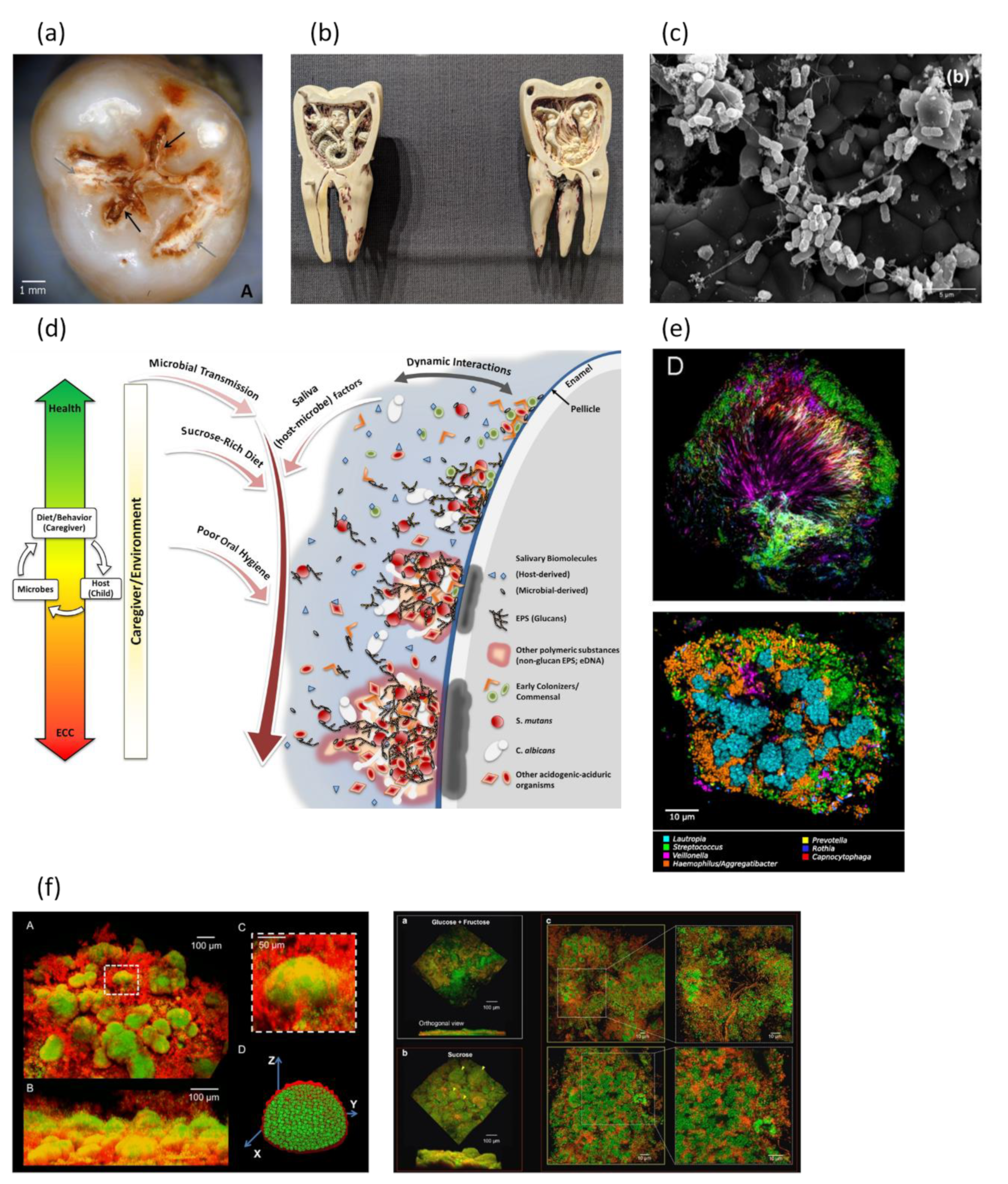
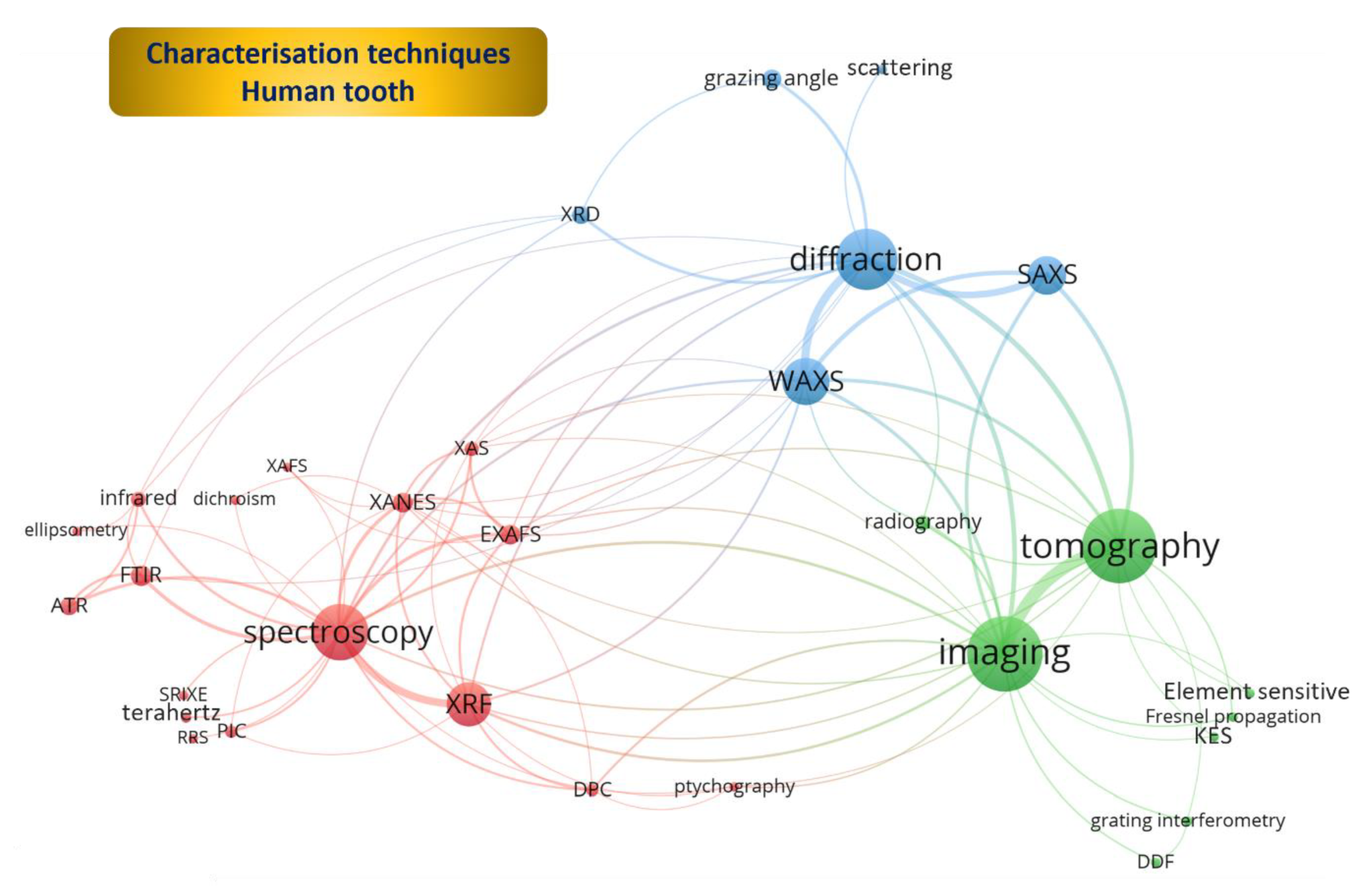
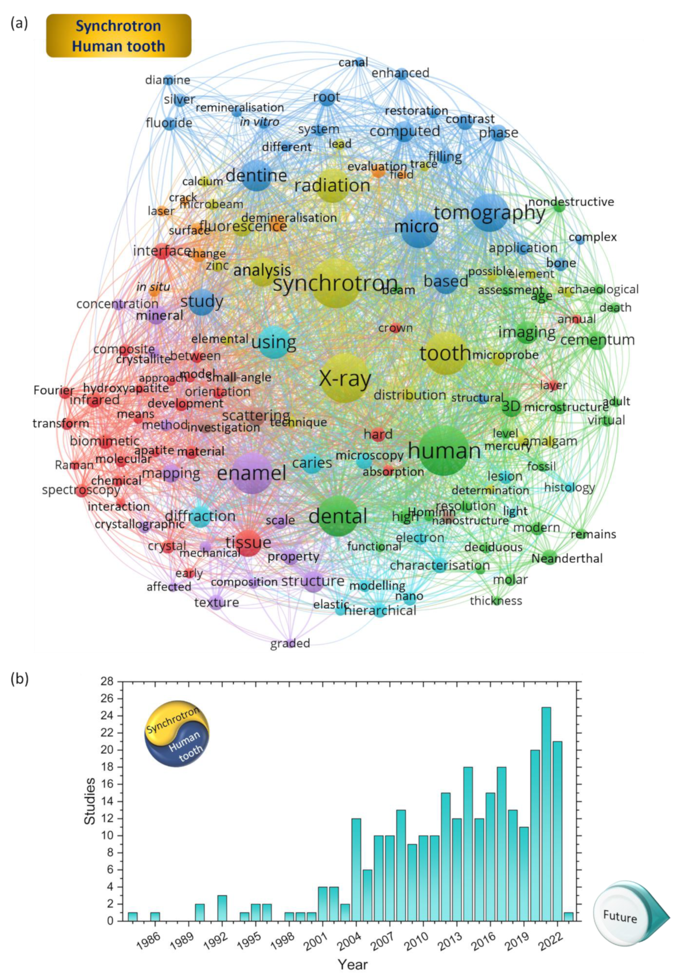
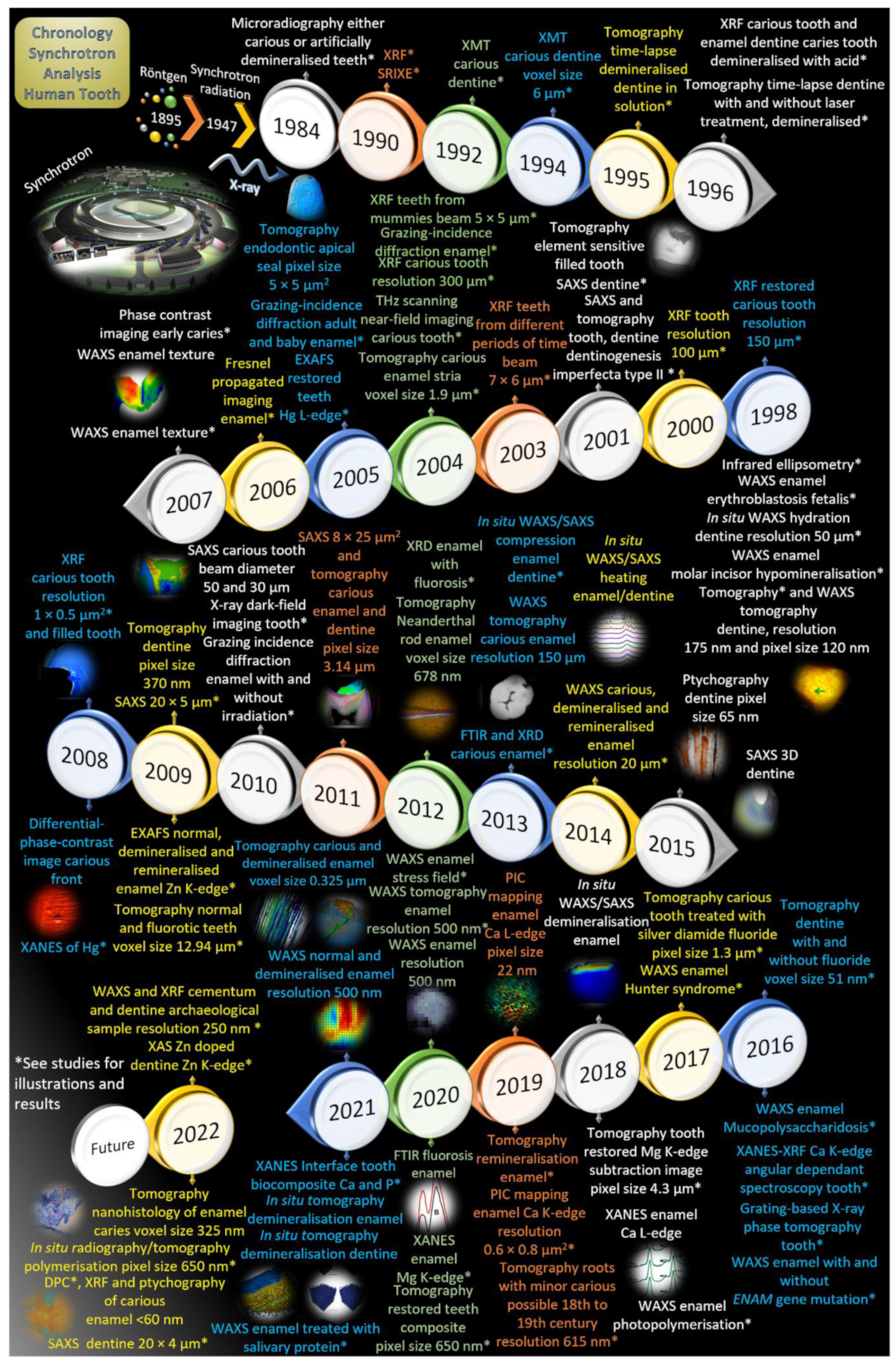
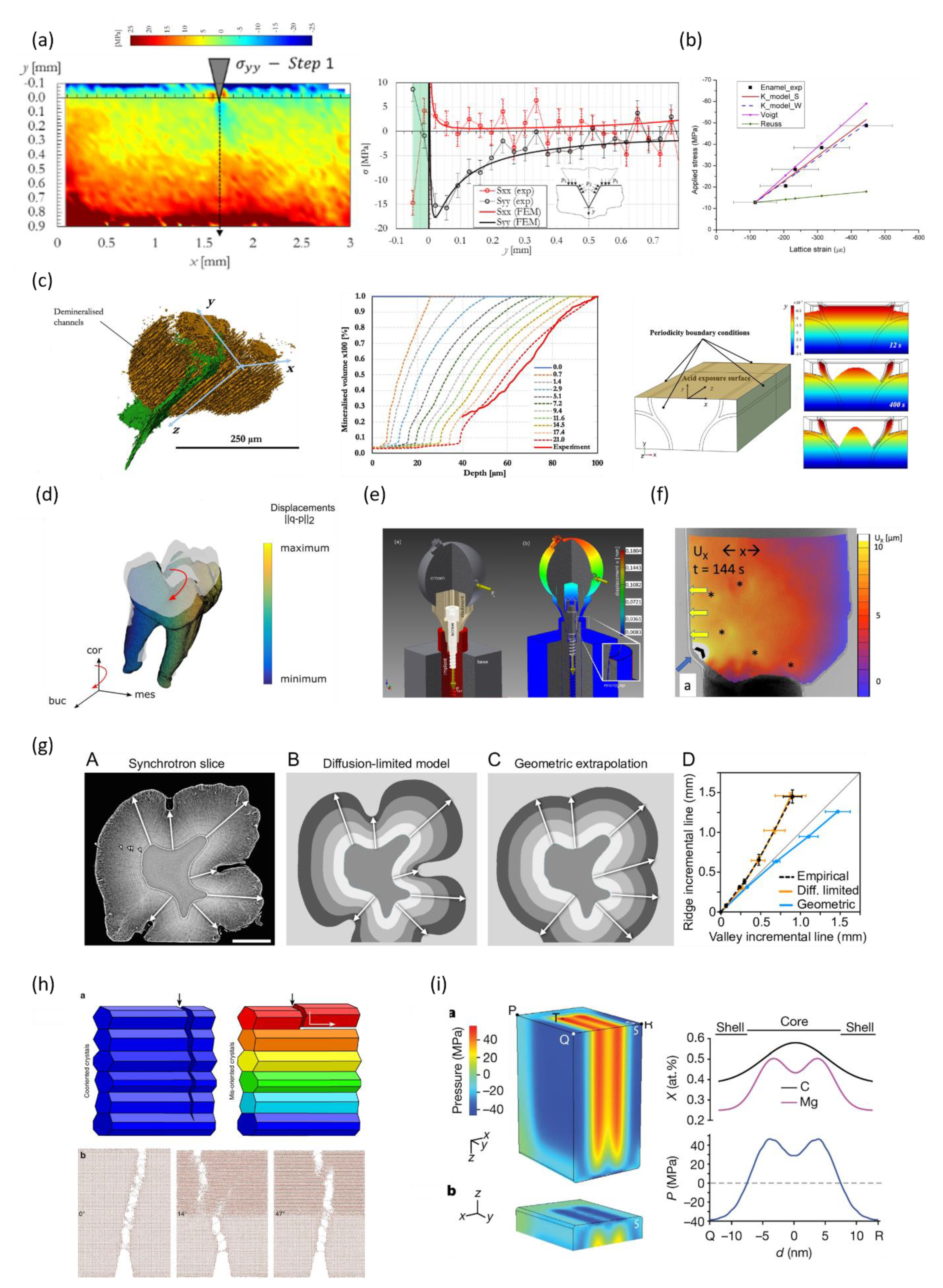
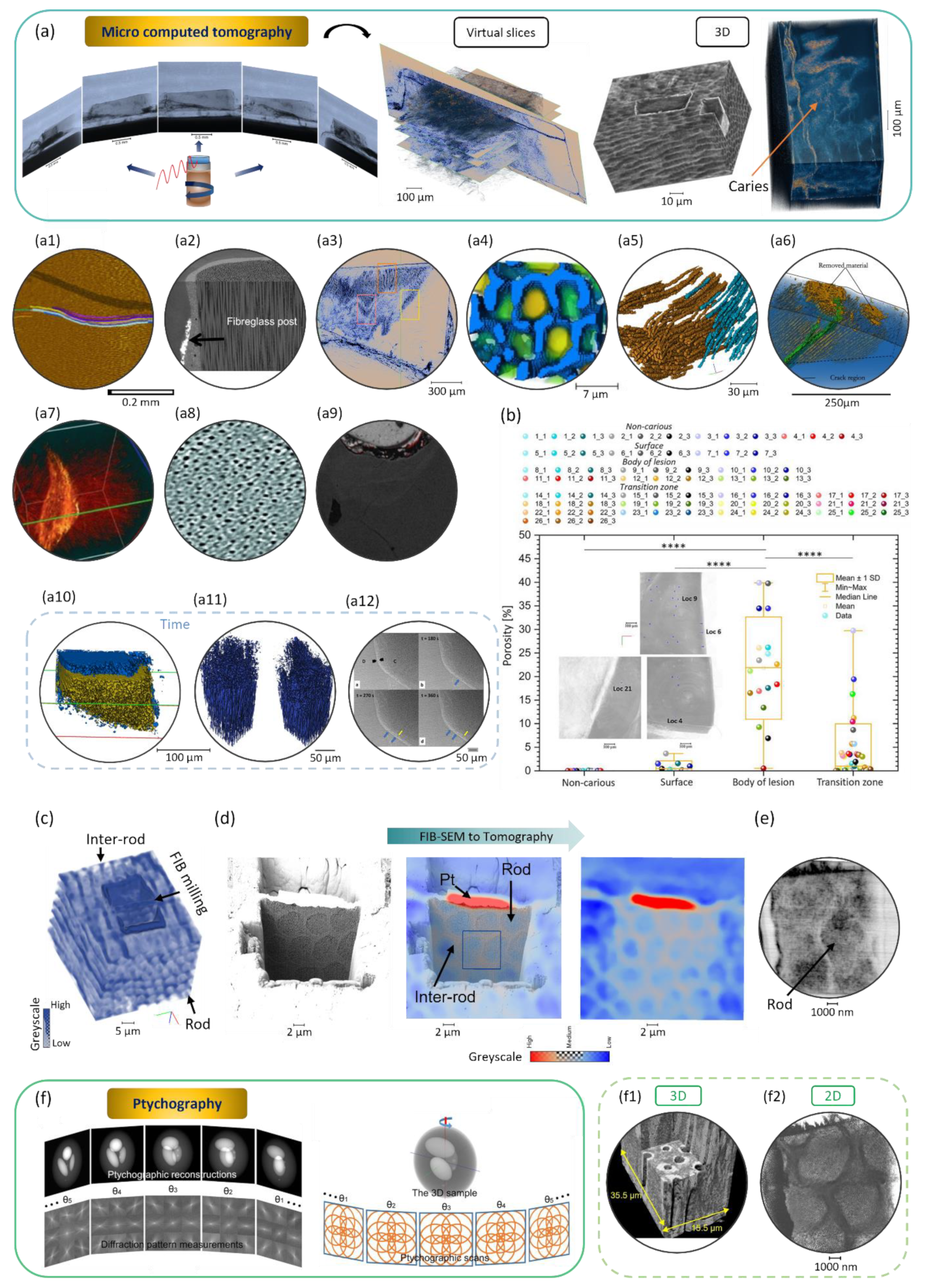
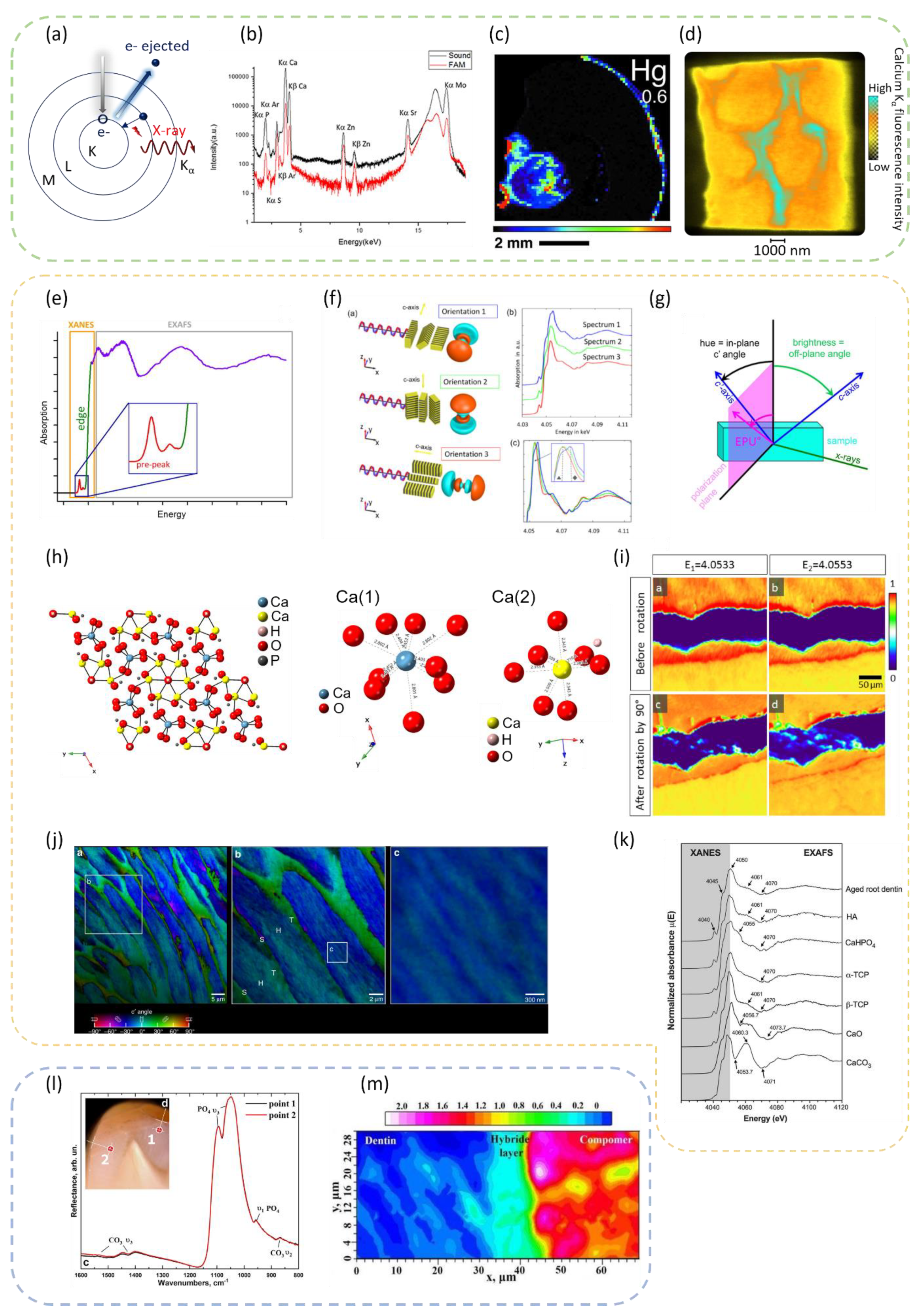
| References | Samples | Studies |
|---|---|---|
| [39,57,319] | Human enamel, carious and non-carious | Details of the pathways of demineralisation, striations, and surface zone at a voxel size of 325 nm, with more details revealed than with conventional tomography (Figure 8a) |
| [121] | Human dentine | Details for the tubule density at a pixel size of 370 nm |
| [359] | Human dentine | 3D rendering of cracks in dentine, with an effective pixel size of 300 nm and the influence of dentine tubules on the trajectory of the cracks (Figure 8a) |
| [318] | Human teeth | Fillings analysis of the quality of the composite in restorations (presence of pores), pixel size of 650 nm |
| [313] | Human teeth | Reconstruction of the cementum with the observation of tooth cementum annulations (which can be used to estimate age) with a resolution of 615 nm, which has not yet been observed at this resolution with other techniques |
| [323] | Human dentine | Silver diamine fluoride applied on dentine, to study the mineral precipitation on the sample, tomography taken at a pixel size of ~1.44 µm considered low compared with a vast number of conventional tomography equipment |
| [314] | Human dentine | Remineralisation of root dentine exposed to demineralising solutions, followed by a fluoride solution with and without EDTA. Tomography was taken at a pixel size of 1.44 µm, and a layer of mineral at the surface of the sample was detected |
| [311] | Human tooth, cementum | The cementum was imaged and reconstructed with a resolution of 650 nm, with the identification and analysis of incremental layers |
| [453] | Human enamel and dentine | Data reconstructed with a pixel size of 370 nm with the observation of the tubular structures and enamel region |
| [391] | Human teeth | Analysis of reconstructed data with a pixel size of 660 nm and 330 nm with details of the microstructure of the cementum (archaeological tooth) and the incremental lines. |
| References | Samples | Resolution or Voxel Size or Effective Pixel Size | Techniques |
|---|---|---|---|
| [357,365] | Human dentine (Figure 8a) | 320 nm | Phase-contrast images |
| [359] | Human dentine | 300 nm | X-ray tomography |
| [109] | Rhinoceros teeth | 280 nm | Holotomography |
| [275] | Cementum of Kuehneotherium and Morganucodon | 280 nm | Propagation distance tomography |
| [420] | Human dentine | ~175 nm | Phase contrast enhanced tomography |
| [275] | Cementum of Morganucodon | 10 to 130 nm | Holotomography |
| [605] | Tooth from a dog, region close to the root | 60 nm | Transmission X-ray microscope |
| [412] | Dentine of human | 51 nm | Tomography with Fresnel zone plate setup |
| [606] | Enamel and dentine of mus musculus and Sorex minutissimus | 25 nm | Holotomography |
| [607] | Human bone | 60 nm | Nano-tomography |
| [608] | Human bone | 60 nm | X-ray phase nano-tomography |
| [609] | Mouse bone | 50 nm | Holographic nano-tomography |
| [610] | Human bone | 50 nm | Phase nano-tomography |
Disclaimer/Publisher’s Note: The statements, opinions and data contained in all publications are solely those of the individual author(s) and contributor(s) and not of MDPI and/or the editor(s). MDPI and/or the editor(s) disclaim responsibility for any injury to people or property resulting from any ideas, methods, instructions or products referred to in the content. |
© 2023 by the authors. Licensee MDPI, Basel, Switzerland. This article is an open access article distributed under the terms and conditions of the Creative Commons Attribution (CC BY) license (https://creativecommons.org/licenses/by/4.0/).
Share and Cite
Besnard, C.; Marie, A.; Sasidharan, S.; Harper, R.A.; Shelton, R.M.; Landini, G.; Korsunsky, A.M. Synchrotron X-ray Studies of the Structural and Functional Hierarchies in Mineralised Human Dental Enamel: A State-of-the-Art Review. Dent. J. 2023, 11, 98. https://doi.org/10.3390/dj11040098
Besnard C, Marie A, Sasidharan S, Harper RA, Shelton RM, Landini G, Korsunsky AM. Synchrotron X-ray Studies of the Structural and Functional Hierarchies in Mineralised Human Dental Enamel: A State-of-the-Art Review. Dentistry Journal. 2023; 11(4):98. https://doi.org/10.3390/dj11040098
Chicago/Turabian StyleBesnard, Cyril, Ali Marie, Sisini Sasidharan, Robert A. Harper, Richard M. Shelton, Gabriel Landini, and Alexander M. Korsunsky. 2023. "Synchrotron X-ray Studies of the Structural and Functional Hierarchies in Mineralised Human Dental Enamel: A State-of-the-Art Review" Dentistry Journal 11, no. 4: 98. https://doi.org/10.3390/dj11040098
APA StyleBesnard, C., Marie, A., Sasidharan, S., Harper, R. A., Shelton, R. M., Landini, G., & Korsunsky, A. M. (2023). Synchrotron X-ray Studies of the Structural and Functional Hierarchies in Mineralised Human Dental Enamel: A State-of-the-Art Review. Dentistry Journal, 11(4), 98. https://doi.org/10.3390/dj11040098