Abstract
The aim of this study was to compare the positional information transfer accuracy of palatal temporary anchorage devices (TADs) of two different brands of transfer caps: PSM and Leone. Thirty plaster casts of maxillary dental arches were chosen for master models. A couple of Leone TADs were inserted in each master model. For each master model, two analysis models were created: using two transfer caps, Leone and PSM, the impressions were taken, the analogues were connected on the transfer caps, and the casts were poured. Using digital methods and equipment, such as a 3D scanner, a 3D analysis and a comparison of the accuracy of the two transfer caps in transferring the positional information of the TADs was then made. The data obtained were analyzed using the Mann–Whitney U-test at a significance level of α = 0.05. PSM transfer caps showed higher error frequency in almost all measurements. Only two measurements had a larger error in the analysis models made with Leone transfer caps. The Mann–Whitney U-test found a significant difference between the error levels of TADs found in the analysis models created with PSM transfer caps. Leone transfer caps showed greater reliability in TADs positional information transmission.
1. Introduction
The need to precisely transfer any type of intraoral information to laboratory technicians has always been the most important aspect in every branch of dental clinical practices, and as such, it has been pushing the research ahead in these fields [1]. Customized impression trays, optimized transfers of specific sizes and types of insertion, dental impression materials of increasingly high performance, and the use of high precision digital technologies [2] are aimed all with reproducing intraoral characteristics in the laboratory in the most accurate and precise way. This also applies to the positioning of palatal temporary anchorage devices (TADs) for which precision between planning and actual positioning is crucial to produce stable and effective skeletally anchored orthodontic devices [3,4].
The use of palatal TADs is gaining ground in orthodontics. Adding a skeletal support to a dental anchorage or creating a fully skeletal one is often needed to carry out expansion in adult patients, distalizations, or other movements, which can be too complex to be managed exclusively with dental anchorages [5,6,7]. Digital technologies provide high accuracy in transmitting positional information, besides providing a simplification of elaborative procedures. The newest methods include the creation of templates from the analysis of cone-beam computed tomography (CBCT) and it’s matching with the intraoral scan: after choosing the position of TADs on a computer-aided design (CAD) project, a template for TADs insertion is created and then used as a guide during the clinical session. The result is a greater accuracy in reproducing the insertion information and a reduction in operational steps [8,9]. Despite these advantages, palatal TADs free-hand positioning and subsequent transfer of their position is still very popular and widely used [10].
If TADs are placed free-hand, according to precise criteria regarding the palatal anatomy [11], it will be necessary to accurately transfer positional information to the dental technician. It is essential that the device can be fitted passively without being affected by directional forces or the stability of the TADs. To achieve this goal, analog transfer caps are usually applied on TADs and then embedded in dental impressions [12]; another way includes the use of scan bodies capable of being detected by intraoral scanners [13]. As it is widely known from different studies performed in implant dentistry, these two positional information transfer techniques provide a similar level of precision [14,15,16]. This, together with the lower economic investment required, are probably the reasons why many clinicians still turn to the analogue method for transferring positional information to the laboratory.
In the orthodontic field, several manufacturers propose their own systems for manufacturing TADs. It sometimes happens that some components of kits from different manufacturers are interchangeable. This came in particularly handy during the first two years of the recent SARS-CoV-2 pandemic period due to a general shortage of materials from various suppliers [17]. This shortage also affected the dental sector [18]. In that period, many orthodontists and orthodontic technicians, as well as for professionals working in our university’s dental clinic, sought different solutions to be able to continue producing skeletally anchored appliances. We have, therefore, noticed the interchangeability offered from the analog transfer caps produced by two manufacturers: PSM (PSM Medical Solutions, Gunningen, Germany), and Leone (Leone SpA, Sesto Fiorentino, Florence, Italy). They are indeed interchangeable when used with Leone-brand TADs for expanders and their laboratory analogues.
The aim of our study was to see whether this interchangeability could lead to a decrease in transfer accuracy, and investigate which of the above-mentioned two analog transfer caps is more accurate for transferring Leone TADs positional information. Through digital processes, analyzing data obtained using scan bodies to avoid any interference and positional changes, the authors have analyzed the positions of TADs in the final models in order to obtain information about the predictability of these systems. The null hypothesis was that both PSM and Leone transfer caps had the same level of accuracy in TADs positional information transmission.
2. Materials and Methods
The entire study was performed using materials that are normally used in dental offices to simulate the conditions of daily clinical practice.
2.1. Analog Stage
The planning of a skeletal anchorage for maxillary expansion was simulated. Thirty plaster casts of maxillary dental arch were selected from the patient archive of the university dental clinic. The inclusion criterion was good identification of palatal anatomy on the models. The thirty selected models have been catalogued as master models and referred to as codes from 1M00 to 1M029.
Two Leone TADs of 2 mm diameter and 9 mm length were inserted in each master model. The TADs have been placed in the anterior paramedian region, at a 4–5 mm distance from the palatal midline, between the second and third palatal rugae. This is usually considered the most appropriate anatomical position for this type of procedures [4]. A pilot hole was prepared in the chosen position before of the TADs insertion. The pilot hole had 1.5 mm diameter and 9 mm length, long enough to host the TADs and narrow enough to allow stable insertion. Insertion of the TADs was made permanent by applying cyanoacrylate glue to the pilot holes before placement.
From each master model, two analysis models were reproduced: one using the Leone transfer cap and, the other using the PSM transfer cap. The Leone transfer caps were positioned and fixed, using the appropriate screwdriver tool, on the TADs’ heads on the master models, then a polyvinyl siloxane impression using Elite HD Putty and Light (ZHERMACK GmbH, Marl am Dümmer, Germany) with double-phase two-component technique was taken. A plastic impression tray was used for the impression. The impression tray was previously modified by making a window at the TADs on the palate of the master model so that the transfer caps screwed onto the TADs could be accommodated. After hardening of the impression material, the Leone transfer caps were unscrewed from the TADs and the impression was then removed from the master models. The Leone transfer caps remained included in the impression. A couple of the Leone laboratory analogues for each impression were then connected and screwed onto the Leone transfer caps, and the impressions thus composed were used to realize the first thirty analysis models. A type 4 extra hard resin reinforced dental plaster was used to process this last step. A similar process was made using the PSM transfer caps to obtain the other thirty analysis models. The PSM transfer caps, due to their clip-on mechanism, remained directly embedded in the impression material without needing to modify the impression tray or unscrew the caps before removing of the impression. This was simply necessary when inserting the Leone laboratory analogues into the impression at the PSM transfer coping holes in order to be ready to realize the second cast. At the end of the whole process, to distinguish the two sets of analysis models, the letters L and P were added before their respective names thus naming from L-1M00 to L-1M29 for the analysis models made with Leone transfer caps and from P-1M00 to P-1M29 for the analysis models made with PSM transfer caps.
The Leone TADs and the two transfer caps, Leone and PSM, used in this study are shown in Figure 1.
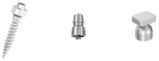
Figure 1.
The Leone TADs and the two transfer caps, Leone and PSM, used in the study.
2.2. Digital stage
At the end of the preparation process described above, the thirty master models and their respective analysis models containing the laboratory analogues were sent to the 3D Leone department (Sesto Fiorentino, Firenze, Italy) where the following steps took place:
- Digitization of the models
- Leone scan bodies for TADs were screwed onto the emerging head of the TADs and TAD analogues.
- Models were digitally scanned using a MDS-500 dental scanner and MAESTRO 3D dental scan software (AGE Solution S.r.l., Pisa, Italy) (Figure 2 and Figure 3).
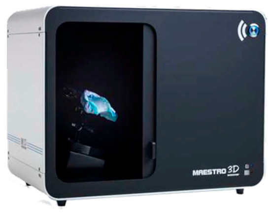 Figure 2. The benchtop scanner used for scanning models: MDS-500 (AGE Solution S.r.l., Pisa, Italy).
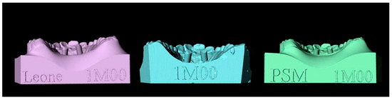 Figure 3. Models processed with Maestro 3D Dental Studio: in the center the master model, on the left the analysis model made with Leone transfer, on the right the analysis model made with PSM transfer.
 - Each digital master model was superimposed with its respective digital analysis models using some teeth and palatine rugae as reference points (Figure 4). In this way, a single reference system for each set of models was defined.
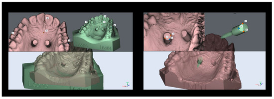 Figure 4. The left image shows the matching between models, the right shows the matching between the TAD and the scan body.
- Geometry alignment
- From the Leone digital library, stereolithography files (STLs) of the scan body and TAD were selected and then coupled together using MAESTRO 3D software (version 5).
- A coupled scan body and TAD unit were matched with each scan body of every set of models previously embedded as single reference systems using MAESTRO 3D software (Figure 4).
- Setting the measurement method
To define the position of the TADs in the area, their axis have been outlined and the angle between their axis and the reference planes were measured and compared (Figure 5). The identification of the center of the TADs’ heads on the master models was used to define the displacement of the analysis models TADs. Rhinoceros 4 software (Robert McNeel & Associates, Seattle, WA, USA) was used for the whole process and measurements.
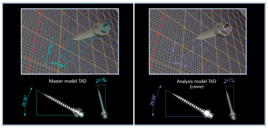
Figure 5.
Starting from the axis of the TADs in the master models (in the left image), the degrees of rotation and deviation from the x and y axes of the TADs in the analysis models (in the right image) were defined.
- 4.
- Measurements
For all TADs of the master models and their analysis models, the angles were measured and the difference between the measurements was calculated (Figure 6). The displacements of the centers of the TADs’ heads of the analysis models and of the master models were measured (Figure 7). Deviation between the STLs of the TADs and the levels of error in terms of angles and displacements were revealed (Figure 8).
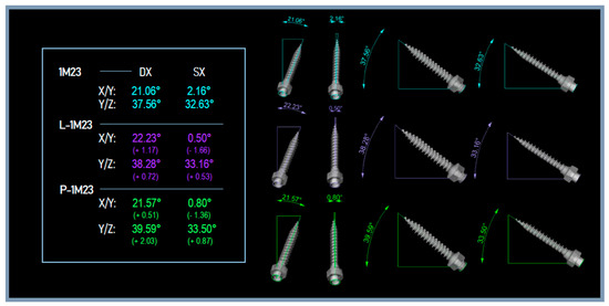
Figure 6.
The angles of the TADs of the master model 1M23 and of the related analysis models (L-1M23, P-1M23).
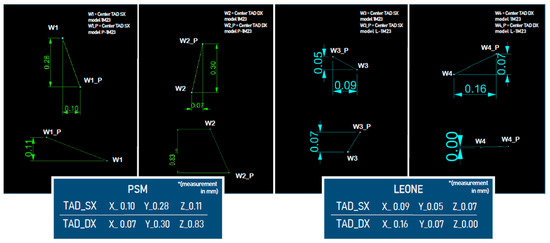
Figure 7.
The distance of the centers of the TADs’ heads of the master model 1M23 and of the related analysis models (L-1M23, P-1M23).
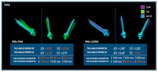
Figure 8.
TADs error levels for the analysis models L-1M16, P-1M16.
2.3. Data Collection
The errors, in terms of angles and displacements, that occurred on different planes and in different spatial directions were listed as Angle_xy, Angle_yz, Disp_x, Disp_y, Disp_z. To differentiate the parameters of the right and left TADs, dx and sx indications, respectively, have been added to the previously listed terms. The values for the error parameters described above were collected for all sixty analysis models (thirty for those made with Leone transfer caps and thirty for those made with PSM transfer caps).
2.4. Statistical Analysis
The error values of the two transfer caps have all been reported as positive values because the point was to record the absolute deviation of each TAD from its reference sample. We wanted to compare error values produced by the transfer caps of the two different manufacturers, therefore the data were statistically analysed using Mann–Whitney U-test at a significance level of α = 0.05. The statistical analysis was performed using IBM SPSS Statistics software, version 28.0 (IBM Corp, Armonk, NY, USA).
3. Results
The mean ranks related to the error levels produced by the two transfer caps in terms of angles and displacements that have occurred on different planes and in different spatial directions are shown in Figure 9. The absolute frequency of error is greater in the analysis models made with PSM transfer caps for almost all measurements, except for the angle on the yz-plane and the deviation in the x-axis direction regarding the left TADs. For the latter two measurements, the frequency of error is greater in the analysis models made with Leone transfer caps, with 31.83 in Angle_sx_yz and 33.60 in Disp_sx_x.
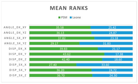
Figure 9.
The mean ranks related to the error levels produced by the transfer caps in terms of angles and displacements in the different planes and spatial directions.
According to the data shown in Table 1, the Mann–Whitney U-test found a significant difference between error levels of the TADs in the two sets of analysis model for almost all measurements, except for the angle on the yz-plane and the deviation in the x-axis direction regarding the left TADs. For the latter two measurements, the Mann–Whitney U-test did not find significant differences between error levels of the TADs in the two set of analysis model.

Table 1.
Statistical data of the Mann–Whitney U-test performed on grouping variables.
4. Discussion
As previously shown in the results, statistical significance exists for measurements whose frequency of error is greater in the analysis models made with PSM transfer caps than for measurements whose frequency of error is greater in the analysis models realized with Leone transfer caps. This suggests that PSM transfer caps have a lower level of accuracy in transferring TAD position information than Leone transfer caps. This could lead to a few problems, mainly related to the difficulty of coupling the appliance during placement in the patient’s mouth, and to potential instability of the appliance in the long term [19,20].
The authors performed the experiment in such a way as to reduce the risk of introducing inaccuracies and errors of various kinds as much as possible. The manufacturer’s directions were strictly followed for all materials used. In addition, impressions were taken with the double-phase two-component technique which, as described in the literature [10], yields optimal results in terms of accuracy. As demonstrated by Iodice et al. [8], the positioning of TADs by the direct method is safe and accurate when performed in the anterior region of the palate. It is therefore reasonable to assume that it is the different characteristics possessed by different transfer caps that determine the greater or lower accuracy in the transfer of positional information.
Regarding these insights, given the paucity of studies in the orthodontic field, we are forced to examine the implantology literature field. Several studies have attempted to compare positional information transfers performed using transfer caps with a screw attachment method versus those with a clip-on mechanism [21,22,23]. They all concluded that the clip-on mechanism is less accurate than the screw attachment method. Some factors that can play a major role in the loss of accuracy of the clip-on mechanism are the tactile sensation and the snap mechanism that indicate proper seating of transfer cap. In some cases, the dentist feels no snap and improperly assumes that the transfer cap is properly seated [24]. Furthermore, the connection of the analog to the transfer cap with the clip-on mechanism may result in movement of the cap into the impression material. Certainly, the stability and repeatability of coupling achievable using transfer caps with a screw attachment method can ensure greater accuracy in transferring the positional information of TADs. From a clinical point of view, however, the lower level of accuracy in transferring TAD position information demonstrated by the PSM transfer cap may not be significant. Fitting defects could easily be resolved in the case of appliances with a less rigid structure, incorporating fewer tooth elements, or for those intended for less complex orthodontic movements [25,26]. However, these are assumptions that cannot be clinically verified in an in vitro study.
In the context of our experiment, we should point out that PSM transfer caps showed some difficulties during coupling to the TADs on some of the master models because of the size of the head of the transfer caps. This problem has been highlighted when PSM transfer caps were used in models with a contracted palate, where a transfer cap on one side bumps against the contralateral, not allowing a complete insertion to be achieved. It is important to specify that this problem is dependent on the head size of the PSM’s transfer caps and not on the coupling with Leone’s TADs, as it can also occur during coupling with the PSM’s TADs.
Study Limitations
The use of a plaster model for making master models may have posed a risk for the potential instability of Leone TADs following their insertion and during subsequent phases of the study. However, this option represented the easiest way to perform an in vitro study.
5. Conclusions
The Leone transfer caps, which are less bulky and are screwed, guarantee a greater accuracy in the TADs positional information transmission even if their management is slightly more difficult due to the need of modifying the tray before taking the impression and of unscrewing to allow the impression removal.
The PSM transfer caps, thanks to their clip-on mechanism, do not require any special maneuvers when taking the impression, but due to an imperfect fit, they can influence the accuracy in the positional information transmission of the TADs. Furthermore, due to their larger rectangular head, in those clinical conditions where there is a significant maxilla transverse contraction, they may bump each other, and thus their insertion could be more difficult, making them even less secure.
Author Contributions
Conceptualization, V.Q., S.E.S. and G.M.; Methodology, V.B., G.T. and S.E.S.; Validation, V.Q. and S.E.S.; Investigation, G.T., V.B. and G.F.F.; Data Curation, G.T., S.E.S., V.Q. and G.F.F.; Writing-Original Draft Preparation, G.T., V.B. and G.F.F.; Writing-Review & Editing, S.E.S. and V.Q.; Visualization, S.E.S., G.M. and V.Q.; Supervision, V.Q. and S.E.S. All authors have read and agreed to the published version of the manuscript.
Funding
This research received no external funding.
Institutional Review Board Statement
Not applicable.
Informed Consent Statement
Not applicable.
Data Availability Statement
Data are available from the corresponding Author upon request.
Acknowledgments
This research did not receive any specific grant from funding agencies in the public, commercial, or not-for-profit sectors. This cost estimation study was entirely funded by institutional funds and no external funding was received.
Conflicts of Interest
The authors declare no conflict of interest.
References
- Mejía, J.B.C.; Wakabayashi, K.; Nakamura, T.; Yatani, H. Influence of abutment tooth geometry on the accuracy of conventional and digital methods of obtaining dental impressions. J. Prosthet. Dent. 2017, 118, 392–399. [Google Scholar] [CrossRef]
- Blatz, M.B.; Conejo, J. The Current State of Chairside Digital Dentistry and Materials. Dent. Clin. N. Am. 2019, 63, 175–197. [Google Scholar] [CrossRef]
- Raghis, T.R.; Alsulaiman, T.M.A.; Mahmoud, G.; Youssef, M. Efficiency of maxillary total arch distalization using temporary anchorage devices (TADs) for treatment of Class II-malocclusions: A systematic review and meta-analysis. Int. Orthod. 2022, 20, 100666. [Google Scholar] [CrossRef]
- Pozzan, L.; Migliorati, M.; Dinelli, L.; Riatti, R.; Torelli, L.; Di Lenarda, R.; Contardo, L. Accuracy of the digital workflow for guided insertion of orthodontic palatal TADs: A step-by-step 3D analysis. Prog. Orthod. 2022, 23, 27. [Google Scholar] [CrossRef] [PubMed]
- Chang, H.; Tseng, Y. Miniscrew implant applications in contemporary orthodontics. Kaohsiung J. Med. Sci. 2014, 30, 111–115. [Google Scholar] [CrossRef] [PubMed]
- Jasoria, G.; Shamim, W.; Rathore, S.; Kalra, A.; Manchanda, M.; Jaggi, N. Miniscrew Implants as Temporary Anchorage Devices in Orthodontics: A Comprehensive Review. J. Contemp. Dent. Pract. 2013, 14, 993–999. [Google Scholar] [CrossRef]
- Magkavali-Trikka, P.; Emmanouilidis, G.; Papadopoulos, M.A. Mandibular molar uprighting using orthodontic miniscrew implants: A systematic review. Prog. Orthod. 2018, 19, 1. [Google Scholar] [CrossRef] [PubMed]
- Iodice, G.; Nanda, R.; Drago, S.; Repetto, L.; Tonoli, G.; Silvestrini-Biavati, A.; Migliorati, M. Accuracy of direct insertion of TADs in the anterior palate with respect to a 3D-assisted digital insertion virtual planning. Orthod. Craniofacial Res. 2022, 25, 192–198. [Google Scholar] [CrossRef]
- Suzuki, E.Y.; Suzuki, B. Accuracy of Miniscrew Implant Placement With a 3-Dimensional Surgical Guide. J. Oral Maxillofac. Surg. 2008, 66, 1245–1252. [Google Scholar] [CrossRef]
- Schenz, N.; Schwarz, V.; Hörmann, R.; Crismani, A.G. Impression material accuracy for palatal orthodontic miniscrews. J. Orofac. Orthop. 2020, 81, 427–439. [Google Scholar] [CrossRef]
- Ahn, H.; Kang, Y.; Jeong, H.; Park, Y. Palatal temporary skeletal anchorage devices (TSADs): What to know and how to do? Orthod. Craniofacial Res. 2021, 24 (Suppl. S1), 66–74. [Google Scholar] [CrossRef] [PubMed]
- Rajapur, A.; Dixit, S.; Hoshing, C.; Raikar, S.P. The Influence of Tray Space and Repeat Pours on the Accuracy of Monophasic Polyvinylsiloxane Impression. J. Contemp. Dent. Pract. 2012, 13, 824–829. [Google Scholar] [CrossRef]
- Möhlhenrich, S.C.; Brandt, M.; Kniha, K.; Prescher, A.; Hölzle, F.; Modabber, A.; Wolf, M.; Peters, F. Accuracy of orthodontic mini-implants placed at the anterior palate by tooth-borne or gingiva-borne guide support: A cadaveric study. Clin. Oral Investig. 2019, 23, 4425–4431. [Google Scholar] [CrossRef] [PubMed]
- Lie, A.; Jemt, T. Photogrammetric measurements of implant positions. Description of a technique to determine the fit between implants and superstructures. Clin. Oral Implant. Res. 1994, 5, 30–36. [Google Scholar] [CrossRef]
- Jemt, T.; Lie, A. Accuracy of implant-supported prostheses in the edentulous jaw. Analysis of precision of fit between cast gold-alloy frameworks and master casts by means of a three-dimensional photogrammetric technique. Clin. Oral Implant. Res. 1995, 6, 172–180. [Google Scholar] [CrossRef] [PubMed]
- Bergin, J.M.; Rubenstein, J.E.; Mancl, L.; Brudvik, J.S.; Raigrodski, A.J. An in vitro comparison of photogrammetric and conventional complete-arch implant impression techniques. J. Prosthet. Dent. 2013, 110, 243–251. [Google Scholar] [CrossRef] [PubMed]
- Okeagu, C.N.; Reed, D.S.; Sun, L.; Colontonio, M.M.; Rezayev, A.; Ghaffar, Y.A.; Kaye, R.J.; Liu, H.; Cornett, E.M.; Fox, C.J.; et al. Principles of supply chain management in the time of crisis. Best Pract. Res. Clin. Anaesthesiol. 2021, 35, 369–376. [Google Scholar] [CrossRef]
- Abrar, E.; Abduljabbar, A.S.; Naseem, M.; Panhwar, M.; Vohra, F.; Abduljabbar, T. Evaluating the Influence of COVID-19 Among Dental Practitioners After Lockdown. Inq. J. Health Care Organ. Provis. Financ. 2021, 58. [Google Scholar] [CrossRef]
- Muñoz-Pereira, M.; Haas-Junior, O.; Meirelles, L.D.S.; Machado-Fernández, A.; Guijarro-Martínez, R.; Hernández-Alfaro, F.; de Oliveira, R.; Pagnoncelli, R. Stability and surgical complications of tooth-borne and bone-borne appliances in surgical assisted rapid maxillary expansion: A systematic review. Br. J. Oral Maxillofac. Surg. 2021, 59, e29–e47. [Google Scholar] [CrossRef]
- Durham, J.N.; King, J.W.; Robinson, Q.C.; Trojan, T.M. Long-term skeletodental stability of mandibular symphyseal distraction osteogenesis: Tooth-borne vs hybrid distraction appliances. Angle Orthod. 2017, 87, 246–253. [Google Scholar] [CrossRef]
- Papaspyridakos, P.; Chen, C.-J.; Gallucci, G.O.; Doukoudakis, A.; Weber, H.-P.; Chronopoulos, V. Accuracy of Implant Impressions for Partially and Completely Edentulous Patients: A Systematic Review. Int. J. Oral Maxillofac. Implant. 2014, 29, 836–845. [Google Scholar] [CrossRef]
- Nakhaei, M.; Madani, A.S.; Moraditalab, A.; Haghi, H.R. Three-dimensional accuracy of different impression techniques for dental implants. Dent. Res. J. 2015, 12, 431–437. [Google Scholar] [CrossRef]
- Tafti, A.F.; Hatami, M.; Razavi, F.; Ebadian, B. Comparison of the accuracy of open-tray and snap-on impression techniques of implants with different angulations. Dent. Res. J. 2019, 16, 413–420. [Google Scholar] [CrossRef]
- Alikhasi, M.; Siadat, H.; Monzavi, A.; Momen-Heravi, F. Three-Dimensional Accuracy of Implant and Abutment Level Impression Techniques: Effect on Marginal Discrepancy. J. Oral Implantol. 2011, 37, 649–657. [Google Scholar] [CrossRef]
- Barone, T.R.; Cahali, M.B.; Vasconcelos, C.; Barone, J.R. A comparison of tooth-borne and bone-anchored expansion devices in SARME. Oral Maxillofac. Surg. 2020, 24, 181–187. [Google Scholar] [CrossRef]
- Lombardo, L.; Occhiuto, G.; Paoletto, E.; Maino, B.G.; Siciliani, G. Class II treatment by palatal miniscrew-system appliance: A case report. Angle Orthod. 2020, 90, 305–313. [Google Scholar] [CrossRef] [PubMed]


Disclaimer/Publisher’s Note: The statements, opinions and data contained in all publications are solely those of the individual author(s) and contributor(s) and not of MDPI and/or the editor(s). MDPI and/or the editor(s) disclaim responsibility for any injury to people or property resulting from any ideas, methods, instructions or products referred to in the content. |
© 2023 by the authors. Licensee MDPI, Basel, Switzerland. This article is an open access article distributed under the terms and conditions of the Creative Commons Attribution (CC BY) license (https://creativecommons.org/licenses/by/4.0/).