Article

Fibroblast Response to Cyclo- and Organic Phosphate Solutions: A Cytotoxicity Study

by
Leonardo Antônio de Morais
,
Alberto Carlos Botazzo Delbem
,
Caio Sampaio
,
Vitória Bittencourt de Aguiar
,
Luigi Pedrini Guisso
,
Lucas Fernando Oliveira Tomáz Ferraresso
,
Juliano Pelim Pessan
and
Thayse Yumi Hosida
*
Department of Preventive and Restorative Dentistry, School of Dentistry, São Paulo State University (UNESP), Rua José Bonifácio, 1193, Aracatuba 16015-050, São Paulo, Brazil
*
Author to whom correspondence should be addressed.
Inorganics 2025, 13(9), 309; https://doi.org/10.3390/inorganics13090309
Submission received: 8 April 2025 / Revised: 3 September 2025 / Accepted: 9 September 2025 / Published: 18 September 2025

Abstract

Although the anticaries properties of phosphate-based compounds have been extensively investigated in recent years, their potential cytotoxic effects remain underexplored. This study evaluated the cytotoxicity of solutions containing sodium trimetaphosphate (TMP), sodium hexametaphosphate (HMP), or calcium glycerophosphate (CaGP). NIH/3T3 fibroblasts were cultured in Dulbecco’s Modified Eagle Medium (DMEM) supplemented with 10% fetal bovine serum and maintained at 37 °C, 100% humidity, and 5% CO2. The cells were seeded in 96-well plates at a density of 104 cells per well and incubated for 24 h. Subsequently, different dilutions of 10% TMP, HMP, or CaGP solutions were applied to the cells. Cell viability was assessed at 24/48 h using the MTT assay. The data were subjected to two-way ANOVA and Fisher’s LSD test. Spearman’s rank correlation was performed. HMP dilutions led to significantly lower cell viability compared to the other compounds, regardless of the incubation period. TMP maintained higher cell viability from 1/8 dilution onwards, regardless of the incubation time. For CaGP, an increase in cell viability was observed at 1/8 dilution after 24 h. In conclusion, TMP and CaGP demonstrated reduced cytotoxicity at higher dilutions compared to HMP, suggesting their potential as promising candidates for the development of novel biomaterials.

Graphical Abstract

1. Introduction

Dental caries is considered the most prevalent chronic oral disease in childhood and remains a global public health problem [1,2]. This disease is biofilm-mediated and is associated with the frequent consumption of carbohydrates, which promotes pH reduction and leads to the progressive loss of minerals from dental hard tissues [3]. It is a disease determined by biological, behavioral, psychosocial, and environmental factors [3]. Strong evidence demonstrates the effectiveness of fluoride-containing products in controlling dental caries [4,5,6], particularly fluoride toothpaste, which is considered the best non-professional method for its management. This is because fluoride toothpaste combines the periodic removal or disruption of dental biofilm with the cariostatic properties of fluoride [7]. In this context, measures aimed at increasing intraoral ionic reservoirs should be based on studies that evaluate the ability of these ions to remain in the dental biofilm for extended periods. Among the available strategies, it is believed that supplementing fluoridated products with phosphate salts enhances the efficacy of fluoride. This is because these salts seem to act as a partial barrier to bacterial acids due to their affinity for the enamel surface [8]. Phosphates are generally classified as cyclic phosphates, polyphosphates, or branched inorganic phosphates. Polyphosphates have the general formula M(n+2)PnO(3n+1) with phosphorus ions arranged in linear chains. These phosphates are nominated according to the number of phosphorus atoms, for instance, tripolyphosphates, tetrapolyphosphates, although monophosphates and diphosphates are still called orthophosphate and pyrophosphate, respectively. Cyclic phosphates (also known as metaphosphates) have a composition represented by the formula (MPO3)n [9,10]. Until recently, only cyclo-triphosphate (sodium trimetaphosphate—TMP) and cyclo-hexaphosphate (sodium hexametaphosphate—HMP) have been employed in mineralization processes [10,11].
TMP is the most studied cyclic phosphate for dental applications [12] due to effects on dental hard tissues [13,14,15] mediated by the adsorption of the cyclic phosphate onto the dental enamel surface, which facilitates the diffusion of cations to the dental enamel [8]. Prior evidence indicates that TMP can enhance the in vitro remineralization of artificial caries-affected dentin [15]. The use of phosphates leaves the substrate surface with a greater number of electron donor sites, promoting increased adsorption of calcium phosphate on the surface [16]. Cell adhesion is also influenced by electrostatic interaction with biomaterial surfaces and is related to the polar components of surface free energy, as higher values of electron donor sites lead to greater cell adhesion [17].
In addition to TMP, in vitro data has shown that toothpastes with reduced fluoride concentration supplemented with HMP exhibited a similar effect to conventional toothpaste (1100 ppm F) on enamel demineralization [8]. The effects of combining HMP and fluoride depend on the molar ratio between these compounds [8], with the optimal concentration of HMP in toothpaste formulations containing 1100 ppm F being generally 1% [8]. Regarding the effects on dental biofilm, it was demonstrated that the combination of 1% HMP with 1100 ppm F resulted in lower calcium and higher fluoride uptake compared to the control group (1100 ppm F), while phosphorus values were similar between treatments. Additionally, this combination significantly reduced the concentration of extracellular polysaccharides and influenced the biofilm’s inorganic composition [18,19].
Unlike TMP and HMP, calcium glycerophosphate (CaGP) consists of an organic phosphate that has also demonstrated remarkable effects on dental hard tissues, as well as on dental-related biofilms. Previous data have shown that a fluoride-containing mouthrinse supplemented with CaGP promoted significantly higher enamel recovery after an in vitro pH-cycling protocol in comparison to a mouthrinse containing only F [20]. Regarding the effects of CaGP on biofilm, the phosphate incorporated in solutions led to an increase in F, Ca, and P concentrations in the biofilm, and its presence promoted an enhanced biofilm pH, even after exposure to sucrose [21]. A literature review on the anticaries effects of this polyphosphate demonstrated that the increase of calcium levels in plaque is the most likely explanation for the anticaries potential of CaGP [22].
In light of the above, the aforementioned phosphates have been demonstrated to enhance the anticaries effect of fluoride found in commonly used dental products. Despite in vivo studies showing that a concentration of 10% of TMP and HMP exhibits chronic toxicity [23], and the concentrations used in studies being lower than 10%, it is important to evaluate the cytotoxicity of solutions containing these phosphates. In addition, information on the toxicity of CaGP is still lacking. While there is currently no literature evaluating the cytotoxicity of these compounds, their incorporation into fluoridated products necessitates close contact with oral cavity tissues, raising concerns about potential adverse effects. Within this context, fibroblasts are essential connective tissue cells, responsible for the synthesis of extracellular matrix and fibers. They play a determinant role in the development and differentiation of connective tissue and its derivatives, such as bones, cartilage, and blood. Furthermore, these mesenchyme-derived cells have versatile functions, including responding to inflammation by recruiting immune cells, producing cytokines and chemokines, and modifying tissue architecture during wound healing [24].
Based on the above, and due to the lack of studies in the literature that evaluate the cytotoxicity of phosphates, this study aimed to evaluate the cytotoxicity, effects of solutions containing TMP, HMP or CaGP, administered in different concentrations. The null hypotheses of the study are that (1) there are no differences in the cytotoxic potential of the phosphates evaluated, and (2) there is no difference in the cytotoxicity of phosphates according to the concentrations administered.

2. Results

2.1. Evaluation of Cell Viability

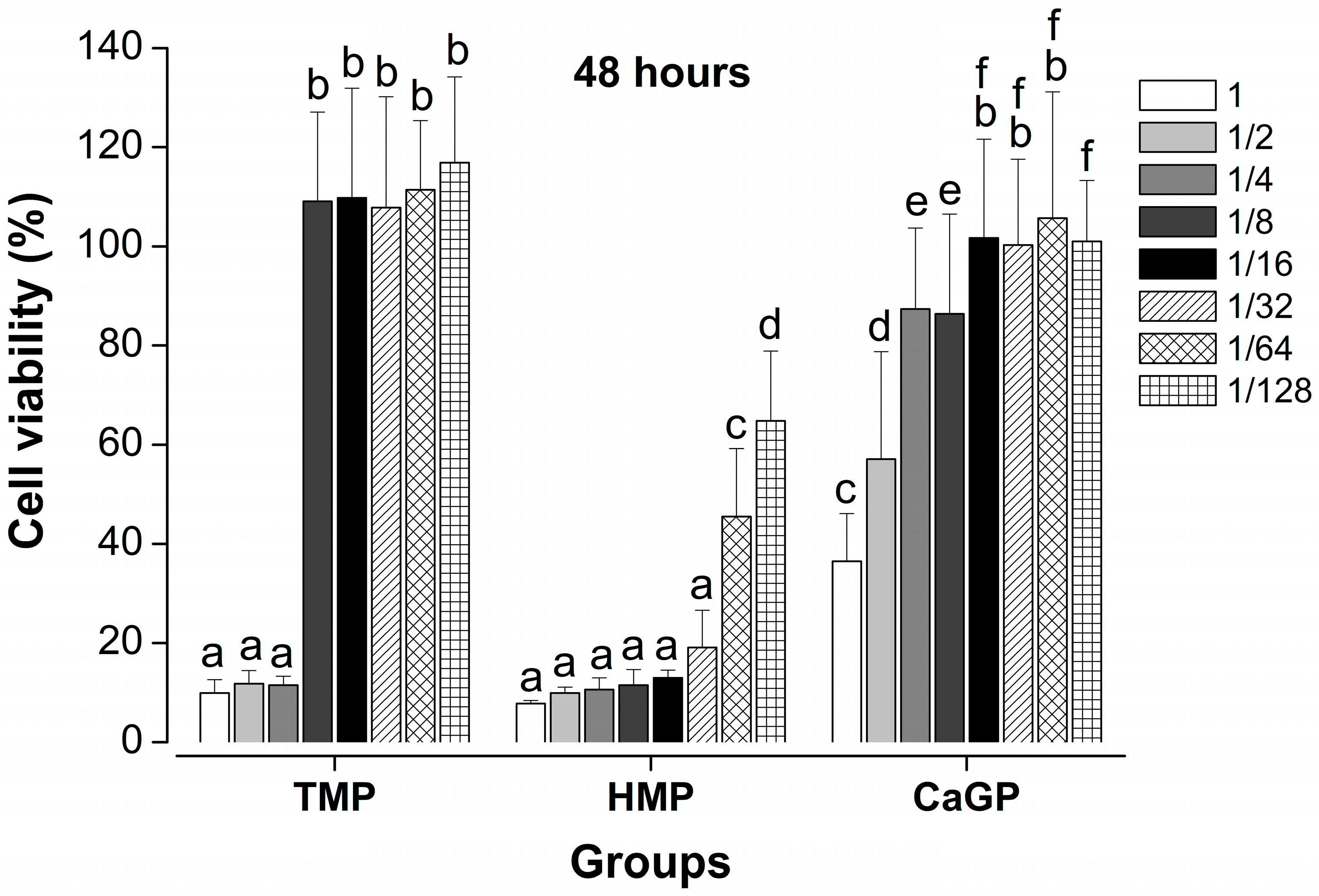
Cell viability was significantly higher in the 48 h evaluation period when compared to 24 h with TMP and CaGP solutions (p < 0.001), except for HMP (p = 0.786). Cell viability was significantly greater for solutions containing CaGP followed by TMP and HMP (p < 0.001). Unlike HMP, TMP showed significantly higher cell viability starting from the 1/8 dilution, regardless of the evaluation period (p < 0.001). Regarding CaGP, there was an increase in cell viability from the 1/8 dilution at the 24 h period and 1/4 dilution at the 48 h period (p < 0.001). Other comparisons are described in Figure 1 and Figure 2.

2.2. Determination of the pH of the Dilutions

The mean pH values and standard deviations are presented in Table 1. TMP maintained stable pH values across all dilutions. For HMP and CaGP, mean pH values > 8.00 were observed from 1/16 onwards. Spearman’s rank analysis showed weak negative correlations between culture-medium pH and the concentrations of TMP (ρ = −0.334, p = 0.00423) and CaGP (ρ = −0.334, p = 0.00432). By contrast, HMP exhibited a strong negative correlation with pH (ρ = −0.804, p < 0.001). Spearman’s rank correlation between culture-medium pH and cell viability showed no significant correlation for TMP at 24 h (p = 0.216) or 48 h (p = 0.410), nor for CaGP at 24 h (p = 0.404) or 48 h (p = 0.788). In contrast, HMP exhibited a weak positive correlation at both time points (24 h: ρ = 0.259, p = 0.0281; 48 h: ρ = 0.264, p = 0.0252). The mean (standard deviation) pH of pure DMEM was 8.11 (0.32).

3. Discussion

Technological advancement has driven research and development of new biomaterials for treating and recovering from injuries caused by trauma and degenerative diseases [25,26,27]. In the field of dentistry, the focus has been on controlling dental caries, a disease related to the demineralization of the hard tooth structure (enamel and dentin) [28]. Phosphates have the ability to modify hydroxyapatite surfaces through adsorption, potentially reducing enamel dissolution, besides interfering with biofilm composition [19,21,29]. This study evaluated the cytotoxicity of two cyclophosphates (i.e., HMP and TMP) and an organic phosphate (CaGP), showing that TMP and CaGP exhibited significantly lower cytotoxicity at higher dilutions than HMP. Also, the higher the phosphate concentration in the medium, the higher the cytotoxicity effects. Based on this, the study’s null hypotheses were rejected.
Overall, TMP demonstrated significantly lower cytotoxicity at the lowest concentrations, while presenting cytotoxicity similar to HMP and higher than CaGP at the highest concentrations. Based on this trend, it can be suggested that cell death could be attributed to the presence of precipitate in the medium, as well as the elevated concentration of phosphates. The use of phosphates leaves the substrate surface with a greater number of electron donor sites, enhancing the adsorption of calcium phosphate on the surface [16]. Cellular adhesion is also influenced by electrostatic interaction with biomaterial surfaces and is related to the polar components of surface free energy; higher values of electron donor sites lead to greater cellular adhesion [17].
CaGP exhibited significantly higher cell viability than HMP at all dilutions and TMP at the most concentrated dilutions. In a literature review on the anticaries effects of CaGP, increases in plaque calcium levels are suggested to be the most likely explanation for CaGP’s anticaries potential [22]; in other words, CaGP increases calcium levels in the environment. This trend was further confirmed in a recent study [30], in which cariogenic-related biofilms treated with CaGP combined with F showed the highest biofilm metabolic activity. These findings can be explained by the cumulative effect of Ca (from CaGP) and F on such microorganisms. This greater availability of calcium tends to increase the pH [21], making the environment more basic or alkaline, which can stimulate greater cell proliferation over time. In fact, in the present study, even at very high concentrations, the pH of the growth medium remained close to neutral. In addition to this assumption, it can be considered that high calcium availability can increase calcium binding to the bacterial cell surface. This calcium interacts with surface proteins, forming ionic bridges between negatively charged macromolecules, which enhances cell aggregation and strengthens biofilm matrices. The proximity of neighboring cells has been shown to depend on these calcium bridges [31], suggesting that calcium facilitates molecular connections within the biofilm [32]. Given the observed effect on biofilm, calcium binding may similarly enhance cell viability by facilitating the retention of new compounds.
HMP led to the highest cytotoxicity among the phosphates, regardless of the evaluation period. The chelating property of this phosphate might justify this trend, as HMP exhibits strong affinity for metal ions (Mg2+, Ca2+, K+, Al+, Fe3+), forming ionic complexes [33,34,35]. One example of the impact of this trait on biological cells is that this property allows HMP to bind to Ca2+ and Mg2+ in microbial cell walls, increasing cell permeability, which is related to its antimicrobial activity. In the study conducted by Sampaio et al. [19], HMP resulted in significant reductions in both biofilm metabolism and biomass production. Additionally, it had a remarkable impact on the structure and composition of the biofilm’s extracellular matrix. The authors suggested that the chelating properties of HMP likely account for these observed effects [19]. These trends are further supported by Omelon et al. [36], where the concentrations of phosphate and calcium ions exceed the supersaturation limits for hydroxyapatite. Polyphosphates can function as an ion reservoir, locally reducing the concentration of mineral ions (as polyphosphates sequester calcium from the medium) to levels below supersaturation. Given these factors, it is feasible that HMP’s ability to form complexes with cations, such as Ca2+, may have impacted metabolic activity and cell viability. It is important to note, however, that although the assumption that the chelating property of HMP may have influenced cellular physiology is reasonable, this variable was not directly assessed in the present study. Accordingly, future protocols specifically addressing variables related to the cell membrane may help to clarify the actual impact of this factor on the activity of the phosphate tested. Another noteworthy aspect of HMP’s cytotoxic profile concerns the pH of HMP-containing growth media. In this study, culture-medium pH was correlated with both HMP concentration and cell viability, suggesting that HMP-driven acidification may have contributed to the observed cytotoxic trend. While this interpretation is consistent with the data, causality cannot be inferred from correlation alone.
While the promising cytotoxicity results contribute valuable data to the literature on the safety of these compounds, some limitations warrant consideration. The absence of salivary components restricts extrapolation to human conditions, as saliva’s interaction with phosphate may influence compound activity [37,38]. Furthermore, the static model limits the applicability of these findings to clinical settings, failing to replicate the dynamic salivary clearance of the oral environment. Also, calcium and phosphate ion availability was not assessed at each concentration; future studies should investigate their roles in fibroblast behavior. Finally, given the study’s specific aims, the effects of prolonged exposure of the phosphates to the cells were not investigated, especially using complementary analytical tools such as cell morphology evaluation and differentiation assay. Based on this, further research using comprehensive tests and dynamic models mimicking in vivo conditions is needed to fully elucidate the remineralizing and cytotoxic actions of these compounds.

4. Materials and Methods

4.1. Preparation of Solutions

The experimental solutions based on HMP (71600-250G-CAS: 68915-31-1 assay: 65–70% P2O5 basis; Sigma Aldrich, St. Louis, MO, USA), TMP (PCode: 1002959054; Purity: 95%; CAS: 7785-84-4; T5508-500G) and calcium glycerophosphate (CaGP; G6626-100G-CAS: 58409-70-4-Pcode:1002316750; Sigma Aldrich) were prepared by mixing the powder of these materials with Dulbecco’s Modified Eagle Medium (DMEM; Sigma-Aldrich), both weighed and prepared at a ratio of 1:10 powder to water.

4.2. Assessment of Cell Viability

For this study, NIH/3T3 fibroblast from American Type Culture Collection (ATCC, Manassas, VA, USA; CRL-1658; isolated from a mouse NIH/Swiss embryo). The cells were cultured under standard cell culture conditions in Dulbecco’s Modified Eagle Medium (DMEM) supplemented with 10% fetal bovine serum (FBS; GIBCO, Grand Island, NE, USA), penicillin, and streptomycin (GIBCO), at 37 °C, 100% humidity, 95% air, and 5% CO2. The cells were then seeded into 96-well plates (104 cells/well) and incubated for 24 h under standard cell culture conditions to allow cell adherence before adding the solutions. Subsequently, various dilutions of TMP, HMP, and CaGP solutions (10%) were applied to the cells, including undiluted, 1/2 dilution, 1/4 dilution, 1/8 dilution, 1/16 dilution, 1/32 dilution, 1/64 dilution, and 1/128 dilution. Cell viability was assessed using the 3-(4,5-dimethylthiazol-2-yl)-2,5-diphenyltetrazolium bromide (MTT) assay after 24 and 48 h. After these times, the culture medium and the dilutions of each well were removed, and 100 μL of MTT solution (0.5 mg/mL in DMEM without FBS, 1:10) was added to each well. The MTT solution was removed after 4 h of incubation, and the formazan crystals were dissolved in 200 μL of isopropyl alcohol. The plate was kept at room temperature in a dark chamber for 30 min on a rotary shaker. Absorbance was measured at 570 nm in a plate reader (EON Spectrophotometer of EON, Biotek, Winooski, VT, USA) [39,40]. The simplified MTT method is illustrated in Figure 3.

4.3. Determination of pH of Dilutions

The pH of each dilution was determined using a pH electrode connected to an analyzer (PHR-146 MicroCombination pH Electrode—Fisher Scientific; Hampton, NH, USA) with a measurement resolution of ±0.01 pH units. The device was previously calibrated using standard buffer solutions at pH 4.0 and 7.0. Three independent readings were taken for each dilution.

4.4. Statistical Analysis

The normality of the data was assessed using the Shapiro–Wilk test. Data was subjected to two-way ANOVA, considering the analysis period and the compound, and dilution and compound in each analysis period. Fisher’s LSD test was used as the post hoc test. The pH data of the dilutions were presented descriptively as mean and standard deviation. Spearman’s rank correlation was used to assess the correlation between culture-medium pH and (1) compound concentration, and (2) cell viability at the different time points (24 and 48 h). Statistical analysis was performed using SigmaPlot 12.0 software (Systat Software Inc., San Jose, CA, USA), adopting the significance level of 5%. All experiments were conducted in triplicate on three different occasions (n = 9/each group).

5. Conclusions

Based on the information above, it can be concluded that:
  • HMP dilutions resulted in significantly lower cell viability compared to the other compounds, regardless of the incubation period.
  • TMP maintained higher cell viability from the 1/8 dilution onward, regardless of the incubation time.
  • TMP and CaGP show lower cytotoxicity at higher dilutions than HMP, suggesting that they may be promising compounds for the development of new biomaterials.

Author Contributions

Conceptualization, L.A.d.M., T.Y.H. and A.C.B.D.; methodology, L.A.d.M., A.C.B.D., C.S., V.B.d.A., L.P.G., L.F.O.T.F., J.P.P. and T.Y.H.; formal analysis, L.A.d.M., C.S., T.Y.H. and A.C.B.D.; investigation, L.A.d.M., A.C.B.D., C.S., V.B.d.A., L.P.G., L.F.O.T.F., J.P.P. and T.Y.H.; writing—original draft preparation, L.A.d.M. and C.S.; writing—review and editing, L.A.d.M., A.C.B.D., C.S., V.B.d.A., J.P.P. and T.Y.H.; supervision, A.C.B.D. and T.Y.H.; project administration, A.C.B.D. and T.Y.H.; funding acquisition, A.C.B.D. All authors have read and agreed to the published version of the manuscript.

Funding

The Coordination for the Improvement of Higher Education Personnel (CAPES), Finance Code 001 (scholarship) and the National Council for Scientific and Technological Development (CNPq), Finance Code 4831/2021-04 (scholarship).

Institutional Review Board Statement

Not applicable.

Informed Consent Statement

Not applicable.

Data Availability Statement

The original contributions presented in this study are included in the article. Further inquiries can be directed to the corresponding author.

Conflicts of Interest

The authors declare no conflict of interest.

References

  1. World Health Organization. Global Oral Health Status Report: Towards Universal Health Coverage for Oral Health by 2030; World Health Organization: Geneva, Switzerland, 2022. [Google Scholar]
  2. Jain, N.; Dutt, U.; Radenkov, I.; Jain, S. WHO’s global oral health status report 2022: Actions, discussion and implementation. Oral Dis. 2024, 30, 73–79. [Google Scholar] [CrossRef] [PubMed]
  3. Machiulskiene, V.; Campus, G.; Carvalho, J.C.; Dige, I.; Ekstrand, K.R.; Jablonski-Momeni, A.; Maltz, M.; Manton, D.J.; Martignon, S.; Martinez-Mier, E.A.; et al. Terminology of Dental Caries and Dental Caries Management: Consensus Report of a Workshop Organized by ORCA and Cariology Research Group of IADR. Caries Res. 2020, 54, 7–14. [Google Scholar] [CrossRef] [PubMed]
  4. Marinho, V.C.; Worthington, H.V.; Walsh, T.; Chong, L.Y. Fluoride gels for preventing dental caries in children and adolescents. Cochrane Database Syst. Rev. 2015, 15, CD002280. [Google Scholar] [CrossRef]
  5. Marinho, V.C.; Chong, L.Y.; Worthington, H.V.; Walsh, T. Fluoride mouthrinses for preventing dental caries in children and adolescents. Cochrane Database Syst. Rev. 2016, 7, CD002284. [Google Scholar] [CrossRef]
  6. Walsh, T.; Worthington, H.V.; Glenny, A.M.; Marinho, V.C.; Jeroncic, A. Fluoride toothpastes of different concentrations for preventing dental caries. Cochrane Database Syst. Rev. 2019, 3, CD007868. [Google Scholar] [CrossRef]
  7. Tenuta, L.M.; Nóbrega, D.F.; Mei, M.L. Chapter 9.1: The Use of Fluorides in the Control of Coronal Caries. Monogr. Oral Sci. 2023, 31, 129–148. [Google Scholar]
  8. Mohammadipour, H.S.; Maghrebi, Z.F.; Ramezanian, N.; Ahrari, F.; Daluyi, R.A. The effects of sodium hexametaphosphate combined with other remineralizing agents on the staining and microhardness of early enamel caries: An in vitro modified pH-cycling model. Dent. Res. J. 2019, 16, 398–406. [Google Scholar]
  9. Greenfield, S.; Clift, M. General properties of the condensed phosphates. In Analytical Chemistry of the Condensed Phosphates; Belcher, R., Freiser, H., Eds.; Elsevier: San Diego, CA, USA, 1975; pp. 1–36. [Google Scholar]
  10. Kulaev, I.; Vagabov, V.; Kulakovskaya, T. The Biochemistry of Inorganic Polyphosphates, 2nd ed.; John Wiley & Sons: Newark, NJ, USA, 2005. [Google Scholar]
  11. Schröder, H.C.; Müller, W.E.G. Inorganic Polyphosphates: Biochemistry, Biology, Biotechnology; Springer: Berlin/Heidelberg, Germany, 1999. [Google Scholar]
  12. Manna, C.M.; Nassar, M.Y.; Tofan, D.; Chakarawet, K.; Cummins, C.C. Facile synthesis of mononuclear early transition-metal complexes of κ3cyclo-tetrametaphosphate ([P4O12]4−) and cyclo-trimetaphosphate ([P3O9]3−). Dalton Trans. 2014, 28, 1509–1518. [Google Scholar] [CrossRef]
  13. Durmuş, E.; Kölüş, T.; Çoban, E.; Yalçınkaya, H.; Ülker, H.E.; Çelik, I. In vitro determination of the remineralizing potential and cytotoxicity of non-fluoride dental varnish containing bioactive glass, eggshell, and eggshell membrane. Eur. Arch. Paediatr. Dent. 2023, 24, 229–239. [Google Scholar] [CrossRef]
  14. Nagata, M.E.; Delbem, A.C.B.; Báez-Quintero, L.C.; Danelon, M.; Sampaio, C.; Monteiro, D.R.; Wiegand, A.; Pessan, J.P. Effect of fluoride gels with nano-sized sodium trimetaphosphate on the in vitro remineralization of caries lesions. J. Appl. Oral Sci. 2023, 31, e20230155. [Google Scholar] [CrossRef] [PubMed]
  15. Liu, Y.; Li, N.; Qi, Y.; Niu, L.N.; Elshafiy, S.; Mao, J.; Breschi, L.; Pashley, D.H.; Tay, F.R. The use of sodium trimetaphosphate as a biomimetic analog of matrix phosphoproteins for remineralization of artificial caries-like dentin. Dent. Mater. 2011, 27, 465–477. [Google Scholar] [CrossRef] [PubMed]
  16. Neves, J.G.; Danelon, M.; Pessan, J.P.; Figueiredo, L.R.; Camargo, E.R.; Delbem, A.C.B. Surface free energy of enamel treated with sodium hexametaphosphate, calcium and phosphate. Arch. Oral Biol. 2018, 90, 108–112. [Google Scholar] [CrossRef]
  17. Harnett, E.M.; Alderman, J.; Wood, T. The surface energy of various biomaterials coated with adhesion molecules used in cell culture. Colloids Surf. B Biointerfaces 2007, 55, 90–97. [Google Scholar] [CrossRef] [PubMed]
  18. Hosida, T.Y.; Pessan, J.P.; Cavazana, T.P.; Sampaio, C.; de Morais, L.A.; Monteiro, D.R.; Delbem, A.C.B. Effects of Sodium Hexametaphosphate and Fluoride on the pH and Inorganic Components of Streptococcus mutans and Candida albicans Biofilm after Sucrose Exposure. Antibiotics 2022, 3, 1044. [Google Scholar] [CrossRef]
  19. Sampaio, C.; Delbem, A.C.B.; Hosida, T.Y.; de Morais, L.A.; Fernandes, A.V.P.; Souza Neto, F.N.; de Camargo, E.R.; Monteiro, D.R.; Pessan, J.P. Effects of nano-sized sodium hexametaphosphate on the viability, metabolism, matrix composition, and structure of dual-species biofilms of Streptococcus mutans and Candida albicans. Biofouling 2022, 38, 321–330. [Google Scholar] [CrossRef]
  20. Torsakul, P.; Rirattanapong, P.; Prapansilp, W. The Remineralization Effect of Calcium Glycerophosphate in Fluoride Mouth Rinse on Demineralized Primary Enamel: An in vitro Study. J. Int. Soc. Prev. Community Dent. 2023, 13, 410–415. [Google Scholar] [CrossRef]
  21. Cavazana, T.P.; Hosida, T.Y.; Sampaio, C.; de Morais, L.A.; Monteiro, D.R.; Pessan, J.P.; Delbem, A.C.B. Calcium glycerophosphate and fluoride affect the pH and inorganic composition of dual-species biofilms of Streptococcus mutans and Candida albicans. J. Dent. 2021, 115, 103844. [Google Scholar] [CrossRef]
  22. Lynch, R.M. Calcium glycerophosphate and caries: A review of the literature. Int. Dent. J. 2004, 54, 310–314. [Google Scholar] [CrossRef]
  23. Lanigan, R.S. Final report on the safety assessment of sodium metaphosphate, sodium trimetaphosphate, and sodium hexametaphosphate. Int. J. Toxicol. 2001, 20, 75–89. [Google Scholar] [CrossRef]
  24. Kalluri, R. The biology and function of fibroblasts in cancer. Nat. Rev. Cancer 2016, 16, 582–598. [Google Scholar] [CrossRef] [PubMed]
  25. O’Brien, F.J. Biomaterials & Scaffolds for tissue engineering. Mater. Today 2011, 14, 88–95. [Google Scholar] [CrossRef]
  26. Haggerty, A.E.; Oudega, M. Biomaterials for spinal cord repair. Neurosci. Bull. 2013, 29, 445–459. [Google Scholar] [CrossRef] [PubMed]
  27. Zadpoor, A.A. The evolution of biomaterials research. Mater. Today 2013, 16, 408–409. [Google Scholar] [CrossRef]
  28. Gu, L.S.; Kim, J.; Kim, Y.K.; Liu, Y.; Dickens, S.H.; Pashley, D.H.; Ling, J.Q.; Tay, F.R. A chemical phosphorylation-inspired design for type I collagen biomimetic remineralization. Dent. Mater. 2010, 26, 1077–1089. [Google Scholar] [CrossRef]
  29. Luka, B.; Duerrschnabel, A.; Neumaier, S.; Schlueter, N.; Vach, K. Interaction between Hexametaphosphate, Other Active Ingredients of Toothpastes, and Erosion-Abrasion in Enamel in vitro. Caries Res. 2023, 57, 265–275. [Google Scholar] [CrossRef]
  30. Cavazana, T.P.; Hosida, T.Y.; Sampaio, C.; de Morais, L.A.; Monteiro, D.R.; Pessan, J.P.; Delbem, A.C.B. The Activity of Calcium Glycerophosphate and Fluoride against Cariogenic Biofilms of Streptococcus mutans and Candida albicans Formed In Vitro. Antibiotics 2023, 20, 422. [Google Scholar] [CrossRef]
  31. Rose, R. The role of calcium in oral streptococcal aggregation and the implications for biofilm formation and retention. Biochim. Biophys. Acta (BBA)-Gen. Subj. 2000, 1475, 76–82. [Google Scholar] [CrossRef]
  32. Körstgens, V.; Flemming, H.-C.; Wingender, J.; Borchard, W. Influence of calcium ions on the mechanical properties of a model biofilm of mucoid Pseudomonas aeruginosa. Water Sci. Technol. 2001, 43, 49–57. [Google Scholar] [CrossRef] [PubMed]
  33. Changgen, L.; Yongxin, L. Selective flotation of scheelite from calcium minerals with sodium oleate as a collector and phosphates as modifiers. II. The mechanism of the interaction between phosphate modifiers and minerals. Int. J. Miner. Process. 1983, 10, 219–235. [Google Scholar] [CrossRef]
  34. Choi, I.K.; Wen, W.W.; Smith, R.W. The effect of a long chain phosphate on the adsorption of collectors on kaolinite. Miner. Eng. 1993, 6, 1191–1197. [Google Scholar] [CrossRef]
  35. Kura, G.; Ohashi, S.; Kura, S. Complex formation of cyclic phosphate anions with bivalent cátions. J. Inorg. Nucl. Chem. 1974, 36, 1605–1609. [Google Scholar] [CrossRef]
  36. Larsen, M.J.; Fejerskov, O. Chemical and structural challenges in remineralization of dental enamel lesions. Scand. J. Dent. Res. 1989, 97, 285–296. [Google Scholar] [CrossRef]
  37. Enax, J.; Fandrich, P.; Schulze Zur Wiesche, E.; Epple, M. The Remineralization of Enamel from Saliva: A Chemical Perspective. Dent. J. 2024, 12, 339. [Google Scholar] [CrossRef] [PubMed]
  38. Hannig, C.; Hannig, M. Interaction between Saliva, Pellicle, and Dental Erosion. Monogr. Oral Sci. 2025, 33, 128–148. [Google Scholar] [PubMed]
  39. Omelon, S.; Georgiou, J.; Henneman, Z.J.; Wise, L.M.; Sukhu, B.; Hunt, T.; Wynnyckyj, C.; Holmyard, D.; Bielecki, R.; Grynpas, M.D. Control of vertebrate skeletal mineralization by polyphosphates. PLoS ONE 2009, 4, e5634. [Google Scholar] [CrossRef] [PubMed]
  40. Neto, F.N.S.; Morais, L.A.; Gorup, L.F.; Ribeiro, L.S.; Martins, T.J.; Hosida, T.Y.; Francatto, P.; Barbosa, D.B.; Camargo, E.R.; Delbem, A.C.B. Facile Synthesis of PVP-Coated Silver Nanoparticles and Evaluation of Their Physicochemical, Antimicrobial and Toxic Activity. Colloids Interfaces 2023, 7, 66. [Google Scholar] [CrossRef]
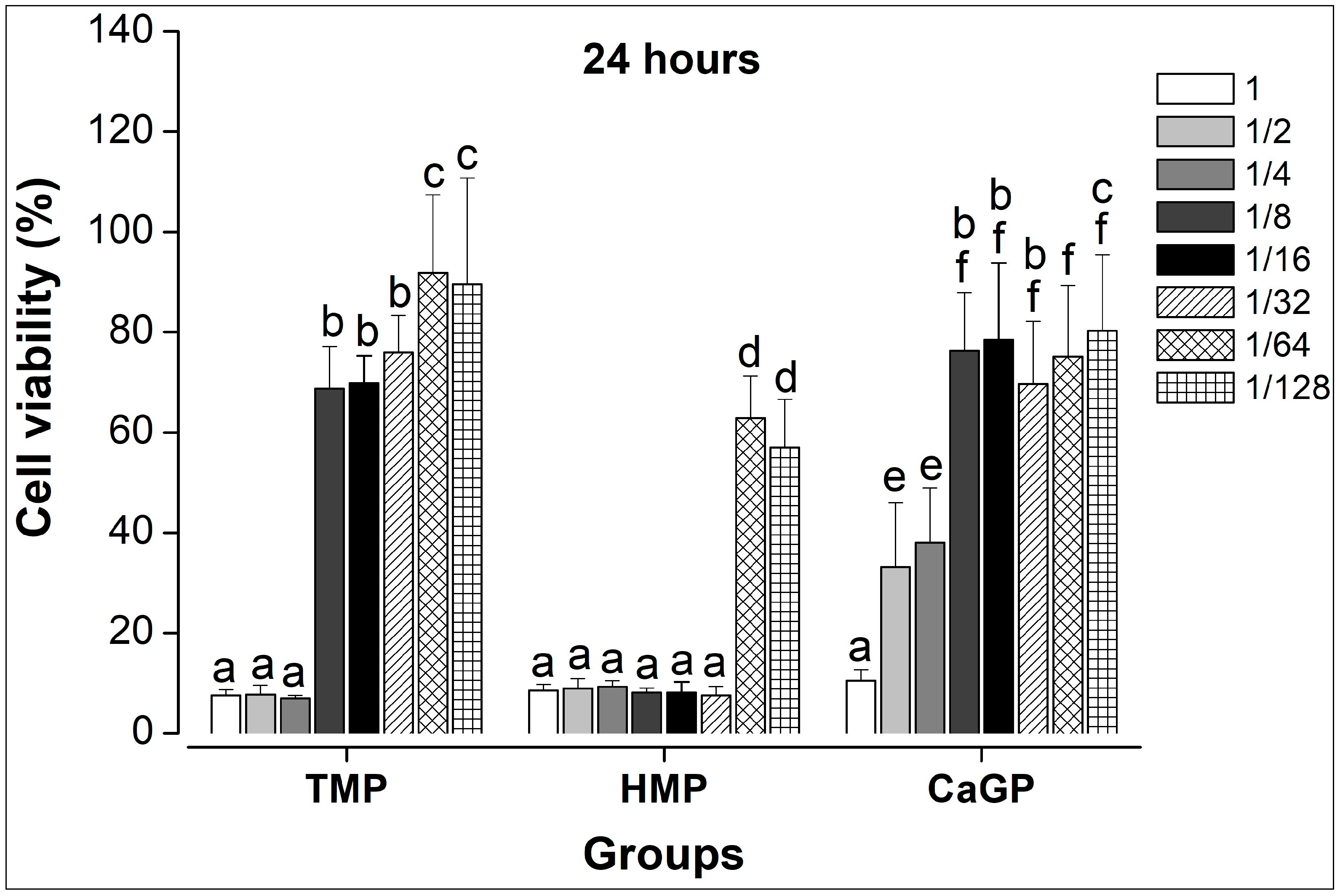
Figure 1. Viability of NIH/3T3 fibroblasts determined by MTT assay within 24 h. Distinct lowercase letter (a, b, c, d, e, f) indicates significance among materials and dilutions (Two-way ANOVA, Fisher’s LSD test, n = 9/per group). TMP = Sodium trimetaphosphate; HMP = Sodium hexametaphosphate; CaGP = Calcium glycerophosphate. The serial dilution of the compounds represents: 1 (100%), 1/2 (50%), 1/4 (25%), 1/8 (12.5%), 1/16 (6.25%), 1/32 (3.125%), 1/64 (1.56%), and 1/128 (0.78%).
Inorganics 13 00309 g001
Figure 2. Viability of NIH/3T3 fibroblasts determined by MTT assay within 48 h. Distinct lowercase letter (a, b, c, d, e, f) indicates significance among materials and dilutions (Two-way ANOVA, Fisher’s LSD test, n = 9/per group). TMP = Sodium trimetaphosphate; HMP = Sodium hexametaphosphate; CaGP = Calcium glycerophosphate. The serial dilution of the compounds represents: 1 (100%), 1/2 (50%), 1/4 (25%), 1/8 (12.5%), 1/16 (6.25%), 1/32 (3.125%), 1/64 (1.56%), and 1/128 (0.78%).
Inorganics 13 00309 g002
Figure 3. Simplified schematic representation of the MTT assay. (A) Fibroblast cell line (NIH/3T3) cells were cultured in cell culture flasks and incubated in a CO2 incubator until reaching 80% confluence. (B) Once confluence was reached, the cells were adjusted to the desired concentration and transferred to 96-well microtiter plates, where they were incubated for 24 h in a CO2 incubator. (C) After this period, the medium was removed, and the treatment groups were added in different dilutions. Following 24 and 48 h of exposure, MTT solution was added, and the cells were incubated again for 4 h. (D) After incubation, the medium was discarded, isopropyl alcohol was added, and the plates were agitated for 30 min to allow formazan formation. Finally, absorbance was measured using a spectrophotometer (EON Spectrophotometer of EON, Biotek, Winooski, VT, USA). In summary, viable cells with active metabolism are capable of reducing MTT to formazan, resulting in color variations depending on the metabolic activity influenced by the applied treatment.
Inorganics 13 00309 g003
Table 1. Mean (standard deviation) pH values according to the dilutions of the experimental groups.
GroupsDilutions
11/21/41/81/161/321/641/132
TMP7.68
(0.43)
7.86
(0.38)
7.95
(0,37)
7.98
(0.37)
8.01
(0.34)
8.02
(0.35)
8.04
(0.35)
8.02 (0.36)
HMP6.38
(0.10)
6.77
(0.15)
7.10
(0.22)
7.47
(0.27)
7.76
(0.32)
7.94
(0.35)
7.98
(0.37)
7.78
(0.49)
CaGP7.60
(0.20)
7.57
(0.15)
7.59
(0.16)
7.68 (0.19)7.75 (0.24)7.81 (0.26)7.89 (0.27)7.79 (0.41)
TMP = Sodium trimetaphosphate; HMP = Sodium hexametaphosphate; CaGP = Calcium glycerophosphate. Mean (standard deviation) pure DMEM pH value = 8.11 (0.32). The serial dilution of the compounds represents: 1 (100%), 1/2 (50%), 1/4 (25%), 1/8 (12.5%), 1/16 (6.25%), 1/32 (3.125%), 1/64 (1.56%), and 1/128 (0.78%). n = 9/per group.
Disclaimer/Publisher’s Note: The statements, opinions and data contained in all publications are solely those of the individual author(s) and contributor(s) and not of MDPI and/or the editor(s). MDPI and/or the editor(s) disclaim responsibility for any injury to people or property resulting from any ideas, methods, instructions or products referred to in the content.