Review

Functional Diversity of the Oxidative Stress Sensor and Transcription Factor SoxR: Mechanism of [2Fe-2S] Cluster Oxidation

The Institute of Scientific and Industrial Research, Sanken, Osaka University, Mihogaoka 8-1, Ibaraki 567-0047, Osaka, Japan
Inorganics 2025, 13(9), 307; https://doi.org/10.3390/inorganics13090307
Submission received: 1 June 2025 / Revised: 7 September 2025 / Accepted: 11 September 2025 / Published: 12 September 2025
(This article belongs to the Section Bioinorganic Chemistry)

Abstract

The [2Fe-2S] transcription activator SoxR, a member of the MerR family, functions as a bacterial stress response sensor. The response governed by SoxR is activated by the oxidation of the [2Fe-2S]. In this review, I describe functional differences between Escherichia coli SoxR (EcSoxR) and Pseudomonas aeruginosa SoxR (PaSoxR). Pulse radiolysis demonstrated that the reduced form of EcSoxR reacts directly with O2 with a second-order rate constant of 5.0 × 10⁸ M−1s−1. PaSoxR was found to undergo a similar reaction, although with a 10-fold smaller rate constant (4.0 × 10⁷ M−1s−1). This difference in rate constants may reflect distinct regulatory features of EcSoxR and PaSoxR. Specifically, mutagenesis studies have shown that Lysine residues―which are located close to [2Fe-2S] clusters, in EcSoxR, but are not conserved in PaSoxR―are essential for EcSoxR activation. In contrast, both EcSoxR and PaSoxR were found to react with various redox-active compounds (RACs), including viologens, phenazines, and quinones, with no apparent differences in the kinetic behavior or specificity of the two proteins. Importantly, both O2 and RACs oxidize SoxR with the same rate constants. soxR regulon may be induced through multiple pathways, and the activation may depend on the cellular concentration of O2 and RACs.

Graphical Abstract

1. Introduction

Iron–sulfur clusters are universal cofactors that play many important roles in biological processes [1,2,3,4,5]. Iron–sulfur clusters in signal-transducing sensor proteins are functionally distinct from most iron–sulfur clusters, which act as redox centers for electron transfer reactions. In contrast, iron–sulfur clusters in sensor proteins respond to small signaling molecules in the environment [4,6,7,8], resulting in structural changes such as oxidation state and cluster type. These changes may alter gene expression in cells. The SoxR, one example of such iron–cluster-containing sensor proteins, proceeds reversible oxidation and reduction between SoxRox and SoxRred, according to the following equation.
SoxR ox e SoxR red
SoxR is a 34 kDa homodimer containing one essential [2Fe-2S] cluster per polypeptide chain. Because SoxRred can be observed by EPR spectra, its redox changes can be monitored quantitatively by EPR spectroscopy. In vivo EPR spectra show that SoxR in E. coli (EcSoxR) is maintained in a reduced form during normal aerobic growth [8]. When O2 generating compounds such as methyl viologen (MV2+) were added to E. coli cells, EPR signals disappeared rapidly, indicating oxidation of the EcSoxR cluster. Thus, SoxR senses oxidative stress. The resulting activated redox state is propagated to the SoxR DNA-binding domain, resulting in the activation of target gene promoters [9,10,11,12,13].
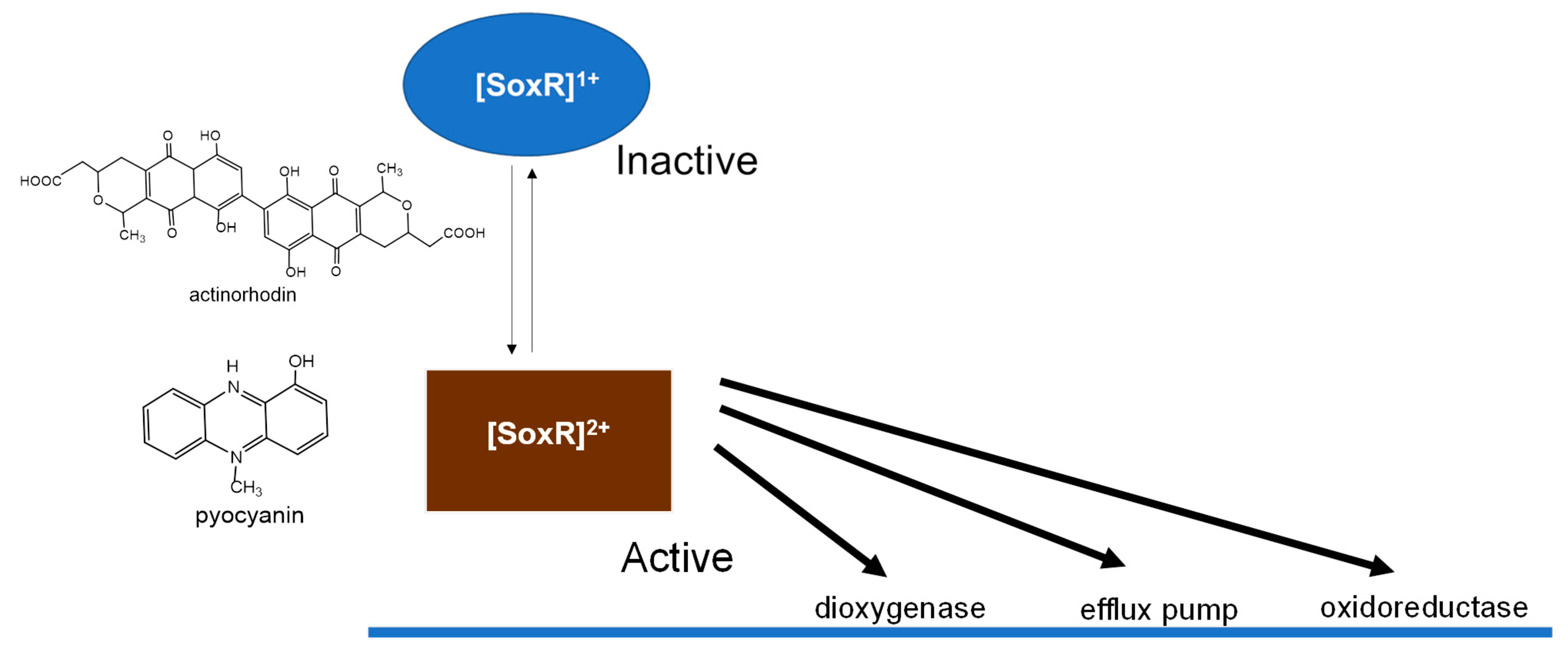
The target of activated SoxR is a single adjacent soxS gene. Increased SoxS levels induce expression of various antioxidant proteins and repair proteins [14]. This two-step soxRS-based regulatory mechanism controls the expression of genes encoding antioxidant defense proteins such as Mn-containing superoxide dismutase (SOD), glucose-6-phosphate dehydrogenase, endonuclease IV, and outer membrane drug efflux pump, as shown in Figure 1.
A number of SoxR homologs have been identified in various bacteria [15], all of which contain a SoxR-specific cysteine motif and DNA binding sites. In contrast to those in E. coli and related enteric bacteria, the majority of SoxR regulons in non-enteric bacteria such as P. aeruginosa and Streptomyces coelicolor lack genes involved in oxidative stress and detoxification [15]. Some of these bacteria produce redox-active compounds (RACs) independent of oxidative stress [15,16]. PaSoxR is activated by endogenous RAC antibiotic compounds, such as pyocyanin (Pyo) and actinorhodin (Act) [15,16,17,18], which are the physiological signals for the regulation of quorum-sensing-regulated genes. These SoxR target genes regulate genes involved in export (via the mexGHI-opmD RND efflux pump [19]) and the metabolism of small molecules [19], including RAC antibiotics and endogenous pigments (Figure 2). In these species, SoxR appears to function as a defense against the toxicity of RACs.

2. Direct Oxidation of [2Fe-2S] Cluster in SoxR by O2

An important question relates to the chemical nature of the signal sensed by SoxR. The first proposal SoxR-activating signal identified was O2 [20], but the mechanism by which O2 activates SoxR remains unclear [21]. Gu and Imaly [22] reported that EcSoxR is not directly activated by O2, but RACs. To investigate the oxidation of EcSoxR by O2, I followed the reactions that occurred following pulse radiolysis of oxygen-saturated aqueous solutions in the presence of EcSoxRox, which were previously determined [13]. Under these experimental conditions, most of the hydrated electrons (eaq) are converted to O2, while a part of the eaq population rapidly reduced the [2Fe-2S] cluster. The initial rapid absorption changes due to reduction in the [2Fe-2S] cluster partially recovered on a millisecond time scale. It is noted that the recovery was inhibited by the addition of Cu/Zn-SOD. From pulse radiolysis results, the reaction schemes can be summarized by reactions (1) and (2):
e aq + [ 2 Fe - 2 S ] 2 + [ 2 Fe - 2 S ] 1 +
O 2 + [ 2 Fe - 2 S ] 1 + + 2 H + [ 2 Fe - 2 S ] 2 + + H 2 O 2
where [2Fe-2S]2+ and [2Fe-2S]1+ are oxidized and reduced forms of SoxR, respectively.
Reaction (2), observed in SoxR, represents the first reported example of O2 oxidation, a reduced form of iron–sulfur protein. Whether other [2Fe-2S] proteins show similar reactivity was tested by examining their reactions with O2 using spinach ferredoxin and adrenodoxin as examples. However, unlike the case of SoxR, after rapid reduction in these [2Fe-2S] proteins by eaq, the recovery of absorption changes was not observed. On the other hand, in the reaction of the oxidized form of iron–sulfur proteins, reactions between iron–sulfur proteins and O2 with second-order rate constants ranging from 106 to 107 M−1 s−1 [23,24,25] were reported. In these cases, the reactions were irreversible and were accompanied by the degradation of the iron–sulfur cluster. By contrast, reaction (2) in SoxR is reversible. Therefore, it can be said that reaction (2) of SoxR is a specific characteristic of SoxR protein.
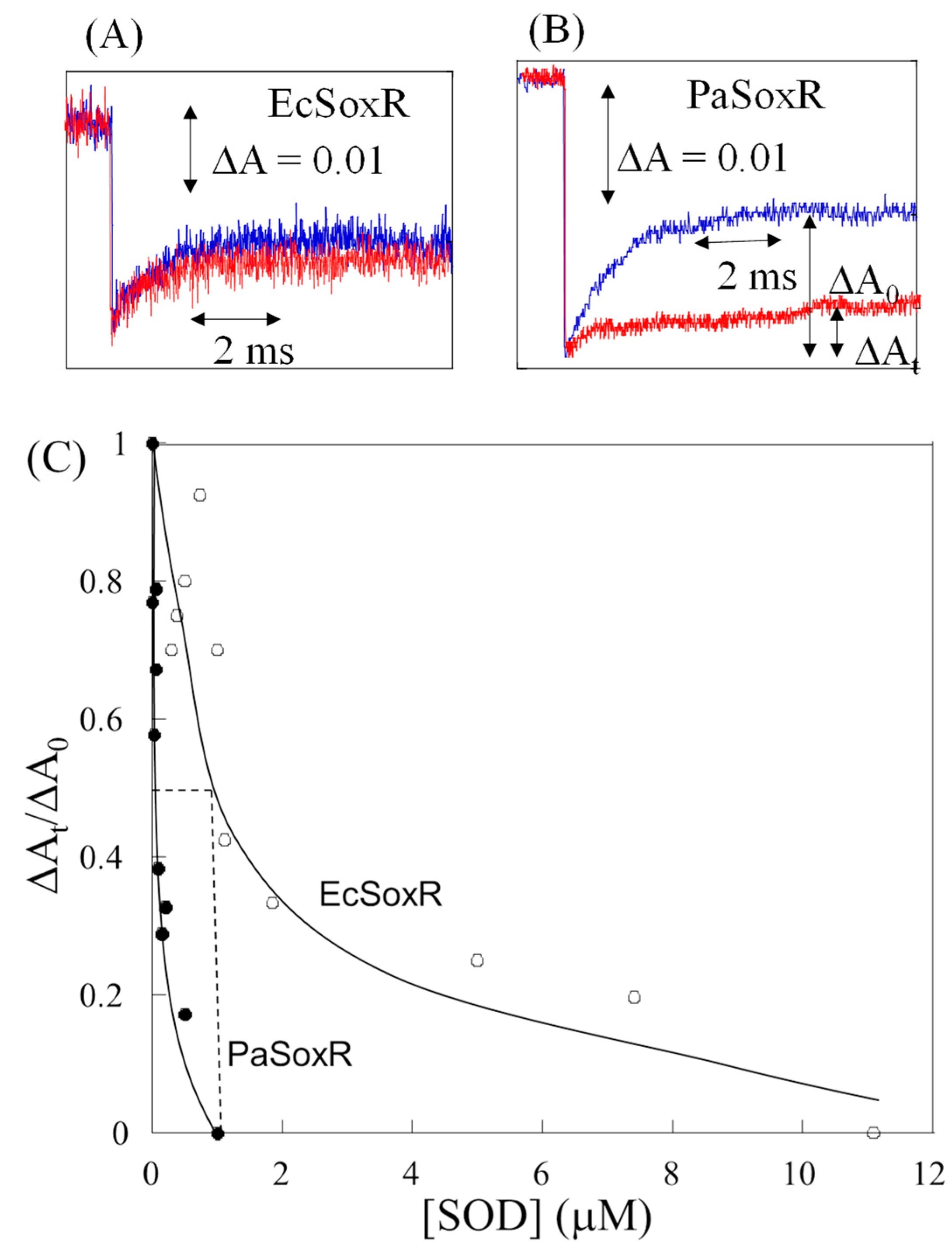
Pulse radiolysis revealed that a similar process was at work in PaSoxR. To compare the reactions of EcSoxR and PaSoxR, we studied the inhibition by SOD oxidation of SoxRred (Figure 3). The oxidation of PaSoxR was completely inhibited by 0.5 μM SOD (Figure 3B), whereas the same concentration of SOD inhibited EcSoxR by only 20% (Figure 3A). This suggests that the reactivity of O2 toward EcSoxR is much greater than toward PaSoxR. The SOD dependence of absorption changes at 420 nm for EcSoxR is presented in Figure 3C. Based on the concentration of SOD with the half-maximal inhibition of SoxRred and the rate constant for the reaction of O2 with SOD, we obtained a second-order rate constant of 5.0 × 10⁸ M−1s−1 for the reaction of O2 with EcSoxRred. In contrast, the rate constant for PaSoxRred with O2 was calculated to be 4.0 × 10⁷ M−1s−1. The difference in rate constant reflects the distinct regulatory roles for EcSoxR and PaSoxR in response to their physiological activation by O2. The mechanism underlying the different sensitivities of various SoxRs to O2 remains an important question.
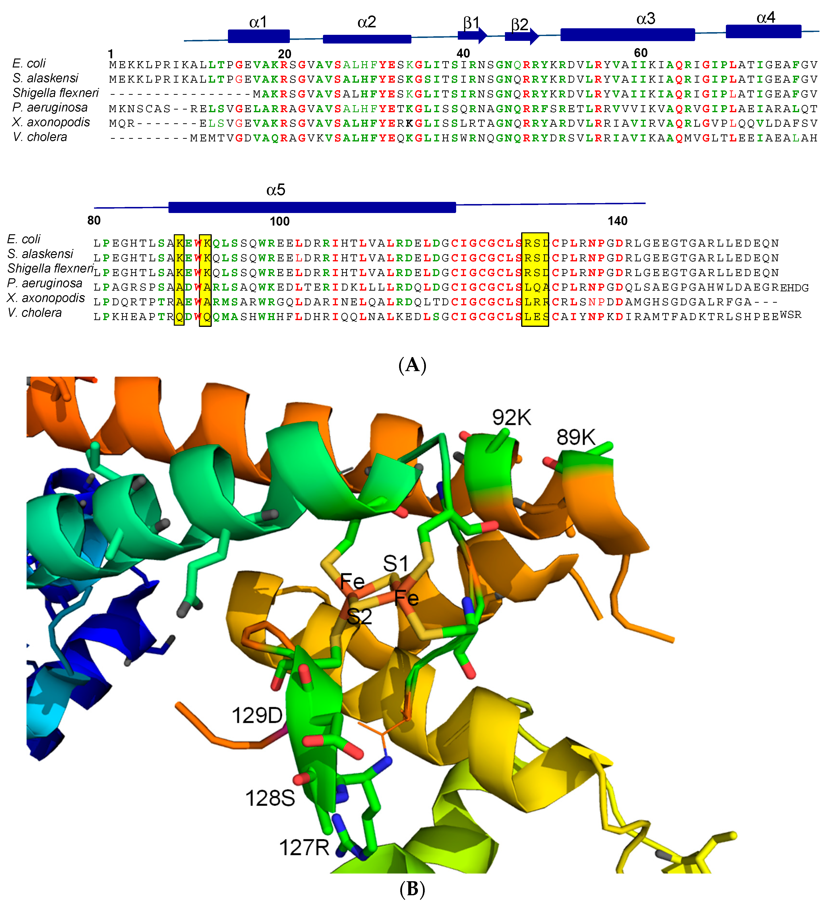
These data clearly suggest that slight alteration in structure between highly homologous SoxR proteins can lead to functional differences. Figure 4A compares sequence alignments for SoxR proteins from different enteric and non-enteric bacteria. SoxR homologs from E. coli, an enteric species, contain two lysine residues, which are substituted with alanine residues in the SoxR of most non-enteric species [26]. In the crystal structure of EcSoxR [27], Lys89 and Lys92, located immediately upstream of dimerization helix 5, are close to the [2Fe-2S] cluster (Figure 4B). In addition, SoxRs from most species of enteric bacteria contain a conserved three-residue hydrophilic motif (R127S128D129) in the proximity of [2Fe-2S] clusters that affect SoxR sensitivity to RACs [26] and it is not conserved in SoxR from other non-enteric bacteria [26].
Mutagenesis experiments have investigated the sensitivity to O2, focusing on several amino acids that are not conserved among SoxR homologues. Specifically, the EcSoxR mutants (K89A, K92A, K89AK92A, D129A, and R127LS128QD129A) and PaSoxR mutants (A87K, A90K, and L125RQ126SA127D) were prepared, and their O2 sensitivity was determined by examining the effects of concentration on SOD inhibition [26]. Table 1 shows the rate constants for EcSoxR and PaSoxR mutants. Although the rate constants for the K89A (3.8 × 10⁸ M−1 s−1) and K92A (2.2 × 10⁸ M−1s−1) mutants were only slightly affected by the indicated substitutions, the rate constant for the double-Lys K89AK92A mutant (3.3 × 10⁷ M−1s−1) was dramatically affected. The rate constant of 3.3 × 10⁷ M−1s−1 was 10 times smaller than that of wild-type SoxR. Conversely, the corresponding substitution of K to A in PaSoxR at residues (A87K) or (A90K) increased rate constants from approximately 10-fold to 2.1 × 10⁸ and 5.4 × 10⁸ M−1s−1, respectively (Table 1). Therefore, substituting an alanine residue for a lysine residue in the [2Fe-2S] region of EcSoxR is sufficient to transform the O2-sensitivity of EcSoxR such that it mimics that of PaSoxR. The effects of the electrostatic charge of amino acid residues were further analyzed by replacing both positive charged both Lys89 and Lys92 residues in EcSoxR with negatively charged glutamate (K89EK92E). The resulting charge reversal from positive to negative decreased the rate constant; in contrast, substituting arginine, another positively charged amino acid, for these Lysine (K89RK92R) did not change rate constants (Table 1). Thus, these Lysine residues are responsible for the high diffusion-limited rate constant. Collectively, these results demonstrate that these electrostatically positive Lysine residues are critical for the reaction of O2 with the [2Fe-2S] cluster in EcSoxR and suggest that they guide O2 into the active site on the surface of SoxR. These findings are consistent with the previously reported involvement of Lysine residues in enzymatic reactions with O2 [28,29,30].
SoxR from most species of enteric bacteria contains a three-residue hydrophilic motif (R127S128D129) in the vicinity of [2Fe-2S] clusters. These residues are not conserved in the SoxR of other bacteria. It has been proposed that the presence of this conserved motif affects the sensitivity of SoxR to RACs [18,31] and is responsible for the observed species-specific differences in RAC sensitivity. However, unlike the above Lys substitutions, the RSD→LQA substitution in EcSoxR and LQA→RSD substitution in PaSoxR did not affect O2 reaction rates (Table 1).

3. Response of SoxR [2Fe-2S] to RACs

It has been shown that EcSoxR responds to endogenous, as well as various synthetic RACs [17,18,31,32], suggesting that EcSoxRred is oxidized directly, but nonspecifically, by RACs. In contrast, PaSoxR is only activated by endogenous molecule RACs and appears to respond to more restricted molecules. The sensitivity and specificity of various RACs were examined by investigating the kinetics of the oxidation of the [2Fe-2S] cluster in SoxR using pulse radiolysis [33]. Pulse radiolysis enables reduction of [2Fe-2S]2+ in SoxRox with a hydrated electron (eaq) within 1 μs (reaction 1), which permits subsequent oxidation of [2Fe-2S]red by RACs such as MV2+ (reaction 3).
Inorganics 13 00307 i001
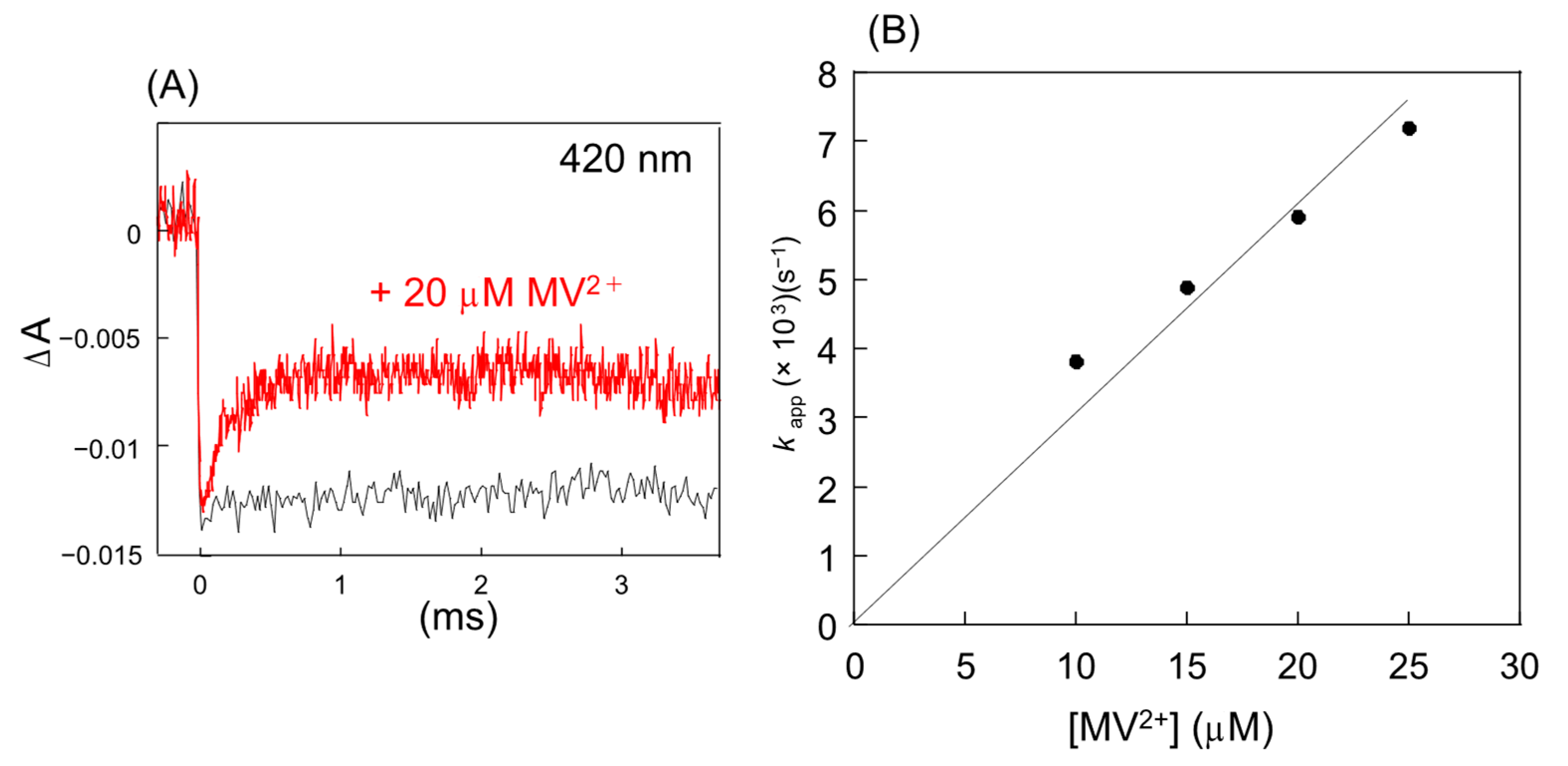
Figure 5 shows the absorption changes during pulse radiolysis of PaSoxR in the absence and presence of MV2+. After the initial decreasing absorbance, a subsequent partially reverse process was observed in the presence of MV2+ on a millisecond time scale. From these findings, it can be inferred that the [2Fe-2S] cluster of SoxRox is initially reduced by eaq, and subsequently becomes partially reoxidized, implying that the recovery process reflects the oxidation of SoxRred by MV2+ (reaction 3). Similar absorption changes were observed in EcSoxR.
To determine the second-order rate constants in the reaction of MV2+ with PaSoxRred, we used conditions in which the concentrations of the RAC (10–25 μM) was much higher than that of SoxRred (0.5–1 μM). Under these conditions, the oxidation of SoxRred obeyed pseudo-first-order kinetics, with the observed rate constant increasing linearly with RAC concentration. The rate constant of the reaction, obtained from the slope in Figure 5B was determined to be 3.0 × 10⁸ M−1s−1. Similar absorption changes were observed in the presence of other RACs, including pyocyanin (Pyo), phenazine methosulfate (PMS), duroquinone (Dur), and diquat (Dq). The second-order rate constants increased in the order Dur <MV2+ < Dq < Pyo < PMS (Table 2) and were found to correlate with the electrode potential of the RAC. In all cases, a rapid decrease phase was followed by a slower recovery phase. Notably, absorbance changes during the slower reoxidation phase varied depending on the specific RACs. The incomplete reoxidation of the [2Fe-2S] cluster may be explained by a rapid equilibrium for electron transfer between the RAC and the [2Fe-2S] cluster (reaction 3).
Table 2. Rate constants of [2Fe-2S] cluster of EcSoxR, and PaSoxR, with RACs and the electrode potentials E0 (vs NHE) at pH 7.0.
RACsE0 (vs. NHE) (mv)EcSoxR
k (× 108 M−1s−1)
PaSoxR
k (× 108 M−1s−1)
MV2+−440 [34]3.03.0
Dq−358 [35]5.75.5
Dur−260 [35]2.12.6
Pyo−34 [36]7.16.8
PMS+80 [36]16.014.0
The pulse radiolysis results presented here show that the [2Fe-2S]red cluster of EcSoxR and PaSoxR react with RACs with electrode potentials in the range of −440 mV (MV2+) [34] to +80 mV (PMS) [36]. Importantly, these results are consistent with the responsiveness of SoxR to RACs observed in vivo [18,22,32]. In comparison of the sensitivity profiles of SoxR towards RACs, Singh et al. showed that both EcSoxR and PaSoxR responded to all RACs, except Act [18].
SoxR homologs exhibit different selectivity toward RACs with each displaying distinctive effective concentrations and cellular response times [31]. However, our study found that these differences are not reflected in the kinetics of the reactions. Specifically, pulse radiolysis revealed no difference in the reactivity of the [2Fe-2S] cluster between EcSoxR and PaSoxR, despite their differing cellular response times to MV2+ [22].

4. Physiological Significance of SoxR Response to O2 and RACs

In this review, from the in vitro data, O2 directly oxidizes purified SoxR [11,13,26], suggesting that O2 itself is the signal that activates SoxR. However, it is not known whether O2 can interact with SoxR under high levels of SOD and a low level of SoxR constitutively under normal cellular conditions [25,37]. In addition, the following in vivo experiments have shown that O2 is not the signal for SoxR. Firstly, endogenous O2 in SOD- mutants does not activate SoxR in SOD-deficient cells [22,38]. Secondly RACs such as MV2+ can activate SoxR in anoxic conditions, in the presence of alternative respiratory acceptors [22]. The soxR regulon may be induced through multiple pathways. Since both O2 and RACs oxidize SoxR with the same rate constants, the activation depends on the cellular concentration of O2 and RACs.
The data presented here provide new insight into electron transfer between RACs and the [2Fe-2S] cluster of SoxR homologs. The observed oxidation of the [2Fe-2S] in SoxR proceeds via direct electron transfer from the [2Fe-2S] cluster to the RAC. It is likely that structurally distinct RACs can gain access through the solvent-exposed surface area allowing rapid electron transfer between the [2Fe-2S] cluster and RACs. An X-ray analysis of DNA-free EcSoxR [24] showed that the upper sulfur atom (S2) and one of the Fe atoms are completely exposed to the solvent. A similar mechanism may take place at the SoxR active site in PaSoxR. The oxidizability of SoxRred is assisted by the low potential of its [2Fe-2S] cluster in EcSoxR (−285 mV) [39] and PaSoxR (−290 mV) [39], which provide a strong driving force for electron transfer to even the moderate oxidant (−440 mV), MV2+ [33].

5. Conclusions and Future Perspectives

In this review, I followed reactions of the [2Fe-2S] cluster of SoxR with O2 and RACs directly. Both rate constants were almost a diffusion-controlled rate and characteristic of the individual SoxRs. The next major goal now is to investigate the factors that control the iron–sulfur cluster. To understand how iron–sulfur clusters respond to the molecules in the environment, future progress at the boundary of physical chemistry, biological chemistry, and inorganic chemistry will be crucial.

Funding

This research received no external funding.

Data Availability Statement

No new data were created or analyzed in this study. Data sharing is not applicable to this article.

Conflicts of Interest

The author declare no conflict of interest.

References

  1. Boncella, A.E.; Sabo, E.T.; Santore, R.M.; Carter, J.; Whalen, J.; Hudspeth, J.D.; Morrison, C.N. The expanding utility of iron-sulfur clusters: Their functional roles in biology, synthetic small molecules, maquettes and artificial proteins, biomimetic materials, and therapeutic strategies. Coord. Chem. 2022, 453, 214229. [Google Scholar] [CrossRef]
  2. Gupta, M.; Outten, C.E. Iron-Sulfur Cluster Signaling; The Common Thread in Fungal Iron Regulation. Curr. Opin. Chem. Biol. 2020, 55, 189–201. [Google Scholar] [CrossRef] [PubMed]
  3. Crack, J.C.; Green, J.; Thomson, A.J.; le Brun, N.E. Iron-sulfur cluster sensor-regulators. Curr. Opin. Chem. Biol. 2012, 16, 35–44. [Google Scholar] [CrossRef] [PubMed]
  4. Beinert, H.; Holm, R.H.; Münck, E. Iron-sulfur clusters: Nature’s modular, multipurpose structures. Science 1997, 277, 653–659. [Google Scholar] [CrossRef]
  5. Read, A.D.; Bentrley, R.E.T.; Archer, S.L.; Dunham-Snary, K.J. Mitochondrial iron-sulfur clusters: Structure, function, and an emerging role in vascular biology. Redox Biol. 2021, 47, 12164. [Google Scholar] [CrossRef]
  6. Bian, S.M.; Cowan, J.A. Protein-bound iron-sulfur centers. Form, function, and assembly. Coord. Chem. Rev. 1999, 190, 1049–1066. [Google Scholar] [CrossRef]
  7. Kiley, P.J.; Beinert, H. The role of Fe-S proteins in sensing and regulation in bacteria. Curr. Opin. Microbiol. 2003, 6, 181–185. [Google Scholar] [CrossRef]
  8. Ding, H.; Demple, B. In vivo kinetics of a redox-regulated transcriptional switch. Proc. Natl. Acad. Sci. USA. 2000, 97, 5146–5150. [Google Scholar] [CrossRef]
  9. Amábile-Cuevas, C.F.; Demple, B. Molecular characterization of the soxRS genes of Escherichia coli: Two genes control a superoxide stress regulon. Nucleic Acids Res. 1991, 19, 4479–4484. [Google Scholar] [CrossRef]
  10. Pomposiello, P.J.; Bennik, M.H.J.; Demple, B. Genome-wide transcriptional profiling of the Escherichia coli responses to superoxide stress and sodium salicylate. J. Bacteriol. 2001, 183, 3890–3892. [Google Scholar] [CrossRef]
  11. Kobayashi, K.; Fujikawa, M.; Kozawa, T. Oxidative stress sensing by the iron-sulfur cluster in the transcription factor, SoxR. J. Inorg. Biochem 2014, 133, 87–91. [Google Scholar] [CrossRef]
  12. Gaudu, P.; Weiss, B. SoxR, a [2Fe-2S] transcription factor, is active only in its oxidized. Proc. Natl. Acad. Sci. USA 1996, 93, 10094–10098. [Google Scholar] [CrossRef] [PubMed]
  13. Fujikawa, M.; Kobayashi, K.; Kozawa, T. Direct Oxidation of the [2Fe-2S] Cluster in SoxR Protein by Superoxide. Distinct Differential Sensitivity to Superoxide-Mediated Signal Transduction. J. Biol. Chem. 2012, 287, 35702–35708. [Google Scholar] [CrossRef] [PubMed]
  14. Blanchard, J.L.; Wholey, W.Y.; Conlon, E.M.; Pomposiello, P.J. Rapid changes in gene expression dynamics in response to superoxide reveal soxRS-dependent and -independent transcriptional networks. PLoS ONE 2007, 2, e1186. [Google Scholar] [CrossRef] [PubMed]
  15. Dietrich, L.E.P.; Teal, T.K.; Price-Whelan, A.; Newman, A.D.K. Redox-Active Antibiotics Control Gene Expression and Community Behavior in Divergent Bacteria. Science 2008, 321, 1203–1206. [Google Scholar] [CrossRef] [PubMed]
  16. Cruz, R.D.; Gao, Y.; Penumetcha, S.; Sheplock, R.; Weng, K.; Chander, M. Expression of the Streptomyces coelicolor SoxR Regulon Is Intimately Linked with Actinorhodin Production. J. Bactriol. 2010, 192, 6428–6438. [Google Scholar] [CrossRef]
  17. Shin, J.H.; Singh, A.K.; Cheon, D.J.; Roe, J.H. Activation of the SoxR Regulon in Streptomyces coelicolor by the Extracellular Form of the Pigmented Antibiotic Actinorhodin. J. Bacteriol. 2011, 193, 75–81. [Google Scholar] [CrossRef]
  18. Singh, A.K.; Shin, J.-H.; Lee, K.-L.; Imlay, J.A.; Roe, J.-H. Comparative study of SoxR activation by redox-active compounds. Mol. Microbiol. 2013, 90, 983–996. [Google Scholar] [CrossRef]
  19. Ma, Z.; Jacobsen, F.E.; Giedroc, D.P. Coordination Chemistry of Bacterial Metal Transport and Sensing. Chem. Rev. 2009, 109, 4644–4681. [Google Scholar] [CrossRef]
  20. Liochev, S.I. Is superoxide able to induce SoxRS? Free Radic. Biol. Med. 2011, 50, 1813. [Google Scholar] [CrossRef]
  21. Dietrich, L.E.P.; Kiley, P.J. A shared mechanism of SoxR activation by redox-cycling compounds. Mol. Microbiol. 2011, 79, 1119–1122. [Google Scholar] [CrossRef] [PubMed]
  22. Gu, M.; Imaly, J.A. The SoxRS response of Escherichia coli is directly activated by redox-cycling drugs rather than by superoxide. Mol. Microbiol. 2011, 79, 1136–1150. [Google Scholar] [CrossRef] [PubMed]
  23. Kuo, C.F.; Mashino, T.; Fridovich, I. Dihydroxyisovalerate Dehydratase. J. Biol. Chem. 1987, 262, 4724–4727. [Google Scholar] [CrossRef] [PubMed]
  24. Gardner, P.R.; Fridovich, I. Superoxide Sensitivity of the Escherichia coli Aconitase. J. Biol. Chem. 1991, 266, 19328–19333. [Google Scholar] [CrossRef]
  25. Gardner, P.R.; Fridovich, I. Superoxide Sensitivity of the Escherichia coli 6-Phosphogluconate Dehydratase. J. Biol. Chem. 1991, 266, 1478–1483. [Google Scholar] [CrossRef]
  26. Fujikawa, M.; Kobayashi, K.; Tsutsui, Y.; Tanaka, T.; Kozawa, T. Rational Tuning of Superoxide Sensitivity in SoxR, the [2Fe-2S] Transcription Factor: Implication of Species-Specific Lysine Residues. Biochemistry 2017, 56, 403–410. [Google Scholar] [CrossRef]
  27. Watanabe, S.; Kita, A.; Kobayashi, K.; Miki, K. Crystal structure of the [2Fe-2S] oxidative-stress sensor SoxR bound to DNA. Proc. Natl. Acad. Sci. USA 2008, 105, 4121–4126. [Google Scholar] [CrossRef]
  28. Getzoff, E.D.; Tainer, J.A.; Weiner, P.K.; Kollman, P.A.; Richardson, J.S.; Richardson, D.C. Electrostatic recognition between superoxide and copper, zinc superoxide dismutase. Nature 1983, 306, 287–290. [Google Scholar] [CrossRef]
  29. Ludwig, M.L.; Metzger, A.L.; Pattrige, K.A.; Stallings, W.C. Manganese superoxide dismutase from Thermus thermophilus: A structural model refined at 1.8 Å resolution. J. Mol. Biol. 1991, 219, 335–358. [Google Scholar] [CrossRef]
  30. Sheng, Y.; Abreu, I.A.; Cabelli, D.E.; Maroney, M.J.; Miller, A.-F.; Teixeira, M.; Valentine, J.S. Superoxide Dismutases and Superoxide Reductases. Chem. Rev. 2014, 114, 3854–3918. [Google Scholar] [CrossRef]
  31. Lee, K.L.; Singh, A.K.; Heo, L.; Seok, C.; Roe, J.-H. Factors affecting redox potential and differential sensitivity of SoxR to redox-active compounds. Mol. Microbiol. 2015, 97, 808–821. [Google Scholar] [CrossRef]
  32. Sheplock, R.; Recinos, D.A.; Macknow, N.; Dietrich, L.E.P.; Chander, M. Species-specific residues calibrate SoxR sensitivity to redox-active molecules. Mol. Microbiol. 2013, 87, 368–381. [Google Scholar] [CrossRef]
  33. Kobayashi, K.; Tanaka, T.; Kozawa, T. Kinetics of the Oxidation of the [2Fe-2S] Cluster in SoxR by Redox-Active Compounds as Studied by Pulse Radiolysis. Biochemistry 2025, 64, 895–902. [Google Scholar] [CrossRef] [PubMed]
  34. Steckhan, E.; Kuwana, T. Spectrochemical study of mediators. I Bipyridyum salts and their electron transfer to cytochrome C. Ber. Bunsenges. Phys. Chem. 1974, 78, 253–258. [Google Scholar] [CrossRef]
  35. Wardman, P. Reduction Potentials of One-Electron Couples involving Free Radicals in Aqueous Solution. J. Phys. Chem. Ref. 1989, 18, 1637–1655. [Google Scholar] [CrossRef]
  36. Moffet, D.A.; Foley, J.; Hecht, M.H. Midpoint reduction potentials and heme binding stoichiometries of de nova proteins from designed combinatorial libraries. Biophys. Chem. 2003, 105, 231–239. [Google Scholar] [CrossRef]
  37. Pomposiello, P.J.; Demple, B. Redox-operated genetic switches: The SoxR and OxyR transcription factors. Trends Biotechnol. 2001, 19, 109–114. [Google Scholar] [CrossRef]
  38. Gort, A.S.; Imaly, J.A. Balance between endogenous superoxide stress and antioxidant defenses. J. Bacteriol. 1998, 180, 1402–1410. [Google Scholar] [CrossRef]
  39. Kobayashi, K.; Tagawa, S. Activation of SoxR-dependent transcription in Pseudomonas aeruginosa. J. Biochem. 2004, 136, 607–615. [Google Scholar] [CrossRef]
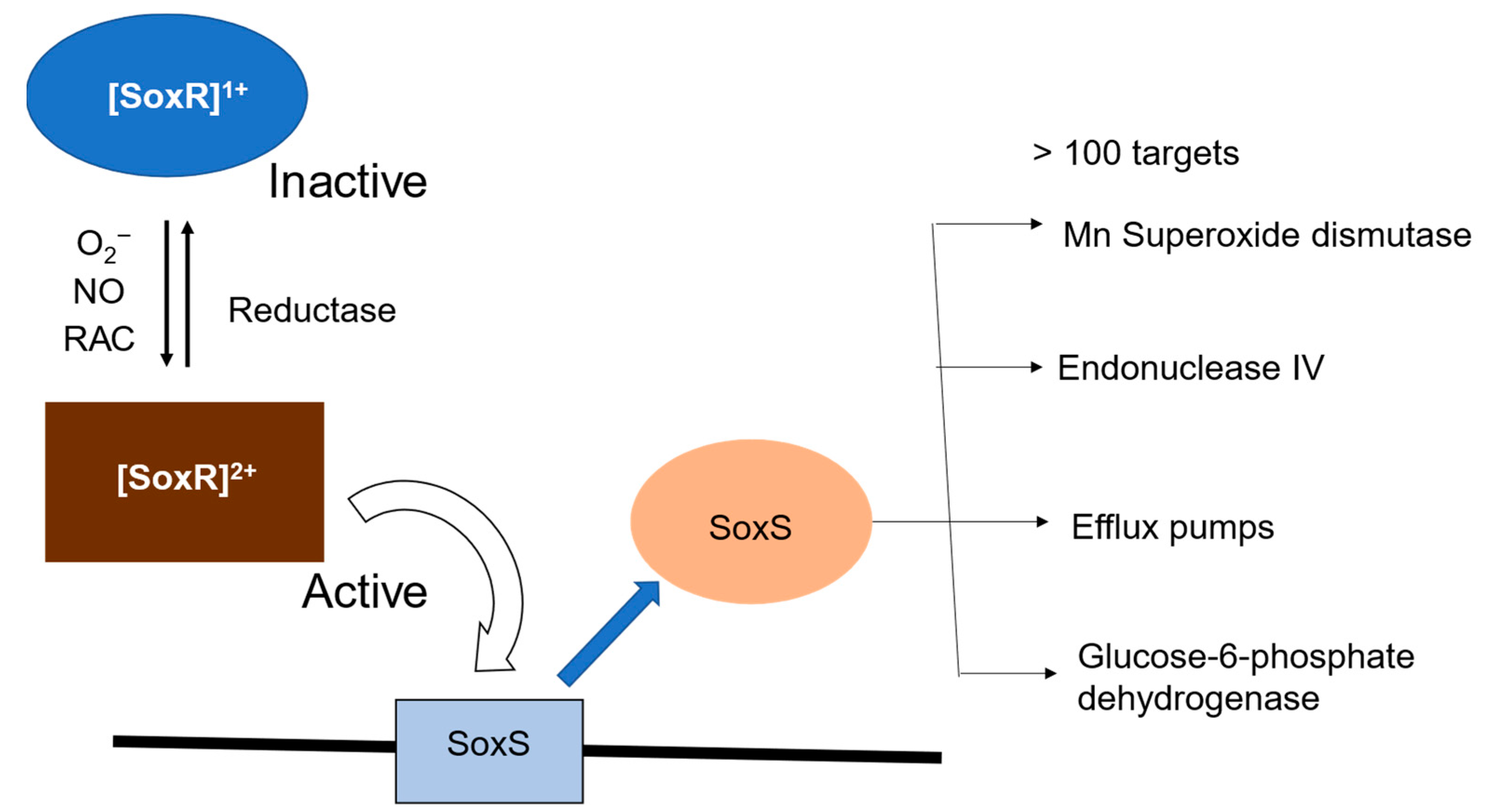
Figure 1. The mechanism underlying transcription of soxRS regulon in E. coli. SoxR is activated by exposure to O2-generating compounds, redox active compounds (RACs), or nitric oxide (NO). The target of activated SoxR is the soxS gene. Increased SoxS induces expression of various antioxidant proteins.
Inorganics 13 00307 g001
Figure 2. SoxR response in non-enteric bacteria.
Inorganics 13 00307 g002
Figure 3. SOD effects on the oxidation of SoxR. Changes in absorbance measured after pulse radiolysis of EcSoxR (A) and PaSoxR (B) in the absence (blue line) or presence (red line) of 0.5 μM SOD. (C) The absorbance changes of (o) EcSoxR and (●) PaSoxR at time (∆At), normalized to the total absorbance change (∆A0) plotted against the concentration of SOD, Reprinted from [13].
Inorganics 13 00307 g003
Figure 4. (A) Sequence alignments of SoxR proteins from enterics E. coli, Sphingopyxis alaskensis, and Shigella flexneri and non-enteric (P. aeruginosa, Xanthomonas axonopodi, Vibrio cholerae) bacteria. Identical and conserved residues among SoxR proteins are denoted in red and green, respectively. Hypervariable residues and the hypervariable motif are highlighted in yellow. (B) Structure of EcSoxR in the region near the [2Fe-2S] cluster. The structure was produced with PyMol using a structure from the Protein Data Bank (entry 2ZHG). Reproduced from [26]. Copyright 2016 American Chemical Society.
Inorganics 13 00307 g004
Figure 5. (A) Changes in absorbance at 420 nm after pulse radiolysis of PaSoxR. Samples contained 70 μM PaSoxR, 20 mM potassium phosphate buffer (pH 7.0), 10 mM potassium/sodium tartrate, 0.5 M KCl, and 0.1 M t-butanol, to which 20 μM MV2+ was further added in red colored. (B) Concentration dependence of MV2+ versus kapp in PaSoxR.
Inorganics 13 00307 g005
Table 1. Second-order rate constants in O2 reactions with the reduced forms of wild-type and mutant SoxR proteins.
k × 108 (M−1 s−1)
E. coli
   WT5.0
   R127LS128QD129A4.8
   D129A5.0
   K89A3.8
   K92A2.2
   K89AK92A0.33
   K89RK92R4.7
   K89EK92E0.31
P. aeruginosa
   WT0.4
   A87K2.1
   A90K5.4
   L125RQ126SA127D0.4
Disclaimer/Publisher’s Note: The statements, opinions and data contained in all publications are solely those of the individual author(s) and contributor(s) and not of MDPI and/or the editor(s). MDPI and/or the editor(s) disclaim responsibility for any injury to people or property resulting from any ideas, methods, instructions or products referred to in the content.