Abstract
In recent times, the global landscape of disease detection and monitoring has been profoundly influenced by the convergence of nanotechnology and biosensing techniques. Biosensors have enormous potential to monitor human health, with flexible or wearable variants, through monitoring of biomarkers in clinical and biological behaviors and applications related to health and disease, with increasing biorecognition, sensitivity, selectivity, and accuracy. The emergence of nanomaterial-based biosensors has ushered in a new era of rapid and sensitive diagnostic tools, offering unparalleled capabilities in the realm of disease identification. Even after the declaration of the end of the COVID-19 pandemic, the demand for efficient and accessible diagnostic methodologies has grown exponentially. In response, the integration of nanomaterial biosensors into breathalyzer devices has gained considerable attention as a promising avenue for low-cost, non-invasive, and early detection of COVID-19. This review delves into the forefront of scientific advancements, exploring the potential of emerging nanomaterial biosensors within breathalyzers to revolutionize the landscape of COVID-19 detection, providing a comprehensive overview of their principles, applications, and implications.
1. Introduction
Coronaviruses (CoVs) represent a class of enveloped viruses characterized by their single-stranded RNA genome [1]. These viruses possess the capability to infect a broad spectrum of hosts, including mammals and avian species, often exhibiting respiratory and enteric pathologies [2]. The process of urbanization, coupled with inadequate urban planning, suboptimal sanitation and water provisions, high population density, and encroachment upon formerly undisturbed ecosystems, collectively foster the proliferation of zoonotic infectious diseases [3]. Despite the presence of established protocols for detection and mitigation of established coronaviruses through various initiatives, the escalating occurrence of newly emergent diseases, such as the recent Coronavirus disease 2019 (COVID-19) pandemic, caused by the severe acute respiratory syndrome coronavirus 2 (SARS-CoV-2), serves as a reminder of the massive costs to the global healthcare systems. Severe cases of COVID-19 can lead to life-threatening situations and are typically linked with risk factors such as age, co-morbidities, and immune status [4].
Timely and early diagnosis of COVID-19 is of paramount importance to minimize the likelihood of encountering severe complications. Diagnosis relies heavily on expensive techniques such as real-time reverse-transcriptase polymerase chain reaction (rRT-PCR) positivity, lateral flow tests, rapid antigen tests, and enzyme-linked immunosorbent assays (ELISA), in addition to clinical symptoms and radiological diagnostics [5]. rRT-PCR, which detects viral nucleic acid from a nasopharyngeal swab, is regarded as the gold standard due to its specificity and accuracy [6]. However, accuracy often depends on the method of sample collection and preservation, prior to testing as well as at the time point of collection. Minor mistakes can lead to false positives. Additionally, these techniques cannot be readily employed in remote or resource-constrained environments. Hence, timely medical intervention assumes critical significance in curbing the progression of the disease and preventing fatalities. Even after the declaration of the end of the pandemic, fast and precise diagnostic testing of COVID-19 continues to be the main strategy in determining isolation or medical intervention.
In the quest for enhanced diagnostic tools to combat the ongoing COVID-19 pandemic, the amalgamation of nanomaterial-based biosensors with breathalyzer technology has emerged as a frontier approach [7]. The convergence of nanotechnology and biosensing has paved the way for innovative and sensitive detection methodologies, holding the promise of revolutionizing disease identification. With the imperative need for rapid, reproducible, non-invasive, and accurate diagnostic techniques, the incorporation of nanomaterial biosensors into breathalyzers offers a promising avenue for addressing these challenges in COVID-19 detection [8]. This scientific research review embarks on an exploration of the scientific progress in this field, elucidating the potential of emerging nanomaterial biosensors within breathalyzers to establish a paradigm shift in the landscape of COVID-19 diagnostics. By delving into the underlying principles, experimental methodologies, and anticipated implications, this study endeavors to contribute to the evolving discourse surrounding the application of nanomaterial biosensors for the early and efficient detection of COVID-19 through breath analysis.
2. Nano-Biosensors as Diagnostic Tools
Nano-biosensors have emerged as a groundbreaking class of diagnostic tools, offering remarkable potential for precise and timely disease detection. These sensors harness the unique properties of nanomaterials such as high surface area and the ability to be functionalized with specific molecules to enable highly sensitive and specific detection of biomolecular interactions, and thus have the potential to be used in a wide variety of diagnostic applications, including the detection of diseases, toxins, and pollutants [9,10,11]. Their miniaturized scale allows for efficient integration into various diagnostic platforms, offering advantages such as rapid response times, reduced sample volumes, and enhanced portability. Nano-biosensors exhibit exceptional versatility, and are capable of detecting a diverse range of analytes, including proteins, nucleic acids, and small molecules. The incorporation of nanomaterials, such as nanoparticles, nanowires, and nanotubes, into biosensor designs enhances surface area interactions and signal transduction mechanisms, thereby amplifying the detection signal and improving overall sensor performance [10].
In totality, an optimal biosensor should encompass several critical attributes, such as being amenable to mass production, autonomous operation, notable sensitivity and selectivity, rapid response kinetics, capacity for multiplexing, multiple sensing modalities, disposability, extended shelf-life, cost-effectiveness, and user-friendly operation.
Nano-biosensors have been used for the detection of cancer, infectious diseases, and toxins. Studies have shown significant potential in the early detection of cancer biomarkers. Wang et al. (2019) devised a methodology to detect the tumor marker carcinoembryonic antigen (CEA) through the application of a solid-state nanopore as an instrumental component [12]. This innovative approach capitalizes on the inherent binding affinity between CEA and aptamer-modified magnetic Fe3O4 nanoparticles, obviating the need for direct monitoring of CEA translocation through the nanopore. The aptamer modified magnetic Fe3O4 nanoparticles were intricately combined with tetrahedral DNA nanostructures (TDNs) to facilitate identification of a low concentration (0.1 nm) of CEA, affirming the specificity of the interaction between the aptamer and the target molecule. Thus, the nanopore sensing approach demonstrated its capability to detect CEA in actual samples. Another study evaluated human epidermal growth factor receptor-2 (HER2) status, which is routinely used in the screening, diagnosis, and surveillance of breast cancer (BC). They employed an innovative electrochemical biosensor specifically targeting the detection of HER2-positive circulating tumor cells. The hybrid nanocomposite, formed through the amalgamation of reduced graphene oxide nanosheets and rhodium nanoparticles onto the surface of a graphite electrode, yielded enhancements in surface area, electrochemical activity, and biocompatibility with outstanding performance in discerning HER2-overexpressing SKBR3 cancer cells. It exhibited a linear dynamic range, an impressively low analytical limit of detection at 1.0 cell/mL, and a limit of quantification of 3.0 cells/mL [13].
Nano-biosensors have also demonstrated their utility in point-of-care (POC) infectious disease detection. Chowdhary et al. developed a rapid and quantitative biosensor for the specific detection of Dengue virus (DENV) serotypes 1 to 4 through the optimization of a stable interaction between cadmium selenide tellurium sulfide fluorescent quantum dots and gold nanoparticles, and were able to successfully detect target virus DNA at standard dilutions ranging from 10−15 to 10−10 M. This versatile biosensor could detect and quantify serotypes, thereby exhibiting significant potential as a diagnostic tool for point-of-care DENV detection [14]. Pang et al. developed a fluorescent nano-biosensor employing magnetic nanoparticles and aptamers for the detection of avian influenza virus H5N1 [15]. The fluorescent aptasensor employed metal-enhanced fluorescence and core-shell Ag@SiO2 nanoparticles for the highly sensitive detection of recombinant hemagglutinin protein associated with the H5N1 influenza virus. This system capitalizes on the dual advantages of the rHA aptamer’s exceptional selectivity and the substantial fluorescence enhancement capabilities inherent to core-shell Ag@SiO2 nanoparticles, with robust selectivity and remarkable sensitivity when applied in the presence of diverse proteins or within complex matrices, achieving a detection limit of 2 ng/mL in buffer and 3.5 ng/mL in the specified context [15].
Nano-biosensors are also powerful tools for the sensitive and selective detection of various toxins across diverse applications. They combine the high specificity of biological recognition elements with the enhanced properties of nanomaterials, resulting in improved sensitivity and rapid response times. For instance, He et al. developed a nano-biosensor utilizing gold nanoparticles and aptamers to detect ochratoxin A (OTA), a mycotoxin commonly found in food products, with exceptional sensitivity, achieving a remarkable detection limit of 0.05 U·L−1 [16]. The method was effectively used to develop a target-responsive aptasensor for colorimetric detection of OTA, associated with food poisoning and various adverse health effects in humans, and thus providing substantial potential for applications in the field of mycotoxin-related food safety monitoring.
Skotadis et al. developed an electrochemical hybrid biosensor using a combination of noble nanoparticles and DNAzymes for the simultaneous detection of Pb2+, Cd2+, and Cr3+ ions. The double-helix structure of DNAzymes undergoes disintegration into smaller fragments upon encountering specific heavy metal ions, with discernable alteration in device resistance due to collapse of conductive inter-nanoparticle DNAzymes. The biosensor allowed successful detection of the three target ions at concentrations significantly below their maximum allowable levels in tap water, with considerable promise for integration into autonomous and remote sensing systems to detect water pollution [17].
Nano-biosensors have also emerged as a promising class of diagnostic tools in the battle against COVID-19. Broadly, biosensor platforms designed for SARS-CoV-2 detection center on the following pivotal facets: specified target for identification (such as viral RNA, proteins, or human antibodies), the modality of identification (leveraging aptamers, immunoglobulins, nucleic acids, and receptors), and augmentation of the signal generation and transduction mechanisms (involving electrical, surface plasmon resonance, electrochemical, optical, mechanical systems, and fluorescence signals). Leveraging the unique properties of nanomaterials, these biosensors offer a powerful platform for the rapid and sensitive detection of the SARS-CoV-2 virus. Through strategic functionalization of nanomaterials with specific biomolecular recognition elements, such as antibodies or aptamers targeting viral antigens, nano-biosensors enable precise and early identification of COVID-19 infection [18]. The amplified surface area provided by nanomaterials enhances the interaction between the sensor and the target virus, resulting in enhanced sensitivity. Moreover, the integration of nano-biosensors into portable and point-of-care devices facilitates efficient and real-time diagnostics, thereby addressing the urgent need for rapid testing and screening strategies and contributing to effective pandemic management.
A study by Karakuş et al. reported the development of a colorimetric assay for the rapid detection of SARS-CoV-2 using gold nanoparticles functionalized with SARS-CoV-2 spike antigens. The SARS-CoV-2 spike antigens trigger rapid and irreversible aggregation of gold nanoparticles due to antibody–antigen interactions, resulting in a noticeable change in color, observable with the naked eye or measured using UV-Vis spectrometry. It exhibits the capability to identify SARS-CoV-2 spike antigens at an astonishingly low concentration and does not cross-react with other antigens, including influenza A (H1N1), MERS-CoV, and Streptococcus pneumoniae, even at high concentrations [19]. Another nano-biosensor array for SARS-CoV-2 detection involved the surface-enhanced Raman scattering-immune substrate, which was meticulously engineered using an innovative oil/water/oil three-phase liquid–liquid interface self-assembly technique. The method yielded two layers of compact and homogeneous gold nanoparticle films. The detection procedure entailed an immunoreaction involving the SARS-CoV-2 spike antibody-modified surface-enhanced Raman scattering (SERS) immune substrate, the spike antigen protein, and Raman reporter-labeled immuno-Ag nanoparticles. This process demonstrated the ability to identify the presence of the SARS-CoV-2 spike protein at concentrations as low as 0.77 fg mL−1 in phosphate-buffered saline and 6.07 fg mL−1 in untreated saliva. These findings underscore its potential utility as a viable option for the early diagnosis of COVID-19 [20].
These examples highlight the versatility and effectiveness of nano-biosensors in various diagnostic applications. As the field continues to evolve, nano-biosensors hold immense promise for revolutionizing disease diagnostics by providing earlier detection, more accurate results, and greater accessibility, thereby addressing critical challenges in effective disease management and healthcare, and fostering advancements in personalized medicine.
3. Uses of Breath Analysis
Breath analysis has emerged as a promising diagnostic strategy with broad applications across various medical fields. By analyzing the composition of exhaled breath, known as the breathome, this non-invasive approach offers valuable insights into an individual’s lifestyle and physiological and pathological states, as well as microbiome metabolism, including the presence and/or abundance of pathogens [21]. The breathome contains a complex mixture of volatile organic compounds (VOCs) originating from metabolic processes, reflecting the underlying clinical state and overall health condition of the individual [8]. Volatolomics denotes the intricate chemical mechanisms associated with VOCs discharged from bodily fluids, including blood, urine, sweat, feces, nasal and skin secretions, and exhaled breath [22]. The respiratory system is a prominent substrate for the investigation of these VOCs which originate endogenously within the human body, undergo systemic circulation through the bloodstream, and ultimately traverse the alveolar interface, depending upon solubility to manifest in exhaled breath. Human breath contains up to 3500 VOCs. Nonpolar VOCs are characterized by limited solubility in blood (indicated by a blood–air partition coefficient, λb:a, of less than 10) and undergo exchange primarily in the alveoli. Conversely, blood-soluble VOCs (with λb:a values exceeding 100) are predominantly exchanged within the lung airways. VOCs exhibiting intermediate solubility, with blood–air partition coefficients falling within the range of 10 to 100, participate in pulmonary gas exchange in both the alveoli and the lung airways [21]. These VOCs can be quantified at minute concentrations, typically within the parts-per-million by volume (ppmv) and parts-per-billion by volume (ppbv) thresholds or even lower levels. This strategy holds the potential to detect and monitor a range of diseases, including respiratory disorders, metabolic diseases, and infectious illnesses. High-humidified exhaled gas consists of more than one compound quantifiable as concentration of ppbv to ppmv [23]. Studies have assessed fluctuations in exhaled breath as vital indicators of a variety of disease conditions, including metabolic disorders, infections, and cancers [24,25,26]. Human breath is composed of VOCs such as acetone, isoprene, ethane, and pentane, as well as inorganic gases such as carbon dioxide (CO2), carbon monoxide (CO), and nitric oxide (NO), in addition to non-volatile compounds and exhaled breath condensates such as peroxynitrite, cytokines, and isoprostanes [21]. Various exhaled gases, such as NO, ammonia (NH3), hydrogen sulfide (H2S), and volatile organic compounds such as acetone (CH3COCH3), methane (CH4), toluene (C6H5CH3), and pentane are recognized as vital indicators of disease [27,28].
A study by Shorter et al. reported that breath analysis is a useful technique for diagnosing and monitoring asthma and chronic obstructive pulmonary disease (COPD) [29]. Carbon monoxide (CO) and nitric oxide in exhaled breath are signs of airway inflammation and can indicate the development of respiratory diseases. The change in NO levels of exhaled air between exacerbations coincide with the collected spirometry data. Breath analysis is important to understand the dynamic of exchange of NO and other types in the lungs and airways. It was found that NO flows are higher for those who suffer from simple asthma and during development. The concentration of NO level was higher in adults with asthma and COPD, as well as in children [29].
Boots et al. investigated two hundred samples of the region of gas immediately surrounding a bacterial sample or headspace, originating from four distinct microorganisms: Escherichia coli, Pseudomonas aeruginosa, Staphylococcus aureus, and Klebsiella pneumonia, and found a significant level of disparity in the occurrence of VOCs among various bacterial cultures [30]. Furthermore, the study successfully delineated a demarcation between methicillin-resistant and methicillin-sensitive isolates of Staphylococcus aureus, potentially offering a valuable diagnostic tool within the field of bacterial resistance.
Neurodegenerative conditions such as Alzheimer’s disease (AD) and Parkinson’s disease (PD) exhibit pathophysiological processes that commonly initiate well in advance of any detectable cognitive impairment symptoms [21]. A study used an assortment of metal oxide nanosensors integrated onto an SnO2 thin film, layered with Au, Cr, or Wo, configuring these sensors in two distinct ways. Analysis of exhaled breath samples using GC-MS and sensor systems revealed distinct dissimilarities in the chemical constituents present in the AD and PD patient cohorts (alkanes, benzene, and acetamide) as compared to those in healthy individuals. The configuration encompassing a greater number of sensors demonstrated enhanced distribution patterns and a superior capacity for discrimination based on factors such as oxidative stress and corresponding metal homeostasis [31].
Electronic nose (e-nose/EOS) devices employ multisensor arrays to differentiate intricate VOC combinations present in clinical air samples. They generate distinct sensor-response patterns, known as “smellprint signatures”, which are disease-specific. Specialized statistical models and pattern recognition algorithms are then utilized to aid in discriminating among various samples. A study utilized a commercial metaloxide semiconductor chemiresistive e-nose (aeoNoseTM), comprising three distinct non-specific micro-hotplate metal-oxide nanosensors. Exposure of the sensors to exhaled gas prompts a redox reaction, leading to a conductivity alteration. Through the quantification of these alterations, the breath print was acquired which effectively differentiated individuals with the neurological disease multiple sclerosis, not undergoing medication, from healthy subjects, with a sensitivity of 93%, a specificity of 74%, and an overall accuracy of 80% [32].
A customized nanoscale artificial olfactory system, NA-Nose, comprising an assemblage of cross-reactive gas sensors composed of spherical gold nanoparticles, was employed to discern various odors within the alveolar exhaled breath of both cancer patients and healthy counterparts [33]. The nanosensors exhibited the capability to differentiate between individuals with head and neck cancer (HNC) and healthy subjects (83%), lung cancer (LC) patients (95%), and LC patients versus healthy controls (87%), relying on detection of some common volatile biomarkers, thus positioning it as a promising prospect for future screening applications [33].
Chronic kidney disease (CKD) poses a significant public health challenge, with its global prevalence on the rise, and is often undetected by patients. Specific compounds found in exhaled breath are associated with biochemical processes linked to CKD or the buildup of toxins resulting from impaired kidney function. To address this, carbon-nanotube-based sensors and breath sample analysis were employed as non-invasive tools for diagnosing end-stage renal disease in an animal model [34]. In an experimental model involving bilateral nephrectomy in rats, researchers differentiated between exhaled breath from healthy individuals versus those with chronic renal failure (CRF) through 15 common volatile organic compounds present in both healthy and CRF states, and twenty-seven VOCs unique to the CRF condition. Utilizing an array of organically coated single-walled carbon chemiresistive nanotubes, online breath analysis demonstrated accurate discrimination between different breath states. This discrimination could be further improved by reducing breath humidity before sensor analysis. In a separate study, gold nanoparticle-based sensors in conjunction with support vector machine analysis successfully detected CKD in breath samples. The sensor effectively distinguished between the exhaled breath of CKD patients and that of healthy individuals. Combining 2–3 gold nanoparticle sensors yielded notable differentiation between early-stage CKD and healthy individuals (79% accuracy) as well as between stage 4 and 5 CKD (85% accuracy). Furthermore, single sensor exhibited the capability to diagnose accurately (76%) both early- and late-stage CKD [35].
Numerous published studies have established associations between specific elements present in exhaled breath and various diseases. However, the practical implementation of these findings for diagnostic purposes has been hindered by the limited application, largely attributable to the inconsistent and conflicting identification of breath biomarkers and lack of large-scale trials. However, the potential of nano-biosensors in the field of nanomedicine, for testing and determination of an individual’s health and application for a varied range of disease diagnosis, based on VOC emissions in exhaled breath analysis, is ever expanding.
4. Breathomics in COVID-19 Diagnostics
Expiratory breathomics, an emerging field at the intersection of metabolomics and exhaled breath analysis, has also gained traction as a promising avenue for advancing COVID-19 diagnostics. It can be used to evaluate inflammatory and oxidative tension in the respiratory system [36]. In the context of COVID-19, breathomics explores the unique quantifiable VOCs profiles associated with SARS-CoV-2 infection, including those arising from viral replication, host immune responses, and potential co-infections [37].
Remy et al. identified that the majority of volatile organic compound concentrations (hydrogen sulfide, methanol, acetic acid, butyric acid, and crotonaldehyde) exhibited suppression in individuals with COVID-19 [38]. Distinctions in VOC concentrations were also noted between symptomatic and asymptomatic cases. The breath-based markers reflected the impact of infections on the host’s cellular metabolism and microbiome, with reduced levels of specific VOCs attributed to the suppressive influence of SARS-CoV-2 on microbial metabolism within the gastrointestinal or pulmonary systems.
A research investigation identified a set of volatile organic compounds comprising ethanal, 2-butanone, octanal, acetone, and methanol that effectively distinguished COVID-19 cases from other disease states [39]. In addition to these VOC biomarkers, other noteworthy differentiators between COVID-19-positive and/or negative states encompassed propanal, propanol, heptanal, and isoprene. Remarkably, an unidentified VOC denoted as M7-VOC exhibited remarkably strong predictive capabilities for assessing the severity of the disease or the risk of mortality. Furthermore, heptanal was also deemed significant in this context. The study’s findings collectively suggest that these nine biomarker VOCs provide clear indications of COVID-19-related alterations in breath biochemistry, reflecting processes associated with ketosis, gastrointestinal effects, and inflammatory responses [39]. The predominant and recurring impact of SARS-CoV-2 infection on human metabolic pathways primarily pertains to certain aspects of amino acid metabolism. Notably, tryptophan metabolism has strong correlation with COVID-19 through a metabolic pathway, in which the production of nicotinamide adenine dinucleotide (Kynurenine pathway) has been observed in recent investigations [40].
It is worth noting that in a retrospective cohort study carried out at a single tertiary care hospital in Northern Italy during the initial wave of the pandemic, dual-energy computed tomography was used to quantitatively evaluate pulmonary gas and blood distribution [41]. The findings of the study revealed a significant difference between patients who underwent invasive ventilation and those receiving non-invasive respiratory support. Specifically, invasively ventilated patients exhibited a lower percentage of normally aerated tissue, amounting to 33% of their lung volume. In contrast, patients receiving non-invasive respiratory support had a substantially higher percentage of normally aerated tissue, accounting for 63% of their lung volume. A disparity in gas-to-blood volume mismatch between the two groups was also observed. In patients subjected to invasive ventilation, the extent of this mismatch was notably higher, constituting 43% of their lung volume compared to non-invasively ventilated patients, at 25% of their lung volume. Interestingly, the poorly aerated tissue in both groups exhibited distinctive characteristics, with some regions displaying a high gas-to-blood volume ratio and others presenting a low gas-to-blood volume ratio [41].
Diffusing capacity (DLCO) is a vital parameter that quantifies the volume of carbon monoxide transferred from alveolar gas to blood per minute. It is calculated by dividing this volume by the difference between the mean pressure of carbon monoxide in alveolar-capillary spaces and the mean pressure in pulmonary capillaries. DLCO serves as a valuable measure to evaluate the lungs’ ability to facilitate the transfer of gases from inspired air into the bloodstream. A study aimed at investigating the underlying pathophysiology of alveolar-to-capillary gas exchange in patients recovering from COVID-19 pneumonia utilized both decreased lung-diffusing capacity for nitric oxide (DLNO) and DLCO [42]. The findings from this study revealed that DLCO exhibited variable results among subjects during the initial 3 months of recovery from severe COVID-19. In contrast, DLNO was more frequently and persistently impaired when compared to the standard DLCO measurement. This disparity suggests a specific impairment in diffusive conductance, likely attributed to damage to the alveolar unit alveolar-capillary interface, while the capillary volume remains relatively preserved. Notably, alterations in gas transport were detected even in subjects who had experienced mild COVID-19 pneumonia and displayed no or minimal lingering abnormalities in computed tomography scans [42]. These findings underscore the potential of both DLCO and DLNO as valuable functional biomarkers for COVID-19 breathomics-based point-of-care diagnostics, even in follow-up post-acute care facilities.
Another interesting study explored the potential of exhaled volatile organic compounds to differentiate COVID-19 vaccine recipients from non-vaccinated individuals with respect to the metabolic adjustments prompted by vaccination and the underlying mechanisms of immune-driven metabolic reprogramming [43]. The study looked at breath samples from both COVID-19 vaccine recipients and non-vaccinated controls analyzed for VOCs using comprehensive two-dimensional gas chromatography coupled with time-of-flight mass spectrometry. Specific VOCs were identified as biomarkers, forming a distinct VOC fingerprint through alveolar gradients and machine learning. This signature effectively distinguished vaccine recipients from the control group with an impressive prediction accuracy of 94.42% [43]. Further analysis of the metabolic pathways associated with these biomarkers revealed that host–pathogen interactions during vaccination enhanced enzymatic activity and microbial metabolism in the liver, lung, and gut, representative of the mechanisms behind vaccine-induced metabolic regulation. In comparison to non-vaccinated individuals, the levels of biomarkers—6-methyl-5-hepten-2-one, CPTO, methanesulfonyl chloride, and benzothiazole—as well as benzene/3-MHeptane levels, increased by approximately 70% in vaccine recipients, notably affecting ketone synthesis pathways such as β-oxidation of fatty acids with branched chains. Five other individual biomarkers (hexanal, benzene, acetonitrile, 2-MOctane, and phenol) decreased to half the levels observed in the control group among vaccine recipients. The findings indicated that vaccination accelerated the metabolism of aromatic hydrocarbons and monomethylated alkanes [43]. This study supports the potential use of breathomics in aspects of vaccine-induced metabolic regulation and in advancing the development of early clinical diagnostics.
Nurputra et al. outlined the selection of acetone as a volatile organic compound model in their investigation. Acetone was chosen due to its presence in rebreathed breath, typically found within the range of 0.8 to 2.0 parts per million (ppm), alongside other VOCs such as alcohol and carbon monoxide. Additionally, acetone stands out as a significant COVID-19 biomarker carried in the breath. Furthermore, in clinical settings, the presence of acetone in exhaled breath has been extensively studied for diagnosing various other conditions, including lung cancer, diabetes mellitus, starvation, and adherence to ketogenic diets [44].
Although the translation of breathomics into clinical practice warrants standardization and validation, its potential for non-invasive, rapid, and scalable COVID-19 diagnosis positions it as a promising tool for complementing existing testing methodologies and advancing our capacity to combat the disease.
5. Nano-Biosensor Breathalyzers for COVID-19 Detection
The nanobody-centered electrochemical methodology has enabled accelerated virus detection owing to its independence from requirements of reagents or extensive processing stages.
A. Promising bio-functionalization techniques and nanomaterials used in bio marker detection in COVID-19 diagnostics:
Biofunctionalization refers to the process of modifying a biomaterial to confer a particular biological function. This is typically achieved by integrating biomimetic molecules, serving as mediators or imitators of cellular systems. These mediators can be introduced into the scaffolds through surface adsorption or via nanospheres, and scaffold-bound complexes, which are subsequently released during scaffold degradation. Surface biofunctionalization entails either the immobilization of bioactive molecules on scaffold surfaces or the introduction of biofunctional surface groups to induce specific cellular responses.
Owing to their distinctive attributes, including novel optical characteristics, biodegradability, low toxicity, biocompatibility, size, and highly catalytic properties, these materials are deemed superior [9,10,11]. Nonetheless, functionalized nanoparticles face challenges such as rapid aggregation, oxidation, and related issues that impede their effective utilization. Effective surface fabrication of nanoparticles through the application of organic and inorganic compounds leads to enhanced physicochemical properties, surmounting these obstacles and potentially allowing a wide range of diagnostic applications. The underlying principle of development of COVID-19 breathalyzers relies on evidence indicating that the virus microenvironment emits VOCs detectable in exhaled air (Table 1). The appearance of VOCs in exhaled breath occurs during the early stages of infection, enabling the immediate detection of COVID-19.

Table 1.
Sensor arrays for biomarker detection in COVID-19 breathalyzers.
The study by Shan et al. described a sensor array based on nanomaterials with multiplexed detection and monitoring abilities for COVID-19 in expirated air [45]. These sensing elements consisted of diverse spherical gold nanoparticles associated with organic ligands, forming a versatile and expandable or contractable silicon wafer sensing layer subsequent to VOC exposure, leading to changes in electrical resistance. The inorganic nanomaterials within the layers exhibited electrical conductance, while the organic elements adsorped VOCs. Upon exposure, VOCs diffused into the sensor or settled on the surface, reacting with either organic segments or functional groups (thermal silicon oxide) on the inorganic nanomaterials. These interactions resulted in volume changes in the nanomaterial film, and the contacts among the inorganic nanomaterials restrained the changes in conductivity. When the nanomaterial layer was exposed to VOCs, rapid transfer of charge with the inorganic nanomaterial occurred, leading measurable conductivity variations, even in the absence of sensor structural changes. Assorted functional group(s) coating the nanoparticles creates sensor variability, which could be utilized as cross-reactive semi-selective sensor arrays, imitating the olfactory repertoire of human systems. The resultant adaptability and potential for training for identification of chemical patterns, or “fingerprints,” associated with diverse health conditions using pattern realization and machine learning are the primary focus for diagnostics development, relevant to both different stages of COVID-19 as well as other disease states.
Exline et al. introduced a specialized NO nanoprobe within an electronic COVID-19 breathalyzer, employing a solitary catalytically active, resistive sensor highly specific for NO [46]. Nitric oxide is an oxidizing gas known for its strong affinity towards metal oxides possessing the perovskite modified cubic ReO3 structure. Among the various metal oxides that conform to this polymorphic structure, γ-phase WO3 stands out as a widely utilized material for resistive gas sensors. Accordingly, the developed WO3 sensor demonstrated a remarkable sensitivity to NO in human exhaled breath, even in the presence of various concentrations of NO and prevalent breath VOCs such as acetone, isoprene, and ammonia. The monoclinic phase nanostructure of WO3 was prepared through the alcoholic hydrolysis of tungsten VI isopropoxide, a metal alkoxide precursor. This nanostructure was then deposited as a thin film on alumina substrates with Pt electrodes and heaters, which were coated using the sol–gel technique.
Wintjens et al. detailed a sampling apparatus designed to detect SARS-CoV-2 in the oropharyngeal environment. This device utilizes machine learning classifiers alongside an electronic portable nose, known as Aeonose (The Aeonose Company in Zutphen, the Netherlands) [47]. The Aeonose is equipped with three micro-hotplate metal-oxide sensors: carbon monoxide (AS-MLC), nitrogen dioxide (AS-MLN), and VOC (AS-MLX) sensors. Each measurement cycle involves the sensors undergoing a sinusoidal temperature variation ranging from 260 to 340 °C, with 32 intervals comprising a single breath analysis. Exhaled volatile organic compounds interact with these sensors through a redox reaction, eliciting changes in conductivity and producing distinctive numerical patterns. These numerical data are transmitted to a data center for storage and subsequent analysis utilizing Aethena software. The software employs pattern recognition techniques to assess and indicate the probability of infection based on the captured sensor data.
The PEN3 eNose used by Snitz et al. in their study differentiates between different types of organics based on the specific functionalization of each of the 10 sensors in the device, each functionalized to detect specific VOCs or gases at different concentration levels [48]. These sensors include W1C detecting up to 5 ppm aromatics; W5S detecting up to 1 ppm ammonia and aromatic molecules; W3C detecting up to 5 ppm broad-nitrogen oxide; W6S detecting up to 5 ppm hydrogen; W5C detecting up to 1 ppm methane, propane, and aliphatics; W1S detecting up to 5 ppm broad-methane; W1W detecting up to 0.1 ppm sulfur-containing organics; W2S detecting up to 5 ppm broad-alcohols and broad-carbon chains; W2W detecting up to 1 ppm aromatics, sulfur- and chlorine-containing organics; and W3S detecting up to 5 ppm methane and aliphatics. The response patterns of each sensor to a given intranasal sample are analyzed using pattern recognition algorithms. By identifying and differentiating between various odor profiles or chemical signatures, the eNose can recognize unique patterns associated with specific organic compounds or mixtures.
Bax et al. utilized the self-powered “electronic nose” developed by SACMI S.C known as EOS-AROMA, which is used for measuring odors in various industries. The instrument is equipped with metal oxide semiconductor (MOS) sensors that respond to the presence of odorous compounds in the analyzed air [50]. The MOS sensor chamber is assisted by an electrical signal processing system that recognizes the “olfactory footprint” of a particular odor and quantifies its concentration. The EOS system is characterized by outstanding stability and high sensitivity, which allows it to measure even very diluted odors at very low olfactory thresholds. The EOS version of the electronic nose used in this study is equipped with three MOS sensors with an active film consisting of a tin oxide thin layer and tin oxide catalyzed with palladium [50]. The specific types of MOS sensors used in the EOS device include SnO2, SnO2 + SiO2, SnO2 + Mo, and SnO2 + MoO2 [49]. These sensors are designed to detect and quantify odors in various industrial and environmental, and even clinical, applications.
An Indonesian study by Nurpurta et al. used the GeNose C19, which is an electronic nose that integrates an array of metal oxide semiconductor gas sensors, optimized feature extraction, and machine learning models to rapidly sniff out COVID-19 based on exhaled breath-print recognition [44]. These nano-biosensors are antibody-based or DNA-based and allow for optical, electrochemical, or field effect transistor (FET)-based transduction. The transducer in the nano-biosensors is modified to capture the target element, convert the biological response into electrical signals, and quickly detect it with high accuracy. They offer several benefits, including high sensitivity, fast response, miniaturization, portability, and accuracy, making detection highly effective, even at very low concentrations. GeNose C19 consists of gas sensors and an artificial intelligence-based pattern recognition system, consisting of four different machine learning algorithms, enabling it to be used as a rapid, non-invasive screening tool in less than two minutes.
B. Device architecture
COVID-19 nano-biosensor breathalyzers have emerged as innovative diagnostic tools in the fight against the pandemic, due to their versatility in the detection of a range of gases and VOCs emitted during the different stages of infection. Figure 1 illustrates the structural layout utilized in constructing a typical breathalyzer setup. This model comprises a compact handheld apparatus adaptable for a disposable tube mouthpiece connected to the sensor chamber via a HEPA filter. Alternatively, a cost-effective single-use device, potentially 3D printed, with an incorporated mouthpiece, has also been developed, eliminating the need for disinfection after each use. The procedure involves exhaling into the mouthpiece briefly and observing the outcome on a separate device.
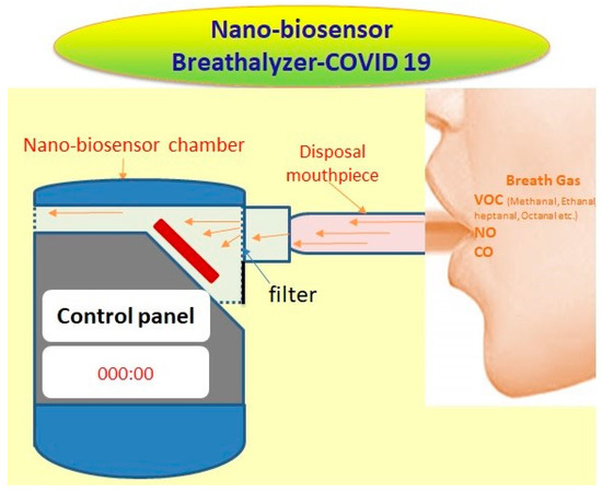
Figure 1.
Potential nano-biosensor breath-analyzer for COVID-19.
Within the sensor chamber, expiratory breath condensation gathers on a dielectric cold mirror, usually coated with a hydrophobic film, and angled at 45 degrees, facilitating breath interaction. Exhaled breath transforms into minute droplets that envelop an electrochemical biosensor. The efficacy of the breathalyzer’s detection hinges on the nano-biosensor type employed, featuring distinct electrodes for various targets. Developing a nanosensor tailored for COVID-19-associated gases or VOCs is pivotal.
Hybrid organic/inorganic configurations featuring 3D nano-architectures such as nanofibers, nanowires, and nanofins are instrumental in augmenting the active surface-area-to-volume ratios. In this context, the surfaces of semiconductor nanostructures frequently undergo functionalization with specific self-assembled monolayers or polymers. Organic materials exhibit limited durability. They have been widely recognized for their tendency to degrade relatively quickly during usage, leading to alterations in their chemical compositions and consequently compromising sensor performance. Consequently, sensor manufacturing prioritizes the production of pure inorganic materials, specifically metal oxide semiconductors, which remain extensively employed in gas-sensing applications due to their superior stability and reliability over time [44]. Electrodes are connected to contacts to fabricate the sensor. The crafted differentially sensitive and selective sensor layers and arrays facilitate precise detection of the targeted gas molecules and/or VOCs. Sensing assessments are often conducted within a quartz chamber comprising a regulated electric furnace and electrical terminals linked to computerized data acquisition. The resultant signal is converted into an electric circuit. Most prototypes integrate an electrical readout integrated circuit linked to a microsystem of sensor arrays capable of detecting exhaled breath and analyzing gas/VOC content. Test evaluation outcomes would be accessible to end-users through a smartphone application.
C. Breath analysis studies for COVID-19
A study from the early months of the pandemic in Wuhan, China focused on a handheld nanomaterial-based sensor array-based breathalyzer called the NaNose electronic nose (e-nose) developed with wider functionality for detection and continuous monitoring of COVID-19 in expirated air [45]. Gold nanoparticles were used in the sensors, which were connected to organic ligands, forming a diverse sensing layer capable of expanding or contracting upon exposure to VOCs, resulting in varying electrical resistance. Within the sensor, inorganic nanomaterials conduct electricity, while the organic elements adsorp VOCs. The system was reliant on a training machine learning algorithm. The cohort consisted of 49 COVID-19 patients, 58 healthy controls, and 33 individuals with non-COVID lung infections. Utilizing the test data, the system achieved an accuracy of 94% and 76%, respectively, in distinguishing patients from controls, and it exhibited accuracy rates of 90% and 95% in distinguishing patients with COVID-19 from those with other lung infections.
Exline and colleagues introduced an innovative electronic breathalyzer technology featuring a solitary sensor composed of γ-phase tungsten trioxide (WO3) as a catalytically active semiconductor sensing film [46]. This film was fabricated through sol–gel processing employing tungsten alkoxide precursors, and the sensor was designed for the rapid detection of NO and ammonia in exhaled breath, providing diagnostic results within a mere 15 s. The breathalyzer demonstrated the detection of elevated exhaled NO levels, exhibiting a unique pattern characteristic of individuals suffering from active COVID-19 pneumonia. Remarkably, it achieved an 88% accuracy rate in identifying COVID-19 pneumonia patients upon their admission to the intensive care unit (ICU). Furthermore, the sensitivity index associated with the breath print, which is closely linked to the concentration of the critical biomarker ammonia, appeared to exhibit a correlation with the duration of COVID-19 infection.
Among the many electronic nose device-based studies thus far, the majority are equipped with metal oxide semiconductor (MOS) sensors, typically containing 10 or fewer MOS sensors [47,48,49]. However, there are a few exceptions with sensor array type instruments that stand out. For instance, the handy EOS Cyranose 320 is noteworthy for its use of thirty-two sensors of carbon black polymer composite, while the NaNose utilizes eight sensors of gold nanoparticle [51,52]. It is worth noting that all these devices, except for one, were in existence and had undergone prior developmental stages before the emergence of the pandemic. Although not originally designed for COVID-19, however, evaluation for their potential utility in identifying other respiratory diseases and cancer was successfully carried out. Kwiatkowski and colleagues engineered an e-nose with innovative sensor array architecture and that is a cheap POC testing and screening device. This array comprises three micro-electro-mechanical system (MEMS) sensors and a solitary MOS sensor. MEMS are chip-sensors, constructed with a suspended mass between two capacitive plates. When sensors are tilted, this configuration generates a disparity in electrical potential, a phenomenon that is quantified as a variation in capacitance [53]. Thus, nano-breathalyzers have the potential to revolutionize the COVID-19 testing process with cheap, non-invasive, point-of-care detection devices with wide employability to service sectors such as schools, workplaces, hospitals, and large public gatherings/events (Figure 2).
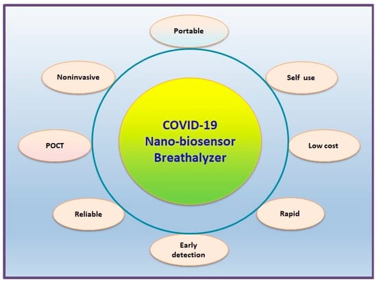
Figure 2.
Advantages and uses of nano-biosensor breathalyzer for COVID-19.
D. Future prospects
Following the stabilization of the acute phase of the COVID-19 pandemic, scientists are now exploring ways to continuously monitor indoor spaces for airborne transmission of viruses in real-time without necessitating specialized expertise. Leveraging recent advancements in aerosol sampling technology and highly sensitive nano-biosensing techniques, researchers are directing efforts towards creating tools capable of promptly detecting the SARS-CoV-2 virus in indoor environments.
A compact, cost-effective, and real-time proof-of-concept air quality (pAQ) monitor with the capability to identify airborne SARS-CoV-2 virus variants within indoor settings within a 5 min timeframe has been engineered by Puthussery et al. [54] This innovative device holds potential applications in hospitals, healthcare facilities, schools, and public areas, aiding in the detection of CoV-2 and potentially monitoring other respiratory virus aerosols. The system integrates a high-volume, high-flow (approximately 1000 L per minute) wet cyclone air sampler with an ultrasensitive micro-immunoelectrode (MIE) previously utilized in Alzheimer’s disease detection. The nanobody, derived from llamas and specifically targeting the SARS-CoV-2 spike antigen, is bonded covalently to the MIE biosensor. Employing screen-printed carbon electrodes, the biosensor utilizes the MIE technique to detect the presence of virus aerosols. Through square wave voltammetry, it identifies oxidation of tyrosine amino acids within the spike protein, offering a quantitative measure of the virus concentration in the sampled air. Comparative assessments demonstrate that the wet cyclone device exhibits similar/superior virus identification compared to commercial samplers, boasting a sensitivity ranging from 77% to 83% and a detection limit of 7–35 viral RNA copies per cubic meter of air [54]. This air quality monitor is tailored for immediate surveillance of virus variants that are detectable indoors and holds potential for adaptation to simultaneously detect various other respiratory pathogens of interest.
Artificial intelligence (AI) and machine learning (ML) play a significant role in enhancing the capabilities of nanomaterial-based biosensors. They have provided new avenues in biomedical monitoring, clinical diagnosis, and high-precision detection of diseases such as COVID-19 [55]. ML-enabled biosensors can offer real-time analysis of breath samples, leading to intelligent healthcare management and early disease detection. In the context of COVID-19, the combination of nano-biosensors with AI and ML can lead to the development of highly sensitive and specific breathalyzers for the rapid and accurate detection of the virus. These advancements hold promise for the future, offering the potential for widespread and accessible screening and early detection of COVID-19 and other respiratory viruses [56,57].
To address limitations associated with variations in test accuracy related to sample composition and storage in breath analysis, research efforts should prioritize standardizing sampling protocols and procedural methodologies [58]. Enhancements in systems should target mitigating technical, physiological, and pathophysiological variables that might affect the results. This approach aims to identify endogenous VOCs and establish reliable exhaled biomarker patterns [8].
Efforts in this direction involve establishing standardized correlations between concentrations of VOCs in blood and breath [8]. Additionally, development of handy and cost-effective nanomaterial sensors that resist humidity should be prioritized. These sensors should fulfill clinical requirements, displaying selectivity for VOCs and inorganic gases identified in specific diseases, and ensuring swift population screening. Moreover, optimizing sensor training and validation using diverse subjects becomes crucial for devising sensors applicable in clinical diagnosis [59]. A noteworthy aspect is the emerging capacity of such systems to differentiate between various diseases, providing diagnoses of conditions that manifest similar clinical presentations. This aspect, previously overlooked, now demands considerable attention in the pursuit of advancing diagnostic capabilities [59].
The ongoing advancements in innovative nanomaterials present a significant avenue for enhancing sensing components, particularly for the creation of sensors that exhibit both selectivity and cross-reactivity. This progress holds promise, particularly in the realms of POC diagnosis and treatment. Several fundamental challenges within this emerging area impede practical application of breathomics in medical settings, necessitating focused attention.
6. Conclusions
Rapid advancements in sensor technologies, coupled with sophisticated data analysis techniques, have paved the way for accurate and sensitive detection of gases as well as specific VOC patterns associated with different diseases. The utilization of large, costly, and intricate analytical devices for identifying exhaled volatile organic compounds faces limitations in hospital settings, and their integration into portable point-of-care systems remains unachievable. The simplicity of sample collection, coupled with the ability to provide real-time results, positions breath analysis as an attractive tool for early COVID-19 diagnosis, disease progression monitoring, and treatment response evaluation. Electronic-nose technology and related VOC detection methods hold the potential for early and noninvasive identification of COVID-19 individuals who are pre-symptomatic or asymptomatic carriers of the virus, displaying no overt symptoms following infection. The multitude of advantages associated with early COVID-19 detection through noninvasive means offers substantial improvements in precision medicine. These benefits encompass reduced transmission rates, enhanced effectiveness of regional epidemiological management, expanded choices for more efficacious treatments, improved prognostic capabilities, and potentially lower COVID-19-related mortality rates across various age groups, racial backgrounds, and ethnic populations. However, validation of breath analysis results is important due to the impact of trace levels of expired VOCs on accuracy. Concurrently, the absence of well-defined breath sampling protocols and procedures for breath collection and storage pose crucial challenges, potentially altering sample composition and consequently affecting analysis outcomes. Identifying the specific VOCs that consistently serve as reliable chemical biomarkers for SARS-CoV-2 infections in individuals with diverse ailment histories, health conditions, immune responses, and nutritional statuses greatly enhances the effectiveness of COVID-19 diagnoses through the utilization of nano-biosensor breathalyzer devices. Fulfilment of these objectives hinges upon interdisciplinary research and collaboration as fundamental prerequisites. Breath analysis emerges as a potent diagnostic tool that, upon surmounting prevailing challenges, holds the potential for adoption in clinical settings or within portable, compact health monitoring systems.
Author Contributions
Conceptualization, S.R., S.S., F.A. and N.S.; writing—original draft preparation, S.R., S.S. and F.A.; writing—review and editing, A.A., K.A.-M. and M.W.A.K.; supervision, S.S.; funding acquisition, S.S. All authors have read and agreed to the published version of the manuscript.
Funding
This research has been funded by Scientific Research Deanship at University of Ha’il-Saudi Arabia through project number MDR-22 016.
Data Availability Statement
Not applicable.
Conflicts of Interest
The authors declare no conflict of interest.
References
- Guan, W.J.; Ni, Z.Y.; Hu, Y.; Liang, W.H.; Ou, C.Q.; He, J.X.; Liu, L.; Shan, H.; Lei, C.L.; Hui, D.S.C.; et al. Clinical characteristics of coronavirus disease 2019 in China. N. Engl. J. Med. 2020, 382, 1708–1720. [Google Scholar] [CrossRef] [PubMed]
- Sherwani, S.; Khan, M.W.A.; Mallik, A.; Khan, M.; Saleem, M.; Raafat, M.; Shati, A.A.; Alam, N. Seroprevalence of Anti-S1-RBD Antibodies in Pre-pandemic and Pandemic Subjects From Hail Region, KSA. Front. Public Health 2022, 10, 874741. [Google Scholar] [CrossRef] [PubMed]
- Alluwaimi, A.M.; Alshubaith, I.H.; Al-Ali, A.M.; Abohelaika, S. The coronaviruses of animals and birds: Their zoonosis, vaccines, and models for SARS-CoV and SARS-CoV-2. Front. Vet. Sci. 2020, 7, 582287. [Google Scholar] [CrossRef] [PubMed]
- Sherwani, S.; Khan, M.W.A. Cytokine response in SARS-CoV-2 infection in the elderly. J. Inflamm. Res. 2020, 13, 737–747. [Google Scholar] [CrossRef] [PubMed]
- Peaper, D.R.; Kerantzas, C.A.; Durant, T.J. Advances in molecular infectious diseases testing in the time of COVID-19. Clin. Biochem. 2023, 117, 94–101. [Google Scholar] [CrossRef] [PubMed]
- Dutta, D.; Naiyer, S.; Mansuri, S.; Soni, N.; Singh, V.; Bhat, K.H.; Singh, N.; Arora, G.; Mansuri, M.S. COVID-19 diagnosis: A comprehensive review of the RT-qPCR method for detection of SARS-CoV-2. Diagnostics 2022, 12, 1503. [Google Scholar] [CrossRef] [PubMed]
- Gowri, A.; Kumar, N.A.; Anand, B.S. Recent advances in nanomaterials based biosensors for point of care (PoC) diagnosis of COVID-19–a minireview. TrAC Trends Anal. Chem. 2021, 137, 116205. [Google Scholar] [CrossRef]
- Das, S.; Pal, M. Non-invasive monitoring of human health by exhaled breath analysis: A comprehensive review. J. Electrochem. Soc. 2020, 167, 037562. [Google Scholar] [CrossRef]
- Chamorro-Garcia, A.; Merkoçi, A. Nanobiosensors in diagnostics. Nanobiomed 2016, 3, 1849543516663574. [Google Scholar] [CrossRef]
- Naresh, V.; Lee, N. A Review on Biosensors and Recent Development of Nanostructured Materials-Enabled Biosensors. Sensors 2021, 21, 1109. [Google Scholar] [CrossRef]
- Willner, M.R.; Vikesland, P.J. Nanomaterial enabled sensors for environmental contaminants. J. Nanobiotechnology 2018, 16, 95. [Google Scholar] [CrossRef] [PubMed]
- Tian, R.; Weng, T.; Chen, S.; Wu, J.; Yin, B.; Ma, W.; Liang, L.; Xie, W.; Wang, Y.; Zeng, X.; et al. DNA nanostructure-assisted detection of carcinoembryonic antigen with a solid-state nanopore. Bioelectrochemistry 2023, 149, 108284. [Google Scholar] [CrossRef] [PubMed]
- Sadeghi, M.; Kashanian, S.; Naghib, S.M.; Askari, E.; Haghiralsadat, F.; Tofighi, D. A highly sensitive nanobiosensor based on aptamer-conjugated graphene-decorated rhodium nanoparticles for detection of HER2-positive circulating tumor cells. Nanotechnol. Rev. 2022, 11, 793–810. [Google Scholar] [CrossRef]
- Chowdhury, A.D.; Takemura, K.; Khorish, I.M.; Nasrin, F.; Tun, M.M.N.; Morita, K.; Park, E.Y. The detection and identification of dengue virus serotypes with quantum dot and AuNP regulated localized surface plasmon resonance. Nanoscale Adv. 2020, 2, 699–709. [Google Scholar] [CrossRef] [PubMed]
- Pang, Y.; Rong, Z.; Wang, J.; Xiao, R.; Wang, S. A fluorescent aptasensor for H5N1 influenza virus detection based-on the core–shell nanoparticles metal-enhanced fluorescence (MEF). Biosens. Bioelectron. 2015, 66, 527–532. [Google Scholar] [CrossRef]
- He, Y.; Tian, F.; Zhou, J.; Zhao, Q.; Fu, R.; Jiao, B. Colorimetric aptasensor for ochratoxin A detection based on enzyme-induced gold nanoparticle aggregation. J. Hazard. Mater. 2020, 388, 121758. [Google Scholar] [CrossRef]
- Skotadis, E.; Aslanidis, E.; Tsekenis, G.; Panagopoulou, C.; Rapesi, A.; Tzourmana, G.; Kennou, S.; Ladas, S.; Zeniou, A.; Tsoukalas, D. Hybrid Nanoparticle/DNAzyme Electrochemical Biosensor for the Detection of Divalent Heavy Metal Ions and Cr3+. Sensors 2023, 23, 7818. [Google Scholar] [CrossRef]
- Samson, R.; Navale, G.R.; Dharne, M.S. Biosensors: Frontiers in rapid detection of COVID-19. 3 Biotech 2020, 10, 385. [Google Scholar] [CrossRef]
- Karakuş, E.; Erdemir, E.; Demirbilek, N.; Liv, L. Colorimetric and electrochemical detection of SARS-CoV-2 spike antigen with a gold nanoparticle-based biosensor. Anal. Chim. Acta 2021, 1182, 338939. [Google Scholar] [CrossRef]
- Zhang, M.; Li, X.; Pan, J.; Zhang, Y.; Zhang, L.; Wang, C.; Yan, X.; Liu, X.; Lu, G. Ultrasensitive detection of SARS-CoV-2 spike protein in untreated saliva using SERS-based biosensor. Biosens. Bioelectron. 2021, 190, 113421. [Google Scholar] [CrossRef]
- Kaloumenou, M.; Skotadis, E.; Lagopati, N.; Efstathopoulos, E.; Tsoukalas, D. Breath Analysis: A Promising Tool for Disease Diagnosis—The Role of Sensors. Sensors 2022, 22, 1238. [Google Scholar] [CrossRef] [PubMed]
- Vishinkin, R.; Haick, H. Nanoscale Sensor Technologies for Disease Detection via Volatolomics. Small 2015, 11, 6142–6164. [Google Scholar] [CrossRef] [PubMed]
- Corradi, M.; Mutti, A. Exhaled breath analysis: From occupational to respiratory medicine. Acta Bio-Medica Atenei Parm. 2005, 76 (Suppl. S2), 20. [Google Scholar]
- Güntner, A.T.; Abegg, S.; Königstein, K.; Gerber, P.A.; Schmidt-Trucksäss, A.; Pratsinis, S.E. Breath Sensors for Health Monitoring. ACS Sens. 2019, 4, 268–280. [Google Scholar] [CrossRef] [PubMed]
- de Lacy Costello, B.; Amann, A.; Al-Kateb, H.; Flynn, C.; Filipiak, W.; Khalid, T.; Osborne, D.; Ratcliffe, N.M. A review of the volatiles from the healthy human body. J. Breath Res. 2014, 8, 14001. [Google Scholar] [CrossRef]
- Kabir, E.; Raza, N.; Kumar, V.; Singh, J.; Tsang, Y.F.; Lim, D.K.; Szulejko, J.E.; Kim, K.-H. Recent Advances in Nanomaterial-Based Human Breath Analytical Technology for Clinical Diagnosis and the Way Forward. Chem 2019, 5, 3020–3057. [Google Scholar] [CrossRef]
- Wallace, M.A.G.; Pleil, J.D. Evolution of clinical and environmental health applications of exhaled breath research: Review of methods and instrumentation for gas-phase, condensate, and aerosols. Anal. Chim. Acta 2018, 1024, 18–38. [Google Scholar] [CrossRef]
- Cikach, F.S., Jr.; Dweik, R.A. Cardiovascular Biomarkers in Exhaled Breath. Prog. Cardiovasc. Dis. 2012, 55, 34–43. [Google Scholar] [CrossRef]
- Shorter, J.H.; Nelson, D.D.; McManus, J.B.; Zahniser, M.S.; Sama, S.R.; Milton, D.K. Clinical study of multiple breath biomarkers of asthma and COPD (NO, CO2, CO and N2O) by infrared laser spectroscopy. J. Breath Res. 2011, 5, 037108. [Google Scholar] [CrossRef]
- Boots, A.W.; Smolinska, A.; van Berkel, J.J.B.N.; Fijten, R.R.R.; Stobberingh, E.E.; Boumans, M.L.L.; Moonen, E.J.; Wouters, E.F.M.; Dallinga, J.W.; Van Schooten, F.J. Identification of microorganisms based on headspace analysis of volatile organic compounds by gas chromatography–mass spectrometry. J. Breath Res. 2014, 8, 027106. [Google Scholar] [CrossRef]
- Lau, H.-C.; Yu, J.-B.; Lee, H.-W.; Huh, J.-S.; Lim, J.-O. Investigation of Exhaled Breath Samples from Patients with Alzheimer’s Disease Using Gas Chromatography-Mass Spectrometry and an Exhaled Breath Sensor System. Sensors 2017, 17, 1783. [Google Scholar] [CrossRef] [PubMed]
- Ettema, A.R.; Lenders, M.W.P.M.; Vliegen, J.; Slettenaar, A.; Tjepkema-Cloostermans, M.C.; de Vos, C.C. Detecting multiple sclerosis via breath analysis using an eNose, a pilot study. J. Breath Res. 2020, 15, 027101. [Google Scholar] [CrossRef] [PubMed]
- Hakim, M.; Billan, S.; Tisch, U.; Peng, G.; Dvrokind, I.; Marom, O.; Abdah-Bortnyak, R.; Kuten, A.; Haick, H. Diagnosis of head-and-neck cancer from exhaled breath. Br. J. Cancer 2011, 104, 1649–1655. [Google Scholar] [CrossRef] [PubMed]
- Haick, H.; Hakim, M.; Patrascu, M.; Levenberg, C.; Shehada, N.; Nakhoul, F.; Abassi, Z. Sniffing Chronic Renal Failure in Rat Model by an Array of Random Networks of Single-Walled Carbon Nanotubes. ACS Nano 2009, 3, 1258–1266. [Google Scholar] [CrossRef] [PubMed]
- Marom, O.; Nakhoul, F.; Tisch, U.; Shiban, A.; Abassi, Z.; Haick, H. Gold nanoparticle sensors for detecting chronic kidney disease and disease progression. Nanomedicine 2012, 7, 639–650. [Google Scholar] [CrossRef]
- Ibrahim, W.; Carr, L.; Cordell, R.; Wilde, M.J.; Salman, D.; Monks, P.S.; Thomas, P.; Brightling, C.E.; Siddiqui, S.; Greening, N.J. Breathomics for the clinician: The use of volatile organic compounds in respiratory diseases. Thorax 2021, 76, 514–521. [Google Scholar] [CrossRef]
- Lichtenstein, M.; Turjerman, S.; Pinto, J.M.; Barash, O.; Koren, O. Pathophysiology of SARS-CoV-2 Infection in the Upper Respiratory Tract and Its Relation to Breath Volatile Organic Compounds. mSystems 2021, 6, e00104-21. [Google Scholar] [CrossRef]
- Remy, R.; Kemnitz, N.; Trefz, P.; Fuchs, P.; Bartels, J.; Klemenz, A.-C.; Rührmund, L.; Sukul, P.; Miekisch, W.; Schubert, J.K. Profiling of exhaled volatile organics in the screening scenario of a COVID-19 test center. iScience 2022, 25, 105195. [Google Scholar] [CrossRef]
- Ruszkiewicz, D.M.; Sanders, D.; O’Brien, R.; Hempel, F.; Reed, M.J.; Riepe, A.C.; Bailie, K.; Brodrick, E.; Darnley, K.; Ellerkmann, R.; et al. Diagnosis of COVID-19 by analysis of breath with gas chromatography-ion mobility spectrometry—A feasibility study. EClinicalMedicine 2020, 29–30, 100609. [Google Scholar] [CrossRef]
- Badawy, A.A.B. The kynurenine pathway of tryptophan metabolism: A neglected therapeutic target of COVID-19 pathophysiology and immunotherapy. Biosci. Rep. 2023, 43, BSR20230595. [Google Scholar] [CrossRef]
- Ball, L.; Robba, C.; Herrmann, J.; Gerard, S.E.; Xin, Y.; Mandelli, M.; Battaglini, D.; Brunetti, I.; Minetti, G.; Seitun, S.; et al. Lung distribution of gas and blood volume in critically ill COVID-19 patients: A quantitative dual-energy computed tomography study. Crit. Care 2021, 25, 214. [Google Scholar] [CrossRef] [PubMed]
- Fuschillo, S.; Ambrosino, P.; Motta, A.; Maniscalco, M. COVID-19 and diffusing capacity of the lungs for carbon monoxide: A clinical biomarker in postacute care settings. Biomarkers Med. 2021, 15, 537–539. [Google Scholar] [CrossRef] [PubMed]
- Cen, Z.; Lu, B.; Ji, Y.; Chen, J.; Liu, Y.; Jiang, J.; Li, X.; Li, X. Virus-induced breath biomarkers: A new perspective to study the metabolic responses of COVID-19 vaccinees. Talanta 2023, 260, 124577. [Google Scholar] [CrossRef] [PubMed]
- Nurputra, D.K.; Kusumaatmaja, A.; Hakim, M.S.; Hidayat, S.N.; Julian, T.; Sumanto, B.; Mahendradhata, Y.; Saktiawati, A.M.I.; Wasisto, H.S.; Triyana, K. Fast and noninvasive electronic nose for sniffing out COVID-19 based on exhaled breath-print recognition. NPJ Digit. Med. 2022, 5, 115. [Google Scholar] [CrossRef] [PubMed]
- Shan, B.; Broza, Y.Y.; Li, W.; Wang, Y.; Wu, S.; Liu, Z.; Wang, J.; Gui, S.; Wang, L.; Zhang, Z.; et al. Multiplexed Nanomaterial-Based Sensor Array for Detection of COVID-19 in Exhaled Breath. ACS Nano 2020, 14, 12125–12132. [Google Scholar] [CrossRef]
- Exline, M.C.; Stanacevic, M.; Bowman, A.S.; Gouma, P.-I. Exhaled nitric oxide detection for diagnosis of COVID-19 in critically ill patients. PLoS ONE 2021, 16, e0257644. [Google Scholar] [CrossRef]
- Wintjens, A.G.W.E.; Hintzen, K.F.H.; Engelen, S.M.E.; Lubbers, T.; Savelkoul, P.H.M.; Wesseling, G.; van der Palen, J.A.M.; Bouvy, N.D. Applying the electronic nose for pre-operative SARS-CoV-2 screening. Surg. Endosc. 2021, 35, 6671–6678. [Google Scholar] [CrossRef]
- Snitz, K.; Andelman-Gur, M.; Pinchover, L.; Weissgross, R.; Weissbrod, A.; Mishor, E.; Zoller, R.; Linetsky, V.; Medhanie, A.; Shushan, S.; et al. Proof of concept for real-time detection of SARS-CoV-2 infection with an electronic nose. PLoS ONE 2021, 16, e0252121. [Google Scholar] [CrossRef]
- Eusebio, L.; Derudi, M.; Capelli, L.; Nano, G.; Sironi, S. Assessment of the Indoor Odour Impact in a Naturally Ventilated Room. Sensors 2017, 17, 778. [Google Scholar] [CrossRef]
- Bax, C.; Robbiani, S.; Zannin, E.; Capelli, L.; Ratti, C.; Bonetti, S.; Novelli, L.; Raimondi, F.; Di Marco, F.; Dellacà, R.L. An Experimental Apparatus for E-Nose Breath Analysis in Respiratory Failure Patients. Diagnostics 2022, 12, 776. [Google Scholar] [CrossRef]
- Zamora-Mendoza, B.N.; de León-Martínez, L.D.; Rodríguez-Aguilar, M.; Mizaikoff, B.; Flores-Ramírez, R. Chemometric analysis of the global pattern of volatile organic compounds in the exhaled breath of patients with COVID-19, post-COVID and healthy subjects. Proof of concept for post-COVID assessment. Talanta 2022, 236, 122832. [Google Scholar] [CrossRef] [PubMed]
- VR, N.; Mohapatra, A.K.; VK, U.; Lukose, J.; Kartha, V.B.; Chidangil, S. Post-COVID syndrome screening through breath analysis using electronic nose technology. Anal. Bioanal. Chem. 2022, 414, 3617–3624. [Google Scholar] [CrossRef]
- Kwiatkowski, A.; Borys, S.; Sikorska, K.; Drozdowska, K.; Smulko, J.M. Clinical studies of detecting COVID-19 from exhaled breath with electronic nose. Sci. Rep. 2022, 12, 15990. [Google Scholar] [CrossRef] [PubMed]
- Puthussery, J.V.; Ghumra, D.P.; McBrearty, K.R.; Doherty, B.M.; Sumlin, B.J.; Sarabandi, A.; Mandal, A.G.; Shetty, N.J.; Gardiner, W.D.; Magrecki, J.P.; et al. Real-time environmental surveillance of SARS-CoV-2 aerosols. Nat. Commun. 2023, 14, 3692. [Google Scholar] [CrossRef] [PubMed]
- Manickam, P.; Mariappan, S.A.; Murugesan, S.M.; Hansda, S.; Kaushik, A.; Shinde, R.; Thipperudraswamy, S.P. Artificial Intelligence (AI) and Internet of Medical Things (IoMT) Assisted Biomedical Systems for Intelligent Healthcare. Biosensors 2022, 12, 562. [Google Scholar] [CrossRef]
- Velumani, M.; Prasanth, A.; Narasimman, S.; Chandrasekhar, A.; Sampson, A.; Meher, S.R.; Rajalingam, S.; Rufus, E.; Alex, Z.C. Nanomaterial-Based Sensors for Exhaled Breath Analysis: A Review. Coatings 2022, 12, 1989. [Google Scholar] [CrossRef]
- Junaid, S.B.; Imam, A.A.; Abdulkarim, M.; Surakat, Y.A.; Balogun, A.O.; Kumar, G.; Shuaibu, A.N.; Garba, A.; Sahalu, Y.; Mohammed, A.; et al. Recent Advances in Artificial Intelligence and Wearable Sensors in Healthcare Delivery. Appl. Sci. 2022, 12, 10271. [Google Scholar] [CrossRef]
- Bikov, A.; Lazar, Z.; Horvath, I. Established methodological issues in electronic nose research: How far are we from using these instruments in clinical settings of breath analysis? J. Breath Res. 2015, 9, 034001. [Google Scholar] [CrossRef]
- Koureas, M.; Kirgou, P.; Amoutzias, G.; Hadjichristodoulou, C.; Gourgoulianis, K.; Tsakalof, A. Target Analysis of Volatile Organic Compounds in Exhaled Breath for Lung Cancer Discrimination from Other Pulmonary Diseases and Healthy Persons. Metabolites 2020, 10, 317. [Google Scholar] [CrossRef]
Disclaimer/Publisher’s Note: The statements, opinions and data contained in all publications are solely those of the individual author(s) and contributor(s) and not of MDPI and/or the editor(s). MDPI and/or the editor(s) disclaim responsibility for any injury to people or property resulting from any ideas, methods, instructions or products referred to in the content. |
© 2023 by the authors. Licensee MDPI, Basel, Switzerland. This article is an open access article distributed under the terms and conditions of the Creative Commons Attribution (CC BY) license (https://creativecommons.org/licenses/by/4.0/).