Communication

Characterization of a Wide-Band Single-Photon Detector Based on Transition-Edge Sensor

1
Key Laboratory for Particle Astrophysics and Cosmology, Ministry of Education (MoE), Shanghai 201210, China
2
Center for Transformative Science, ShanghaiTech University, Shanghai 201210, China
3
Advanced Energy Science and Technology Guangdong Provincial Laboratory, Huizhou 516007, China
4
Shanghai Institute of Microsystem and Information Technology, Chinese Academy of Sciences, Shanghai 200050, China
*
Author to whom correspondence should be addressed.
Photonics 2025, 12(6), 609; https://doi.org/10.3390/photonics12060609
Submission received: 23 April 2025 / Revised: 18 May 2025 / Accepted: 20 May 2025 / Published: 13 June 2025
(This article belongs to the Special Issue Recent Progress in Single-Photon Generation and Detection)

Abstract

A superconducting transition-edge sensor (TES) as a microcalorimeter detects incoming photons by measuring heat converted from photon energy. With high resolving power and low noise levels, a TES is sensitive to single photons and able to count photons within a wide spectral band from X-ray to near-infrared. We have developed a TES detector aiming at soft X-ray spectroscopy applications. In this work, the performance of this detector is characterized. It is shown that the energy resolution of this detector is about 1.8 eV for 1.5 keV photons. The good resolution is also kept in visible range, enabling photon-number resolving for 405 nm photons.

1. Introduction

Single-photon detectors have been supporting and pushing the frontiers of a variety of science and technology. In recent years, interests in single-photon detectors with a low dark rate, high detection efficiency, high energy resolution or photon-number resolution increase in fields like quantum information, life sciences, materials science, astrophysics and particle physics experiments [1,2,3,4]. Single-photon detectors able to count photon numbers are also demanded in calibrating single-photon sources and other metrology applications [5,6]. With developments in detection techniques and material processing, diverse photodetectors have turned up and been employed practically. Among them, superconducting transition-edge sensors (TESs) operated as microcalorimeters have been proved to be high-performance single-photon detectors with a low dark rate and high resolving power over a wide spectral range from X-ray to near-infrared [7,8,9].
A TES is a superconducting film operated at cryogenic temperature on its superconducting-to-normal transition-edge. Its resistance is extremely sensitive to temperature variation within the steep transition-edge. In general, a TES detector consists of a photon absorber, a TES film as the thermometer and a thermal bath. The energy absorption from the incident photons in the absorber causes a temperature increment, which is sensed by the TES thermometer. The TES resistance increases first and then recovers together with the temperature decreasing. With a voltage-bias applied on the TES, the resistance changing and recovering produces an electrical signal of current pulse to be read out. The feature that the pulse height is proportional to the absorbed photon energy leads to intrinsic energy resolution with the TES [5]. As a result, a TES detector can be used and is advantageous for both single-photon spectroscopy and photon counting. In the spectroscopy application, the TES works in energy-dispersive (ED) mode, which has higher detection efficiency than wavelength-dispersive (WD) devices relying on dispersive optics, while offering much higher energy resolution than conventional ED devices based on semiconductors [9]. In photon counting applications, the TES detects photons with negligible dark counts and can be made sensitive to any wavelength in a broad band via proper design and material choice. Attributed to the above advantages, TES detectors have been widely used in various fields despite the requirements of sophisticated cooling and readout systems.
A TES detector targeting soft X-ray and extreme ultraviolet spectroscopy applications at the Shanghai soft X-ray Free-Electron Laser facility (SXFEL) [10] was built up recently. In this paper, we characterize the response of this detector to photons with different energies, with a focus on its energy and photon-number resolution. Some of the results are compared to a silicon photomultiplier detector. A discussion on the performance and applications is given.

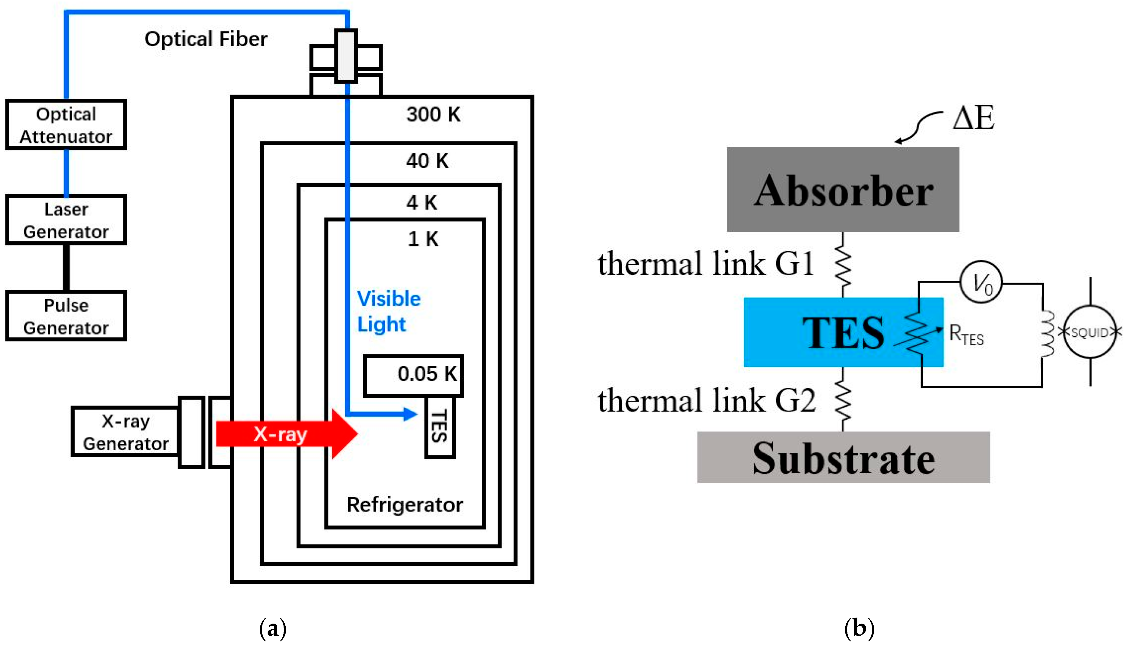
2. Detector Configuration and Experimental Set-Up

The TES detector was optimized for the detection of soft X-ray photons from hundreds to one-kilo electron volt (around several nm wavelength). The active area of the TES is 125 µm × 125 µm. The photon absorber as the top layer of the sensor is 2 µm thick bismuth, which can stop the incident soft X-ray photons (less than 1 keV) with near unity efficiency. The TES thermometer under the absorber is made of molybdenum-copper bilayer film, whose superconducting critical temperature is around 80 mK. A silicon nitride layer serves as weak thermal link between the TES thermometer and the heat sink. The current pulse signal produced by TES is converted to voltage signal and amplified by a two-stage superconducting quantum interference device (SQUID) readout circuit inside the cryogenic refrigerator. On room-temperature side, the signal is further amplified by an operational amplifier then acquired by a digitizer.
Both the TES and SQUID circuit are superconducting devices that should be operated at low temperature, and, especially for TES, lower temperature results in better resolution. A dilution refrigerator is used to provide the required cryogenic environment (tens of mK) for the TES detector. In addition, when the refrigerator is running, it also acts as a dark and vacuum chamber for photon detection. Flanges on the top and side of the refrigerator are interfaces to external photon sources.
Given the mechanical structure of the refrigerator, we characterize the response of the TES detector with two types of photon sources. The first is an X-ray generator coupled to the lateral flange on the refrigerator. X-ray photons pass the apertures and illuminate the sensor directly without going through any optical elements. The energy of the photons depends on the anode material in the X-ray generator and the applied voltage, and the intensity can be controlled by adjusting the power and filament current. Photons from the X-ray generator are composed of a characteristic X-ray with certain energies and bremsstrahlung photons with continuous energy spectrum. The second photon source is a laser diode driven by a pulse generator. Visible light emitted by the laser diode was transmitted via an optical fiber from the outside to the inside of the refrigerator and illuminates the sensor. The optical fiber goes into the refrigerator through a vacuum feedthrough mounted on its top flange. The energy of the visible photons is decided by the diode, and the intensity can be controlled by adjusting the electrical signal output from the pulse generator together with an optical attenuator. The schematic diagrams of the experimental set-up and the structure of the TES detector are shown in Figure 1.

3. Results

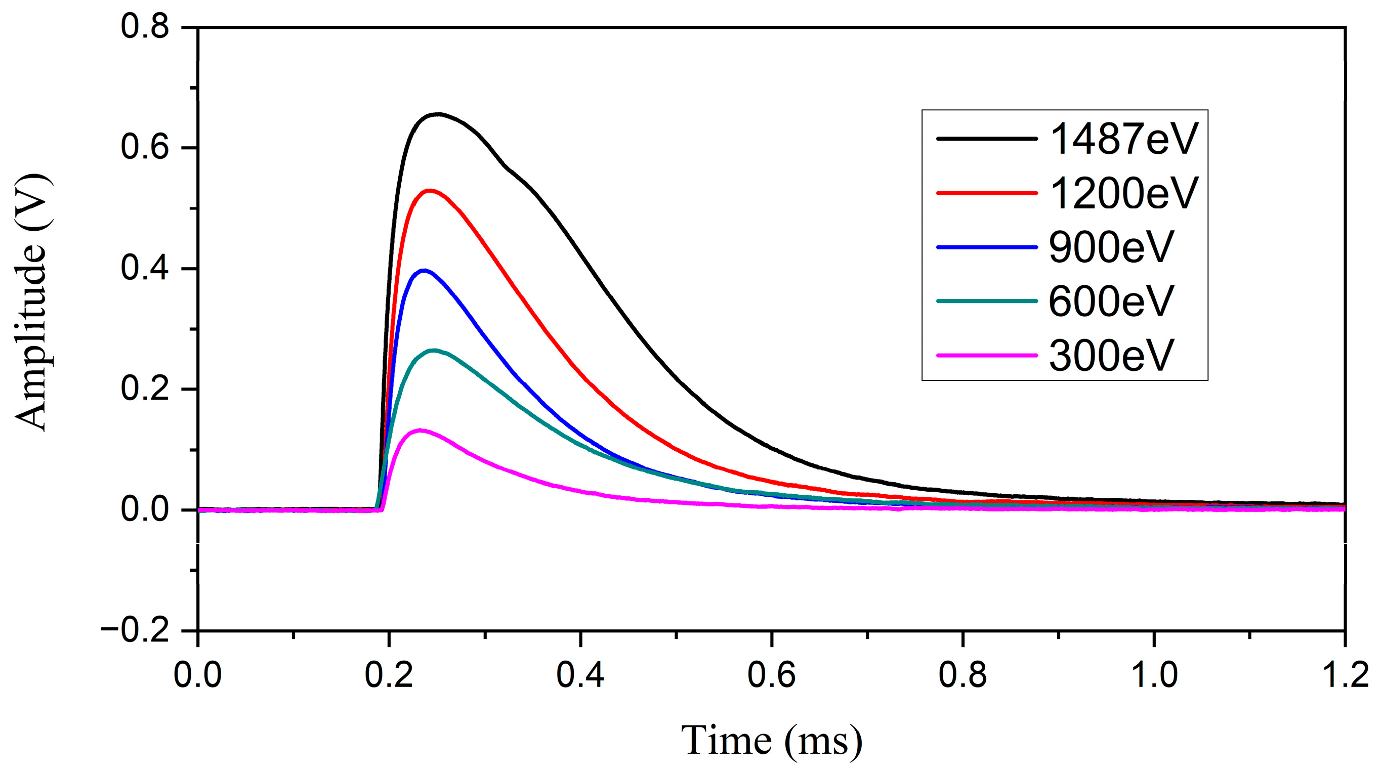
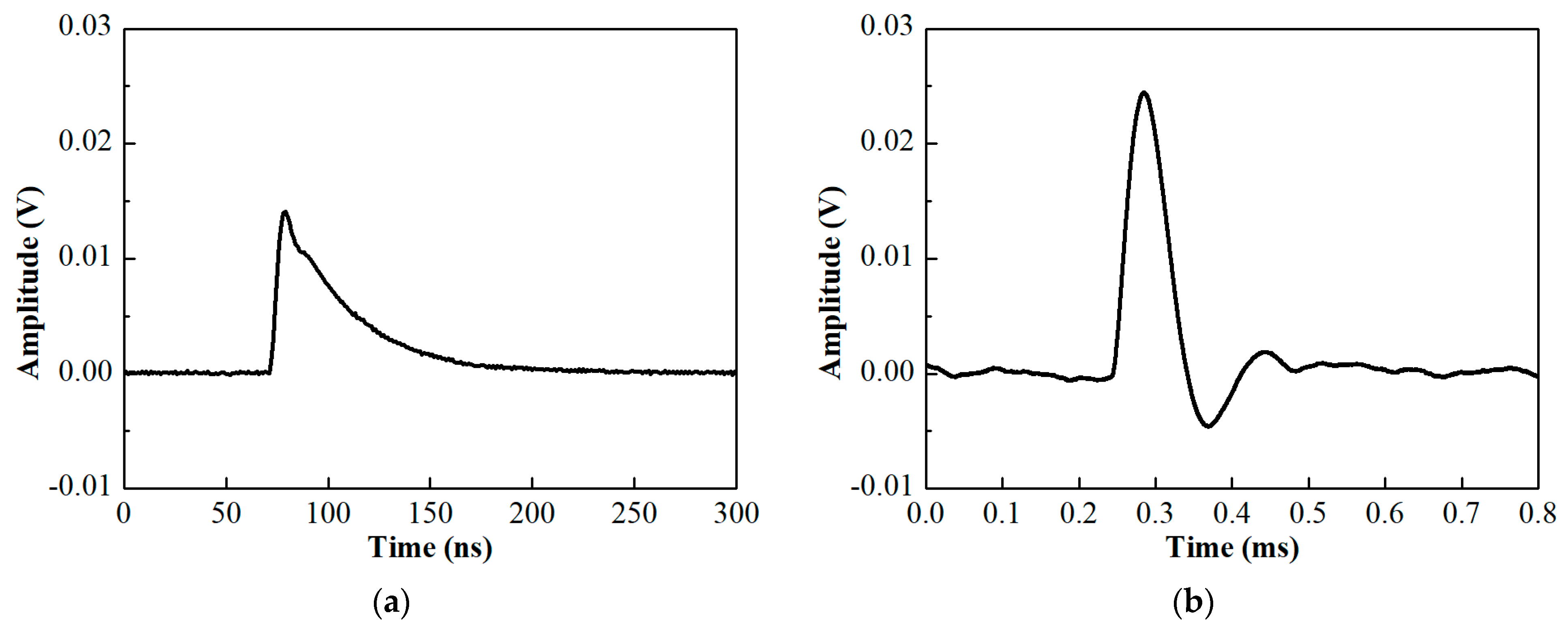
3.1. Single X-Ray Photon Detection

In this work, an X-ray generator with an aluminum anode (X-ray source RS 40B1 from PREVAC, Rogów, Poland) is used as the X-ray photon source. The aluminum Kα characteristic X-ray photons around 1.5 keV (~0.83 nm) are the dominant component and are accompanied by bremsstrahlung photons with lower energies (longer wavelengths) in our test. The intensity is adjusted to a low level in order to let each single photon hit the sensor separately in time. Since the height of the signal pulse is proportional to the photon energy, a single-photon energy spectrum can be obtained after accumulating a number of signal pulse heights and energy calibration. Figure 2 shows the peak of aluminum Kα photons in the acquired spectrum. The natural line of Al Kα consists of several components: Al Kα1 at 1486.94 eV, Al Kα2 at 1486.52 eV, Al Kα3 at 1496.85 eV, Al Kα4 at 1498.70 eV, Al Kα5 at 1507.4 eV, and Al Kα6 at 1510.9 eV [11]. By fitting (chi-square fitting) the measured data with the natural line shape considered, we obtain the energy resolution of the detector, which is about 1.8 eV (FWHM) at 1.5 keV.
Although bremsstrahlung photons with lower energies are fewer than the Kα photons, corresponding signals can be picked out from the data according to the pulse height. Pulses produced by different energies down to 300 eV (~4.1 nm) are shown in Figure 3. Due to the slow thermal decay via the weak thermal link between the TES and the heat sink, the recovery time of the signal pulse is relatively long. With a recovery time of about hundreds of microseconds, the maximum count rate of this detector is limited to several kilo-Hz.

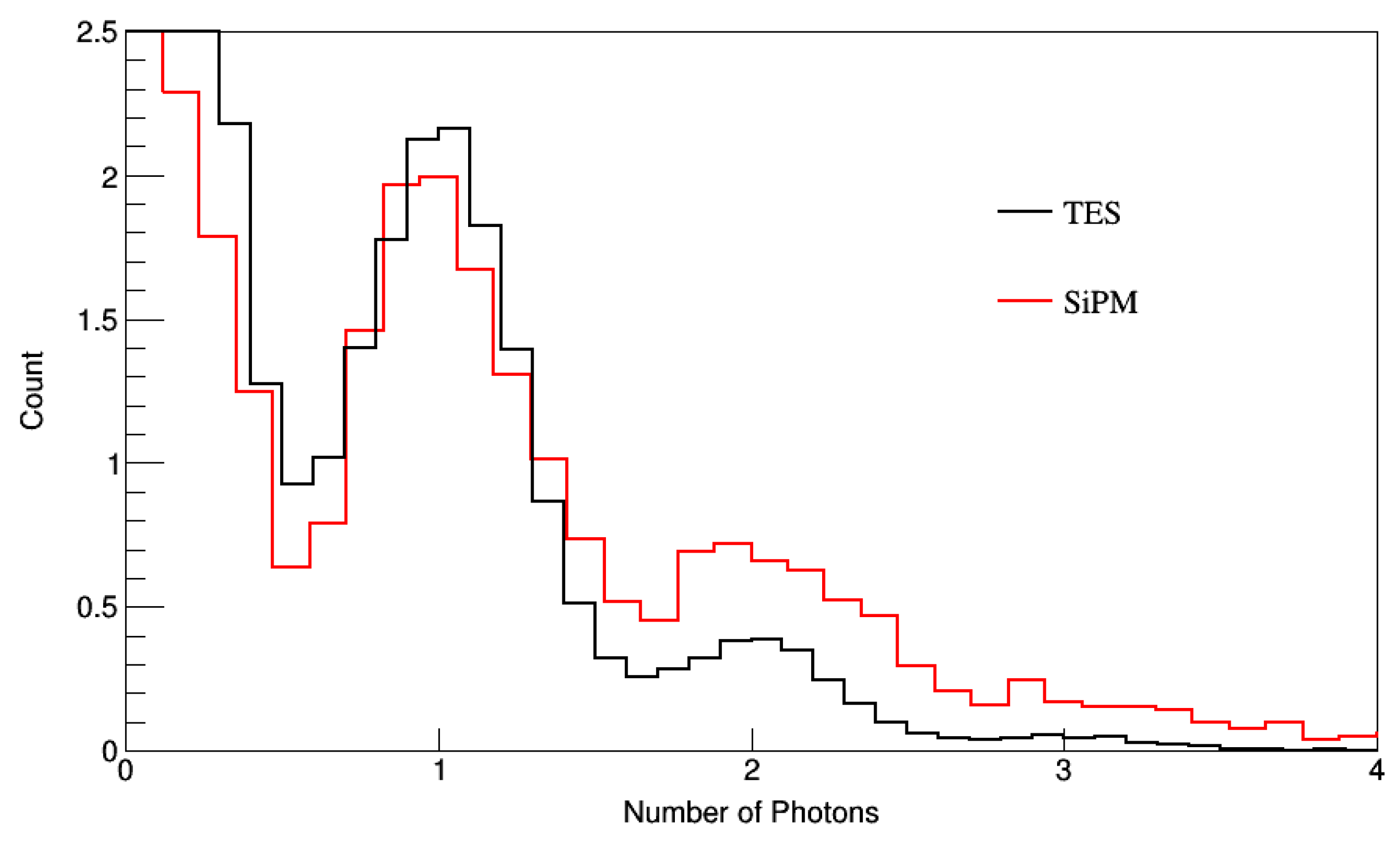
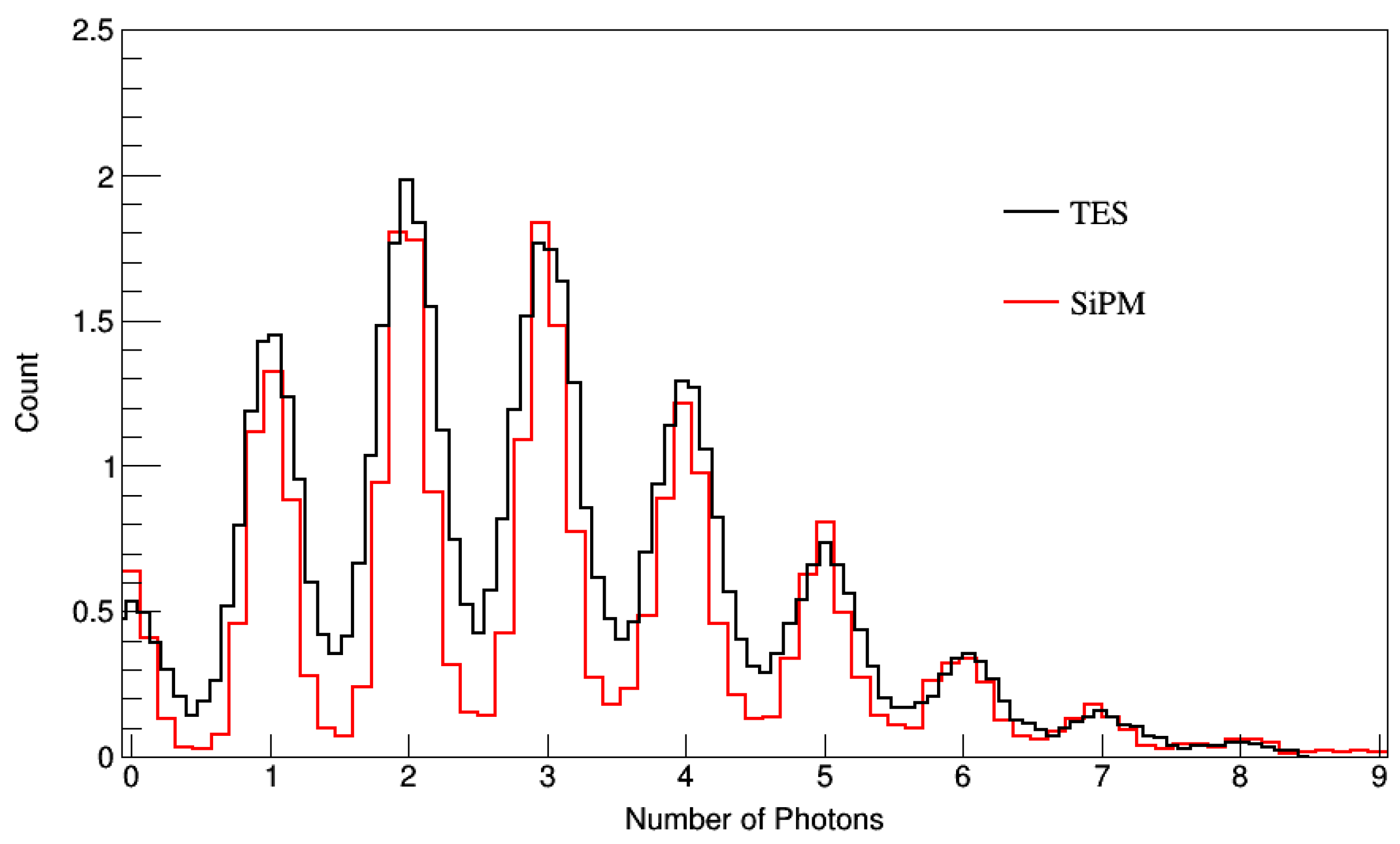
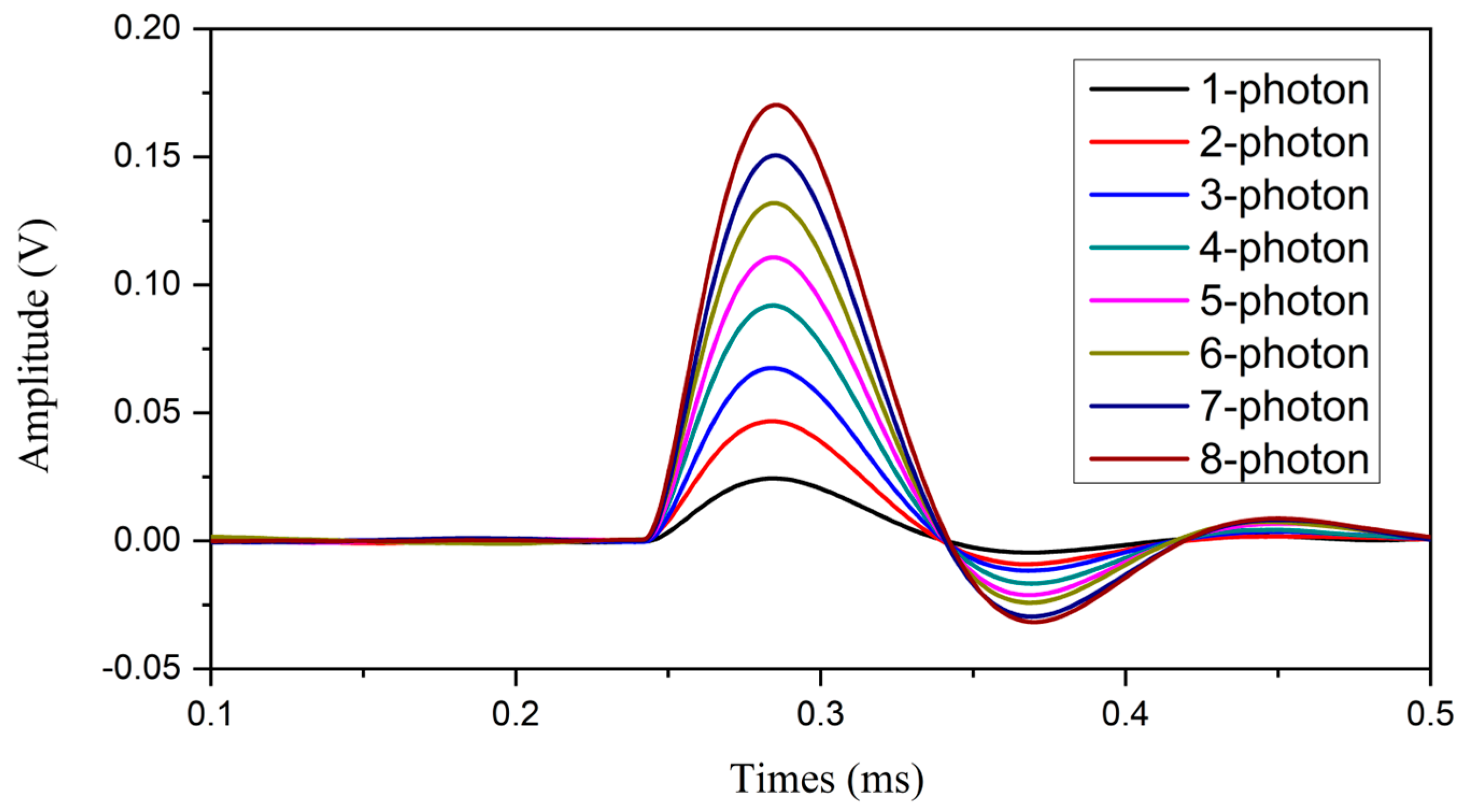
3.2. Single Visible Photon Detection

Properties of high sensitivity and low noise enable the TES to detect photons in a wide wavelength band. Although the TES is optimized for a soft X-ray range in our detector, it is also sensitive to heat caused by the absorption of visible photons. When the readout circuit is operated at a higher amplification gain, the signal resulting from visible photons can be identified. The energy resolution achieved in X-ray detection indicates its photon-number resolving ability in the visible range if the wavelength is fixed and known. That is because different numbers of photons result in different energy absorption and produce signal pulses with different heights in the TES. In this paper, we evaluate the detector response to 405 nm (3.06 eV) photons. Another photon-number resolving detector, a silicon photomultiplier (SiPM) sensitive to this wavelength (Model S13360-1350CS from Hamamatsu, Japan [12]), is also characterized for comparison. The SiPM counts the photon number in a different way. It is an array of avalanche photodiode pixels connected in parallel. Each pixel can detect photons separately and output a multiplied signal through an avalanche process. A pixel works as a binary unit whose response is the same for one or more photons. In faint light conditions, photons are dispersed onto different pixels. In this way, the SiPM is able to identify the photon number by spatial-multiplexing the pixels and counting the responsive pixels. While the dynamic range is limited by the width of the transition edge for a TES, it is limited by the number of pixels for a SiPM.
In our tests, the light intensity is controlled by a pulse generator, which also generates a synchronous signal along with each laser pulse. This synchronous signal can be used to trigger the data acquisition digitizer to capture the detector output signals. After accumulating a sequence of signals, the photon number can be calibrated according to the distribution of the detector response. The results obtained when the majority of light pulses contain a single photon are shown in Figure 4. Spectra in Figure 4 show that both our TES and the SiPM exhibit good single-photon resolution, with relative resolution of 27.9% and 26.4% for the SiPM and TES, respectively. The single-photon peak is remarkably distinguished from the pedestal peak, and the peaks of double or even three photons are also identified in each spectrum. Figure 4 confirms that the signal-to-noise ratio of the two detectors (both better than 3.5 at 405 nm) is good enough for single-photon detection.
After increasing the light intensity, a multi-photon spectrum following a Poissonian distribution can be obtained by both of the two detectors, as shown in Figure 5. In this test, the average photon number detected by the detector is around 3 in a light pulse. Both the TES and SiPM show good photon-number resolution up to eight photons. For the SiPM, the response to N photons is the sum of N pixels, thus, N times of single pixel response. This naturally results in a discrete distribution as shown in Figure 5. For the TES, the good energy resolution is maintained along with the increasing photon number; thus, the photon peaks can be clearly identified as energy peaks.
Figure 6 shows the average of 100 pulse waveforms of the two detectors in response to a single 405 nm photon signal. The timing properties of the pulse indicate the time response of the detector. It can be seen that the time response of the TES is quite slow. The rise time of the SiPM is on a level of several nanoseconds, and that of our TES is tens of microseconds. The recovery times are tens of nanoseconds and hundreds of microseconds, respectively. Since the time scales of the pulses are at different levels, the digitizers used to sample the pulses for the two detectors are different. For the SiPM, a 1 GS/s digitizer is used, while, for the TES, the digitizer sampling rate is 5 MS/s.
The design of spatial-multiplexing pixels in the SiPM enables the detection of multiple photons at a time shorter than the pixel recovery time. However, the number of photons should be less than the pixel number since a single pixel cannot detect other photons before recovering. For the TES, although the intrinsic energy resolution enables it to identify the photon number, the rather slow response is a significant limitation on the count rate. In many applications of the TES detector, an array of TESs is usually equipped in a detector to improve the count rate and enlarge the active area.
In Figure 7, the average of 100 pulse waveforms corresponding to different photon numbers is presented for our TES detector. The waveforms are selected according to the pulse heights, which have been separated in the multiple-photon spectrum. It can be seen that the pulses are well distinguished from each other in height.

4. Discussion

In the absence of a package and a dead layer on the TES, incident photons can be directly absorbed by the sensor without loss in any inactive structure. As a result, the TES detector can achieve high detection efficiency over a wide range of wavelengths, especially for soft X-ray or ultraviolet photons that are easily absorbed in materials. In addition, the intrinsic energy resolution of the TES avoids photon loss on additional dispersive optics. As our TES detector gives a good response to photons from 300 eV (4.1 nm) to 1.5 keV (0.83 nm), it can support a series of spectroscopy applications in the soft X-ray and extreme ultraviolet range. The energy resolution is close to that of the soft X-ray spectrometer with TESs used at the Stanford Synchrotron Radiation Lightsource, which is 1.5 eV in array form [13].
Our TES detector has proved photon-resolving capability for visible photons, although it is optimized for soft X-ray detection. The extremely low dark counts, which are measured to be less than 0.01 Hz, are also an advantage in photon counting. While the achieved performance is suitable in applications like life sciences and particle physics experiments, it can be further optimized to obtain higher detection efficiency in the visible and near-infrared range. It has been reported that the TES detector can be made a near-unity-efficiency detector [2,5], while this can hardly be realized with a semiconductor-based photodetector like the SiPM. In that way, the TES detector will be a competitive photodetector in quantum-information applications.
Another direction of optimization is to increase the number of TESs. With an array of TESs, the shortcoming of slow response can be compensated, and a high count rate is possible.

5. Conclusions

In conclusion, we characterized a newly developed photodetector based on the TES. The detector exhibits good single-photon response across a wide spectral range from soft X-rays to visible photons. For X-ray single-photon spectroscopy, the energy resolution at 1.5 keV is about 1.8 eV, resulting in E/ΔE better than 800. For visible photons, single-photon detection and photon-number resolving capability are also verified, and low dark count is measured. The time response of the detector is evaluated via the signal waveforms and found to be slow. In the next stage, we will employ an array of sensors to improve the count rate and increase the active area.

Author Contributions

Conceptualization, J.X.; methodology, J.X. and S.Z.; software, J.X.; formal analysis, J.X. and B.W.; writing—original draft preparation, J.X.; writing—review and editing, S.Z. and B.W.; funding acquisition, J.X. All authors have read and agreed to the published version of the manuscript.

Funding

This research was funded by the National Key Research and Development Program of China (grant number 2022YFF0608303), the National Natural Science Foundation of China (grant number 12005134), the Shanghai Soft X-ray Free-Electron Laser Beamline Project, and the Open Fund of the Key Laboratory for Particle Astrophysics and Cosmology, Ministry of Education of China.

Data Availability Statement

The data presented in this study are available upon request from the corresponding author.

Conflicts of Interest

The authors declare no conflicts of interest.

References

  1. Hadfield, R. Single-photon detectors for optical quantum information applications. Nat. Photonics 2009, 3, 696–705. [Google Scholar] [CrossRef]
  2. Bartley, T.J. Superconducting detectors count more photons. Nat. Photonics 2023, 17, 8–9. [Google Scholar] [CrossRef]
  3. Charaev, I.; Bandurin, D.A.; Bollinger, A.T.; Phinney, I.Y.; Drozdov, I.; Colangelo, M.; Butters, B.A.; Taniguchi, T.; Watanabe, K.; He, X.; et al. Single-photon detection using high-temperature superconductors. Nat. Nanotechnol. 2023, 18, 343–349. [Google Scholar] [CrossRef] [PubMed]
  4. Ota, R. Photon counting detectors and their applications ranging from particle physics experiments to environmental radiation monitoring and medical imaging. Radiol. Phys. Technol. 2021, 14, 134–148. [Google Scholar] [CrossRef] [PubMed]
  5. Rosenberg, D.; Lita, A.E.; Miller, A.J.; Nam, S.W. Noise-free high-efficiency photon-number-resolving detectors. Phys. Rev. A 2005, 71, 061803. [Google Scholar] [CrossRef]
  6. Zwinkels, J.C.; Ikonen, E.; Fox, N.P.; Ulm, G.; Rastello, M.L. Photometry, radiometry and ‘the candela’: Evolution in the classical and quantum world. Metrologia 2010, 47, R15. [Google Scholar] [CrossRef]
  7. Doriese, W.B.; Abbamonte, P.; Alpert, B.K.; Bennett, D.A.; Denison, E.V.; Fang, Y.; Fischer, D.A.; Fitzgerald, C.P.; Fowler, J.W.; Gard, J.D.; et al. A practical superconducting-microcalorimeter X-ray spectrometer for beamline and laboratory science. Rev. Sci. Instrum. 2017, 88, 053108. [Google Scholar] [CrossRef] [PubMed]
  8. Rauscher, B.J.; Nagler, P.C.; Sadleir, J.E.; Moseley, S.H.; Greenhouse, M.A. Development of transition edge sensor detectors optimized for single-photon spectroscopy in the optical and near-infrared. Proc. SPIE 2018, 10709, 1070931. [Google Scholar] [CrossRef]
  9. Cabrera, B.; Clarke, R.M.; Colling, P.; Miller, A.J.; Nam, S.; Romani, R.W. Detection of single infrared, optical, and ultraviolet photons using superconducting transition edge sensors. Appl. Phys. Lett. 1998, 73, 735–737. [Google Scholar] [CrossRef]
  10. Zhao, Z.; Wang, D.; Gu, Q.; Yin, L.; Gu, M.; Leng, Y.; Liu, B. Status of the SXFEL Facility. Appl. Sci. 2017, 7, 607. [Google Scholar] [CrossRef]
  11. Smith, S.J.; Adams, J.S.; Bailey, C.N.; Bandler, S.R.; Chervenak, J.A.; Eckart, M.E.; Finkbeiner, F.M.; Kelley, R.L.; Kilbourne, C.A.; Porter, F.S.; et al. Small Pitch Transition-Edge Sensors with Broadband High Spectral Resolution for Solar Physics. J. Low. Temp. Phys. 2012, 167, 168–175. [Google Scholar] [CrossRef]
  12. Available online: http://www.hamamatsu.com (accessed on 23 April 2025).
  13. Lee, S.-J.; Titus, C.J.; Mori, R.A.; Baker, M.L.; Bennett, D.A.; Cho, H.-M.; Doriese, W.B.; Fowler, J.W.; Gaffney, K.J.; Gallo, A.; et al. Soft X-ray spectroscopy with transition-edge sensors at Stanford Synchrotron Radiation Lightsource beamline 10-1. Rev. Sci. Instrum. 2019, 90, 113101. [Google Scholar] [CrossRef] [PubMed]
Figure 1. Schematic diagram of (a) the experimental set-up for detector characterization and (b) the structure of the TES detector.
Photonics 12 00609 g001
Figure 2. Aluminum Kα spectrum measured by the TES detector. The blue dotted line is the natural shape of aluminum Kα, and the red solid line is a fit to the data.
Photonics 12 00609 g002
Figure 3. Signal pulses of the TES detector produced by X-ray photons at different energies.
Photonics 12 00609 g003
Figure 4. Single-photon spectra of two detectors with 405 nm photons. The count is scaled for comparison.
Photonics 12 00609 g004
Figure 5. Multiple-photon (up to eight) spectra of two detectors with 405 nm photons. The count is scaled for comparison.
Photonics 12 00609 g005
Figure 6. The 100-averaged pulse waveform of the single-photon (405 nm) signal of the two detectors: (a) SiPM, (b) TES.
Photonics 12 00609 g006
Figure 7. The 100-averaged multiple-photon (405 nm) pulses of the TES detector.
Photonics 12 00609 g007
Disclaimer/Publisher’s Note: The statements, opinions and data contained in all publications are solely those of the individual author(s) and contributor(s) and not of MDPI and/or the editor(s). MDPI and/or the editor(s) disclaim responsibility for any injury to people or property resulting from any ideas, methods, instructions or products referred to in the content.

