Article

The Mass Spectrometry Identification, Antimicrobial Genes Detection, and Proteomics Analysis of Stutzerimonas stutzeri Strain Was Isolated from Industrial Wastewater

1
School of Environmental Engineering, Yellow River Conservancy Technical Institute, Kaifeng Key Laboratory of Food Composition and Quality Assessment, Kaifeng 475004, China
2
National Research Center for Wildlife-Borne Diseases, Key Laboratory of Animal Ecology and Conservation Biology, Institute of Zoology, Chinese Academy of Sciences, Beijing 100101, China
*
Authors to whom correspondence should be addressed.
Separations 2023, 10(9), 461; https://doi.org/10.3390/separations10090461
Submission received: 25 July 2023 / Revised: 14 August 2023 / Accepted: 21 August 2023 / Published: 22 August 2023

Abstract

:
A large amount of organic matter, heavy metals, and even antibiotics are present in industrial wastewater, aquaculture waters, and various types of sewage, along with abundant microorganisms. To date, only a few studies involving the resistance and proteomics of Stutzerimonas stutzeri in high-salt wastewater are available. Herein, a comprehensive assessment of a newly isolated Stutzerimonas stutzeri strain, which is present in high-salt wastewater, was performed using mass spectrometry, genetic identification, and biochemical analysis to characterize the genetic and biochemical properties. Growth experiments revealed that the Stutzerimonas stutzeri strain had a moderate growth rate in nutrient broth, and the bacterial count was not high. Further analysis highlighted an apparent susceptibility of this strain to most antibiotics but some resistance to chloramphenicol and minocycline. A resistance gene assay results showed that the gene gyrB was associated with antibiotic resistance in this Stutzerimonas stutzeri strain. Proteomic analysis revealed for the first time the co-existence of two drug-resistance-related proteins (Multidrug/solvent RND membrane fusion protein and MexE) in Stutzerimonas stutzeri. Moreover, Stutzerimonas stutzeri isolated from high-salt wastewater was subjected to drug resistance gene detection, and the total protein of Stutzerimonas stutzeri was detected by protein mass spectrometry analysis. The subcellular classification shows that the 50 proteins with the highest abundance are divided into cell inner membrane, cell outer membrane, cytoplasm, cytoplasmic side, membrane, multi-pass membrane protein, and peripheral membrane protein, among which the proportion of cytoplasmic components is the highest. Overall, this study’s findings provide a new perspective for further research on the characteristics of Stutzerimonas stutzeri in high-salt wastewater.

1. Introduction

With the rapid development of the global economy, water pollution has become a global problem, affecting economic development and ecological protection [1]. Due to the wide variety of chemical components in the environment, it is necessary to reduce its impact on the environment, especially by reducing its concentration in special areas, such as wastewater treatment plants, which is the main pathway for polluted water to enter the environment. For this purpose, some physical and chemical technologies have been developed to treat wastewater, such as using advanced electrochemical oxidation processes that are efficient, but the microbial treatment of wastewater still has advantages over chemical methods [2,3]. Paracetamol is a commonly used pharmaceutical worldwide, and its widespread use has led to its presence in various environments [4]. Paracetamol in the environment can cause genotoxicity, hepatotoxicity, endocrine interference, and other negative effects on organisms. Therefore, it is of great significance to study efficient removal methods of paracetamol to ensure water quality and safety.
Stutzerimonas, a recently proposed genus within the Pseudomonadaceae family belonging to Pseudomonas stutzeri in the formerly phylogenetic group, which includes at least 16 named species and 22 genomovars of Stutzerimonas stutzeri [5]. Pseudomonas stutzeri is a short, rod-shaped, white, and transparent colony with a denitrification ability that can be isolated from contaminated sediment [6]. A study identified the tetracycline resistance potential of heterotrophic bacteria isolated from 24 freshwater fin-fish farming ponds in Andhra Pradesh, India, and identified a Stutzerimonas stutzeri strain resistant to high tetracycline concentrations [7]. At present, the biological treatment of wastewater containing high concentrations of ammonia and nitrogen has received considerable attention. Previous studies showed that Pseudomonas sp. can remove ammonia and nitrogen from water [8,9,10]. In recent years, an MDR plasmid co-harboring the carbapenem resistance gene bla (VIM-2) and tigecycline resistance gene cluster tmexCD1-toprJ1 was identified in a Pseudomonas stutzeri isolate [11,12]. A Stutzerimonas stutzeri strain isolated from sewage sludge produced in wastewater treatment plants could degrade very high paracetamol concentrations in solutions wherein paracetamol was the sole carbon and energy source, and none of the Stutzerimonas stutzeri strains were previously described as paracetamol degraders [13]. Pseudomonas stutzeri, which can inhibit corrosion by sulfate-reducing bacteria, was isolated from the soil in an enrichment culture [14,15].
In this study, a Stutzerimonas stutzeri strain was isolated from local high-salt wastewater and subjected to biochemical identification, drug sensitivity identification, and growth analysis. Subsequently, a multifaceted analysis was performed using drug-resistance gene detection and proteomics. These experiments provide important implications for the next step in studying the analytical functions and applications of Stutzerimonas stutzeri.

2. Materials and Methods

2.1. Bacterial Isolation and Culture

High-salt wastewater was extracted from the inoculated total samples and subjected to streak culture on nutrient agar medium (HopeBiol, Qingdao, China) in a 37 °C incubator with a nutrient broth as the liquid medium. Subsequently, the bacteria were inoculated into the liquid medium and cultured at 240 rpm for >12 h.

2.2. Extraction of DNA

The Genome and Plasmid Extraction Kit (Tiangen) was used to extract DNA from the bacterial broth. Subsequently, PCR amplification of the 16S rDNA gene was performed using universal primers, 27F (5′-AGAGTTTGATCCTGGCTCAG-3′) and 1492R (5′-GGTTACCTTGTTACGACTT-3′). PCR reaction conditions: 98 °C for 5 min, 98 °C for 15 s, 55 °C for 25 s, and 72 °C for 20 s (40 cycles), then maintenance at 72 °C for 5 min. Additionally, a 1% agarose gel was made using Gelred. Finally, the PCR product size was determined by electrophoresis.

2.3. Construction of Phylogenetic Tree

The phylogenetic tree was constructed by the neighbor-joining method through the NCBI website, and the Max Seq difference was 0.5. All species involved in the evolutionary analysis were sourced from NCBI. B5 was the isolated sequence in this study.

2.4. Identification of Matrix-Assisted Laser Desorption Ionization Time-of-Flight Mass Spectrometry (MALDI-TOFMS)

The MALDI-TOFMS system (MEIHUA, M-Discover 100 Excellence, Zhuhai, China) was used for bacterial identification. Additionally, the samples were pre-treated with the Mass Spectrometer Microbial Sample Pretreatment Reagent (MEIHUA, 2 × 50 model, Zhuhai, China). Specifically, an appropriate number of bacteria were transferred to the target site of the mass spectrum sample target plate and applied uniformly to form a film. Subsequently, 1 μL of lysate M1 was pipetted onto the above-mentioned bacterial film and dried at room temperature. Finally, 1 μL of matrix solution was pipetted onto the same target site and dried at room temperature before being tested on the machine.

2.5. Biochemical Identification

The Microbial Identification and Drug Sensitivity Analysis System (MEIHUA, MA120, Zhuhai, China) were used for the biochemical identification of the microbe. Biochemical experiments first involved picking individual colonies for pure culture. Subsequently, a bacterial suspension of 0.5 Mcfarland turbidity units was prepared. Next, 100 µL of suspension was pipetted into the biochemical identification wells, where some wells required sterile paraffin oil. Additionally, self-adhesive stickers were peeled off and stuck on the biochemical identification plates. Finally, the samples reacted at 37 °C for 24 h and were placed in the machine for interpretation.

2.6. Antibiotic Resistance Analysis

50 µL bacterial suspension (0.5 Mcfarland turbidity units) was added to the M-H broth medium, followed by a drug-sensitive chromogenic solution and adequate mixing. Subsequently, 100 µL of suspension was pipetted into each drug-sensitivity assay well. Afterward, the self-adhesive stickers were peeled off and stuck onto the drug-sensitivity identification plate. The samples were incubated at 37 °C for 24 h and then placed into the machine for interpretation.

2.7. Detection of Bacterial Growth Curve

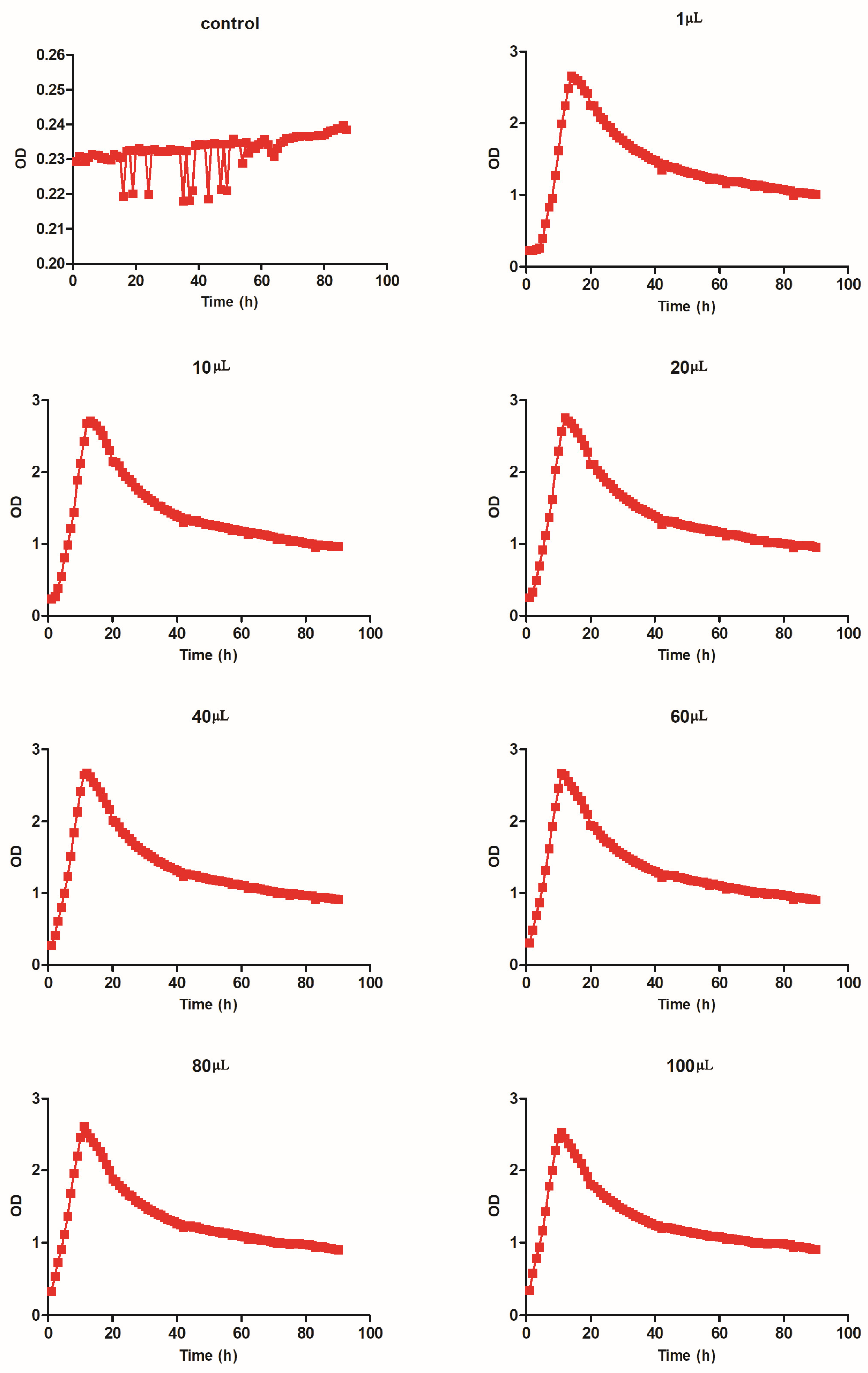
200 µL bacterial broth was added to a 96-well plate, and the absorbance of OD600 was determined using a microplate reader (Tecan, Sunrise, Grödig, Austria). Subsequently, the bacterial broth was diluted to 1 OD for growth experiments as follows: group 1, medium without bacteria; group 2, 1 µL bacterial broth + 999 µL medium; group 3, 10 µL bacterial broth + 990 µL medium; group 4, 20 µL bacterial broth + 980 µL medium; group 5, 40 µL bacterial broth + 960 µL medium; group 6, 60 µL bacterial broth + 940 µL medium; group 7, 80 µL bacterial broth + 920 µL medium; and group 8, 100 µL bacterial broth + 900 µL medium. The above-mentioned diluted 1 mL of medium was added into 48-well deep-well plates and subjected to microbial high-throughput growth assay (JIELING, MicroScreen-HT, Tianjin, China), and the OD value was recorded once every hour. After 90 h of growth, growth curves were constructed from the OD values collected per hour.

2.8. Identification of Drug-Resistant Genes

Primers for AAC(3)-II, cmlA, CTX-M-l, gyrA, gyrB, blaKPC, NDM-1, oqxA, oqxB, OXA, parC, qepA, qnrA, qnrB, qnrC, qnrD, qnrS, and Sul2 genes were synthesized [16,17,18,19]. PCR (BIORAD, C1000 Touch, Foster City, CA, USA) was used to amplify the resistance genes. Migration experiments of nucleic acids were performed by nucleic acid electrophoresis (BIORAD, PowerPac Basic, Hercules, CA, USA). Subsequently, the molecular weight of the genes was performed by a gel imager (BIORAD, GelGel Go, Hercules, CA, USA). Finally, the sequences with correct molecular weight were sequenced (Tsingke Bio, Beijing, China) and aligned with the sequences in the NCBI database.

2.9. Proteomic Analysis

An appropriate amount of protein was added to 50 µL of lyse buffer and heated at 95 °C for 10 min at 1000 rpm with agitation. Samples were cooled to room temperature, and trypsin digestion buffer was added and incubated at 37 °C for 2 h at 500 rpm. The termination buffer was added to terminate the enzymatic hydrolysis reaction. The peptides were desalted using an iST cartridge from the kit, eluted with 2 × 100 µL of elution buffer, and then lyophilized by SpeedVac. The peptides were re-dissolved in 0.1% formic acid in water and analyzed using Q-Exactive Plus coupled with an EASY-nanoLC 1200 system (Thermo Fisher Scientific, Waltham, MA, USA, Dr. Maisch C18). The mass spectrometer was run under data-dependent acquisition (DDA) mode, and automatically switched between MS and MS/MS mode. The survey of full scan MS spectra (m/z 350–1800) was acquired with 70,000 resolution. Tandem mass spectra were processed using PEAKS Studio version 10.6 (Bioinformatics Solutions Inc., Waterloo, IA, Canada). The database was Stutzerimonas stutzeri (strain A1501) (version 2023, 4092 entries), which download from uniprot. PEAKS DB was searched using a fragment ion mass tolerance of 0.02 Da and a parent ion tolerance of 10 ppm. The max missed cleavages was 2. Carbamidomethyl on cysteine was specified as the fixed modification. Oxidation on methionine, deamination on asparagine and glutamine, and acetylation on protein N-term were specified as the variable modifications. Peptides with 1%FDR and the proteins with 1%FDR and containing at least 1 unique peptide were filtered.

3. Results

3.1. Rapid Identification of Stutzerimonas stutzeri Using MALDI-TOFMS

In the MALDI-TOFMS and accompanying software MicroCtrl 1.0 (M-Discover 100 Excellence) analysis, the detected ion peaks (intensity) and the ion mass–charge ratio (m/z) were set as the vertical coordinate and horizontal coordinate, respectively. The protein fingerprints of the isolated bacterium were smooth in the baseline with prominent protein main peaks, which indicated good results. The ion peaks were primarily located near 8200, 7120, and 6631. The protein fingerprints of the isolated bacterium are presented in Figure 1. Compared with the standard spectrum diagram in the supporting software, the isolated bacterium was identified as Stutzerimonas stutzeri.

3.2. Molecular Identification of Stutzerimonas stutzeri

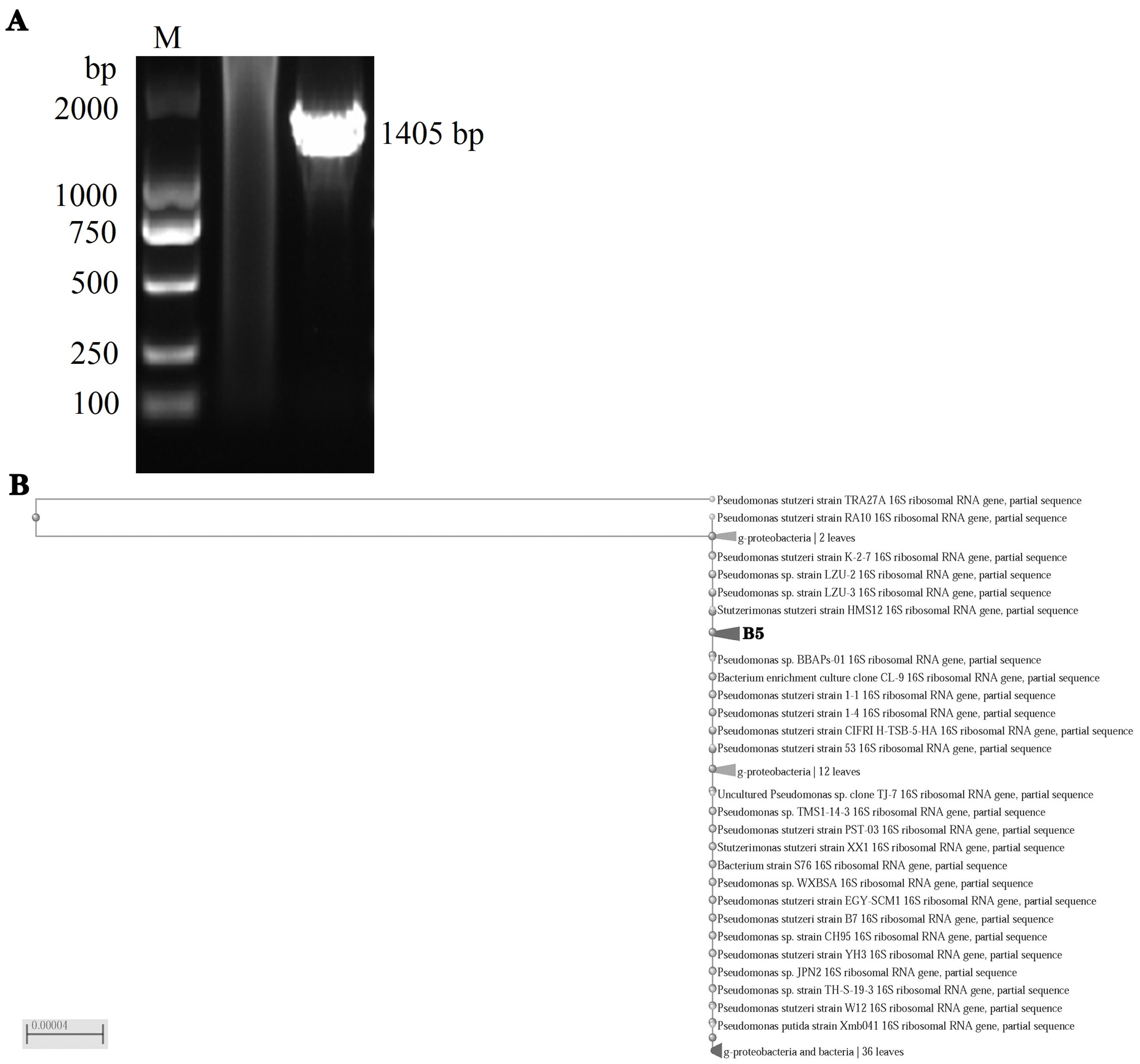
The amplification product of the 16S rDNA gene had a size of 1405 bp with only one specific DNA fragment present and exhibited good specificity (Figure 2A). The isolate was identified as Stutzerimonas stutzeri based on the 16S rDNA sequencing results and the alignment with the NCBI database. Moreover, the molecular evolutionary analysis showed that this Stutzerimonas stutzeri strain, isolated from high-salt wastewater, exhibited high homology with Stutzerimonas stutzeri (Figure 2B).

3.3. Biochemical Identification of Stutzerimonas stutzeri

Biochemical identification experiments showed that ADH, GLUf, NIT, ODC, GLU, LDC, MNE, MTE, MAL, SAC, CIT, XYL, ONPG, MLT, and ACE exhibited positive reactions. However, H2S, C, ESC, URE, IND, GEL, LAC, FRU, and MAN exhibited negative reactions (Table 1).

3.4. Antibiotic Susceptibility of Stutzerimonas stutzeri

Antibiotic susceptibility tests showed that the newly isolated Stutzerimonas stutzeri strain obtained from high-salt wastewater was susceptible to a majority of the tested antibiotics, including GEN, TOB, CAZ, CAZ, etc. However, it was resistant to CHL and intermediate to MIN (Table 2). These results indicated that Stutzerimonas stutzeri had good susceptibility to most antibiotics.

3.5. Growth Test for Stutzerimonas stutzeri

Stutzerimonas stutzeri growth at different dilutions was evaluated after 90 h of growth, which peaked at 11–14 h after incubation. Bacterial proliferation gradually decreased with time and reduced to the lowest point by approximately 90 h. However, differences were observed in the time point at which the peak was reached for Stutzerimonas stutzeri under different inocula. Specifically, the time to peak was longer for the low-inoculum bacteria than for the high-inoculum bacteria. However, the OD values at which the bacteria reached their peak were not significantly higher under low inoculum than under high inoculum. A higher OD value indicated a higher number of bacteria. The results indicated that the highest OD value for bacteria was at the 20 µL inoculum level, and the lowest OD value was at the 100 µL inoculum level (Figure 3 and Table 3).

3.6. Detection of Drug-Resistant Genes

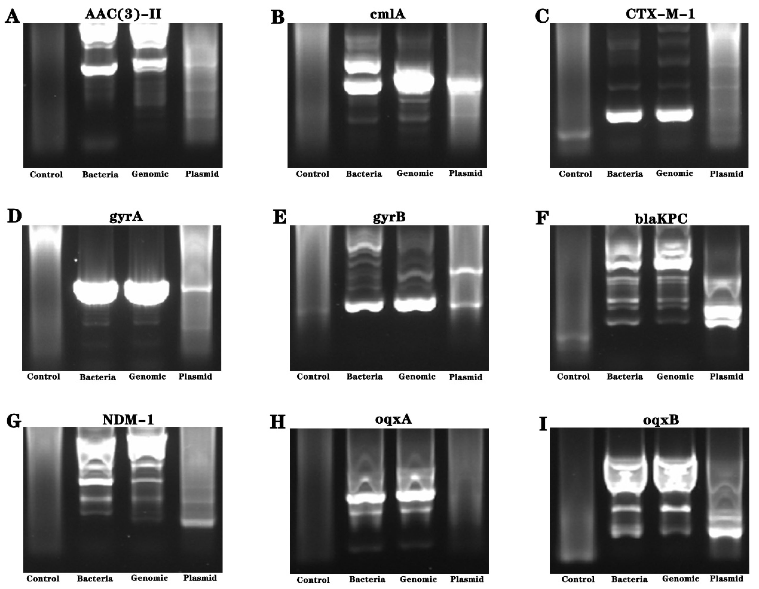
Primer amplification was performed using a bacterium, extracted bacterial genome, and plasmid as templates. Then, nucleic acid gel electrophoresis was performed, and the electrophoretic bands were subjected to sequencing analysis. The results revealed the presence of the resistant gene gyrB (Figure 4 and Figure 5).

3.7. Proteomic Analysis

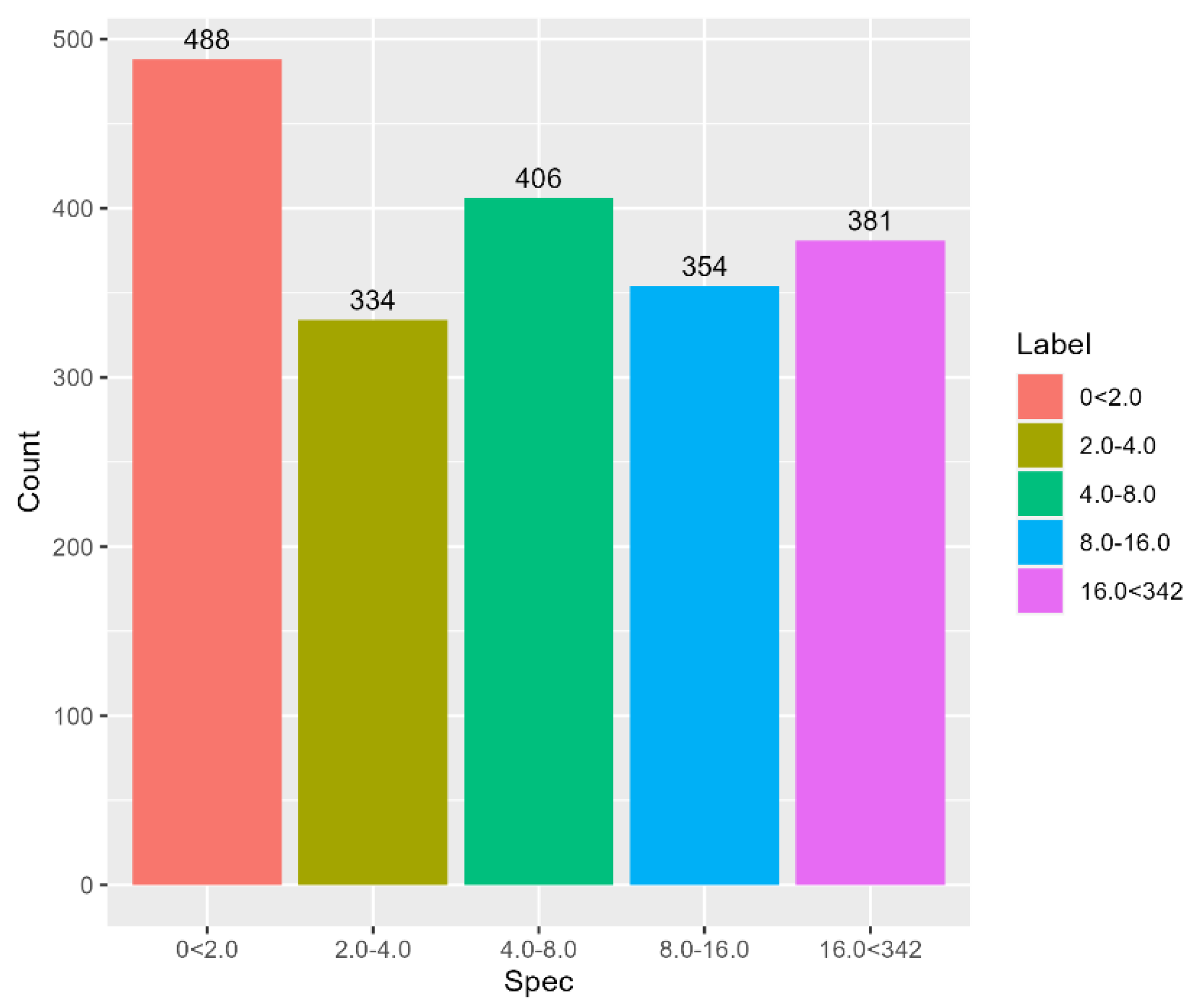
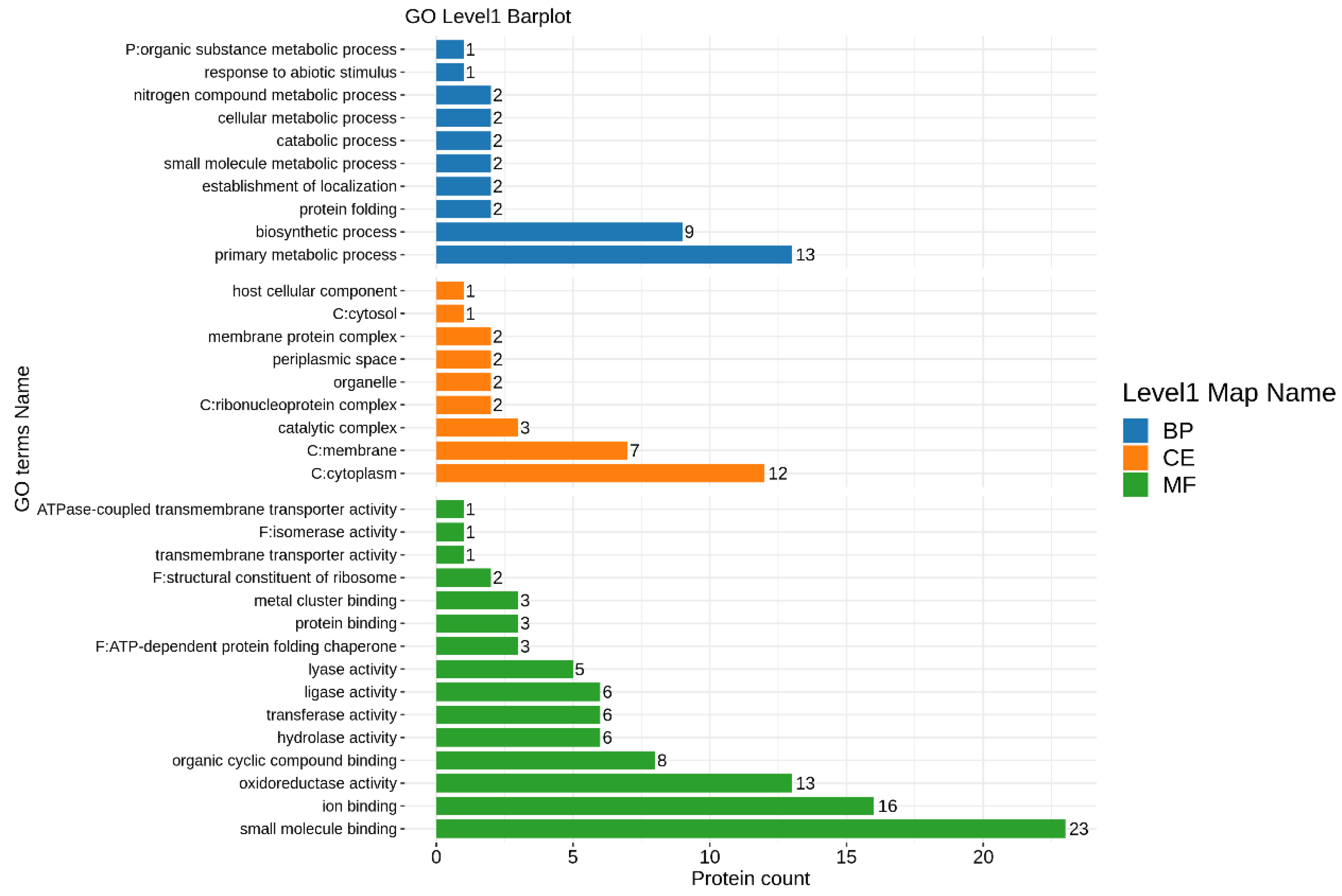
Protein mass spectrometry technology was used to further analyze the expression of drug-resistance-related proteins. The study demonstrated that more than 1900 Stutzerimonas stutzeri proteins were identified through proteomic analysis (Figure 6 and Figure 7). After the screening, two drug-resistance-related proteins were identified, namely, the Multidrug/solvent Resistance-Nodulation-cell Division (RND) membrane fusion protein and the RND multidrug efflux membrane fusion protein, MexE (Table 4). Combined with genetic testing, protein mass spectrometry identification technology further comprehensively analyzed the drug-resistance-related protein composition of Stutzerimonas stutzeri.
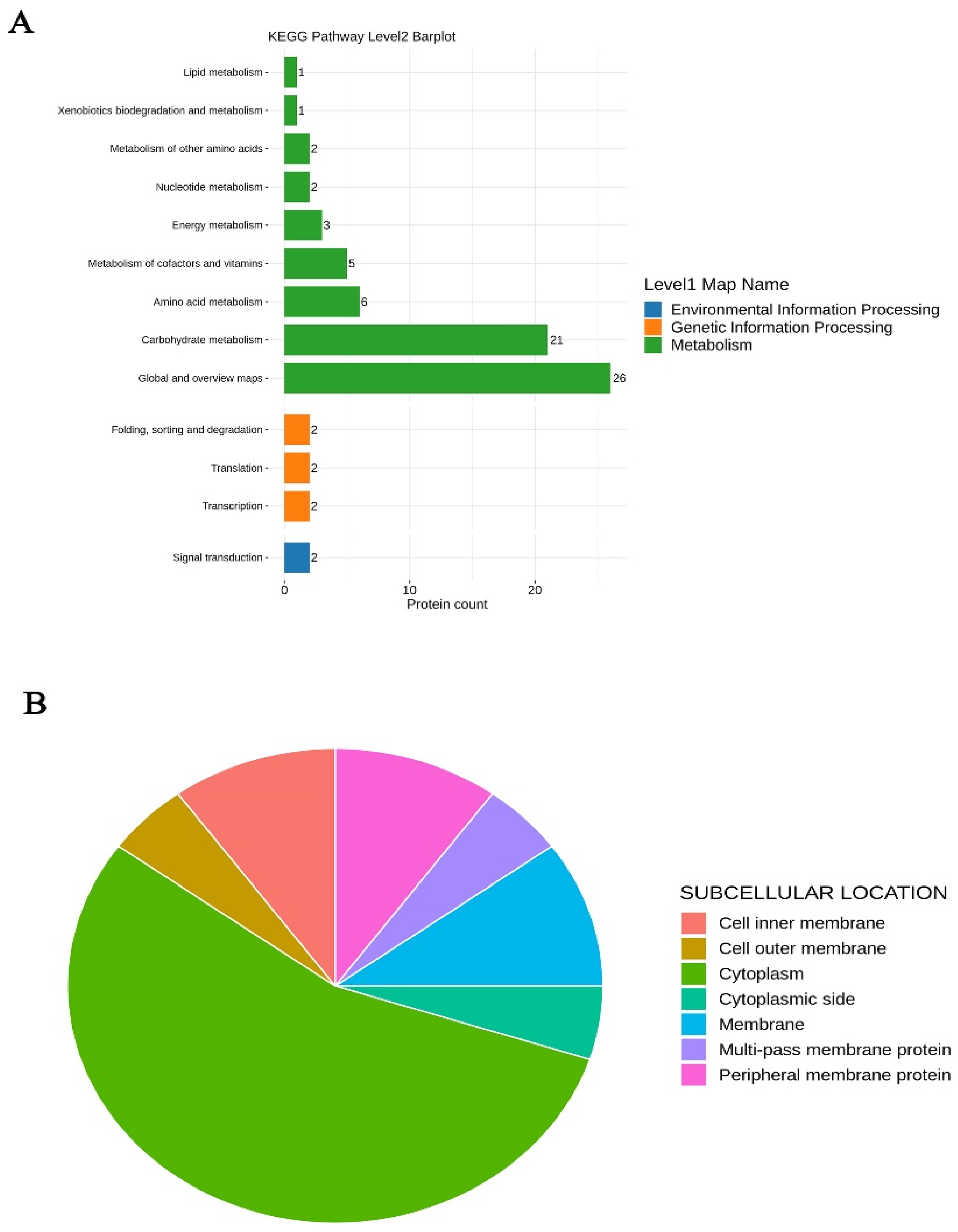
The enrichment analysis of the KEGG pathway revealed that the metabolism of level 1 is still the main component, and level 2 represent the classification of metabolic and function pathway (Figure 8A). The subcellular classification shows that the 50 proteins with the highest abundance are divided into cell inner membrane, cell outer membrane, cytoplasm, cytoplasmic side, membrane, multi-pass membrane protein, and peripheral membrane protein, among which the proportion of cytoplasmic components is the highest. (Figure 8B)

4. Discussion

Water pollution not only aggravates the deterioration of the ecological environment, but also endangers human life and health. In recent years, with the rapid development of random industries, more and more industrial wastewater is being discharged. Therefore, it is of great significance to explore new industrial wastewater treatment methods to improve industrial wastewater pollution. The microbial treatment of wastewater has also become a research hotspot.
Pseudomonas stutzeri, recently reclassified as a new genus Stutzerimonas, has various functions in different environments [20]. Some studies showed that Pseudomonas stutzeri could serve as a probiotic. For example, a previous study showed that the supplementation of 3.0 × 10⁵ CFU/mL of Pseudomonas stutzeri in the rearing water of spotted seabass increased the growth performance and improved the relative abundance of their gut microbiota, paving the way for the application of probiotics [21]. A study indicated that Pseudomonas stutzeri could be a promising species for the bioremediation of heavy crude oil [22]. Polyhydroxyalkanoate is used as an eco-friendly internal and external substrate for the growth regulation of heterotrophic denitrifiers and the promotion of the denitrification process for deep nitrogen removal from wastewater. Previous studies have shown that hydroxy alkanoate can significantly promote the growth of Pseudomonas stutzeri and increase the removal rate of nitrate [23]. A recent study showed that Pseudomonas stutzeri effectively degraded 4-trifluoromethylnicotinamide to 5-TFNA [24]. Pseudomonas stutzeri, Paracoccus sp. and Comamonas nitrativorans achieved a 98.4% total nitrogen removal rate at a COD/N ratio of 3.2. In addition, some studies result showed that Pseudomonas stutzeri was critically involved in NOx reduction [25]. Paracetamol is one of the most used pharmaceuticals in environments, such as surface and ground waters, sediments, soils or even plants, considering it is introduced mainly from the discharge of wastewater. Paracetamol in the environment can cause genotoxicity, hepatotoxicity, endocrine interference, and other negative effects on organisms. A study found the Pseudomonas stutzeri and Pseudomonas extremaustralis were able to degrade very high concentrations of paracetamol in solution as a sole carbon and energy source [13]. Phylogenetic analysis based on the phylogeny of the core genome of the Pseudomonadaceae family and several indices based on shared genes of members of new genera, as well as their phenotypic characteristics, including denitrification characteristics and lack of arginine dihydrogenase activity, support the proposal of the genera. Stutzerimonas stutzeri is the type species of the genus, which is characterized by its subdivision into 22 different genomes. Recently, a Stutzerimonas stutzeri strain was isolated from the surface of seawater, and the complete genome sequence was obtained using Illumina and Oxford Nanopore sequencing. This sequence provided a reference for genomic and metabolic analyses of Stutzerimonas stutzeri [26]. These genomes can be distinguished by experimental DNA-DNA hybridization and genome index, whose value is lower than the species threshold but cannot be distinguished in the phenotype [27,28]. Stutzerimonas stutzeri effectively reduced selenate and selenite into elemental selenium and volatile selenium species [29,30]. Stutzerimonas stutzeri has many applications, such as use in aquaculture as a probiotic, the degradation of heavy metals or organic pollutants, and the elimination of pesticides. However, it has not been studied in high-salt wastewater. Therefore, this study successfully isolated a strain of Stutzerimonas stutzeri from industrial wastewater, which was confirmed by the MALDI-TOFMS and 16S rDNA sequencing results. Further biochemical analysis showed that Stutzerimonas stutzeri was positive for ADH, GLUf, NIT, ODC, GLU, LDC, MNE, MTE, MAL, SAC, CIT, XYL, ONPG, MLT, and ACE. Among them, ADH provides catabolic activity that converts arginine to ornithine, resulting in the concomitant release of carbon dioxide and ammonia [31]. ONPG is mainly divided into ɑ-ONPG and β-ONPG. There is evidence that β-ONPG has the ability to hydrolyze lactose and has great potential for application in the food and pharmaceutical industries [32]. ACE is mainly used as a stabilizer and plasticizer, and can also be used in organic synthesis, pharmaceutical industry and dye preparation [33]. Taken together, the strain isolated from industrial wastewater in this study was identified as Stutzerimonas stutzeri.
Pseudomonas stutzeri develops strong resistance to antibiotics commonly used in clinics [34]. This study found Stutzerimonas stutzeri was significantly sensitive to most antibiotics, including GEN, TOB, CAZ, CAZ, etc., but had some resistance to CHL and intermediate to MIN. These results indicated that Stutzerimonas stutzeri had good susceptibility to most antibiotics. GyrB is a promising target for the discovery and development of a new class of antibiotics, binding ATP in the ATPase domain and catalyzing ATP hydrolysis, which provides energy for DNA superlinearity [35,36]. Current research has found that gyrB is a potential target for breaking the resistance of Staphylococcus aureus [37]. There is evidence that gyrB mutation plays an important role in the in vitro screening of Pseudomonas aeruginosa for fluoroquinolones resistance [38]. In this study, gyrB was amplified in Stutzerimonas stutzeri. Moreover, proteomic analysis revealed for the first time the co-existence of two drug-resistance-related proteins (Multidrug/solvent RND membrane fusion protein and MexE) in Stutzerimonas stutzeri. An RND efflux pump consists of inner membrane protein, periplasmic membrane fusion protein, and outer membrane protein, which play an important role and involve multidrug resistance [39]. Efflux pumps of the RND superfamily contribute to intrinsic and acquired resistance in Gram-negative pathogens, such as Vibrio parahaemolyticus, Pseudomonas aeruginosa, and Escherichia coli, and Gram-negative microbes [40,41]. The MexE, MexF, and OprN genes of RND family members play an important role in the activation of MDR. In recent years, it has been found that the expression of the RND efflux pump is significantly correlated with the resistance of most anti-pseudomonas antibiotics in Pseudomonas aeruginosa [42]. Moreover, growth experiments revealed that the Stutzerimonas stutzeri strain had a moderate growth rate in a nutrient broth, and the bacterial count was not high. In summary, combined with the results of our study, it can be inferred that Stutzerimonas stutzeri could grow in nutrient broth medium and have good sensitivity to most antibiotics, and that the proteins Multidrug/solvent RND membrane fusion protein and MexE may be closely related to the drug resistance of Stutzerimonas stutzeri.

5. Conclusions

In conclusion, Stutzerimonas stutzeri has many applications, such as use in aquaculture as a probiotic, the degradation of heavy metals or organic pollutants, and the elimination of pesticides. Studies on the involvement of Stutzerimonas stutzeri in high-salt wastewater are currently limited. In this study, the characteristics of this newly isolated Stutzerimonas stutzeri strain were analyzed using mass spectrometry, molecular biology, and proteomic analysis. gyrB was amplified from Stutzerimonas stutzeri. In addition, Multidrug/solvent RND membrane fusion protein and MexE were also detected via proteomics analysis. Overall, this study’s findings lay the foundation for future functional studies and the utilization of this bacterium. However, the potential value of Stutzerimonas stutzeri degradation and industrial wastewater treatment needs to be further explored.

Author Contributions

Methodology, Z.W. and X.S.; editing, H.W. and X.C.; conceptualization and review, H.W. and H.H. All authors have read and agreed to the published version of the manuscript.

Funding

This work was supported by the Science and Technology Major Project in Henan Province (No. 221100320200) and the Science and Technology Development Plan of Kaifeng in 2020 (No. 2003048).

Data Availability Statement

Not applicable.

Conflicts of Interest

The authors declare no conflict of interest.

References

  1. Wang, J.; Qian, W.; Du, J.; Liu, Y. Effective allocation of resources in water pollution treatment alternatives: A multi-stage gray group decision-making method based on hesitant fuzzy linguistic term sets. Environ. Sci. Pollut. Res. Int. 2020, 27, 3173–3186. [Google Scholar] [CrossRef] [PubMed]
  2. Torun, M.; Gültekin, O.; Şolpan, D.; Güven, O. Mineralization of paracetamol in aqueous solution with advanced oxidation processes. Environ. Technol. 2014, 36, 970–982. [Google Scholar] [CrossRef]
  3. Villota, N.; Lomas, J.M.; Camarero, L.M. Study of the paracetamol degradation pathway that generates color and turbidity in oxidized wastewaters by photo-Fenton technology. J. Photochem. Photobiol. A Chem. 2016, 329, 113–119. [Google Scholar] [CrossRef]
  4. Culebras, M.; Pishnamazi, M.; Walker, G.M.; Collins, M.N. Facile Tailoring of Structures for Controlled Release of Paracetamol from Sustainable Lignin Derived Platforms. Molecules 2021, 26, 1593. [Google Scholar] [CrossRef] [PubMed]
  5. Gomila, M.; Mulet, M.; García-Valdés, E.; Lalucat, J. Genome-Based Taxonomy of the Genus Stutzerimonas and Proposal of S. frequens sp. nov. and S. degradans sp. nov. and Emended Descriptions of S. perfectomarina and S. chloritidismutans. Microorganisms 2022, 10, 1363. [Google Scholar] [CrossRef]
  6. Li, B.; Jing, F.; Wu, D.; Xiao, B.; Hu, Z. Simultaneous removal of nitrogen and phosphorus by a novel aerobic denitrifying phosphorus-accumulating bacterium, Pseudomonas stutzeri ADP-19. Bioresour. Technol. 2021, 321, 124445. [Google Scholar] [CrossRef] [PubMed]
  7. Kusunur, A.B.; Mogilipuri, S.S.; Moturu, D.; Benala, M.; Vaiyapuri, M.; Panda, S.K.; George, J.C.; Badireddy, M.R. Tetracycline resistance potential of heterotrophic bacteria isolated from freshwater fin-fish aquaculture system. J. Appl. Microbiol. 2023, 134, 12. [Google Scholar] [CrossRef]
  8. Fu, W.-L.; Duan, P.-F.; Wang, Q.; Liao, Y.-X.; Wang, Y.-S.; Xu, M.-J.; Jiang, H.-H.; Zhang, X.; Rao, Z.-M. Transcriptomics reveals the effect of ammonia nitrogen concentration on Pseudomonas stutzeri F2 assimilation and the analysis of amtB function. Synth. Syst. Biotechnol. 2023, 8, 262–272. [Google Scholar] [CrossRef]
  9. Fu, W.; Wang, Q.; Chen, S.; Wang, Y.; Wang, Y.; Duan, P.; Yi, G.; Liu, C.; Zhang, X.; Rao, Z. Isolation and Identification of an Efficient Aerobic Denitrifying Pseudomonas stutzeri Strain and Characterization of Its Nitrite Degradation. Catalysts 2021, 11, 1214. [Google Scholar] [CrossRef]
  10. Yang, Z.; Li, Q.; Yan, Y.; Ke, X.; Han, Y.; Wu, S.; Lv, F.; Shao, Y.; Jiang, S.; Lin, M.; et al. Master regulator NtrC controls the utilization of alternative nitrogen sources in Pseudomonas stutzeri A1501. World J. Microbiol. Biotechnol. 2021, 37, 177. [Google Scholar] [CrossRef]
  11. Zhu, Y.; Xu, Q.; Schwarz, S.; Yu, S.; Xia, L.; Xie, F.; Yang, W.; Zhou, L.; Lin, L.; Zhang, W. Identification of a novel MDR plasmid co-harbouring the carbapenem resistance gene bla(VIM-2) and tigecycline resistance gene cluster tmexCD1-toprJ1 in a Pseudomonas stutzeri isolate. J. Antimicrob. Chemother. 2023, 78, 1309–1311. [Google Scholar] [CrossRef] [PubMed]
  12. Liu, S.X.; Xu, H.; Guo, X.B.; Li, S.; Wang, Q.; Li, Y.; Liu, R.S.; Gou, J.J. Emergence and Genetic Characterization of Plasmid-Encoded VIM-2-Producing Pseudomonas stutzeri with Novel Integron in1998 Isolated from Cerebrospinal Fluid. Infect. Drug Resist. 2021, 14, 3415–3424. [Google Scholar] [CrossRef] [PubMed]
  13. Vargas-Ordóñez, A.; Aguilar-Romero, I.; Villaverde, J.; Madrid, F.; Morillo, E. Isolation of Novel Bacterial Strains Pseudomonas extremaustralis CSW01 and Stutzerimonas stutzeri CSW02 from Sewage Sludge for Paracetamol Biodegradation. Microorganisms 2023, 11, 196. [Google Scholar] [CrossRef]
  14. Qiu, L.; Zhao, D.; Zheng, S.; Gong, A.; Liu, Z.; Su, Y.; Liu, Z. Inhibition Effect of Pseudomonas stutzeri on the Corrosion of X70 Pipeline Steel Caused by Sulfate-Reducing Bacteria. Materials 2023, 16, 2896. [Google Scholar] [CrossRef] [PubMed]
  15. Liu, H.; Chen, W.; Tan, Y.; Meng, G.; Liu, H.; Cheng, Y.; Liu, H. Characterizations of the biomineralization film caused by marine Pseudomonas stutzeri and its mechanistic effects on X80 pipeline steel corrosion. J. Mater. Sci. Technol. 2022, 125, 15–28. [Google Scholar] [CrossRef]
  16. Yigit, H.; Queenan, A.M.; Anderson, G.J.; Domenech-Sanchez, A.; Biddle, J.W.; Steward, C.D.; Alberti, S.; Bush, K.; Tenover, F.C. Novel Carbapenem-Hydrolyzing β-Lactamase, KPC-1, from a Carbapenem-Resistant Strain of Klebsiella pneumoniae. Antimicrob. Agents Chemother. 2001, 45, 1151–1161. [Google Scholar] [CrossRef]
  17. Yu, Y.; Ji, S.; Chen, Y.; Zhou, W.; Wei, Z.; Li, L.; Ma, Y. Resistance of strains producing extended-spectrum β-lactamases and genotype distribution in China. J. Infect. 2007, 54, 53–57. [Google Scholar] [CrossRef]
  18. Kim, H.B.; Wang, M.; Park, C.H.; Kim, E.-C.; Jacoby, G.A.; Hooper, D.C. oqxAB Encoding a Multidrug Efflux Pump in Human Clinical Isolates of Enterobacteriaceae. Antimicrob. Agents Chemother. 2009, 53, 3582–3584. [Google Scholar] [CrossRef]
  19. Liu, J.-H.; Deng, Y.-T.; Zeng, Z.-L.; Gao, J.-H.; Chen, L.; Arakawa, Y.; Chen, Z.-L. Coprevalence of Plasmid-Mediated Quinolone Resistance Determinants QepA, Qnr, and AAC(6′)-Ib-cr among 16S rRNA Methylase RmtB-Producing Escherichia coli Isolates from Pigs. Antimicrob. Agents Chemother. 2008, 52, 2992–2993. [Google Scholar] [CrossRef]
  20. Mulet, M.; Gomila, M.; Lalucat, J.; Bosch, R.; Rossello-Mora, R.; García-Valdés, E. Stutzerimonas decontaminans sp. nov. isolated from marine polluted sediments. Syst. Appl. Microbiol. 2023, 46, 126400. [Google Scholar] [CrossRef]
  21. Fu, W.; Duan, P.; Wang, Q.; Song, J.; Wang, Y.; Zhang, Z.; Wang, P.; Jiang, H.; Zhang, X.; Song, G.; et al. Effect of Pseudomonas stutzeri F2 on rearing water quality and growth, innate immunity, visceral morphology and gut microbiota structure of juvenile spotted seabass (Lateolabrax maculatus). Aquac. Rep. 2023, 30, 12. [Google Scholar] [CrossRef]
  22. Wei, F.; Xu, R.; Rao, Q.; Zhang, S.; Ma, Z.; Ma, Y. Biodegradation of asphaltenes by an indigenous bioemulsifier-producing Pseudomonas stutzeri YWX-1 from shale oil in the Ordos Basin: Biochemical characterization and complete genome analysis. Ecotoxicol. Environ. Saf. 2023, 251, 114551. [Google Scholar] [CrossRef] [PubMed]
  23. Zhao, B.; Cheng, D.Y.; Tan, P.; An, Q.; Guo, J.S.J. Characterization of an aerobic denitrifier Pseudomonas stutzeri strain XL-2 to achieve efficient nitrate removal. Bioresour. Technol. 2018, 250, 564–573. [Google Scholar] [CrossRef] [PubMed]
  24. Jiang, H.; Yuan, P.; Ding, J.; Wu, H.; Wang, L.; Chen, K.; Jiang, N.; Dai, Y. Novel biodegradation pathway of insecticide flonicamid mediated by an amidase and its unusual substrate spectrum. J. Hazard. Mater. 2023, 441, 129952. [Google Scholar] [CrossRef] [PubMed]
  25. Jiang, M.; Wu, Y.; He, P.; Hu, S.; Li, Q.; Chen, S. Assembled denitrifying consortia for efficient nitrate removal under low-COD/N conditions. Chem. Eng. J. 2023, 460, 10. [Google Scholar] [CrossRef]
  26. Guo, J.; Xu, S.; Liu, Y.; Zhang, C.; Hou, S. Complete Genome Sequence of Stutzerimonas stutzeri Strain SOCE 002, a Marine Bacterium Isolated from the Surface Seawater of Dapeng Bay. Microbiol. Resour. Ann. 2023, 12, e0015023. [Google Scholar] [CrossRef]
  27. Wood, H.N.; Venken, T.; Willems, H.; Jacobs, A.; Reis, A.J.; da Silva, P.E.A.; Homolka, S.; Niemann, S.; Rohde, K.H.; Hooyberghs, J. Molecular drug susceptibility testing and strain typing of tuberculosis by DNA hybridization. PLoS ONE 2019, 14, e0212064. [Google Scholar] [CrossRef]
  28. Lalucat, J.; Bennasar, A.; Bosch, R.; García-Valdés, E.; Palleroni, N.J. Biology of Pseudomonas stutzeri. Microbiol. Mol. Biol. Rev. 2006, 70, 510–547. [Google Scholar] [CrossRef]
  29. Kuroda, M.; Sei, K.; Yamashita, M.; Ike, M. Draft Genome Sequence of Stutzerimonas stutzeri NT-I, Which Reduces Selenium Oxyanions into Elemental Selenium and Volatile Selenium Species. Genome Announc. 2022, 11, e0101622. [Google Scholar] [CrossRef]
  30. Ma, N.; Cai, R.; Sun, C. Threonine dehydratase enhances bacterial cadmium resistance via driving cysteine desulfuration and biomineralization of cadmium sulfide nanocrystals. J. Hazard. Mater. 2021, 417, 126102. [Google Scholar] [CrossRef]
  31. Lee, H.; Rhee, S. Structural and mutational analyses of the bifunctional arginine dihydrolase and ornithine cyclodeaminase AgrE from the cyanobacterium Anabaena. J. Biol. Chem. 2020, 295, 5751–5760. [Google Scholar] [CrossRef] [PubMed]
  32. Damin, B.I.S.; Kovalski, F.C.; Fischer, J.; Piccin, J.S.; Dettmer, A. Challenges and perspectives of the β-galactosidase enzyme. Appl. Microbiol. Biotechnol. 2021, 105, 5281–5298. [Google Scholar] [CrossRef] [PubMed]
  33. Martinho, N.; Pires, R.F.; Zloh, M.; Bonifácio, V.D.B. Intrinsic acetamide brush-off by polyurea biodendrimers. J. Mater. Chem. B 2021, 9, 3371–3376. [Google Scholar] [CrossRef]
  34. Padhi, L.; Panda, S.K.; Mohapatra, P.P.; Sahoo, G. Antibiotic susceptibility of cultivable aerobic microbiota from the oral cavity of Echis carinatus from Odisha (India). Microb. Pathog. 2020, 143, 104121. [Google Scholar] [CrossRef]
  35. Collin, F.; Karkare, S.; Maxwell, A. Exploiting bacterial DNA gyrase as a drug target: Current state and perspectives. Appl. Microbiol. Biotechnol. 2011, 92, 479–497. [Google Scholar] [CrossRef]
  36. Zidar, N.; Tomašič, T.; Macut, H.; Sirc, A.; Brvar, M.; Montalvão, S.; Tammela, P.; Ilaš, J.; Kikelj, D. New N-phenyl-4,5-dibromopyrrolamides and N-Phenylindolamides as ATPase inhibitors of DNA gyrase. Eur. J. Med. Chem. 2016, 117, 197–211. [Google Scholar] [CrossRef]
  37. Kaul, M.; Mark, L.; Zhang, Y.; Parhi, A.K.; Lyu, Y.L.; Pawlak, J.; Saravolatz, S.; Saravolatz, L.D.; Weinstein, M.P.; LaVoie, E.J.; et al. TXA709, an FtsZ-Targeting Benzamide Prodrug with Improved Pharmacokinetics and Enhanced In Vivo Efficacy against Methicillin-Resistant Staphylococcus aureus. Antimicrob. Agents Chemother. 2015, 59, 4845–4855. [Google Scholar] [CrossRef] [PubMed]
  38. Feng, X.; Zhang, Z.; Li, X.; Song, Y.; Kang, J.; Yin, D.; Gao, Y.; Shi, N.; Duan, J. Mutations in gyrB play an important role in ciprofloxacin-resistant Pseudomonas aeruginosa. Infect. Drug Resist. 2019, 12, 261–272. [Google Scholar] [CrossRef]
  39. Cacciotto, P.; Basciu, A.; Oliva, F.; Malloci, G.; Zacharias, M.; Ruggerone, P.; Vargiu, A.V. Molecular rationale for the impairment of the MexAB-OprM efflux pump by a single mutation in MexA. Comput. Struct. Biotechnol. J. 2022, 20, 252–260. [Google Scholar] [CrossRef]
  40. Barlow, V.L.; Lai, S.-J.; Chen, C.-Y.; Tsai, C.-H.; Wu, S.-H.; Tsai, Y.-H. Effect of membrane fusion protein AdeT1 on the antimicrobial resistance of Escherichia coli. Sci. Rep. 2020, 10, 20464. [Google Scholar] [CrossRef]
  41. Fabre, L.; Ntreh, A.T.; Yazidi, A.; Leus, I.V.; Weeks, J.W.; Bhattacharyya, S.; Ruickoldt, J.; Rouiller, I.; Zgurskaya, H.I.; Sygusch, J. A “Drug Sweeping” State of the TriABC Triclosan Efflux Pump from Pseudomonas aeruginosa. Structure 2021, 29, 261–274.e6. [Google Scholar] [CrossRef] [PubMed]
  42. Bialvaei, A.Z.; Rahbar, M.; Hamidi-Farahani, R.; Asgari, A.; Esmailkhani, A.; Dashti, Y.M.; Soleiman-Meigooni, S. Expression of RND efflux pumps mediated antibiotic resistance in Pseudomonas aeruginosa clinical strains. Microb. Pathog. 2021, 153, 104789. [Google Scholar] [CrossRef] [PubMed]
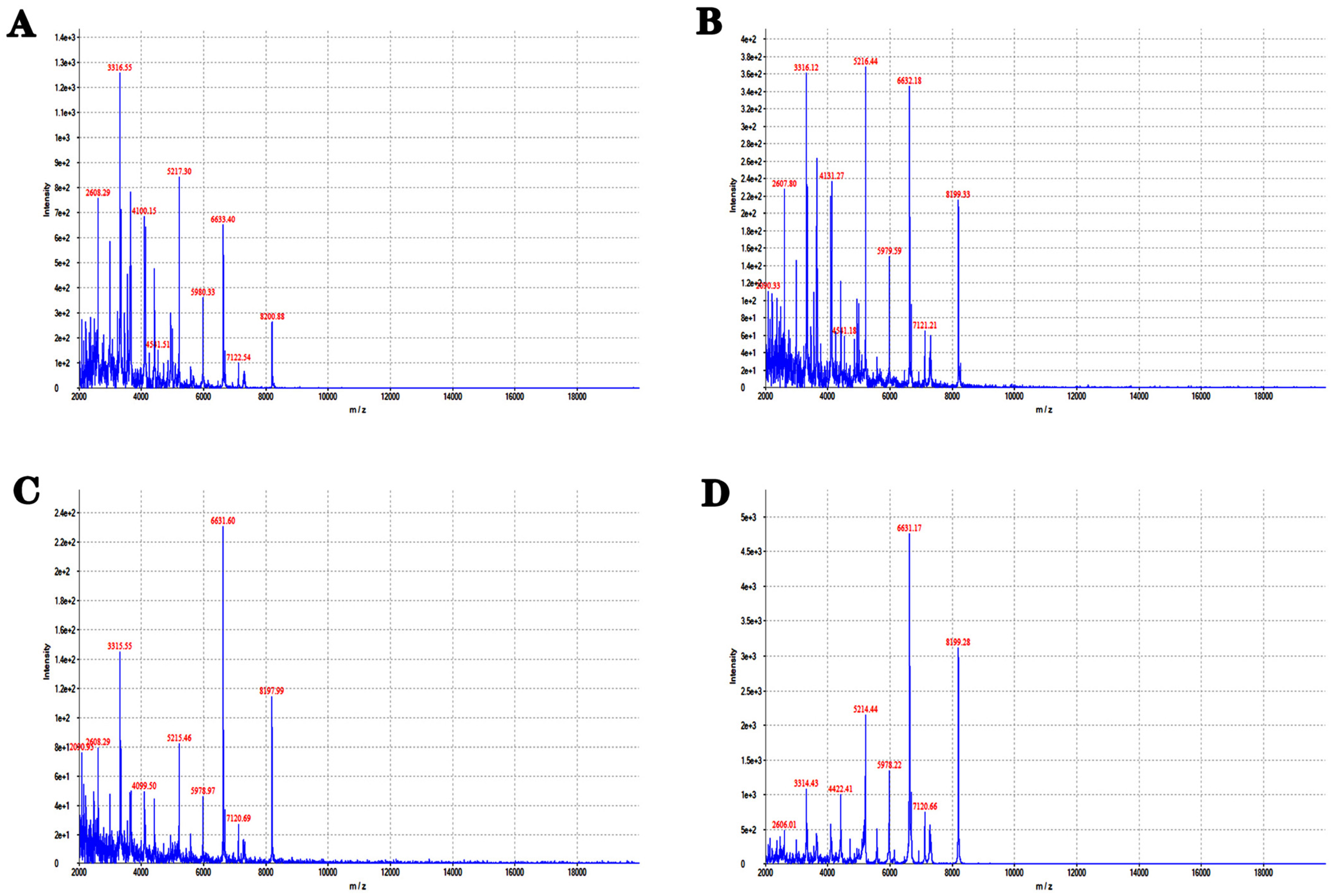
Figure 1. MALDI-TOFMS identification of the isolated strain. (AD) MALDI-TOFMS identification results of colonies incubated for 12, 16, 20, and 24 h, respectively.
Separations 10 00461 g001
Figure 2. Molecular biological identification and phylogenetic tree analysis of Stutzerimonas stutzeri from high-salt wastewater. (A) Gene amplification of colonies using bacterial 16S primers, where the amplification products denote band size by gel electrophoresis. M: DL2000 DNA maker. (B) Phylogenetic tree based on 16S rDNA gene sequences of Stutzerimonas stutzeri.
Separations 10 00461 g002
Figure 3. Growth curve of Stutzerimonas stutzeri. The growth of Stutzerimonas stutzeri was evaluated after 90 h of growth at different dilutions of bacterial broth (control, 1, 10, 20, 40, 60, 80, and 100 μL).
Separations 10 00461 g003
Figure 4. Analysis of drug-resistant genes in Stutzerimonas stutzeri in high-salt wastewater. Primer amplification was performed using bacterium, extracted bacterial genome, and plasmid as templates, followed by sequencing analysis of the electrophoretic bands. (AI): Electrophoresis images of AAC(3)-II, cmlA, CTX-M-l, gyrA, gyrB, blaKPC, NDM-1, oqxA, and oqxB genes.
Separations 10 00461 g004
Figure 5. Analysis of drug-resistant genes in Stutzerimonas stutzeri in high-salt wastewater. Primer amplification was performed using bacterium, extracted bacterial genome, and plasmid as templates, followed by sequencing analysis of the electrophoretic bands. (AI): Electrophoresis images of OXA, parC, qepA, qnrA, qnrB, qnrC, qnrD, qnrS, and Sul2 genes.
Separations 10 00461 g005
Figure 6. Bacteria proteomic data of the Stutzerimonas stutzeri. The x-axis represents the number of identified spectra of protein, and the y-axis represents the protein count.
Separations 10 00461 g006
Figure 7. GO classification of proteomic data of the Stutzerimonas stutzeri. The x-axis represents the protein count, and the y-axis represents the GO terms name.
Separations 10 00461 g007
Figure 8. KEGG pathways and subcellular location of proteomic data of the Stutzerimonas stutzeri. (A): KEGG pathways. Level 1 represents the classification of environmental information processing, genetic information processing, and metabolism. Level 2 represents the classification of the metabolic pathway. (B) Subcellular location of the top 50 proteins with the highest abundance.
Separations 10 00461 g008
Table 1. Biochemical identification of Stutzerimonas stutzeri.
NameAbbreviationResults
Arginine dihydrolaseADHP
Anaerobic glucose fermentationGLUfP
Hydrogen sulfide productionH2SN
Nitrate reductionNITP
Ornithine decarboxylaseODCP
Amino acid controlCN
Acid production of aerobic glucoseGLUP
Aescin hydrolysisESCN
UreaseUREN
Lysine decarboxylaseLDCP
Production of indoleINDN
Acid production of mannoseMNEP
Malic acid utilizationMTEP
Acid production of maltoseMALP
Gelatin hydrolysisGELN
Acid production of sucroseSACP
Acid production of lactoseLACN
Citrate utilizationCITP
Acid production of xyloseXYLP
GalactosidaseONPGP
Acid production of fructoseFRUN
Malonic acid salt utilizationMLTP
AcetamideACEP
Acid production of mannitolMANN
Note: P: positive, N: negative.
Table 2. Antibiotic susceptibility of Stutzerimonas stutzeri.
Drug NameAbbreviationMIC ValueResults
GentamicinGEN≤2S
TobramycinTOB≤1S
CeftazidimeCAZ≤1S
CiprofloxacinCIP≤1S
ImipenemIPM≤1S
MeropeneMMRP≤1S
CefepimeFEP≤2S
LevofloxacinLEV≤2S
Piperacillin/TazobactamP/T≤4/4S
AztreonamATM≤4S
AmikacinAMK≤4S
Compound xinnuominSXT≤2/38S
CeftriaxoneCRO=2S
CefotaximeCTX=4S
ChloramphenicolCHL=32R
Polymyxin BPB≤2/
Polymyxin ECT≤2/
Cefoperazone/SulbactamCPS≤4/2S
Ampicillin/SulbactamAMS=16/8/
DoxycyclineDOX≤4S
PiperacillinPIP≤8S
MinocyclineMIN=8I
Ticarcillin/Clavulanic acidTIM≤8/2S
TetracyclineTET≤4S
Note: S: susceptible, I: intermediate, R: resistant.
Table 3. The growth peak point of Stutzerimonas stutzeri under different concentrations.
Test GroupTime (h)Peak Point (OD)
Stutzerimonas stutzeri-1 μL142.66
Stutzerimonas stutzeri-10 μL132.72
Stutzerimonas stutzeri-20 μL122.76
Stutzerimonas stutzeri-40 μL122.67
Stutzerimonas stutzeri-60 μL112.67
Stutzerimonas stutzeri-80 μL112.61
Stutzerimonas stutzeri-100 μL112.53
Table 4. Proteomic screening of proteins related to drug resistance.
ItemProteomic Analysis
1Multidrug/solvent RND membrane fusion protein
2RND multidrug efflux membrane fusion protein, MexE
Disclaimer/Publisher’s Note: The statements, opinions and data contained in all publications are solely those of the individual author(s) and contributor(s) and not of MDPI and/or the editor(s). MDPI and/or the editor(s) disclaim responsibility for any injury to people or property resulting from any ideas, methods, instructions or products referred to in the content.