Next Article in Journal
Use of a Clinical Decision Support System (CDSS) to Improve Antimicrobial Stewardship Efforts at a Single Academic Medical Center
Previous Article in Journal
Antibiotic Susceptibility of Bacterial Pathogens Recovered from the Hand and Mobile Phones of University Students
 
 
GERMS is published by MDPI from Volume 15 Issue 4 (2025). Previous articles were published by another publisher in Open Access under a CC-BY (or CC-BY-NC-ND) licence, and they are hosted by MDPI on mdpi.com as a courtesy and upon agreement with the former publisher Infection Science Forum.
Case Report

Fusobacterium nucleatum: A Cause of Subacute Liver Abscesses with Extensive Fibrosis Crossing the Diaphragm, Mimicking Actinomycosis

by
Babak Hooshmand
1,*,
Riad Khatib
1,
Ameer Hamza
2,
Daniel Snower
2 and
Anthony L Alcantara
3
1
Division of Infectious Diseases, Department of Medicine, Ascension St John Hospital, 19251 Mack Avenue, Suite 340, Grosse Pointe Woods, MI 48236, USA
2
Department of Pathology, Ascension St John Hospital, 19251 Mack Avenue, Suite 340, Grosse Pointe Woods, MI 48236, USA
3
Department of Radiology, Ascension St John Hospital, 19251 Mack Avenue, Suite 340, Grosse Pointe Woods, MI 48236, USA
*
Author to whom correspondence should be addressed.
GERMS 2019, 9(2), 102-105; https://doi.org/10.18683/germs.2019.1164
Submission received: 13 March 2019 / Revised: 19 April 2019 / Accepted: 17 May 2019 / Published: 3 June 2019

Abstract

Introduction: Fusobacterium nucleatum is a Gram-negative, obligate anaerobic bacterium which predominantly resides within the oral cavity and causes acute abscesses and venous thrombosis, primarily in the head and neck region, but could have unique clinical presentations in different anatomical regions of the body. Case report: We present a case of subacute liver abscesses extending to the lung. The histopathological examination showed extensive necrosis and fibrosis. The chronic course, extensive fibrosis and extension across the anatomic barriers were suggestive of actinomycosis. two sets of blood cultures grew Fusobacterium nucleatum, only 16s rRNA analysis of the liver tissue and pleural fluid revealed F. nucleatum DNA without other organisms. The clinical and pathological features of our case illustrate that F. nucleatum may mimic actinomycosis. Conclusions: This case illustrates that F. nucleatum should be considered in patients with subacute infections with extensive fibrosis that crosses anatomic barriers, mimicking actinomycosis.

Introduction

Fusobacterium nucleatum is part of the oropharyngeal and gastrointestinal flora [1]. It causes a variety of infections including liver abscesses, either mono-bacterial or as co-infection with other pathogens. Usually, infections due to F. nucleatum as well as most other anaerobes cause acute exudative infiltrates and tissue necrosis. In comparison, Actinomyces organisms are characterized by a chronic course and extensive fibrosis, often mimicking malignancy [2]. We describe a case of liver abscesses with indolent presentation, extensive fibrosis and extension across the diaphragm. Blood culture and 16s rRNA analysis of liver tissue plus pleural fluid revealed F. nucleatum only. The chronic course, extensive fibrosis and extension across anatomic barriers, mimicking actinomycosis, has not been previously reported in F. nucleatum liver abscesses.

Case report

A 69-year-old female with a remote history of thyroid cancer presented to her primary care physician with right upper quadrant and right lower chest pain that radiated to the back, difficulty breathing and infrequent dry cough for 2 weeks duration. Her chest X-ray was normal. Laboratory tests showed white blood cells (16,000/cmm), hemoglobin (12.6 g/dL), platelets (443,000/cmm), alkaline phosphatase (194 U/L). She was given a 5 days course of azithromycin 500 mg orally daily for presumed bronchitis. After 3 weeks the symptoms worsened. Computed tomography showed multiple liver cystic lesions and right sided pleural effusion/infiltrates. She was thought to have cystic malignancy and pneumonia (Figure 1). Her pain became pleuritic with intermittent chills, night sweats and dry cough. She presented to the emergency room. Physical examination revealed normal temperature, diminished breathing sounds on the right lung base and distended and tender abdomen. Her laboratory tests showed white blood cells (28,600/cmm), hemoglobin (8.4 g/dL), platelets (778,000/cmm), alkaline phosphatase (336 U/L). Repeated imaging at the time of admission showed that the lesions had enlarged. The blood culture was drawn, and she was started on ampicillin/sulbactam 3000 mg IV every 6 hours, empirically for presumed aspiration pneumonia. Ultrasound guided thoracentesis one day after admission revealed exudative effusion with negative Gram stain and cultures.
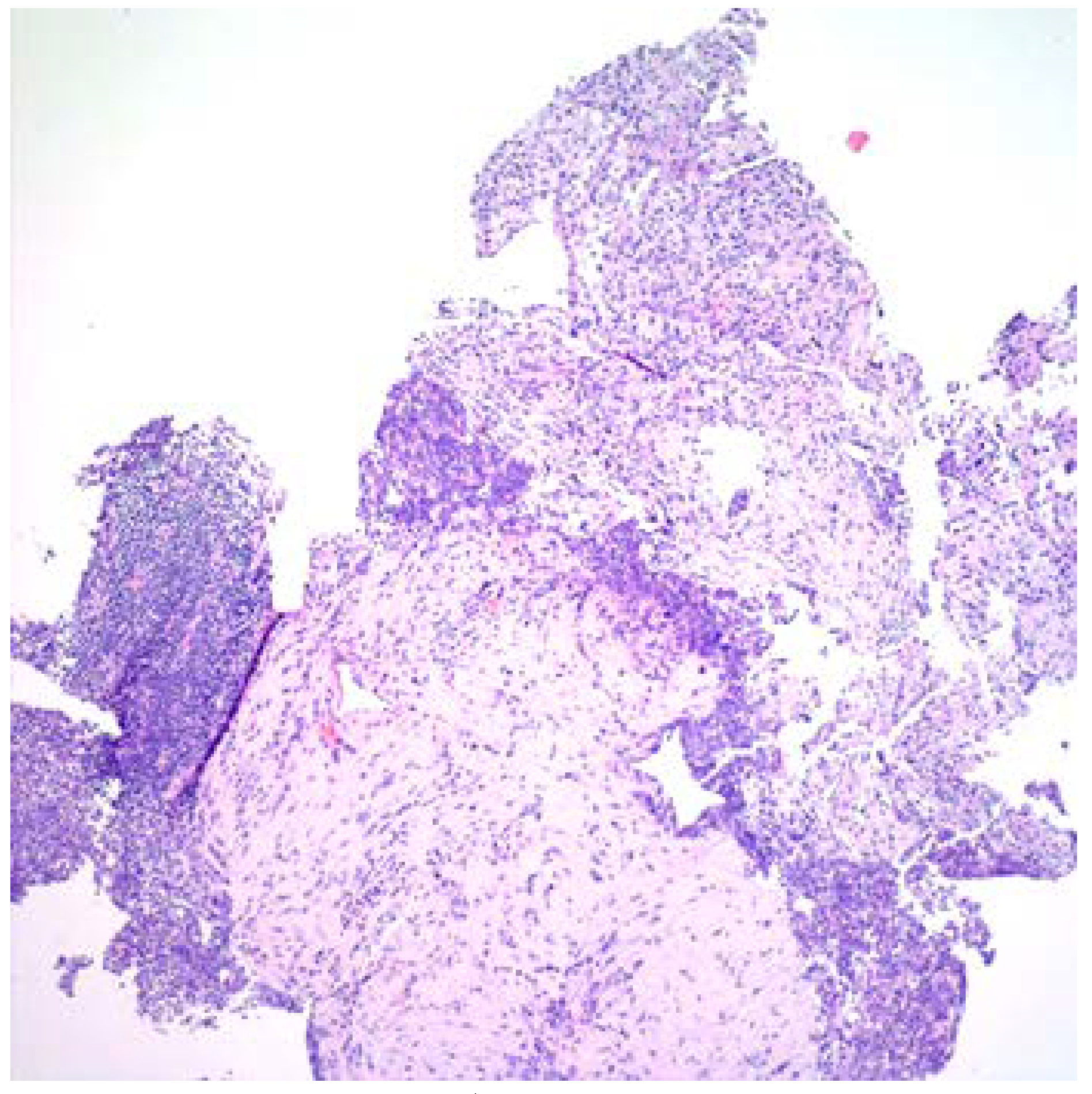
The following day, computed tomography guided core liver biopsy revealed acute inflammation, necrosis and extensive fibrosis without evidence of malignancy (Figure 2). Liver biopsy specimens Gram stain, acid-fast and silver stains and cultures were negative. Five days later, however, 2 sets of blood cultures drawn on admission revealed F. nucleatum. She was suspected of having a mixed infection with Actinomyces species based on the pathological findings. However, 16s rRNA analysis of liver tissue and pleural fluid revealed F. nucleatum DNA without other organisms.
Treatment was changed 6 days later at the time of discharge to ertapenem IV 1000 mg IV every 24 hours for 25 more days and then switched to metronidazole 500 mg orally three times a day for three more months. She continued to improve, one month after discharging home, repeat laboratory tests showed white blood cells (6,200/cmm), hemoglobin (15 g/dL), platelets (373,000/cmm), alkaline phosphatase (67 U/L) and imaging showed progressive resolution of abscesses. On follow up visit 6 months after treatment she remained well without any symptoms.

Discussion

Subacute infection with extensive necrosis and fibrosis that crosses anatomic barriers is characteristic of actinomycosis [3,4,5]. Fusobacterium nucleatum, an obligate anaerobic filamentous Gram-negative rod, is known to cause abscesses and venous thrombosis but has not been reported to cause extensive fibrosis mimicking actinomycosis [6,7,8,9,10,11,12,13,14,15]. Fusobacterium species are indigenous to the oral cavity and the gastrointestinal tract flora [16,17]. They are considered commensal but have been associated with mono-bacterial and mixed infections. The pathogenesis is suspected to be a hematogenous seeding from an upper respiratory tract or a gastrointestinal focus. The histopathology usually reveals acute inflammation, necrosis with abscess formation and rarely, thrombophlebitis. In comparison, actinomycosis is characterized by chronic presentation, acute and chronic inflammatory infiltrates and extensive fibrosis, mimicking malignancy [3,4,5]. It often causes sinus formation and crosses anatomic barriers with or without sulfur granule formation.
Our patient developed several cystic liver lesions with right lower lung infiltrates and pleural effusions. Initially, she was suspected of having cystic liver neoplasm with right lower lung pneumonia and empyema or an atypical granulomatous infection including mycobacteria or fungi. Parasitic causes like echinococcosis and/or amebic liver abscesses were less likely based on the clinical presentation and in the absence of risk factors such as travel or residence in endemic areas. Malignancy and granulomas, however, were not detected in the liver tissues. Blood cultures revealed Fusobacterium nucleatum, but we suspected mixed infection with Actinomyces species based on the histopathological findings. Molecular analysis, however, revealed F. nucleatum DNA without any other organism. Liver tissue and pleural fluid cultures were negative, probably because of antibiotic therapy.
The treatment of F. nucleatum infections is based on susceptibility to β-lactams/β-lactamase-inhibitors and metronidazole whereas Actinomyces species are resistant to metronidazole. Duration of antibacterial treatment for actinomycosis is generally prolonged whereas the length of therapy of F. nucleatum infections is not clear. We elected to continue therapy for 4 months with combination of parenteral and oral antibiotics until resolution of the liver abscesses was documented.

Conclusions

This case illustrates that F. nucleatum should be considered in patients with subacute infection with extensive fibrosis that crosses anatomic barriers, mimicking actinomycosis.

Author Contributions

BH and RK were involved in the diagnosis of the case and writing the manuscript. AH and DS provided pathology slide image and interpretation. AA provided the images and interpretation. All authors revised and approved the final version of the manuscript.

Funding

None to declare.

Acknowledgments

The authors are grateful to Alice Mar for her valuable assistance in figure preparation and for Susan Szpunar for editing the manuscript.

Conflicts of interest

All authors—none to declare.

References

  1. Garcia-Carretero, R.; Lopez-Lomba, M.; Carrasco-Fernandez, B.; Duran-Valle, M.T. Clinical features and outcomes of Fusobacterium species infections. J Crit Care Med (Targu Mures) 2017, 3, 141–147. [Google Scholar] [CrossRef] [PubMed]
  2. Valour, F.; Sénéchal, A.; Dupieux, C.; et al. Actinomycosis: Etiology, clinical features, diagnosis, treatment, and management. Infect Drug Resist 2014, 7, 183–197. [Google Scholar] [PubMed]
  3. Weed, L.A.; Baggenstoss, A.H. Actinomycosis: A pathologic and bacteriologic study of 21 fatal cases. Am J Clin Pathol 1949, 19, 201–216. [Google Scholar] [CrossRef] [PubMed]
  4. Brown, J.R. Human actinomycosis: A study of 181 subjects. Hum Pathol 1973, 4, 319–330. [Google Scholar] [CrossRef] [PubMed]
  5. Amrikachi, M.; Krishnan, B.; Finch, C.J.; Shahab, I. Actinomyces and Actinobacillus actinomycetemcomitans-Actinomyces-associated lymphadenopathy mimicking lymphoma. Arch Pathol Lab Med. 2000, 124, 1502–1505. [Google Scholar] [CrossRef] [PubMed]
  6. Buelow, B.D.; Lambert, J.M.; Gill, R.M. Fusobacterium liver abscess. Case Rep Gastroenterol 2013, 7, 482–486. [Google Scholar] [CrossRef] [PubMed]
  7. Maroun, E.; Chakrabarti, A.; Baker, M.; Canchi, D.; Oehler, R.; Greene, J. Pyogenic liver abscess due to Fusobacterium mimicking metastatic liver malignancy. Infect Dis Clin Pract 2012, 20, 359–361. [Google Scholar] [CrossRef]
  8. Ahmed, Z.; Bansal, S.K.; Dhillon, S. Pyogenic liver abscess caused by Fusobacterium in a 21-year-old immunocompetent male. World J Gastroenterol 2015, 21, 3731–3735. [Google Scholar] [CrossRef] [PubMed]
  9. Rahmati, E.; She, R.C.; Kazmierski, B.; Geiseler, P.J.; Wong, D. A case report of liver abscess and Fusobacterium septicemia. IDCases 2017, 9, 98–100. [Google Scholar] [CrossRef] [PubMed]
  10. Nagpal, S.J.; Mukhija, D.; Patel, P. Fusobacterium nucleatum: A rare cause of pyogenic liver abscess. Springerplus 2015, 4, 283. [Google Scholar] [CrossRef] [PubMed]
  11. Wilson, R.; LeBlanc, R.; Hamour, A.A. Cryptogenic pyogenic liver abscess due to Fusobacterium nucleatum: A case report. BC Med J 2014, 56, 130–134. [Google Scholar]
  12. Kearney, A.; Knoll, B. Myopericarditis associated with Fusobacterium nucleatum-caused liver abscess. Infect Dis (Lond) 2015, 47, 187–189. [Google Scholar] [CrossRef] [PubMed]
  13. Houston, H.; Kumar, K.; Sajid, S. Asymptomatic pyogenic liver abscesses secondary to Fusobacterium nucleatum and Streptococcus vestibularis in an immunocompetent patient. BMJ Case Rep 2017, 2017, bcr–2017. [Google Scholar] [CrossRef] [PubMed]
  14. Wijarnpreecha, K.; Yuklyaeva, N.; Sornprom, S.; Hyman, C. Fusobacterium nucleatum: Atypical organism of pyogenic liver abscess might be related to sigmoid diverticulitis. N Am J Med Sci 2016, 8, 197–199. [Google Scholar] [CrossRef] [PubMed]
  15. Sinave, C.P.; Hardy, G.J.; Fardy, P.W. The Lemierre syndrome: Suppurative thrombophlebitis of the internal jugular vein secondary to oropharyngeal infection. Medicine (Baltimore) 1989, 68, 85–94. [Google Scholar] [CrossRef] [PubMed]
  16. Han, Y.W. Fusobacterium nucleatum: A commensal-turned pathogen. Curr Opin Microbiol 2015, 23, 141–147. [Google Scholar] [CrossRef]
  17. Segata, N.; Haake, S.K.; Mannon, P. Composition of the adult digestive tract bacterial microbiome based on seven mouth surfaces, tonsils, throat and stool samples. Genome Biol 2012, 13, R42. [Google Scholar] [CrossRef] [PubMed]
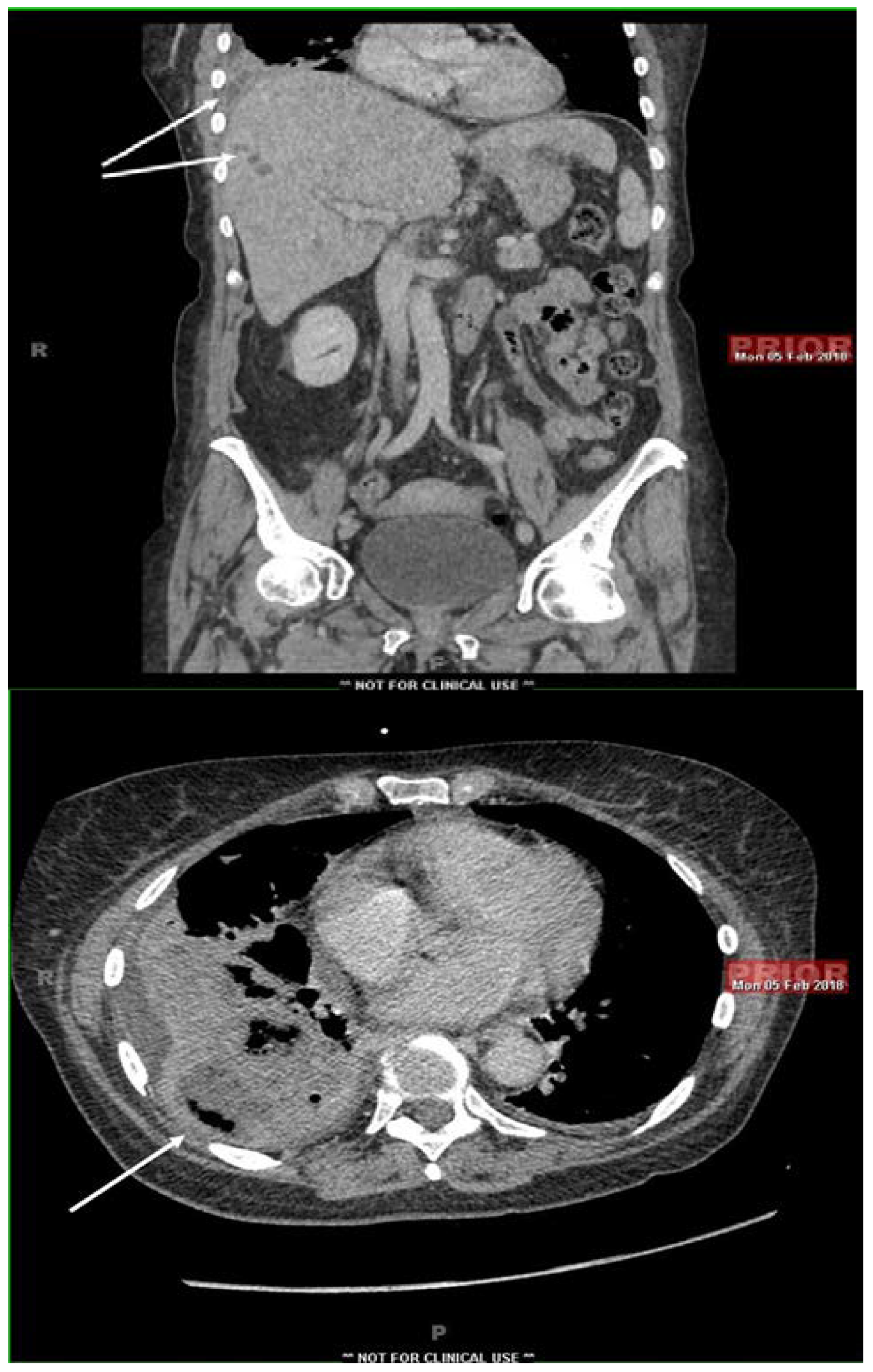
Figure 1. Computed tomography of the abdomen showing multiple liver cysts, extending to the diaphragm with pleural effusion and right lower lung infiltrates (arrows).
Germs 09 00102 g001
Figure 2. Hematoxylin and eosin stain of core liver biopsy showing extensive fibrosis.
Germs 09 00102 g002