Next Article in Journal
Comment on: “High Level Bacterial Contamination of Secondary School Students’ Mobile Phones”
Previous Article in Journal
Evaluation of Clinicians’ Reporting Proficiency and Their Risk Perceptions of Ebola Virus Disease in Ebonyi State, Nigeria
 
 
GERMS is published by MDPI from Volume 15 Issue 4 (2025). Previous articles were published by another publisher in Open Access under a CC-BY (or CC-BY-NC-ND) licence, and they are hosted by MDPI on mdpi.com as a courtesy and upon agreement with the former publisher Infection Science Forum.
Case Report

Hemophagocytic Lymphohistiocytosis (HLH): Elusive Diagnosis of Disseminated Mycobacterium avium Complex Infection

by
Eloy E. Ordaya
1,
Sulieman Abu Jarir
2,
Robert Yoo
3 and
Pranatharthi H. Chandrasekar
3,4,*
1
Division of Infectious Diseases, Henry Ford Hospital, P.O. Box 553920, 2799 W. Grand Blvd, Detroit, MI 48202-3920, USA
2
Hamad Medical Corporation, Hamad General Hospital, P.O. Box 3050, Al Rayyan Rd, Doha, Qatar
3
Karmanos Cancer Institute, 4100 John R, Detroit, MI 48201, USA
4
Division of Infectious Diseases, Wayne State University School of Medicine/Harper University Hospital, 3990 John R, Detroit, MI 48201, USA
*
Author to whom correspondence should be addressed.
GERMS 2017, 7(3), 149-152; https://doi.org/10.18683/germs.2017.1120
Submission received: 9 June 2017 / Revised: 29 July 2017 / Accepted: 30 July 2017 / Published: 1 September 2017

Abstract

Introduction: Hemophagocytic lymphohistiocytosis (HLH) is a clinical syndrome of hyperinflammation leading to an uncontrolled and ineffective immune response, associated with high mortality. Case report: A 26-year-old woman with acute lymphoblastic leukemia, 8 months in remission, was found to have HLH. Without any improvement, stem cell transplantation was considered. Then, Mycobacterium avium complex (MAC) infection was identified as cause of her HLH. With appropriate therapy for infection, HLH improved and transplantation was averted. Conclusions: MAC should be included in the list of potential causes of HLH.

Introduction

Hemophagocytic lymphohistiocytosis (HLH) is a clinical syndrome of hyperinflammation leading to an uncontrolled and ineffective immune response, associated with high mortality. It may develop subsequent to a number of recognized genetic mutations (primary HLH) or in association with infections, malignancy, or autoimmune conditions (secondary HLH) [1,2]. We report an unusual presentation of disseminated Mycobacterium avium complex (MAC) causing secondary HLH in a patient with acute lymphoblastic leukemia (ALL) in remission.

Case report

Our patient is a 26-year-old woman with prior history of ALL status post induction and maintenance treatment in complete remission, who presented eight months later with recurrent episodes of febrile neutropenia. Relapse of ALL was ruled out with appropriate studies. She had a history of possible pulmonary aspergillosis based on fever and non-specific tiny pulmonary nodules in the CT scan of her chest, but with negative serum biomarkers as well as normal bronchoalveolar lavage studies. The patient received empiric treatment with voriconazole for 2 months without clinical or radiologic improvement, so antifungal therapy was discontinued. Four months later, (14 months after the initial diagnosis of ALL), the patient was diagnosed with secondary HLH based on the updated Henter’s criteria [3] including: unexplained fever, chronic pancytopenia (white blood cell count 1.8 K/µL, hemoglobin 7.8 g/dL, platelet count 8 K/µL), hypertriglyceridemia (355 mg/dL), low fibrinogen (189 mg/dL), ferritin (> 2000 ng/mL) and elevated soluble IL-2 receptor 5610 pg/mL. Additionally, liver enzymes were elevated (AST 76 IU/L, ALT 79 IU/L, alkaline phosphatase 640 IU/L). However, bone marrow biopsy did not show hemophagocytosis or relapsing leukemia; therapy was started with bi-weekly etoposide and high-dose dexamethasone with some improvement of her fever. Due to worsening neutropenia, etoposide was stopped but daily dexamethasone was continued. As her condition did not improve over the course of 4 months, a stem-cell transplant was considered.
The patient was re-admitted one month later with complaints of sore throat, dry cough and fever after exposure to a sick contact. On examination, she was found to be febrile (temperature 39.3°C), and the spleen was mildly enlarged. Initial laboratory tests revealed total white blood cell count 0.7 K/µL, absolute neutrophil count 0.0 K/µL, platelet count 31 K/µL, serum creatinine 0.56 mg/dL, serum ALT 67 IU/L, serum alkaline phosphatase 118 IU/L. CT chest showed slight enlargement of multiple irregular pulmonary nodules previously described. Therapy was started with cefepime and oseltamivir. A bone marrow biopsy was repeated with flow cytometry analysis and cytogenetic studies. There was no evidence of relapsing ALL or HLH features, but new poorly-formed, non-caseating granulomas with negative GMS and AFB stains were seen (Figure 1).
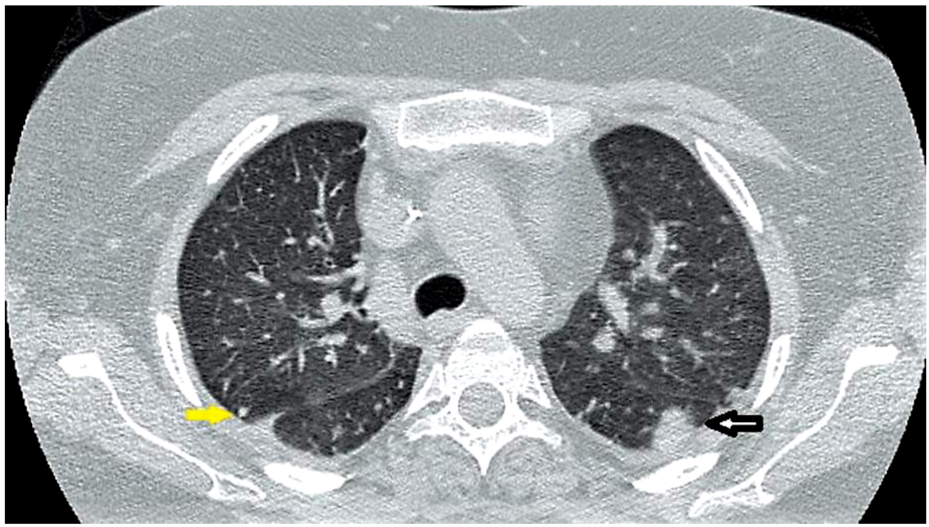
Additional negative tests included serum galactomannan and β-d-glucan, serology for Cryptococcus, Histoplasma, Blastomyces and Coccidioides, CMV PCR, EBV PCR as well as fungal, mycobacterial and bacterial blood cultures. As the patient’s condition slightly improved, antimicrobials were stopped and she was discharged. At regular outpatient evaluation, she continued to complain of episodes of fever and fatigue which partially improved with dexamethasone. Three weeks later, the patient was re-admitted due to a new episode of fever. Physical exam showed mild splenomegaly. Laboratory tests were remarkable for persistent pancytopenia, and the mycobacterial blood culture taken from the previous admission (three weeks earlier) returned positive for Mycobacterium avium complex. A repeat CT chest showed further enlargement of pulmonary nodules and infiltrates (Figure 2). Anti-mycobacterial treatment was started with clarithromycin 500 mg twice daily, ethambutol 15 mg/kg daily and rifabutin 300 mg daily. Also, progressive reduction of dexamethasone dose was scheduled. Within 1 week of treatment, fever subsided and the patient was discharged in stable condition. Six weeks later, the patient was evaluated in the clinic and remained afebrile, neutropenia had resolved and a repeat CT scan of the chest showed marked improvement of pulmonary lesions. She was doing well at 6 months follow up.

Discussion

HLH is a rare, potentially fatal disorder characterized by abnormal macrophages proliferation and hypercytokinemia. It may be divided into primary and secondary forms. Primary HLH consists of monogenic disorders that mainly affect the perforin-mediated cytotoxicity of cytotoxic T-lymphocytes and natural killer cells; the secondary HLH typically occurs in immunocompromised hosts and strong immunologic activation is triggered by immunosuppressant drugs, malignancies or infections [1,2,3,4,5]. Infectious causes of secondary HLH are most commonly viral, but fungal, parasitic and bacterial infections, including tuberculosis, have also been implicated [6,7,8,9,10]. Another case of secondary HLH, similar to ours, reported by Chamsi-Pasha et al. [11] was a female patient with sickle cell anemia who presented with fever of unknown origin. On further testing, nodular pulmonary lesions were noted and MAC was isolated in tissue culture. Despite appropriate therapy, fever persisted with splenomegaly and disseminated intravascular coagulopathy, and laboratory findings and bone marrow biopsy compatible with HLH. The patient died with worsening respiratory failure. Our patient had fever with pulmonary nodules; initial studies were negative. In both cases of MAC causing HLH, correct diagnosis was delayed highlighting the difficulty of promptly identifying MAC infection with currently available diagnostic technology. Late diagnosis of pulmonary MAC after initial negative study including bronchoscopy has been reported and seems to be more frequent in patients with nodular lesions compared to those with fibro-cavitary disease [12]. Absence of hemophagocytosis in bone marrow biopsy of our patient does not preclude the diagnosis of HLH. She received treatment with etoposide and high doses of steroids but no antibiotics for several weeks, without acute clinical deterioration. This may be explained by the role of steroids in modulating the immunological response presented in HLH causing less inflammation and muted clinical manifestations [6]. The cornerstone of therapy for disseminated MAC is a macrolide. Clarithromycin has been shown to more rapidly clear bacteremia compared to azithromycin. Despite the possible drug interactions and side effects of rifamycins, a 3-drug regimen containing clarithromycin, ethambutol and rifabutin has been associated with fewer relapses and better survival compared to 2-drug regimens containing clarithromycin and ethambutol or clarithromycin and rifabutin [13].

Conclusions

Exact reasons for the development of MAC infection and secondary HLH in this patient with lymphoma in remission are unclear. We suspect the patient’s overall immunosuppressed state from prior lymphoma and its therapy led to the aggressive infection and subsequent HLH. HLH is a rare but serious disorder that should prompt a thorough search for a reversible cause. Diagnosis of primary HLH should be considered only after infectious causes are ruled out. We avoided stem cell transplantation in this patient once a correct diagnosis was made.

Consent

Written consent was obtained from the patient prior to publication.

Author Contributions

All authors had equal contributions. All authors read and approved the final version of the manuscript.

Conflicts of Interest

All authors—none to declare.

References

  1. Morimoto, A.; Nakazawa, Y.; Ishii, E. Hemophagocytic lymphohistiocytosis: Pathogenesis, diagnosis, and management. Pediatr Int 2016, 58, 817–825. [Google Scholar] [CrossRef] [PubMed]
  2. Freeman, H.R.; Ramanan, A.V. Review of haemophagocytic lymphohistiocytosis. Arch Dis Child 2011, 96, 688–693. [Google Scholar] [CrossRef] [PubMed]
  3. Henter, J.I.; Horne, A.; Aricó, M.; et al. HLH-2004: Diagnostic and therapeutic guidelines for hemophagocytic lymphohistiocytosis. Pediatr Blood Cancer 2007, 48, 124–131. [Google Scholar] [CrossRef] [PubMed]
  4. Rosado, F.G.; Kim, A.S. Hemophagocytic lymphohistiocytosis: An update on diagnosis and pathogenesis. Am J Clin Pathol 2013, 139, 713–727. [Google Scholar] [PubMed]
  5. Larroche, C. Hemophagocytic lymphohistiocytosis in adults: Diagnosis and treatment. Joint Bone Spine 2012, 79, 356–361. [Google Scholar] [CrossRef] [PubMed]
  6. Sung, L.; Weitzman, S.S.; Petric, M.; King, S.M. The role of infections in primary hemophagocytic lymphohistiocytosis: A case series and review of the literature. Clin Infect Dis 2001, 33, 1644–1648. [Google Scholar] [PubMed]
  7. Hui, Y.M.; Pillinger, T.; Luqmani, A.; Cooper, N. Haemophagocytic lymphohistiocytosis associated with Mycobacterium tuberculosis infection. BMJ Case Rep 2015, 2015, pii–bcr2014208220. [Google Scholar] [CrossRef] [PubMed]
  8. Naha, K.; Dasari, S.; Vivek, G.; Prabhu, M. Disseminated tuberculosis presenting with secondary haemophagocytic lymphohistiocytosis and Poncet’s disease in an immunocompetent individual. BMJ Case Rep 2013, 2013, pii–bcr2012008265. [Google Scholar] [CrossRef] [PubMed]
  9. Tseng, Y.T.; Sheng, W.H.; Lin, B.H.; et al. Causes, clinical symptoms, and outcomes of infectious diseases associated with hemophagocytic lymphohistiocytosis in Taiwanese adults. J Microbiol Immunol Infect 2011, 44, 191–197. [Google Scholar] [CrossRef] [PubMed]
  10. Chou, Y.H.; Hsu, M.S.; Sheng, W.H.; Chang, S.C. Disseminated Mycobacterium kansasii infection associated with hemophagocytic syndrome. Int J Infect Dis 2010, 14, e262–e264. [Google Scholar] [CrossRef] [PubMed]
  11. Chamsi-Pasha, M.A.; Alraies, M.C.; Alraiyes, A.H.; His, E.D. Mycobacterium avium complex-associated hemophagocytic lymphohistiocytosis in a sickle cell patient: An unusual fatal association. Case Rep Hematol 2013, 2013, 291518. [Google Scholar] [CrossRef] [PubMed]
  12. Koyama, K.; Ohshima, N.; Kawashima, M.; et al. Characteristics of pulmonary Mycobacterium avium complex disease diagnosed later in follow-up after negative mycobacterial study including bronchoscopy. Respir Med 2015, 109, 1347–1353. [Google Scholar] [CrossRef] [PubMed]
  13. Benson, C.A.; Williams, P.L.; Currier, J.S.; et al. A prospective, randomized trial examining the efficacy and safety of clarithromycin in combination with ethambutol, rifabutin, or both for the treatment of disseminated Mycobacterium avium complex disease in persons with acquired immunodeficiency syndrome. Clin Infect Dis 2003, 37, 1234–1243. [Google Scholar] [CrossRef] [PubMed]
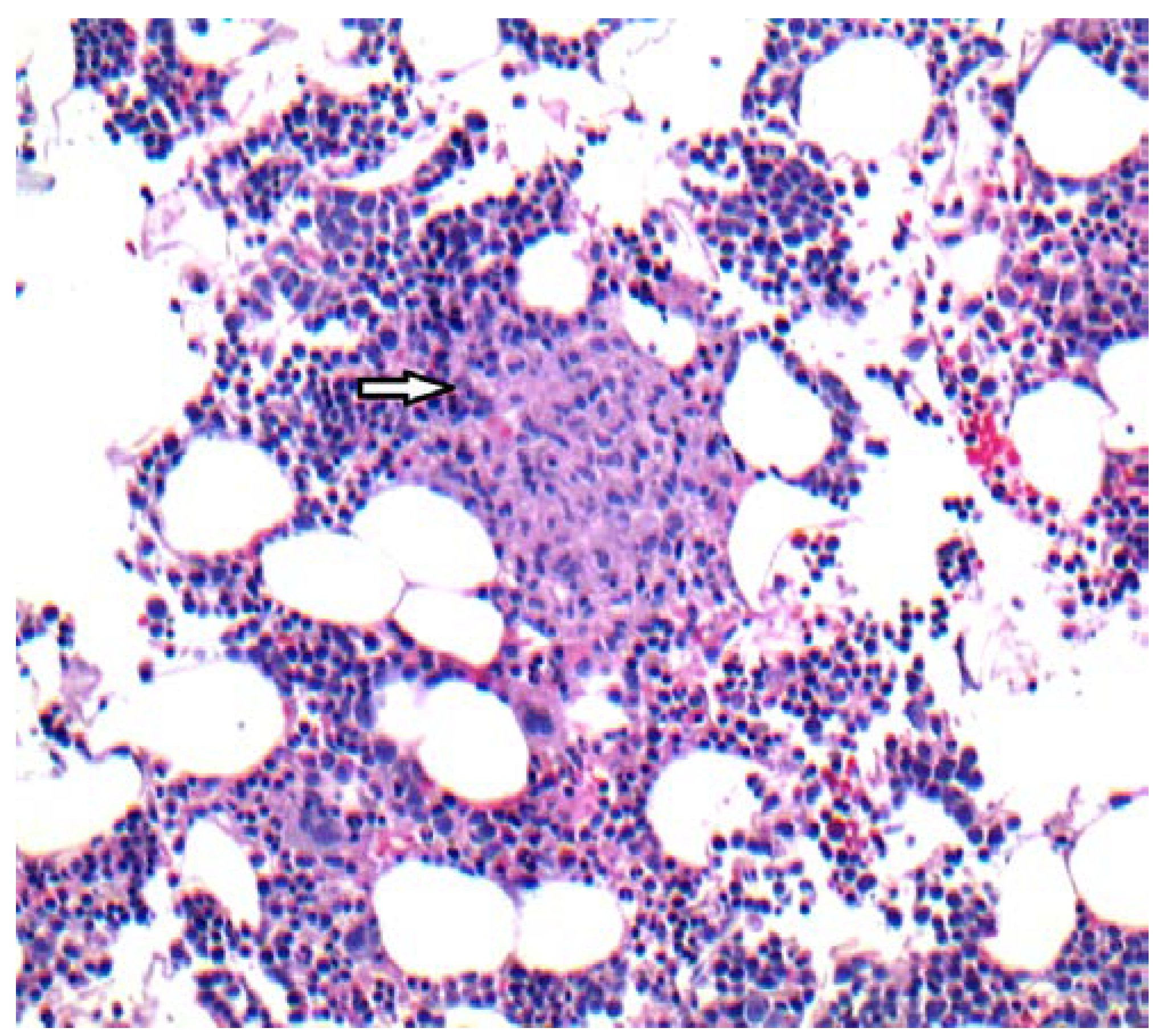
Figure 1. H&E stain, 10X. Bone marrow core biopsy demonstrating a poorly formed, light pink colored non-caseating granuloma (arrow) surrounded by normal hematopoietic cells.
Germs 07 00149 g001
Figure 2. Chest CT showing a peripheral nodule (yellow arrow) and irregular focal infiltrate (white arrow).
Germs 07 00149 g002