Next Article in Journal
Risk Factors Associated with Rabies Incidence in Rabies Endemic Areas in West Kalimantan
Previous Article in Journal
The Mycological Efficacy and Safety of Selenium Sulfide 1.8% Versus Ketoconazole 2% Shampoo in Pityriasis Versicolor: A Double-Blind Randomized Controlled Trial
 
 
GERMS is published by MDPI from Volume 15 Issue 4 (2025). Previous articles were published by another publisher in Open Access under a CC-BY (or CC-BY-NC-ND) licence, and they are hosted by MDPI on mdpi.com as a courtesy and upon agreement with the former publisher Infection Science Forum.
Article

Distribution of Chlorhexidine Resistance Genes Among Staphylococcus aureus Clinical Isolates: The Challenge of Antiseptic Resistance

by
Amira M. Sultan
* and
Mohammad A. Ahmed
Medical Microbiology and Immunology Department, Faculty of Medicine, Mansoura University, Elgomhouria S, Mansoura 35516, Egypt
*
Author to whom correspondence should be addressed.
GERMS 2022, 12(4), 461-471; https://doi.org/10.18683/germs.2022.1352
Submission received: 12 July 2022 / Revised: 14 October 2022 / Accepted: 31 December 2022 / Published: 31 December 2022

Abstract

Introduction: Chlorhexidine is an antiseptic agent which is extensively used to prevent nosocomial infections; however, this could result in reduction of its susceptibility. The aim of this work was to determine chlorhexidine susceptibility among Staphylococcus aureus isolates and to detect qacA/B and smr antiseptic resistance genes among these isolates. Furthermore, we aimed to identify possible risk factors for the reduction of chlorhexidine susceptibility among S. aureus isolates. Methods: Various clinical samples were collected from patients with evidence of S. aureus infection. Antimicrobial susceptibilities of identified S. aureus isolates were determined by disk diffusion method. Resistance to methicillin was identified by cefoxitin disk diffusion test besides mecA gene detection by PCR. Chlorhexidine minimum inhibitory concentration (MIC) values were measured by broth microdilution method while qacA/B and smr resistance genes were detected by multiplex PCR. Results: A total percentage of 25.9% of S. aureus isolates showed reduced susceptibility to chlorhexidine. Methicillin resistant S. aureus (MRSA) had a reported percentage of 39.5%, which was significantly higher than the 11.3% reported for methicillin susceptible S. aureus (MSSA), p < 0.001. S. aureus isolates were found to harbor qacA/B and smr genes at 23.2% and 7.7% respectively. Risk factors for reduced susceptibility to chlorhexidine included; ICU setting (OR = 2.02, 95%CI: 0.3–1.6), prolonged ICU stay (OR = 1.7, 95%CI: 0.4–1.1), presence of central vascular catheter (OR = 2.3, 95%CI: 0.2–1.9), mechanical ventilation (OR = 1.88, 95%CI: 0.4–1.7) and acquisition of qacA/B (OR = 15.7, 95%CI: 3.4–12.1) or smr gene (OR = 15.7, 95%CI: 3.4–12.1). Conclusions: Our work highlighted the current challenge of antiseptic resistance in our locality. The frequencies of qacA/B and smr genes were significantly higher among MRSA than MSSA isolates. About two thirds of chlorhexidine tolerant isolates displayed an MDR profile. To maintain chlorhexidine efficiency, biocidal stewardship program and ongoing surveillance are essential.

Introduction

In the modern world, healthcare associated infections (HAIs) continue to be major health problems of a notable financial burden since they have considerable association with wide variety of morbidities and mortalities [1]. Staphylococcus aureus is considered as a leading bacterial cause of HAIs responsible for bacteremia, pneumonia, skin or soft tissue infections and devices-associated infections [2]. Interestingly, S. aureus was found to be the second leading cause of HAIs in United States [3]. However, it has been reported that up to 60% of nosocomial S. aureus infections were attributed to methicillin resistant isolates [4].
Methicillin resistant S. aureus (MRSA) is considered as a common resistant pathogen to antibiotics particularly in certain settings and countries. Over the last decade, the rates of MRSA infection have increased owing to antibiotics abuse and continuous bacterial evolution. Furthermore, infections attributed to MRSA strains are more challenging to eradicate because of their resistance to many antimicrobials [5].
Infection control guidelines have been established to control HAIs through the proper use of antiseptics such as chlorhexidine aiming for diminishing bacterial load from the infected or colonized body surfaces [2]. Chlorhexidine is a known cationic topical antiseptic agent that is available in different forms such as acetate, gluconate or hydrochloride salts [6]. Following adherence to bacterial cell wall, chlorhexidine molecules act through damaging the cell wall and cytoplasmic membrane leading to intra-cellular components leakage and subsequent bacterial death [1,6].
Chlorhexidine is one of the commonly used antiseptics in healthcare facilities because of its safety and tolerability as it can be tolerated by human tissue with a good safety record. It is also characterized by its broad-spectrum activity and residual effect which is the prolonged antimicrobial activity that extends after the product application. Furthermore, chlorhexidine has a wide range of applications in clinical practice including hand disinfection, skin disinfection prior to surgeries and invasive procedures such as vascular catheters insertion, MRSA decolonization and bathing patients in intensive care units (ICUs) [6]. However, the wide use of chlorhexidine in healthcare settings could lead to reduction of its susceptibility among different bacterial isolates including S. aureus [2,6,7]. Furthermore, various HAIs and outbreaks have been attributed to chlorhexidine resistance [7,8].
Phenotypic susceptibility to chlorhexidine is usually tested by identifying the minimum inhibitory concentration (MIC) values [1,6]. Staphylococcal isolates with MIC≥4 mg/L are considered to have reduced chlorhexidine susceptibility, in other words chlorhexidine tolerance [2,6]. Chlorhexidine MIC values actually identify chlorhexidine tolerance instead of resistance. Chlorhexidine resistance indicates that the bacteria are able to survive at the in-use concentrations of chlorhexidine. On the other hand, when the bacteria are inhibited at lower concentrations, as in MIC tests, it should be defined as tolerance to chlorhexidine (MIC ≥4 mg/L). Therefore, the favored name for phenotypic chlorhexidine resistance is “chlorhexidine tolerance” or “reduced chlorhexidine susceptibility”, while genotypic resistance can be identified by the existence of chlorhexidine resistance genes [6].
Chlorhexidine resistance is mainly caused by multidrug efflux pumps which can carry various antibiotics and antiseptic agents outside the bacterial cells [2,6]. In one report, chlorhexidine was effluxed in 96% of bloodstream S. aureus isolates which highlighted the significance of efflux pumps [9]. Genetic determinants responsible for the efflux-mediated antiseptic resistance include qac (quaternary ammonium compound) and smr (staphylococcal multidrug resistance) resistance genes [6,7].
Chlorhexidine resistance among staphylococcal isolates is frequently caused by qacA and qacB genes. These genes cause chlorhexidine resistance by encoding a proton motive force-dependent efflux pump. It is hard to differentiate between qacA and qacB genes by PCR because they are very similar and closely related. Hence, their PCR products are identified as qacA/B gene negative or positive [1]. In S. aureus isolates, the qacA/B gene is located in chromosomes and on plasmids [7]. Both QacA and QacB proteins belong to the major facilitator superfamily of transport proteins. These large proteins have 14 transmembrane α-helical segments which can export chlorhexidine out of the bacterial cells [1]. The qacA/B gene was previously reported as the most frequent biocidal resistance gene among S. aureus isolates [7].
The smr gene is another antiseptic resistance gene that was initially located on a plasmid recovered from a S. aureus isolate. The protein encoded by smr gene belongs to the small multidrug resistance family of transport proteins and causes an efflux-mediated antiseptic resistance. The smr gene was found to be associated with reduced susceptibility toward chlorhexidine among S. aureus isolates [7].
The extensive usage of chlorhexidine in different healthcare settings raised an alarm about the development of acquired bacterial resistance toward this antiseptic agent [7]. Previous studies have demonstrated reduced chlorhexidine susceptibility among S. aureus isolates and reported different rates for antiseptic resistance genes [6,7]. To our knowledge, none of these studies were conducted in our locality or investigated the possible risk factors for such reduced susceptibility. Antiseptic resistance can compromise the efficiency of prevention strategies for HAIs. Therefore, the aim of our study was to determine chlorhexidine susceptibility among clinical S. aureus isolates and to detect qacA/B and smr antiseptic resistance genes among these isolates. Furthermore, we aimed to identify possible risk factors for the reduction of chlorhexidine susceptibility among S. aureus isolates.

Methods

Selection of patients and samples collection

This cross-sectional study was carried out over a duration of 9 months between January to September 2021. Candidates were recruited from inpatients admitted to Mansoura University Hospitals, Mansoura, Egypt who showed clinical picture suggestive of S. aureus infection. Different infections that are potentially caused by S. aureus were considered eligible for our study such as bloodstream, urinary tract, respiratory tract and surgical site infections. Various clinical samples from the study subjects were collected according to the site of infection including blood, endotracheal aspirates, wound drainages and urine. The possible risk factors for reduced susceptibility to chlorhexidine were recorded from all study subjects such as gender, duration of hospital stay, previous hospital admissions, ICU setting (admission or transfer), duration of ICU stay, previous surgical procedure during hospital stay, previous MRSA decolonization protocol and presence of invasive devices (central vascular catheter, urinary catheter, mechanical ventilator).
All the collected samples were properly transported (as per the correct methods for transport of samples including packaging, transport time and the use of transport media) to the Medical Microbiology and Immunology Department for further processing.

Identification of S. aureus isolates

All collected samples were processed using routine microbiological procedures. Inoculated plates of blood agar and mannitol salt agar were incubated at 37 °C for 24 hours. Isolates of S. aureus were identified by the colony appearance, Gram-stained films, and biochemical reactions (positive catalase and tube coagulase tests) [10]. Identified S. aureus isolates were confirmed by using API Staph (bioMérieux, France).

Detection of antimicrobial susceptibilities

Confirmed S. aureus isolates were subjected to disk diffusion method to determine their antimicrobial susceptibility patterns [11]. The disk diffusion test was conducted by means of Mueller-Hinton agar (MHA) (Oxoid, UK) and a selected set of nine antibiotic-impregnated disks (Oxoid, UK); penicillin (10 units), cefazolin (30 μg), ciprofloxacin (5 μg), tetracycline (30 μg), erythromycin (15 μg), clindamycin (2 μg), gentamicin (10 μg), trimethoprim/ sulfamethoxazole (1.25/23.75 μg) and linezolid (30 μg). The inhibition zone diameter was measured around each antibiotic disk and the results were interpreted as per the guidelines of Clinical and Laboratory Standard Institute (CLSI) [11]. The vancomycin MIC values were detected by Etest strips (bioMérieux, France) using MHA plates as per the manufacturer’s instructions. For results interpretation, the CLSI resistance breakpoints were used [11]. Isolates showing lack of susceptibility to at least one agent in three or more chemical classes of antibiotics were defined as multidrug resistant (MDR) [12,13].

Detection of methicillin resistance

In line with CLSI recommendations, methicillin resistance was determined by using cefoxitin disk diffusion test where cefoxitin disk (30 μg) (Oxoid, UK) was applied as a surrogate for oxacillin [11]. The inhibitory zone diameter was measured after 24 hours incubation of the inoculated plate at 35 °C. Isolates with a diameter of ≤21 mm were reported as positive for methicillin resistance [11]. S. aureus ATCC 29213 and S. aureus ATCC 33591 were used as negative (methicillin sensitive) and positive control strains (methicillin resistant), respectively. Methicillin resistant isolates were confirmed through mecA gene detection by PCR as previously described using the following primers set: F-5’GTGGAAGTTAGATTGGGATCATAGC3’; R-5’GTCAACGATTGTGACACGATAGC3’. These primers resulted in 544 bp amplification products [14].

Determination of susceptibility to chlorhexidine

In order to determine chlorhexidine susceptibility among the isolated S. aureus strains, MIC was identified by broth microdilution method as described previously [15]. Serial dilutions were prepared from a chlorhexidine stock solution of 200 mg/mL (Sigma-Aldrich, USA) by using Mueller–Hinton broth (MHB) (Oxoid, UK) to produce final ten concentrations ranging from 0.12 to 64 mg/L.
Before conducting the procedure, all the S. aureus isolates were inoculated on blood agar plates followed by 24 hours incubation at 37 °C. Isolated colonies were suspended in normal saline (0.9%) to achieve a 0.5 McFarland turbidity, resulting in a suspension containing 1-2 × 10⁸ colony forming units (CFU)/mL. Such suspension was further diluted with saline to reach the concentration of 1 × 10⁶ CFU/mL. The test was conducted in sterile plastic 96-well microdilution trays. One hundred microliters of each chlorhexidine dilution were added to each well. After that, the adjusted bacterial inoculum (100 μL) was inoculated in each well with a final concentration of 5 × 10⁵ CFU/mL. The following control wells were dedicated for each S. aureus isolate: a positive control well that contained only the bacterial inoculum and a negative control well that included only chlorhexidine. The S. aureus ATCC strain 700699, with reduced susceptibility to chlorhexidine, and S. aureus ATCC strain 29213 were used as control strains [2]. All tests were done in duplicates.
Within 15 minutes of inoculation, the trays were incubated at 35 °C for 24 hours. In addition, blood culture plates, inoculated by bacterial suspensions, were similarly incubated in order to check purity. Following incubation, the tray wells were examined for bacterial growth determined by the presence of a single dot of turbidity in the well. The MIC was read as the lowest concentration of chlorhexidine that completely inhibited bacterial growth in the microdilution wells. The procedure was re-executed if duplicate wells produced different readings. The MIC breakpoint (MIC ≥4 mg/L) was used to define reduced chlorhexidine susceptibility among S. aureus isolates. According to this breakpoint, isolates with MIC ≥4 mg/L were defined as chlorhexidine tolerant while isolates with MIC ≤2 mg/L were defined as chlorhexidine susceptible [1,6]. Staphylococcal strains were kept at a temperature of -70 °C in trypticase soy broth (Oxoid, UK) with added 30% glycerol until further molecular testing [5].

Molecular detection of antiseptic resistance qacA/B and smr genes

Isolated S. aureus strains were examined for existence of both qacA/B and smr genes by multiplex PCR using the following specific sets of primers: F-5’ GCAGAAAGTGCAGAGTTCG3’; R-5’CCAGTCCAATCATGCCTG3’ for the qacA/B gene and F-5’GCCATAAGTACTGAAGTTATTGGA3’; R-5’GACTACGGTTGTTAAGACTAAACCT3’ for the smr gene [16]. Genomic DNA extraction was conducted by Thermo Scientific GeneJET Genomic DNA Purification Kit (Thermo Fisher Scientific, USA) as per guidelines of the manufacturer. Polymerase chain reaction was conducted using a final volume of 50μL containing Taq PCR Master Mix (QIAGEN-UK), template DNA and the genes specific primers. Multiplex PCR assay was performed as follows: a single cycle of initial denaturation for 3 minutes at 96 °C, 25 cycles of denaturation (94 °C for 20 seconds), annealing (53 °C for 20 seconds) and extension (72 °C for 20 seconds), followed by one final extension (72 °C for 5 minutes) [16]. The following PCR products were identified by agarose gel electrophoresis; 361 bp for qacA/B gene and 195 bp for smr gene [16].

Statistical analysis

The data processing was conducted by the SPSS program (v 22.0, IBM Corp. USA). The descriptive data was presented in the form of frequency. When categorical variables were tested, the Chi-squared test was applied for the comparison. Student T test was applied to compare parametric data. A univariate analysis was performed to evaluate the possible risk factors for reduced susceptibility toward chlorhexidine among S. aureus isolates. Reduced susceptibility toward chlorhexidine among S. aureus isolates was defined according to the MIC breakpoint (MIC ≥4 mg/L). Statistical significance was considered when p value <0.05.

Ethics statement

The Faculty of Medicine institutional research board at Mansoura University approved the research protocol (R.21.08.1392). A written informed consent was collected from every subject in the study after full explanation. In case of unconscious patients, written informed consent was obtained from their legal guardians.

Results

In our study, 220 S. aureus isolates were identified from the collected samples including 123 (55.9%) blood, 39 (17.7%) endotracheal aspirates, 30 (13.6%) wound drainages and 28 (12.7%) urine. The study subjects included 119 (54.1%) females and 101 (45.9%) males with mean age of 44±18 years.Out of the identified 220 S. aureus isolates, 114 (51.8%) were MRSA and 106 (48.2%) were methicillin susceptible S. aureus (MSSA). All MRSA strains were positive for cefoxitin disk diffusion test as well as mecA gene detection.
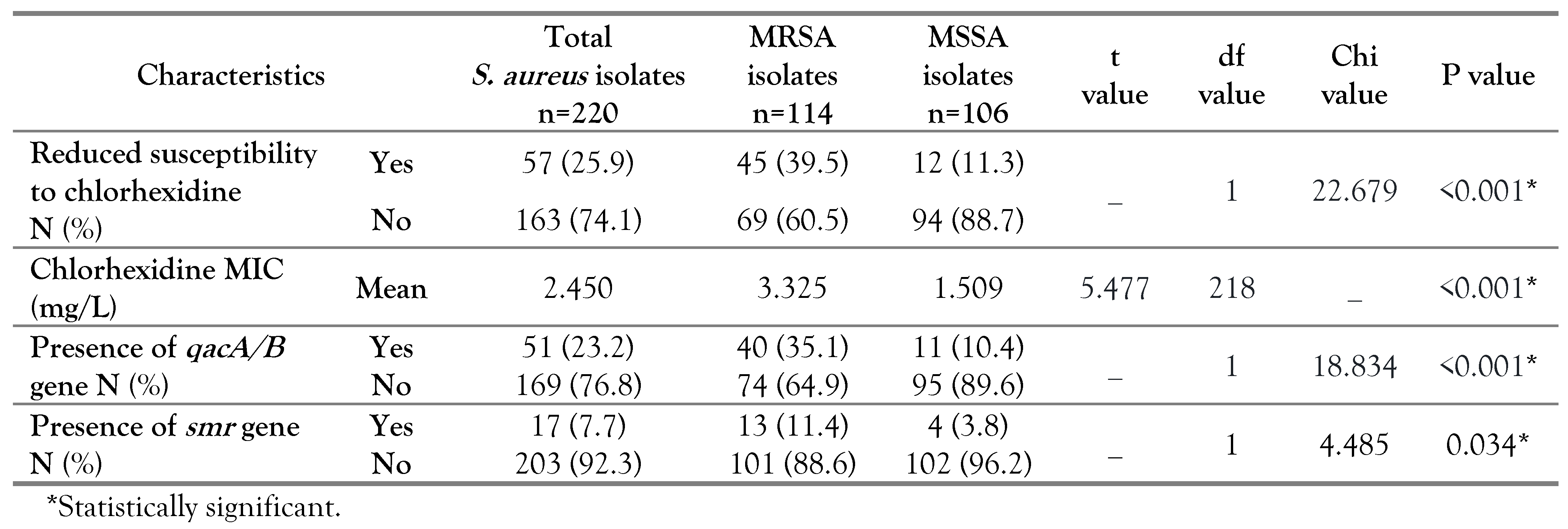
According to MIC results, 57 out of 220 S. aureus isolates had reduced susceptibility to chlorhexidine with a percentage of 25.9%. The percentage of chlorhexidine tolerant isolates among MRSA and MSSA were 39.5% (45/114) and 11.3% (12/106) respectively, with a statistical significance (p<0.001). The MRSA strains had a range of MIC between 0.5 and 16 mg/L, whereas the MIC values for MSSA ranged from 0.5-8 mg/L. In parallel, the mean chlorhexidine MIC among the MRSA (3.325 mg/L) was significantly higher than MSSA isolates (1.509 mg/L), with p<0.001 as shown in Table 1.
In our study, the molecular analysis revealed that 23.2% and 7.7% of S. aureus strains harbored the qacA/B and smr genes, respectively. The percentage of qacA/B gene among MRSA (35.1%) was significantly higher than in MSSA isolates (10.4%), p<0.001. Likewise, the MRSA had a higher percentage of smr gene (11.4%) than the MSSA isolates (3.8%) which was statistically significant (p=0.034) (Table 1). A significant association was found between qacA/B gene existence and reduced chlorhexidine susceptibility as 94.1% of the S. aureus isolates harboring this gene were chlorhexidine tolerant, while 5.9% were chlorhexidine susceptible (p<0.001). Also, an association was noted between the existence of smr gene and reduced chlorhexidine susceptibility (p<0.001) (Table 2).
The antiseptic resistance genes were identified among 61 (27.7%) out of the 220 S. aureus isolates; 44 had the qacA/B gene, 10 had the smr gene and 7 harbored both genes (5 MRSA and 2 MSSA). Our results showed that all 57 S. aureus isolates with reduced susceptibility to chlorhexidine had one or both of the antiseptic resistance genes; 41 isolates carried the qacA/B gene, 9 isolates had the smr gene and 7 isolates harbored both genes (Table 3). On the other hand, among the 163 chlorhexidine susceptible isolates, three had the qacA/B and one carried the smr gene (Table 3). Isolates harboring antiseptic resistance genes displayed a wider range of chlorhexidine MIC (2-16 mg/L) compared to others lacking these genes (0.5-2 mg/L). It should be noted that S. aureus isolates carrying either qacA/B or smr gene exhibited chlorhexidine MIC range of 2-8 mg/L, while isolates harboring both genes displayed higher chlorhexidine MIC values up to 16 mg/L with a chlorhexidine MIC range of 8-16 mg/L (Table 3).
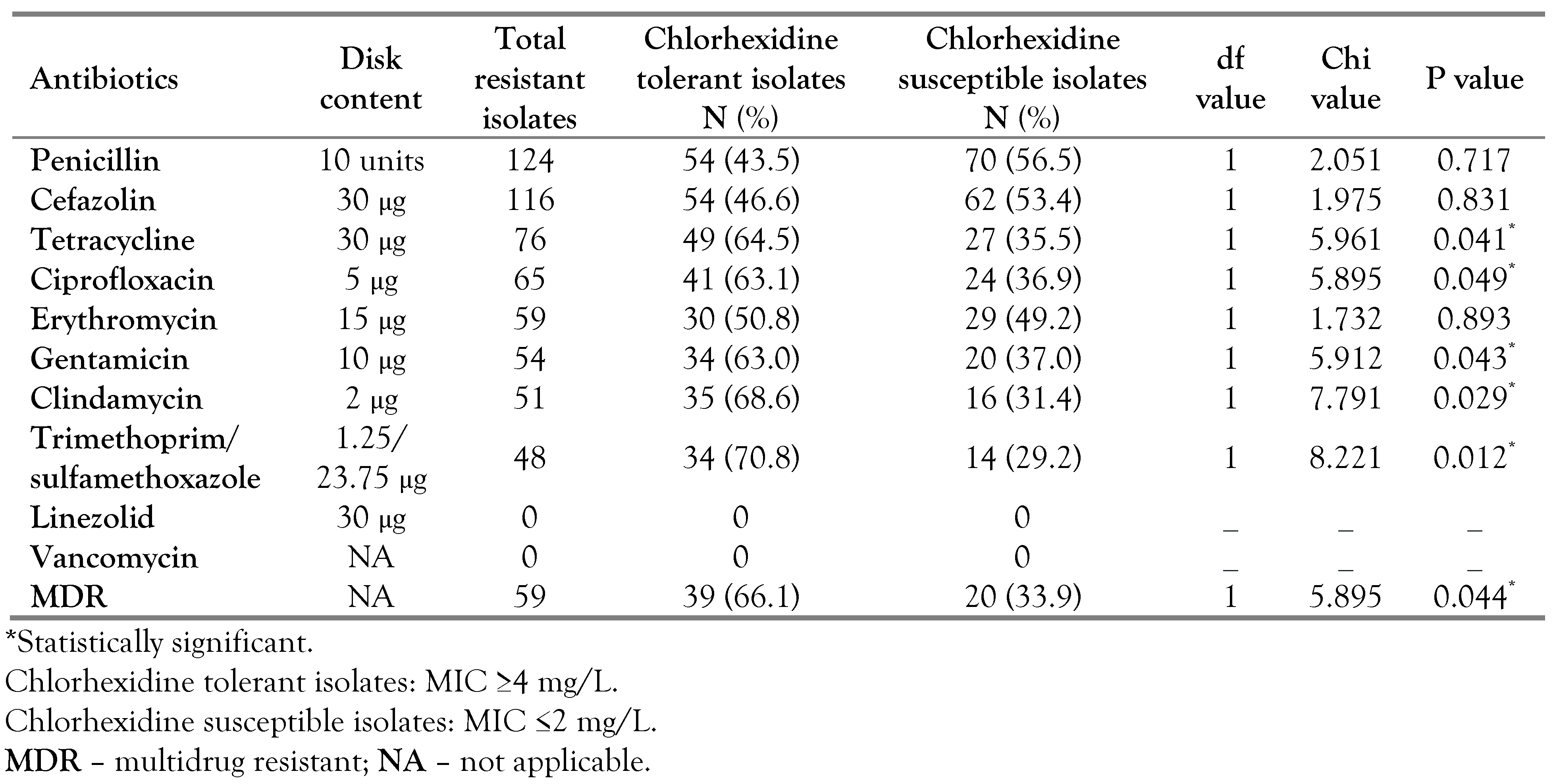
A significant association was found between S. aureus isolates with reduced chlorhexidine susceptibility and resistance toward these antibiotics: tetracycline (p=0.041), ciprofloxacin (p=0.049), gentamicin (p=0.043), clindamycin (p=0.029) and trimethoprim/sulfamethoxazole (p=0.012). Of the 59 isolates showing MDR profiles, 66.1% were chlorhexidine tolerant and 33.9% were chlorhexidine susceptible showing a statistically significant difference (p=0.044). None of the tested S. aureus strains demonstrated vancomycin or linezolid resistance (Table 4).
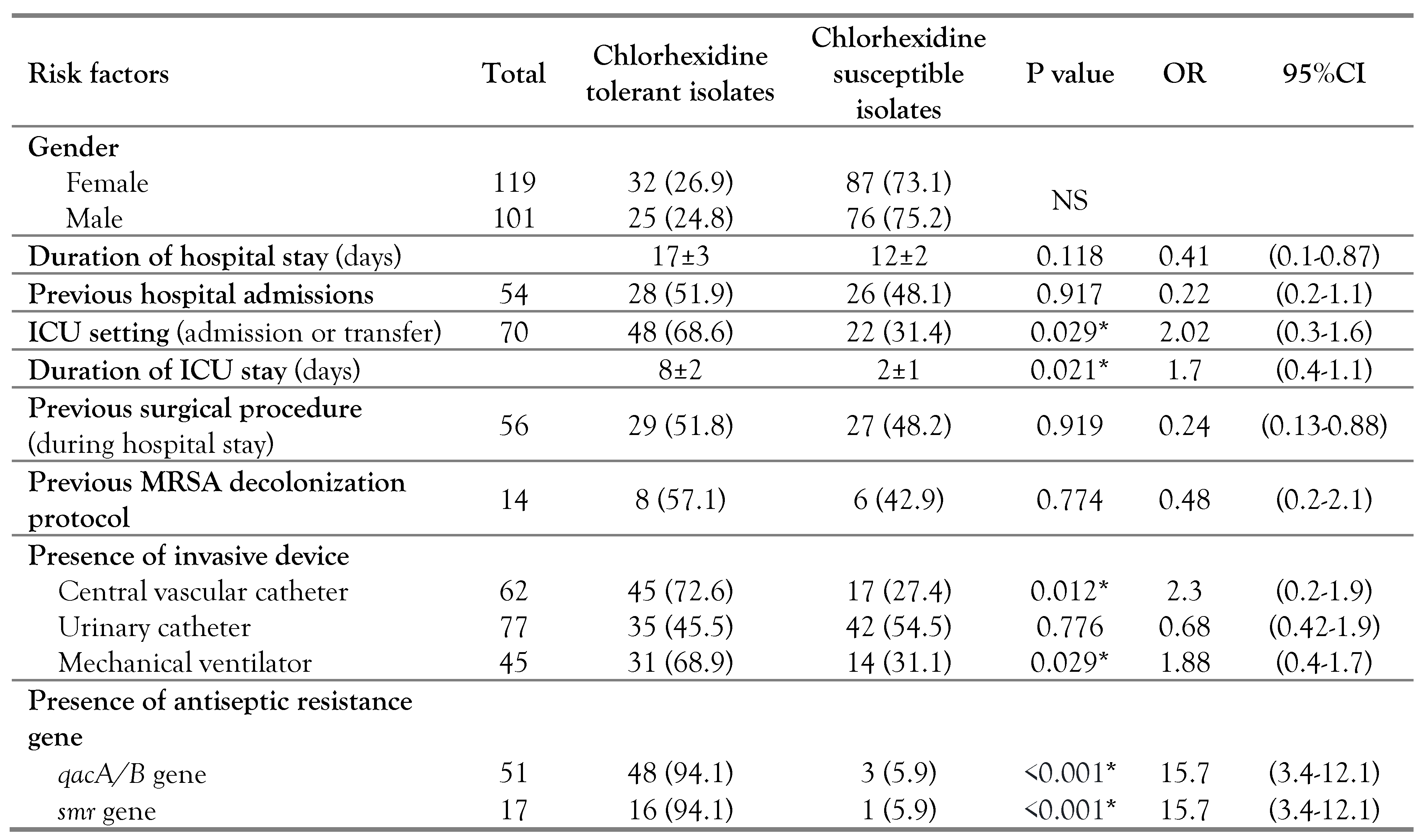
According to univariate analysis, the following were identified as risk factors for reduced susceptibility to chlorhexidine among S. aureus isolates; ICU setting (p=0.029, OR=2.02, 95%CI: 0.3-1.6), prolonged ICU stay (p=0.021, OR=1.7, 95%CI: 0.4-1.1), presence of central vascular line (p=0.012, OR=2.3, 95%CI: 0.2-1.9), usage of mechanical ventilation (p=0.029, OR=1.88, 95%CI: 0.4-1.7) and acquisition of qacA/B (p<0.001, OR=15.7, 95%CI: 3.4-12.1) or smr gene (p<0.001, OR=15.7, 95%CI: 3.4-12.1). On the other hand, none of the following showed a significant association with reduced chlorhexidine susceptibility; gender, duration of hospital stay (p=0.118), previous hospital admissions (p=0.917), previous surgical procedure during hospital stay (p=0.919), previous MRSA decolonization protocol (p= 0.774) and the existence of urinary catheter (p=0.776) (Table 5).

Discussion

The percentage of MRSA in our work with a value of 51.8% was found to be slightly higher than 48.9%, reported in an earlier study conducted in the same locality [17]. Such observation highlighted a steady increment of MRSA infections in our community. In our study, the percentage of S. aureus strains which had reduced chlorhexidine susceptibility, defined by MIC, was 25.9% that was almost two folds that been reported by Azrad et al. and by Hughes and Ferguson at values of 13.5% and 10% respectively [2,18]. The chlorhexidine tolerant isolates had a significantly higher frequency among MRSA than MSSA (p<0.001). In line with our findings, previous studies have reported that MSSA was more susceptible to chlorhexidine than MRSA [2,5]. In addition, MRSA had a significantly higher mean value of chlorhexidine MIC when compared to that of MSSA isolates (p<0.001) which was parallel to the reported results by Azrad and his colleagues [2]. Such findings have raised a great concern because of the extensive use of chlorhexidine in MRSA decolonization protocols. However, the significance of such observation is not fully identified as the in-use concentrations of chlorhexidine were much higher than the MIC tested in vitro [2,6]. Nevertheless, Batra and his colleagues reported that usage of chlorhexidine-based protocol of decolonization in ICU has led to dissemination of MRSA strain with increased MIC value toward this antiseptic agent [8]. These findings suggested that the low chlorhexidine susceptibility among MRSA could be associated with outbreaks inside healthcare settings.
In our study, the total percentage of qacA/B gene among S. aureus strains was 23.2%, which was higher than 13.15% reported by Azrad et al. [2]. We have also found that 35.1% of the MRSA isolates harbored the qacA/B gene. Similar to the findings of our study, the qacA/B gene rates among MRSA isolates were reported at 33.9% in Japan and 37.7% in Korea [14,19]. Higher detection rates of that gene were reported in Turkey at 64.3% and 83% in China [20,21]. The mecA gene, causing methicillin resistance, is positioned on staphylococcal cassette chromosome (SCC) that can integrate other plasmids carrying antiseptic resistance genes, and this can offer an explanation of our findings [5,16]. Sidhu and his colleagues have reported such genetic linkage between qac genes and mecA gene on the same mobile element [22].
Out of S. aureus isolates investigated in our study, 7.7% carried the smr gene with a percentage of 11.4% among MRSA. Similar to the qacA/B gene, the detection rates of the smr gene among MRSA isolates varied as per different regions; 1.4% in Japan, 3.1% in Asia, 44.2% in Scotland, and 77.4% in China [14,16,21,23]. Such variations could be attributed to the geographical differences, clonal spread of tolerant strains, study design, applied infection control programs and most importantly the utilization pressure of chlorhexidine inside healthcare settings. Therefore, it is essential to continuously monitor the development of antiseptic resistance in different settings.
In our work, a significant association was reported between reduced chlorhexidine susceptibility and the existence of qacA/B gene (p<0.001), and the same goes for smr gene (p<0.001). Other studies have reported similar findings [2,5]. It is noteworthy that isolates harboring both genes displayed elevated MIC values compared to isolates carrying only one gene, which was in line with previous findings [5]. The presence of antiseptic resistance genes does not necessarily induce tolerance to chlorhexidine as the gene could be silent and not expressed by the bacteria [1,2]. Therefore, it was not surprising to find that among chlorhexidine susceptible isolates, three isolates were qacA/B gene-positive and one was smr gene-positive.
In our study, resistance to tetracycline, ciprofloxacin, gentamicin, clindamycin and trimethoprim/sulfamethoxazole was significantly associated with chlorhexidine tolerant isolates. Such association between antiseptic resistance genes and antimicrobial resistance was previously reported [1,6]. We have also found that chlorhexidine tolerant strains displayed an MDR profile at a higher frequency than the susceptible strains (p=0.044), which was in line with previous studies [1,24]. These findings can be attributed to co-resistance and cross-resistance. Such cross-resistance occurs when one multidrug efflux pump carries chlorhexidine along with other antibiotics such as fluoroquinolones [9]. On the other hand, the co-resistance is caused by the existence of different resistance genes on one genetic element [2,6]. The genetic link between the qacA/B gene and resistance genes towards aminoglycosides and trimethoprim was reported among staphylococcal isolates [1,6]. In addition, the qacA/B gene was detected in strains harboring SCCmec (types I–V) which carry resistance genes for tetracycline, aminoglycosides and lincosamides [6]. These observations have raised an alarm that excessive exposure to antiseptics might result in selection of strains harboring both antiseptic and antibiotic resistance genes, which impose an additional challenge in controlling HAIs [1]. It is also necessary to mention that the increasing utilization of antiseptics outside the healthcare settings, because of the pandemic of COVID-19, could exaggerate the problem of antiseptic resistance.
To the best of our knowledge, the current study is the first to explore possible risk factors for reduced chlorhexidine susceptibility among S. aureus. The following risk factors were identified: ICU setting, prolonged duration of ICU stay, presence of central vascular catheter, mechanical ventilation and acquisition of qacA/B or smr gene. Our findings reflected the high selective pressure present in the ICU setting where chlorhexidine is frequently used [6]. In addition, prolonged ICU stay is associated with repeated exposure to chlorhexidine building up more selective pressure over bacterial isolates. Vali and his colleagues have reported that recurrent exposure of MRSA to chlorhexidine has led to reduction in chlorhexidine susceptibility and induction of qacA/B gene expression [23]. Therefore, repeated chlorhexidine exposure in the ICU environment can lead to selection of chlorhexidine-tolerant isolates.
We also recognized the presence of central vascular catheter and mechanical ventilator as risk factors for reduced chlorhexidine susceptibility. In order to prevent catheter-associated bloodstream infections, chlorhexidine preparations are used for skin disinfection before the insertion and for the subsequent catheter care. Similarly, chlorhexidine can be used up to 6 times per day as an oral rinse to prevent ventilator-associated pneumonia [6,7]. Such high magnitude of chlorhexidine exposure can lead to selection of antiseptic tolerant organisms. In the UK, a significant correlation was reported between the magnitude of chlorhexidine exposure and the MIC values of clinical isolates [25].
Chlorhexidine is an important antiseptic agent that is effective in preventing HAIs. To reduce selective pressure in healthcare settings, the unnecessary usage of chlorhexidine should be limited. A biocidal stewardship program should also be initiated to preserve the effectiveness of antiseptic agents. In addition, an ongoing surveillance system is necessary for early recognition of the antiseptic tolerant strains and for identification of possible risk factors. The limitation of our study includes lack of detection of other chlorhexidine resistance genes and relatively small sample size. Thus, further researches on a wider scale overcoming these limitations are needed.
Conclusions

In the current study, more than 25% of S. aureus isolates had reduced susceptibility to chlorhexidine with even higher percentage among MRSA, which highlighted this problem in our locality. The frequencies of qacA/B and smr genes were significantly higher among MRSA than among MSSA isolates. Besides, about two thirds of the chlorhexidine tolerant isolates displayed an MDR profile. Risk factors for reduced chlorhexidine susceptibility included: ICU setting, prolonged ICU stay, presence of central vascular line, mechanical ventilation and acquisition of the qacA/B or the smr gene. Unnecessary use of chlorhexidine should be discouraged to limit its resistance. Implementation of a biocidal stewardship program and ongoing surveillance for antiseptic resistance are recommended. Further research to understand genetic determinants of antiseptic resistance are also required.

Author Contributions

AMS developed the study design, processed the samples, drafted the manuscript and analyzed the data. MAA contributed in selection of the study subjects and data collection. All authors read and approved the final version of the manuscript.

Funding

None to declare.

Data Availability Statement

The data supporting the findings of this study are available from the corresponding author, upon reasonable request.

Acknowledgments

The authors would like to show their appreciation to both laboratory and medical staff for their constant help throughout this research.

Conflicts of interest

All authors—none to declare.

References

  1. do Vale, B.C.M.; Nogueira, A.G.; Cidral, T.A.; Lopes, M.C.S.; de Melo, M.CN. Decreased susceptibility to chlorhexidine and distribution of qacA/B genes among coagulase-negative Staphylococcus clinical samples. BMC Infect Dis. 2019, 19, 199. [Google Scholar] [CrossRef] [PubMed]
  2. Azrad, M.; Shmuel, C.; Leshem, T.; et al. Reduced susceptibility to chlorhexidine among Staphylococcus aureus isolates in Israel: Phenotypic and genotypic tolerance. Antibiotics (Basel). 2021, 10, 342. [Google Scholar] [CrossRef]
  3. Magill, S.S.; Edwards, J.R.; Bamberg, W.; et al. Multistate point-prevalence survey of healthcare-associated infections. N Engl J Med. 2014, 370, 1198–1208. [Google Scholar] [CrossRef]
  4. Köck, R.; Becker, K.; Cookson, B.; et al. Methicillin-resistant Staphylococcus aureus (MRSA): Burden of disease and control challenges in Europe. Euro Surveill. 2010, 15, 19688. [Google Scholar] [CrossRef]
  5. Damavandi, M.S.; Safarpour Dehkordi, M.; Dehghan, A.; Heibati, F.; Taghaddosi, R.; Gholipour, A. Detection of antiseptic resistance genes among Staphylococcus aureus colonising nurses and coagulase-negative staphylococci isolated from clinical specimens at teaching hospitals in southwest of Iran. Jundishapur J Microbiol. 2017, 10, e39285. [Google Scholar] [CrossRef]
  6. Horner, C.; Mawer, D.; Wilcox, M. Reduced susceptibility to chlorhexidine in staphylococci: Is it increasing and does it matter? J Antimicrob Chemother. 2012, 67, 2547–2559. [Google Scholar] [CrossRef]
  7. Kampf, G. Acquired resistance to chlorhexidine—Is it time to establish an ‘antiseptic stewardship’ initiative? J Hosp Infect. 2016, 94, 213–227. [Google Scholar] [CrossRef]
  8. Batra, R.; Cooper, B.S.; Whiteley, C.; Patel, A.K.; Wyncoll, D.; Edgeworth, J.D. Efficacy and limitation of a chlorhexidine-based decolonization strategy in preventing transmission of methicillin-resistant Staphylococcus aureus in an intensive care unit. Clin Infect Dis. 2010, 50, 210–217. [Google Scholar] [CrossRef]
  9. DeMarco, C.E.; Cushing, L.A.; Frempong-Manso, E.; Seo, S.M.; Jaravaza, T.A.; Kaatz, G.W. Efflux-related resistance to norfloxacin, dyes, and biocides in bloodstream isolates of Staphylococcus aureus. Antimicrob Agents Chemother. 2007, 51, 3235–3239. [Google Scholar] [CrossRef]
  10. Cheesbrough, M. District laboratory practice in tropical countries, part 2, 2nd ed.; Cambridge University Press: New York, 2006; pp. 157–159. [Google Scholar] [CrossRef]
  11. Clinical and Laboratory Standards Institute. Performance Standards for Antimicrobial Susceptibility Testing, 28th ed.; CLSI supplement M100: Wayne, PA, CLSI; 2018. [Google Scholar]
  12. Prag, G.; Falk-Brynhildsen, K.; Jacobsson, S.; Hellmark, B.; Unemo, M.; Söderquist, B. Decreased susceptibility to chlorhexidine and prevalence of disinfectant resistance genes among clinical isolates of Staphylococcus epidermidis. APMIS. 2014, 122, 961–967. [Google Scholar] [CrossRef]
  13. Seliem, W.A.; Sultan, A.M. Etiology of early onset neonatal sepsis in neonatal intensive care unit—Mansoura, Egypt. J Neonatal Perinatal Med. 2018, 11, 323–330. [Google Scholar] [CrossRef]
  14. Noguchi, N.; Nakaminami, H.; Nishijima, S.; Kurokawa, I.; So, H.; Sasatsu, M. Antimicrobial agent of susceptibilities and antiseptic resistance gene distribution among methicillin-resistant Staphylococcus aureus isolates from patients with impetigo and staphylococcal scalded skin syndrome. J Clin Microbiol. 2006, 44, 2119–2125. [Google Scholar] [CrossRef]
  15. Clinical and Laboratory Standards Institute. Methods for Dilution Antimicrobial Susceptibility Tests for Bacteria That Grow Aerobically, Approved Standard 10th ed.; CLSI document M07-A10: Wayne, PA, CLSI; 2015. [Google Scholar]
  16. Noguchi, N.; Suwa, J.; Narui, K.; et al. Susceptibilities to antiseptic agents and distribution of antiseptic-resistance genes qacA/B and smr of methicillin-resistant Staphylococcus aureus isolated in Asia during 1998 and 1999. J Med Microbiol. 2005, 54, 557–565. [Google Scholar] [CrossRef]
  17. Sultan, A.M.; Nabiel, Y. Association of tsst-1 and pvl with mecA genes among clinical Staphylococcus aureus isolates from a tertiary care hospital. J Pure Appl Microbiol. 2019, 13, 855–864. [Google Scholar] [CrossRef]
  18. Hughes, C.; Ferguson, J. Phenotypic chlorhexidine and triclosan susceptibility in clinical Staphylococcus aureus isolates in Australia. Pathology. 2017, 49, 633–637. [Google Scholar] [CrossRef] [PubMed]
  19. Cho, O.H.; Baek, E.H.; Bak, M.H.; et al. The effect of targeted decolonization on methicillin-resistant Staphylococcus aureus colonization or infection in a surgical intensive care unit. Am J Infect Control. 2016, 44, 533–538. [Google Scholar] [CrossRef] [PubMed]
  20. Duran, N.; Temiz, M.; Duran, G.G.; Eryılmaz, N.; Jenedi, K. Relationship between the resistance genes to quaternary ammonium compounds and antibiotic resistance in staphylococci isolated from surgical site infections. Med Sci Monit. 2014, 20, 544–550. [Google Scholar] [CrossRef] [PubMed]
  21. Liu, Q.; Zhao, H.; Han, L.; Shu, W.; Wu, Q.; Ni, Y. Frequency of biocide-resistant genes and susceptibility to chlorhexidine in high-level mupirocin-resistant, methicillin-resistant Staphylococcus aureus (MuH MRSA). Diagn Microbiol Infect Dis. 2015, 82, 278–283. [Google Scholar] [CrossRef]
  22. Sidhu, M.S.; Heir, E.; Sørum, H.; Holck, A. Genetic linkage between resistance to quaternary ammonium compounds and beta-lactam antibiotics in food-related Staphylococcus spp. Microb Drug Resist. 2001, 7, 363–371. [Google Scholar] [CrossRef]
  23. Vali, L.; Davies, S.E.; Lai, L.L.; Dave, J.; Amyes, S.G. Frequency of biocide resistance genes, antibiotic resistance and the effect of chlorhexidine exposure on clinical methicillin-resistant Staphylococcus aureus isolates. J Antimicrob Chemother. 2008, 61, 524–532. [Google Scholar] [CrossRef]
  24. Schlett, C.D.; Millar, E.V.; Crawford, K.B.; et al. Prevalence of chlorhexidine-resistant methicillin-resistant Staphylococcus aureus following prolonged exposure. Antimicrob Agents Chemother. 2014, 58, 4404–4410. [Google Scholar] [CrossRef] [PubMed]
  25. Block, C.; Furman, M. Association between intensity of chlorhexidine use and micro-organisms of reduced susceptibility in a hospital environment. J Hosp Infect. 2002, 51, 201–206. [Google Scholar] [CrossRef] [PubMed]
Table 1. Susceptibility to chlorhexidine among MRSA versus MSSA isolates. 
Germs 12 00461 i001
Table 2. Distribution of antiseptic resistance genes among chlorhexidine tolerant versus chlorhexidine susceptible S. aureus isolates. 
Germs 12 00461 i002
Table 3. Relation between the presence of antiseptic resistance genes and chlorhexidine MIC value among S. aureus isolates. 
Germs 12 00461 i003
Table 4. Distribution of antibiotic resistance among chlorhexidine tolerant versus chlorhexidine susceptible S. aureus isolates. 
Germs 12 00461 i004
Table 5. Risk factors for reduced susceptibility to chlorhexidine among S. aureus isolates. 
Germs 12 00461 i005