Next Article in Journal
Clinical Factors Predictive of Gram-Negative Bloodstream Infection at the Emergency Department
Previous Article in Journal
Integrated Antibiotic Clinical Decision Support System (Cdss) for Appropriate Choice and Dosage: An Analysis of Retrospective Data
 
 
GERMS is published by MDPI from Volume 15 Issue 4 (2025). Previous articles were published by another publisher in Open Access under a CC-BY (or CC-BY-NC-ND) licence, and they are hosted by MDPI on mdpi.com as a courtesy and upon agreement with the former publisher Infection Science Forum.
Article

A Comparative Study of the Clonal Diversity and Virulence Characteristics of Uropathogenic Escherichia Coli Isolated from Australian and Turkish (Turkey) Children and Adults with Urinary Tract Infections

by
Dylan John Astley
1,
Labolina Spang
1,
Fatemeh Parnian
1,
Tara Vollmerhausen
1,
Huseyin Kilic
2,
Mehmet Hora
3,
Aycan Gundogdu
4 and
Mohammad Katouli
5,*
1
School of Science, Technology and Engineering, University of the Sunshine Coast, Maroochydore DC 4558, Queensland, Australia
2
Department of Microbiology and Clinical Microbiology, Faculty of Medicine, Erciyes University, Kayseri 38039, Turkey
3
Genome and Stem Cell Center (GenKok), Erciyes University, Kayseri, Turkey
4
Department of Microbiology and Clinical Microbiology, Faculty of Medicine, Erciyes University, Turkey and Genome and Stem Cell Center (GenKok), Erciyes University, Kayseri 38039, Turkey
5
School of Science, Technology and Engineering and Centre for Bioinnovation, University of the Sunshine Coast, Maroochydore DC 4558, Queensland, Australia
*
Author to whom correspondence should be addressed.
GERMS 2022, 12(2), 214-230; https://doi.org/10.18683/germs.2022.1324
Submission received: 21 October 2022 / Revised: 30 March 2022 / Accepted: 20 May 2022 / Published: 30 June 2022

Abstract

Introduction: The virulence-associated gene (VAG) repertoire and clonal organization of uropathogenic Escherichia coli (UPEC) strains is influenced by host demographic, geographic locale, and the setting of urinary tract infection (UTI). Nevertheless, a direct comparison of these features among Australian and Turkish UPEC remains unexplored. Accordingly, this study investigated the clonal composition and virulence characteristics of a collection of UPEC isolated from Australian and Turkish UTI patients. Methods: A total of 715 UPEC strains isolated from Australian (n = 361) and Turkish (n = 354) children and adults with hospital (HA)- and community-acquired (CA)-UTIs were included in this study. Typing of the strains using RAPD-PCR and PhPlate fingerprinting grouped all strains into 25 clonal groups (CGs). CG representatives were phylogrouped and screened for the presence of 18 VAGs associated with extraintestinal pathogenic E. coli. Results: Turkish UPEC strains were characterized by high clonal diversity and predominance of the phylogroup D, while few distinct clonal groups with phylogenetic group B2 backgrounds dominated among the Australian strains. Twelve identical CGs were shared between ≥1 patient group from either country. Australian strains, particularly those isolated from children with HA-UTI, showed higher virulence potential than their Turkish counterparts, carrying significantly more genes associated with adhesion, iron acquisition and capsule biosynthesis. Conclusions: This study identified identical CGs of UPEC causing HA- and CA-UTIs among Australian and Turkish UTI patients. These CGs frequently carried VAGs associated with adhesion, iron acquisition, immune evasion, and toxin production, which may contribute to their ability to disseminate internationally and to cause UTI.

Introduction

Urinary tract infections (UTIs) are among the most commonly encountered bacterial infections, worldwide. [1] The clinical spectrum of UTI spans from benign asymptomatic bacteriuria, through to more complicated sequelae including permanent renal scarring and septicemia. [2,3] Among children, UTIs are most common among infants, while as many as 40% of women and 12% of men experience at least one symptomatic UTI within their lifetime. [4,5]
Among the multitude of uropathogens capable of causing UTI, uropathogenic Escherichia coli (UPEC) is the primary etiological agent in 80–90% of cases. [6] Belonging mostly to the phylogenetic groups, B2, and, D, UPEC strains differ from their commensal counterparts by carrying specialized repertoires of virulence- associated genes (VAGs). [7] Synergistic expression of combinations of VAGs facilitates UPECs colonization of extraintestinal sites including the urogenital tract, preceding UTI pathogenesis. [8] For instance, type-1 pili mediate attachment to the urothelium, P-pili have an affinity for renal epithelial cells and hence, are common among UPEC causing pyelonephritis. [9,10] Phase-variable expression of antigen 43 (Ag43) provides UPEC with additive biofilm forming capabilities, while α-hemolysin perforates and lyses host cell outer membranes, liberating key growth-limiting nutrients such as iron. [11,12]
It has been shown previously that host demographic, infection setting (i.e., inpatient vs. outpatient) and geographic locale influence the virulence characteristics and clonal organization of UPEC communities. [13,14,15] For example, several independent Australian studies showed that UPEC isolated from children and adults with hospital-acquired (HA)-UTIs were dominated by few distinct clones with phylogenetic group B2 backgrounds and varying VAG repertoires. [16,17] The predominance of the phylogenetic group B2 was also reported among UPEC isolated from Australian community-acquired (CA)-UTI patients however, the VAG profiles of these strains differed considerably from those of HA- UTI strains. [18,19] In other geographic locales such as Turkey, the available data indicates that UPEC strains causing both HA- and CA-UTIs belong to highly diverse clonal lineages dominated by the phylogenetic group, D. [20,21] While these findings suggest that selection pressures imposed by varying hosts and environments influences the clonal organization and virulence traits of UPEC, a direct comparison of these features among Australian and Turkish UPEC is yet to be explored.
The dissemination of internationally successful clonal groups (CGs) of UPEC has been reported before. For instance, Manges et al. (2008) showed that CGs of UPEC isolated from women with CA-UTIs in California, USA, were identical to those recovered from women with CA-UTIs in Montreal, Canada. [22] Similarly, Johnson et al. (2005) described the dissemination of the E. coli clonal group A throughout the United States, South America, Europe and Asia. [23,24] While these findings demonstrate the adaptability of dominant UPEC CGs to varying patient demographics and geographic locales, data describing differences between the clonal organization and virulence characteristics of UPEC strains causing HA- and CA-UTIs among Australian and Turkish patients is scarce. Moreover, whether dominant CGs of UPEC have disseminated between these countries to cause UTI remains undetermined. In view of the above, this study aimed to: (i) compare and contrast the clonal composition and virulence characteristics of UPEC isolated from Australian and Turkish children and adults with HA- or CA- UTIs, and (ii) to investigate the presence and distribution of shared CGs of UPEC between UTI patients in these countries.

Methods

UPEC strains and their sources

A total of 715 Australian (n=361; male=177, female=84) and Turkish (n=354; male=174, female=180) UPEC strains were included in this study. Australian strains were isolated from hospitalized children (n=102) aged between 1 month and 11 years (median age 14 months), and adults (n=92) aged between 18 and 96 years (median age 65 years) from two metropolitan hospitals in South-East Queensland, Australia between 2016 and 2017. Twenty-nine UPEC strains were isolated from children with CA-UTI aged between 9 months and 10 years (median age 2 years), while a further 138 strains were isolated from adults aged between 18 and 89 years (median age 64 years). Both groups attended the same outpatient clinic in the Sunshine Coast region of Queensland, Australia between 2016 and 2017. None of the CA-UTI patients had a history of hospitalization six weeks prior to attending the outpatient clinics and all patients had symptoms consistent with uncomplicated symptomatic UTI. Upon isolation, all strains were stored in tryptone-soy broth (Oxoid, Australia) containing 25% (v/v) glycerol at -80 °C for further analysis.
Turkish strains were isolated from hospitalized children (n=30) aged between 6 months and 11 years (median age 4 years), and adults (n=98) aged between 18 and 87 years (median age 61 years) from a 1400-bed metropolitan hospital located in Central Anatolia, Turkey, during 2016. Another 127 strains were isolated from children with CA-UTI aged between 10 months and 15 years (median age 7 years) and adults with CA-UTI (n=99) aged between 18 and 90 years (median age 54 years). None of the CA-UTI patients had a history of hospitalization at least six weeks prior to attending the outpatient clinics and all had symptoms consistent with uncomplicated symptomatic UTI.
Urine samples were collected either by patients themselves as clean-catch urine, through suprapubic aspiration in out-patient clinics, or via collection bag in hospitals. All samples were processed by the pathology technicians in the outpatient clinics and in hospitals respectively. Upon isolation of E. coli from the urine samples by pathology technicians, a culture of the strains was saved on nutrient agar for the research group to be collected. These strains were then transferred to the laboratory, purified on MacConkey agar no. 3 and confirmed as E. coli using the universal stress protein A (uspA) gene [25] with the clinical E. coli strain, RBH130, serving as the positive control.
To confirm PCR amplification of the uspA gene (accession number NC 00913), PCR products were purified using a QIAquick PCR purification kit (Qiagen, Australia) and submitted for Sanger Sequencing at the Australian Genome Research Facility (AGRF; University of Queensland, Australia). Sequences showing a 100% match with the sequence encoding uspA served as confirmation of the presence of E. coli. The corresponding strains were stored in tryptone-soy broth (Oxoid) containing 25% glycerol (v/v) at -80 °C for further analysis.

Typing of the isolates

Typing of all E. coli strains was performed using combination of a PhP biochemical fingerprinting method (PhPlate-RE) and randomly amplified polymorphic DNA (RAPD)- polymerase chain reaction (PCR) typing methods. [25,26,27]
a)
PhP typing with the PhPlate-RE system
All strains were typed using a high-resolution biochemical fingerprinting method developed specifically for the typing of E. coli (PhPlate-RE; PhPlte AB, Sweden). Briefly, single colonies were suspended in PhPlate growth medium containing 0.011% (w/v) bromothymol blue and 0.1% (w/v) proteose peptone and dispensed into PhPlates according to the manufacturer’s instructions. Plates were inoculated, incubated at 37 °C and scanned at intervals of 7, 24 and 48 h using a HP Scanjet 4890 scanner. After the final reading, images were imported into PhP software (PhPWin version 4.24, PhPlate AB) and converted to absorbance numerical values. Mean numerical values calculated from all individual readings created the biochemical fingerprint for each isolate. [26] Similarity among the isolates was calculated as a correlation coefficient and clustered according to the unweighted pair-group method using arithmetical averages (UPGMA) to yield a dendrogram. Strains showing an identity (ID) level >0.975 were regarded as identical and assigned to the same biochemical phenotype (BPT).
b)
Randomly amplified polymorphic DNA analysis
Strains were also typed using a RAPD PCR typing method adapted from Corney et al. (1993) using the primer KG (5′- ACACGCACACGGAAGAA-3′). [28] PCR products were separated in a 1.5% agarose gel (Progen, Australia) at 100V for 100 min stained with ethidium bromide and visualized using the Bio-Rad XRS Chemichanger (Bio-Rad Laboratories, Australia). Images were imported into the GelCompar II v 5.1 Software (AppliedMath, Belgium). Resulting banding patterns were compared for their similarities and dendrograms were created using the UPGMA with Dice’s coefficient. BPTs identified in each patient group throughout both countries showing identical RAPD-PCR patterns were regarded as common types (CTs), while the remaining isolates were designated as single types (STs).
Detection of clonal groups in both countries Using the abovementioned typing methods, single representatives of each CT were selected and compared between the various UTI patient subsets within the same country. Strains identified as being identical were assigned to the same clonal group (CG). Single representatives of each CG from Australian and Turkish patient groups were compared and if identical, they were regarded as members of the same clonal cluster (CC). Diversity among the isolates within each patient group was calculated using Simpson’s index of diversity (Di), with Di reflecting the breadth of distribution of isolates within a given CT. [29] All data handling, including calculations of correlations coefficients, diversity indices and clustering was performed using the PhPlate software version 4002 (PhPlate). To identify VAG profiles of CGs, single representatives of each CG were selected and cultivated on MacConkey agar no. 3 (Oxoid) for 24 h at 37 °C for purification and stored in tryptic soy broth (Oxoid) containing 25% (v/v) glycerol at -80 °C for further analysis.

DNA extraction

All strains were recovered onto nutrient agar (Oxoid) and genomic DNA was extracted using an adaptation of the boiling method described previously. [30] Briefly, individual colonies were suspended in 150 L of sterile filtered MiliQ H2O and boiled in a dry heat block for 15 min at 100 °C. Suspensions were homogenized and centrifuged at 25,897 × g for 10 min. Supernatant containing the DNA was collected and stored at − 20 °C for further analysis.

Phylogenetic grouping and PCR detection of VAGs

Strains were tested for their phylogenetic groups using a triplex PCR method described previously. [31] They were also screened for the presence of 18 VAGs associated with E. coli strains causing extraintestinal infections. [32] These included genes encoding P-pili (papAH, papEF, papC, papG allele II and papG allele III) and type 1 pili (fimH); the central region of S fimbriae and F1C fimbriae operons (sfa/focDE), siderophore genes (fyuA, iutA, iroNE.coli); the toxins genes, hlyA and cnf1; genes encoding K1 capsule (kpsMT K1) and group II polysaccharide synthesis (kpsMT II); colicin C (cvaC), invasion of brain endothelium (ibeA) and serum survival (traT). VAGs were arranged into 10 multiplex and 4 uniplex PCRs, with sterile filtered MilliQ H2O serving as the negative control. The primer sequences, amplicon sizes and PCR cycling conditions used were detailed previously. [33] Strains were also screened for the presence of Ag43 (fluA) as previously described. [34] The clinical E. coli strains, RBH130, and RBH136, served as the positive control for papAH, papEF, papC, papG allele II and papG allele III, fimH, sfa/focDE, hlyA, cnf1, fyuA, iutA, iroNE.coli and kpsMT II as previously reported. [16] Clinical strain RBH14 served as the positive control for ibeA and kpsMT K1, RBH133 for afa/draBC and traT, and RBH8 for cvaC 16. All PCR products were separated on a 1.5% agarose gel stained with ethidium bromide at 100 V for 60 min and PCR products were visualized using the Syngene, GeneGenius Gel Light Imaging System (Syngene, India).

VAG scores

VAG scores were calculated from the sum of all VAGs for which all CGs within a patient subset tested positive. Each detected VAG was assigned a score of one with adjustments made for multiple detection of the same operon (i.e., papAH, papC, papEF and papG allele II and III, and capsule genes kpsMT II and kpsMT K1) as described before. [16,35] VAG scores for CGs represented the VAG score for each CT multiplied by the number of isolates it represented, divided by the total number of isolates represented in each shared CG. Differences in the prevalence of VAG functional groups were calculated from the sum of all VAGs for which a patient group tested positive.

Statistical analyses

Statistical analysis was performed using the Graph Pad Prism version 9.01 for Windows (GraphPad software, USA). Fisher’s exact test was used to compare the significance of the difference between the distribution of CGs and the prevalence of VAGs among Australian and Turkish patient groups. Differences were considered statistically significant where p<0.05.

Results

Distribution of UPEC among Australian and Turkish patients with UTI

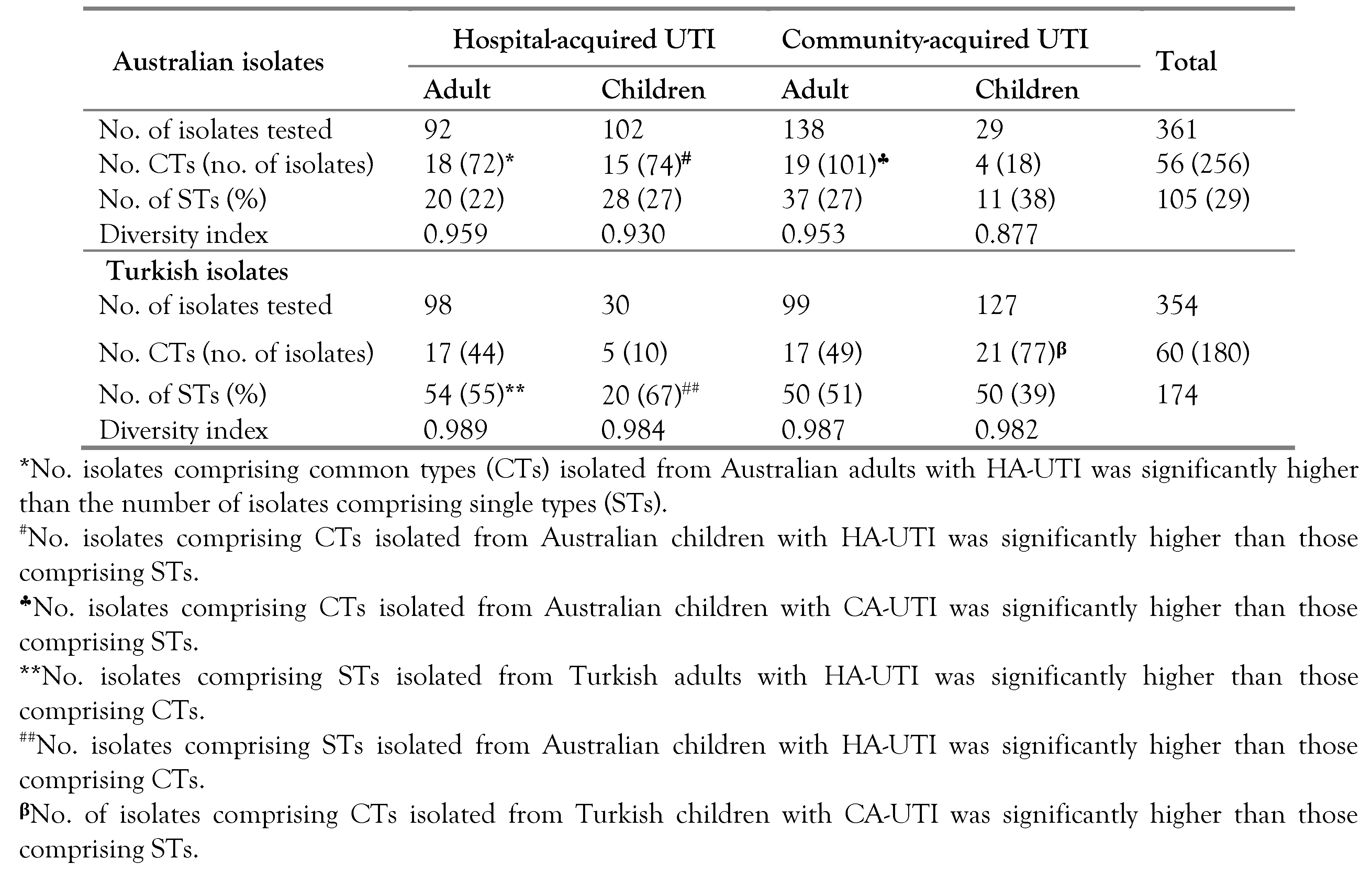
Initial clustering analysis of the 715 Australian and Turkish E. coli isolates resolved 56 CTs comprising 265 isolates (73.4%) and 96 STs among the Australian isolates, and 60 CTs comprising 180 isolates (51%) and 174 STs among the Turkish isolates (Table 1). CT representatives were selected and compared to identify identical UPEC (referred to as CGs) among different patient groups within the same country. Clustering analysis resolved 13 CGs among the Australian strains (CG 1–CG 13) and 14 CGs among the Turkish strains (CG 14–CG 27) (Figure 1A,B). The total number of isolates represented by Australian CGs (n=256) was significantly (p<0.0001) higher than those represented by the Turkish CGs (n=180) (Table 1). Most (85%) of the Australian CGs were found in ≥2 patient groups, with four dominant CGs (CGs 2, 7, 8 and 12) identified in ≥3 patient groups and representing 64% (n=120) of the isolates tested (Figure 1A). CG 7 was present in all Australian patient groups and represented 39% of the isolates tested (Figure 1A). Differences in the distribution of Australian CGs relative to patient age (55% adults vs 45% children), or infection setting were not statistically significant however, 10 (77%) of the 13 CGs were present in both hospital and community settings. Most (93%) of Turkish CGs were detected in ≥2 patient groups, with 57% (8/14) of the strains present among adults and children with CA-UTI, revealing a significant (p=0.019) association between Turkish CTs and community infection settings (Figure 1B).

Identical clonal groups of UPEC are shared between Australian and Turkish patients with UTI

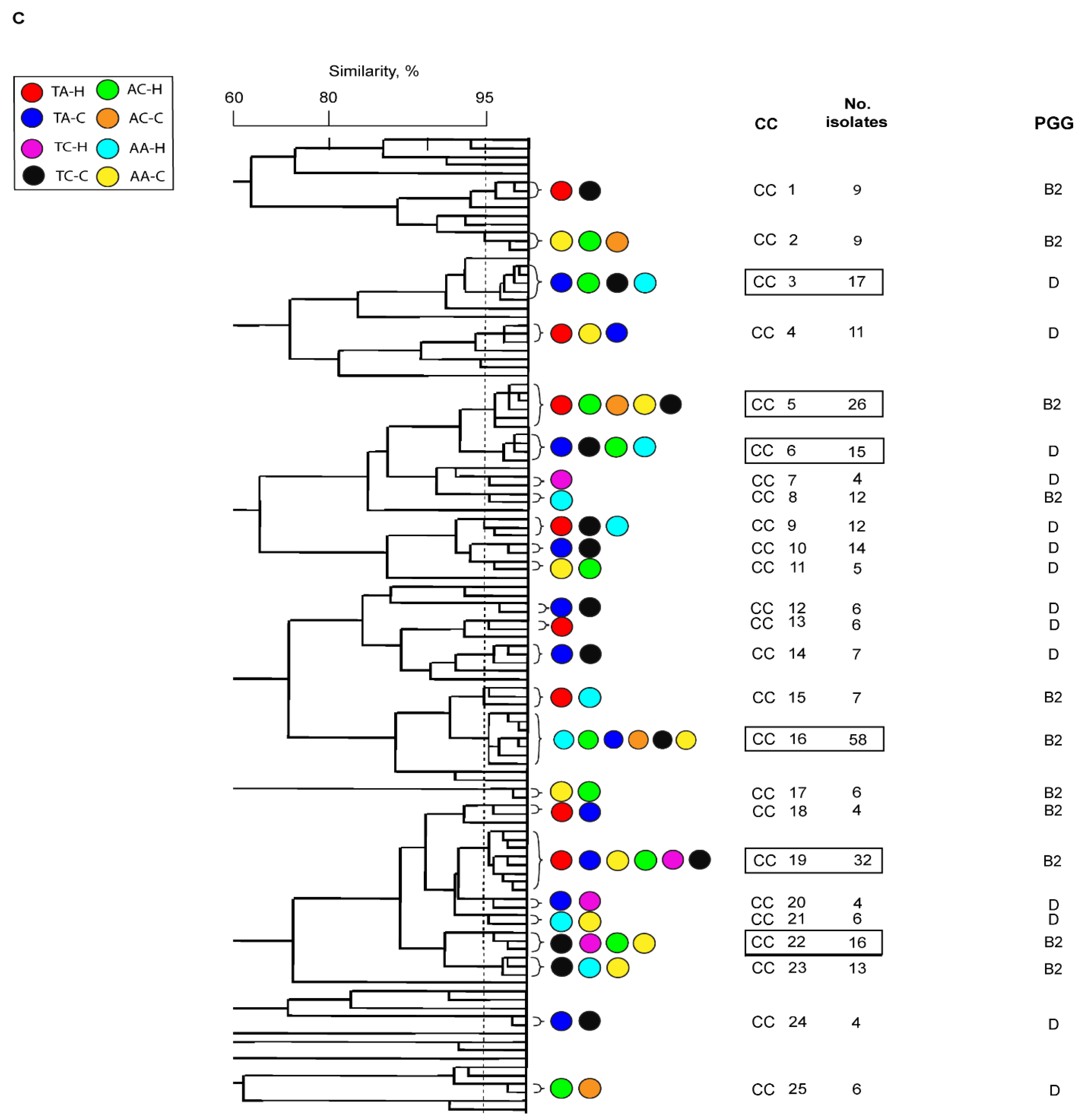
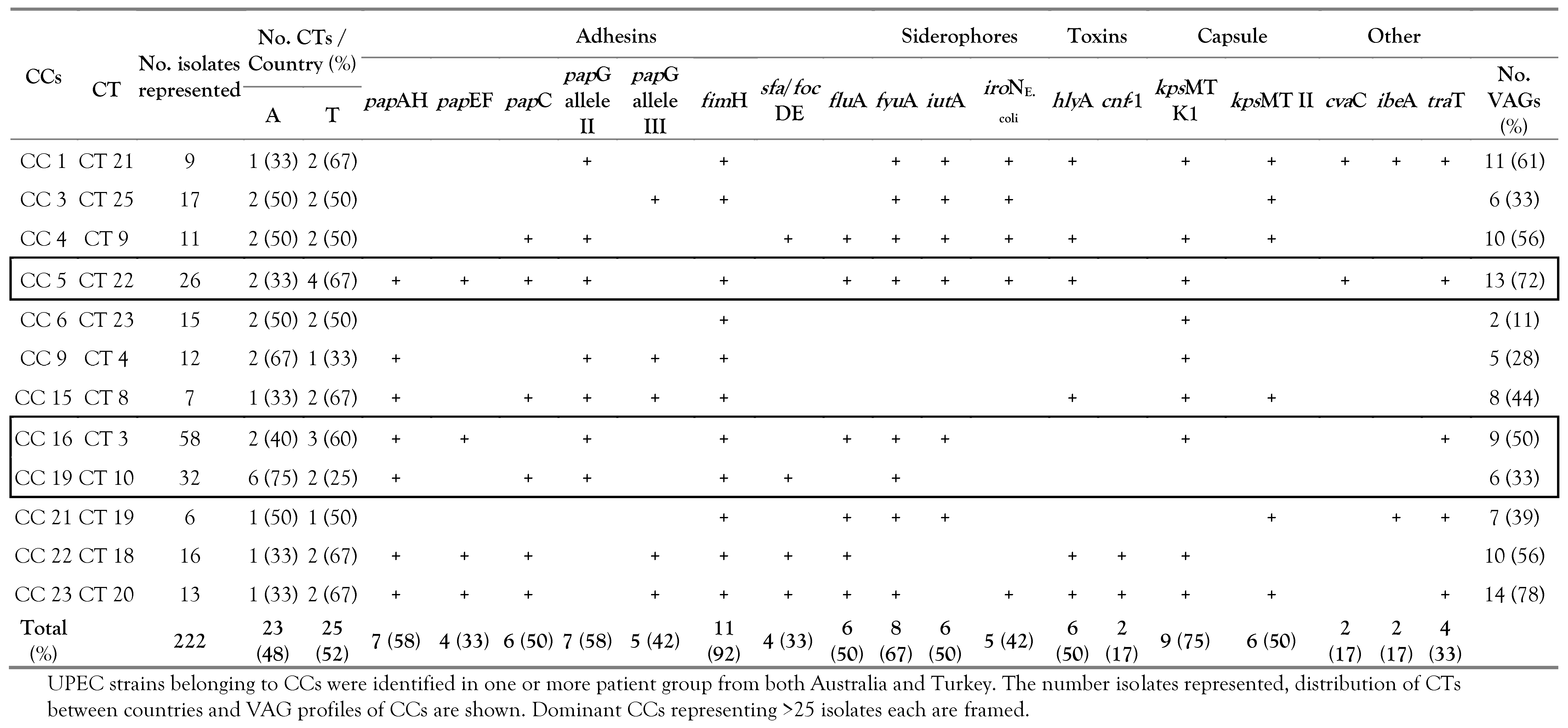
The abovementioned typing methods were used to compare single representatives of each Australian and Turkish CG. In all, 25 CGs representing a total of 309 isolates were resolved (Figure 2). Of these, 12 CGs were detected in one or more patient groups in either country, and we named them as members of the same clonal cluster (CC) (Figure 2). In all, we found that 53% (n=164) of the isolates tested were represented by six CCs, with each being identified in ≥2 patient groups from either country. These include CCs 3, 5, 6 16, 19 and 22 (Figure 2). CG 16 was recovered from all Australian groups and two Turkish groups, while the opposite was true for CG 19 (Figure 2). The distribution of CCs relative to patient age (55% adult vs 45% child) or country of isolation (50% each) did not differ significantly however, CCs were significantly (p<0.050) more prevalent in community settings.

Phylogeny

The phylogenetic grouping of the strains revealed an association between each CTs’ source of isolation and its phylogroup. Whilst the majority of the Australian CTs belonged to the phylogroups B2 (75%) and D (21.4%), most Turkish CTs belonged to the phylogroup D (84%) (Table 2). The prevalence of the phylogroup B2 was not significantly different between the Australian patient groups.

VAG scores

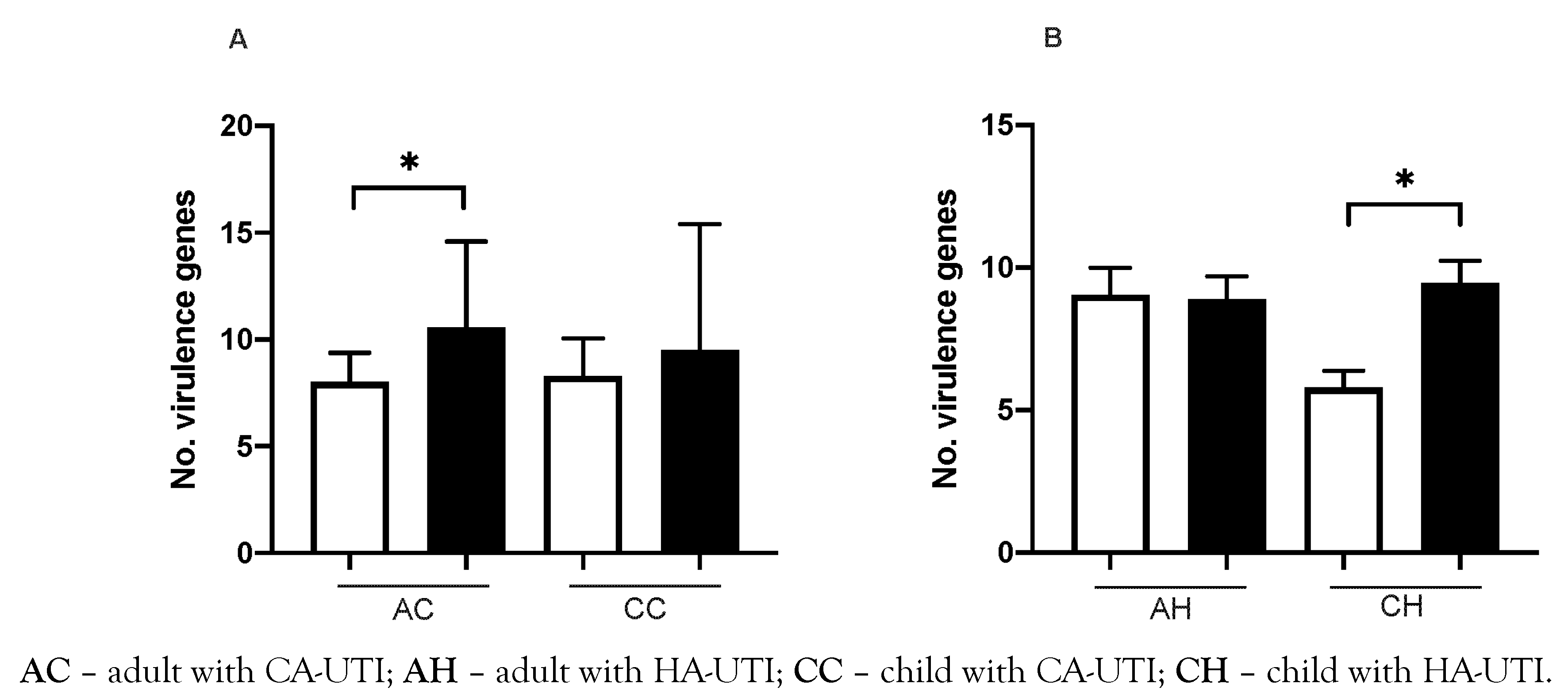
While Australian isolates generally carried more VAGs than the Turkish isolates, Australian children with HA-UTI (9.5±3.0) and adults with CA-UTI (9.4±4.0) had significantly (p=0.015 and p=0.017 respectively) higher VAG scores than Turkish children with HA-UTI (6.8±1.0) and adults with CA-UTI (8.0±1.3) (Figure 3A,B).

Prevalence of VAG functional groups

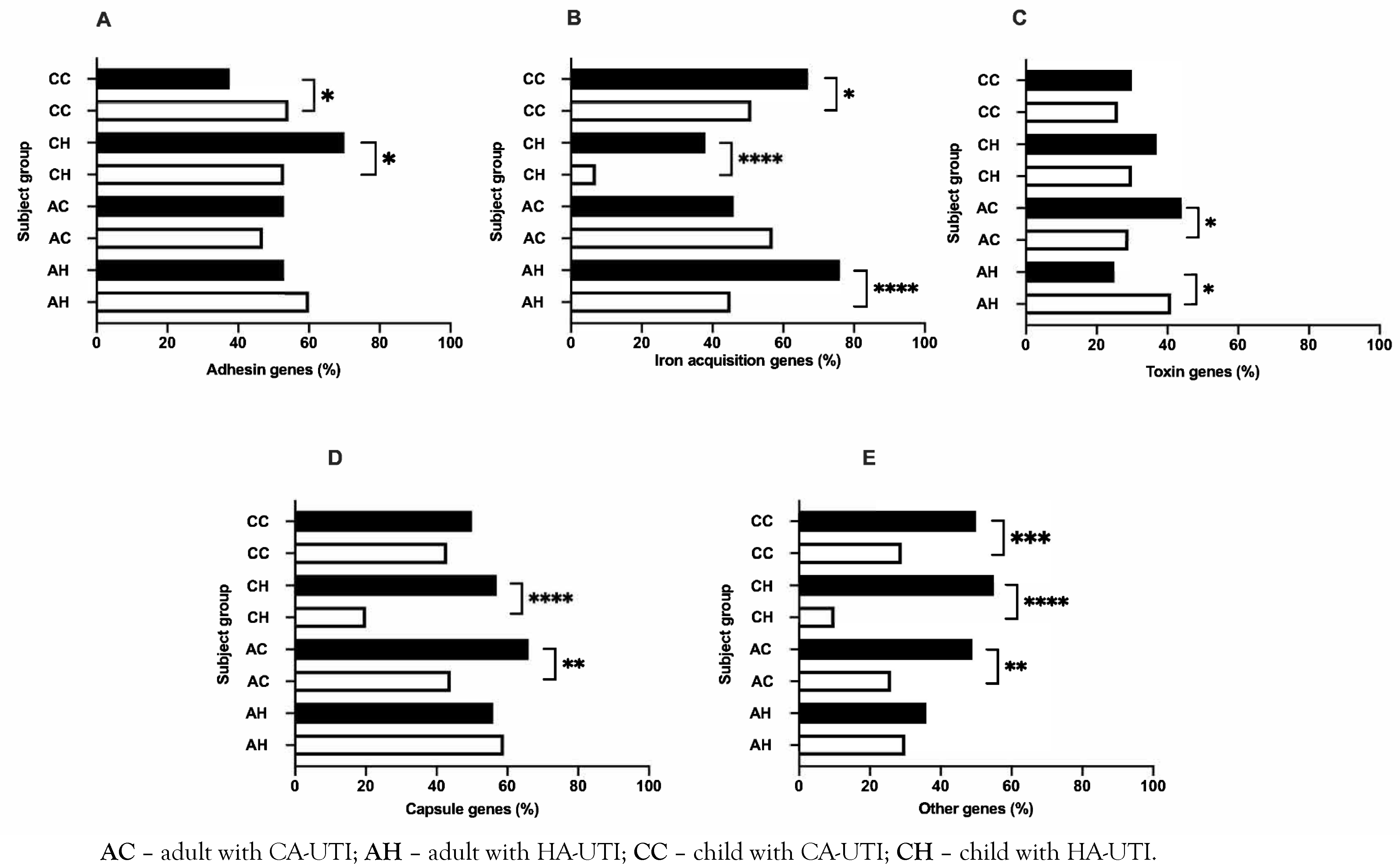
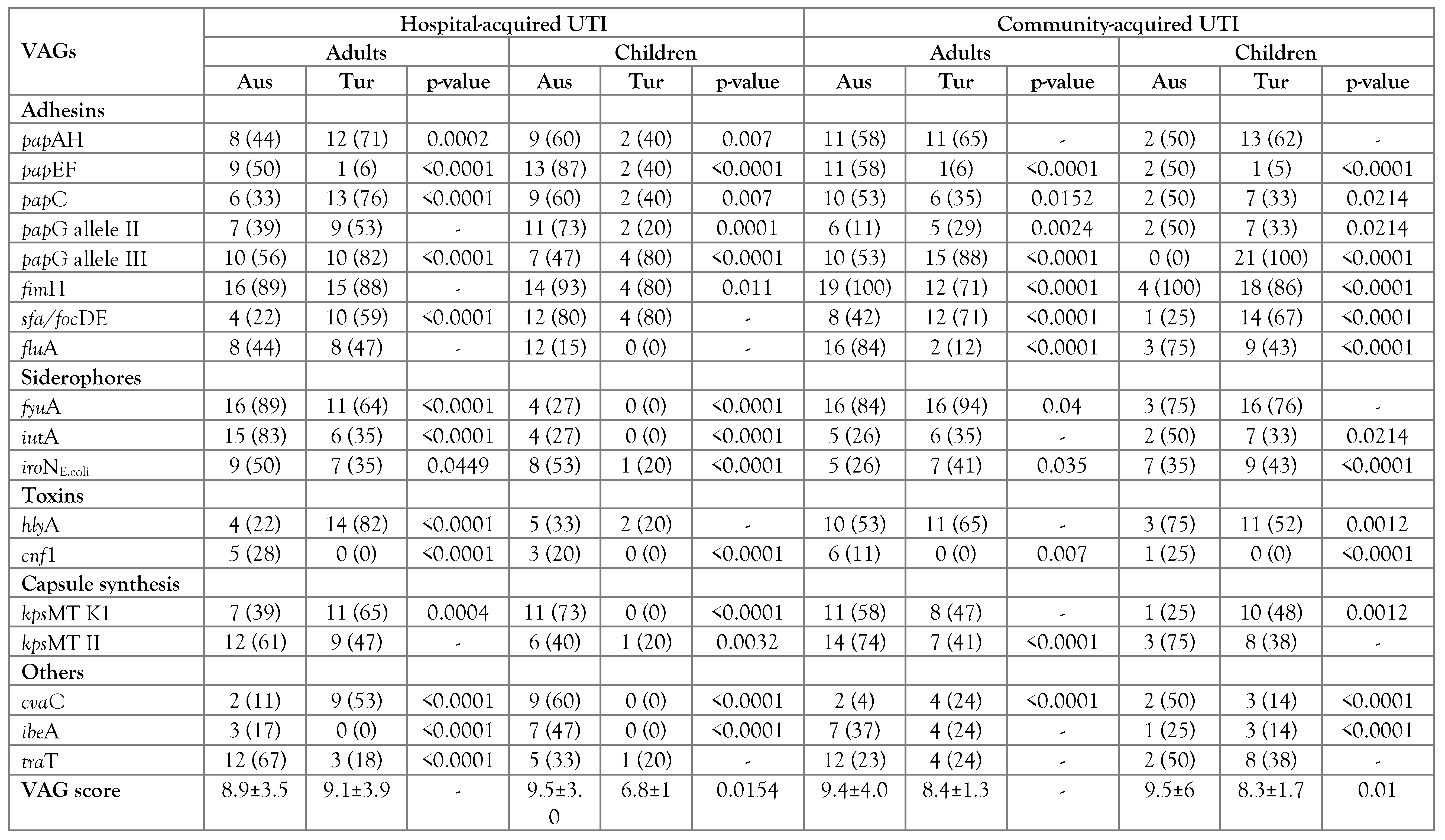
Comparison of VAG functional groups among Australian and Turkish CGs showed that adhesin genes were significantly (p=0.002) more prevalent in Australian children with HA-UTI (70%) than in Turkish children with HA-UTI (54%) (Figure 4A). In outpatient settings, adhesin genes were significantly (p=0.019) more prevalent in Turkish children with CA-UTI (53%) than in Australian children with CA-UTI (37%) (Figure 4A). All Australian patient groups, except for adults with CA-UTI, carried significantly more genes encoding iron acquisition, capsule biosynthesis and other VAGs when compared to their Turkish counterparts (Figure 4B–E).
UPEC shared between Australian and Turkish patients with UTI share common virulence characteristics
Table 3 shows the prevalence of different VAGs among CGs of UPEC between the Australian and Turkish patient groups. The difference was more pronounced among children with both HA- and CA-UTI in these countries (Table 3). Among the CCs, 42% (n=5) carried ≥10 of the 18 VAGs tested (Table 4). The fimH gene was highly conserved (98%), while 58% of CCs carried papAH and papG allele II (Table 4). Fifty percent of CCs encoded fluA, while the prevalence of the siderophore receptor genes, fyuA, and iutA, was 67% and 50% respectively (Table 4). Fifty percent of SCGs encoded hlyA, while the capsule synthesis genes, kpsMT KI and kpsMT KII were carried by 75% and 50% strains, respectively (Table 4). There was no correlation between the total number of VAGs carried and the number of isolates represented by a CC (r [2]=0.04) (Table 4).

Discussion

The emergence and dissemination of globally successful clonal clusters of UPEC represents a formidable and justified public health concern. While the mechanisms that facilitate the dissemination of these strains are yet to be fully elucidated, variations in the carriage of VAGs of these CCs could be partly due to the selective pressures imposed by hospitals and communities. [36,37]
In this study, we were interested to compare UPEC strains isolated from Turkish and Australian patients with HA- or CA-UTIs mainly due to their significant geographic distance and cultural practices while lacking any remarkable socioeconomic differences. Using a combination of two typing methods, the study was extended to determine whether identical clonal groups of UPEC were shared between UTI patients in these countries. We found that the Turkish strains belonged to a highly diverse collection of clonal groups predominated by the phylogroup D, as reported before. [20,21] The fact that most of these strains were detected among both children and adults with CA-UTI suggests that clonally diverse strains of UPEC are readily transmitted between members of the Turkish community, irrespective of patient age. The opposite was true for the Australian strains, with four dominant clonal groups accounting for 64% of the strains tested; most of which belonged to the phylogroup B2. While distinct clonal groups of UPEC have been shown to cause HA-UTIs among Australian children and adults before, [16,38] our results showed that a significant proportion of the Australian CGs originating from HA-UTI settings were also present among CA-UTI patients. Although novel to this study, this finding was not surprising given that the transition of UPEC clones from Australian sewage treatment plants to nearby environmental waters has been reported before.19,27,39
Several key studies have described the endemic and epidemic transmission of dominant UPEC clones. For example, Manges et al. (2008) showed that distinct clonal clusters of UPEC causing CA-UTIs throughout California, USA, were identical to those causing CA-UTIs in Montréal, Canada. [40] Similarly, Johnson and colleagues (2002) reported the dissemination of what was once considered a bona fide European UPEC clone (O15:K52:H1) into the United States. [23] Using two high-resolution typing methods, we identified 12 identical UPEC clonal clusters causing CA- and HA-UTIs among Turkish and Australian patients of varying ages and designated these strains as clonal clusters (CCs). Among these, six CCs represented 53% of the strains tested, with each being detected in two or more patient groups from either country. While the distribution of CCs had no association with patient age or country of isolation, they were significantly more prevalent throughout Australian and Turkish community settings. We postulate that these dominant clusters colonize and circulate among otherwise healthy community members, likely as part of the intestinal reservoir, before being disseminated more broadly through widespread exposure to a common source (i.e., water, food), person-to- person contact or exposure to some other environmental source. [41,42] Nevertheless, we recognize epidemiological studies of higher resolution are required to determine the directionality of transfer for these strains. Due to logistical reasons, we did not investigate the prevalence of important clones of UPEC such as the serotype O25:H4, ST131 that has a universal distribution among UPEC strains.
Whole genome analyses and PCR studies have demonstrated the broad diversity of VAG repertoires encoded by UPEC. [43,44] Indeed, variations in the carriage and expression of VAGs correspond with marked differences in virulence potential and urofitness. [41,45] In the present study, we showed that Australian UPEC strains carried more VAGs than Turkish strains, which could be partly due to the prevalence of the phylogroups, B2 and D, among these countries, respectively. [14] Of the Australian strains, those isolated from children with HA-UTIs and adults with CA-UTIs had significantly higher VAG scores than their Turkish counterparts, possibly reflecting the adaptation of less virulent clones to the less robust immune systems of these Turkish groups, or vice versa. The scarcity of information on patients did not allow us for a detailed comparison of the two groups with regards to the immune response or disease factors among our patient groups in these countries. Of particular interest to us was the fact that several of the more dominant CCs (i.e., CC 16 and 19) identified in this study carried no more than half of the VAGs tested. Accordingly, we suggest that the additive functions of other putative and/or unknown genetic elements contribute to the dominance and/or dissemination of these strains.
The adherence of UPEC to host urothelial cells, mediated by adhesins, is crucial for successful colonization of the urinary tract. [45] Interestingly, this study identified several key differences in the prevalence in adhesin genes among Australian and Turkish UPEC. For example, UPEC trains from Turkish children with CA-UTI and Australian children with HA- UTI encoded significantly more adhesin genes than strains in their respective patient groups, suggesting an increased requirement for adhesins among these patient groups. [16] The fimH gene was highly conserved among all CGs and CCs, reinforcing the importance of type-1 pili in the establishment of UTI, regardless of infection setting, geographic locale or patient demographic. [45] Additionally, most CCs encoded pap-family genes indicating that these strains have the potential to cause upper UTI. [46,47] Similarly, Ag43 was detected among the CCs of UPEC at the same prevalence rate reported elsewhere. [16,48] In view of its demonstrated roles in adhesion, auto-aggregation and biofilm formation, [12,49] we postulate that Ag43 facilitates the long-term survival and persistence of UPEC as observed among CCs in our study.
The importance of iron acquisition systems in UPEC’s establishment of UTI has been unequivocally demonstrated. [50,51] In iron-limited environments such as the urinary tract, UPEC sequesters iron through synthesis and secretion of low molecular weight Fe [3]+-chelating siderophores and their associated receptors. [52,53] Although present in certain Turkish patient groups, almost all Australian strains carried genes associated with iron acquisition. Similar prevalence rates have been reported in studies conducted among strains isolated from comparable Australian and Turkish UTI patient cohorts. [18,21,54] Furthermore, genes encoding siderophore receptors, fyuA, and iutA, were encoded by most of identified CCs. Given the importance of iutA and fyuA in iron acquisition, we suggest that these genes are positively selected among the CCs to provide an overall fitness advantage within the urinary tract. [55]
Secreted toxins are also key virulence factors in various E. coli-mediated pathologies, including UTI 56. α-hemolysin is arguably the most important of these and is produced by ~50% of UPEC strains causing pyelonephritis. [56] In this study, hlyA was significantly more prevalent among Turkish adults with HA- and CA-UTI. Moreover, 50% of the CCs identified in this study encoded hlyA as part of their toxin repertoire. It is widely recognized that α-hemolysin contributes to host cell lysis, the liberation of iron and caspase-1/caspase-4- dependent bladder cell exfoliation. [57,58] α- hemolysin also has recognized roles in perturbation of innate host immune responses through inhibition of phagocyte effector functions, NF-ĸB signaling and IL-6 expression. [59,60,61] Together, the increased prevalence of α- hemolysin among CCs and CGs isolated from Turkish adults may reflect an increased demand for access to growth limiting nutrients and/or to subvert innate host immune responses.
Polysaccharide capsules also contribute to the evasion of host immune defenses, providing resistance towards phagocytosis, complement- mediated killing and molecular mimicry. [62,63] Here, genes encoding group II capsule biosynthesis were significantly more prevalent among strains isolated from Australian adults when compared to Turkish adults. While there is a paucity in data describing the prevalence of capsule synthesis genes among Australian CA- UTI patients, higher prevalence rates of the genes kpsMT K1 and kpsMTII have consistently been reported among Australian HA-UTI patients when compared to Turkish HA-UTI patients. [16,21,38] Capsule synthesis genes were also prominent among the CCs, with kpsMT K1 and kpsMT II genes encoded by 75% and 50% of these strains, respectively. Taken together, these findings suggest that capsule synthesis genes are positively selected among the CCs and UPEC strains isolated from Australian adults where they fulfil an increased need to subvert innate immune defense mechanisms.

Conclusions

In conclusion, this study has shown that UPEC strains causing UTIs throughout Turkey belong to highly diverse clonal groups predominated by the phylogroup D, whereas Australian UPEC belonged to a few distinct B2- dominant clonal clusters. This may be attributed to the fact that Australian strains, particularly those isolated from children, had higher virulence potential than Turkish strains. Interestingly, we identified 12 identical UPEC clonal clusters causing UTIs among both Turkish and Australian patients with UTI of all ages throughout community and hospital settings. These clonal clusters frequently encoded VAGs associated with adhesion, iron acquisition, immune evasion and toxin production, which may have relevance to their ability to disseminate internationally to cause UTI. These strains were most prevalent in community infection settings, implying their colonization and circulation among otherwise healthy community members. If this is the case, our findings may have serious public health ramifications, particularly for individuals predisposed to UTI.

Author Contributions

DJA drafted the original manuscript and collected and analyzed the data. LS, FP, TV, HK, MH and AG collected data. MK reviewed and edited the manuscript and provided conceptualization. All authors read and approved the final version of the manuscript.

Funding

None to declare.

Institutional Review Board Statement

This study used collection of E. coli strains isolated by the staff of the pathology laboratory in hospitals or by the staff of the outpatient clinics as part of their routine processing of urine collected from patients. The research group received isolated E. coli strains and had no access to patients’ information. These strains had been stored at -80 °C until they were tested, which did not require ethical approval.

Acknowledgments

The authors would like to Acknowledge Dr Nicole Masters for her assistance in the preparation of this manuscript.

Conflicts of interest

All authors—none to declare.

References

  1. Behzadi, P.; Urbán, E.; Matuz, M.; Benkő, R.; Gajdács, M. The role of Gram-negative bacteria in urinary tract infections: Current concepts and therapeutic options. Adv Exp Med Biol. 2021, 1323, 35–69. [Google Scholar] [CrossRef]
  2. Russo, T.A.; Johnson, J.R. Medical and economic impact of extraintestinal infections due to Escherichia coli: Focus on an increasingly important endemic problem. Microbes Infect. 2003, 5, 449–456. [Google Scholar] [CrossRef] [PubMed]
  3. Ulett, G.C.; Totsika, M.; Schaale, K.; Carey, A.J.; Sweet, M.J.; Schembri, M.A. Uropathogenic Escherichia coli virulence and innate immune responses during urinary tract infection. Curr Opin Microbiol. 2013, 16, 100–107. [Google Scholar] [CrossRef] [PubMed]
  4. Leung, A.K.C.; Wong, A.H.C.; Leung, A.A.M.; Hon, K.L. Urinary tract infection in children. Recent Pat Inflamm Allergy Drug Discov. 2019, 13, 2–18. [Google Scholar] [CrossRef] [PubMed]
  5. Zare, M.; Vehreschild, M.; Wagenlehner, F. Management of uncomplicated recurrent urinary tract infections. BJU Int. 2022, 129, 668–678. [Google Scholar] [CrossRef] [PubMed]
  6. Boroumand, M.; Sharifi, A.; Manzouri, L.; Khoramrooz, S.S.; Khosravani, S.A. Evaluation of pap and sfa genes relative frequency P and S fimbriae encoding of uropathogenic Escherichia coli isolated from hospitals and medical laboratories; Yasuj City, Southwest Iran. Iran Red Crescent Med J. 2019, 21, e89499. [Google Scholar] [CrossRef]
  7. Hozzari, A.; Behzadi, P.; Kerishchi Khiabani, P.; Sholeh, M.; Sabokroo, N. Clinical cases, drug resistance, and virulence genes profiling in uropathogenic Escherichia coli. J Appl Genet. 2020, 61, 265–273. [Google Scholar] [CrossRef]
  8. Moreno, E.; Andreu, A.; Pigrau, C.; Kuskowski, M.A.; Johnson, J.R.; Prats, G. Relationship between Escherichia coli strains causing acute cystitis in women and the fecal E. coli population of the host. J Clin Microbiol. 2008, 46, 2529–2534. [Google Scholar] [CrossRef]
  9. Sarshar, M.; Behzadi, P.; Ambrosi, C.; Zagaglia, C.; Palamara, A.T.; Scribano, D. FimH and anti-adhesive therapeutics: A disarming strategy against uropathogens. Antibiotics 2020, 9, 397. [Google Scholar] [CrossRef]
  10. Behzadi, P. Classical chaperone-usher (CU) adhesive fimbriome: Uropathogenic Escherichia coli (UPEC) and urinary tract infections (UTIs). Folia Microbiol 2020, 65, 45–65. [Google Scholar] [CrossRef]
  11. Welch, R.A. Uropathogenic Escherichia coli-associated exotoxins. In Urinary Tract Infections, 2nd ed.; Mulvey, M.A., Klumpp, J.D., Stapleton, A.E., Eds.; ASM Press: Washington, DC, USA, 2017; pp. 263–276. [Google Scholar] [CrossRef]
  12. Ulett, G.C.; Valle, J.; Beloin, C.; Sherlock, O.; Ghigo, J.M.; Schembri, M.A. Functional analysis of antigen 43 in uropathogenic Escherichia coli reveals a role in long-term persistence in the urinary tract. Infect Immun. 2007, 75, 3233–3244. [Google Scholar] [CrossRef]
  13. Zoetendal, E.G.; Akkermans, A.D.; Akkermans-van Vliet, W.M.; de Visser, J.A.G.; de Vos, W.M. The host genotype affects the bacterial community in the human gastronintestinal tract. Microb Ecol Health Dis. 2001, 13, 129–134. [Google Scholar] [CrossRef]
  14. Moreno, E.; Johnson, J.R.; Pérez, T.; Prats, G.; Kuskowski, M.A.; Andreu, A. Structure and urovirulence characteristics of the fecal Escherichia coli population among healthy women. Microbes Infect. 2009, 11, 274–280. [Google Scholar] [CrossRef] [PubMed]
  15. Aguilar-Duran, S.; Horcajada, J.P.; Sorlí, L.; et al. Community-onset healthcare-related urinary tract infections: Comparison with community and hospital- acquired urinary tract infections. J Infect. 2012, 64, 478–483. [Google Scholar] [CrossRef]
  16. Vollmerhausen, T.; Katouli, M. Molecular characterisation of Escherichia coli isolated from hospitalised children and adults with urinary tract infection. Eur J Clin Microbiol Infect Dis. 2014, 33, 975–982. [Google Scholar] [CrossRef] [PubMed]
  17. Vollmerhausen, T.L.; Woods, J.L.; Faoagali, J.; Katouli, M. Interactions of uroseptic Escherichia coli with renal (A- 498) and gastrointestinal (HT-29) cell lines. J Med Microbiol. 2014, 63 Pt 12, 1575–1583. [Google Scholar] [CrossRef]
  18. Owrangi, B.; Masters, N.; Kuballa, A.; O’Dea, C.; Vollmerhausen, T.L.; Katouli, M. Invasion and translocation of uropathogenic Escherichia coli isolated from urosepsis and patients with community-acquired urinary tract infection. Eur J Clin Microbiol Infect Dis. 2018, 37, 833–839. [Google Scholar] [CrossRef]
  19. Astley, D.; Masters, N.; Kuballa, A.; Katouli, M. Commonality of adherent-invasive Escherichia coli isolated from patients with extraintestinal infections, healthy individuals and the environment. Eur J Clin Microbiol Infect Dis. 2021, 40, 181–192. [Google Scholar] [CrossRef]
  20. Yumuk, Z.; Afacan, G.; Nicolas-Chanoine, M.H.; Sotto, A.; Lavigne, J.P. Turkey: A further country concerned by community-acquired Escherichia coli clone O25-ST131 producing CTX-M-15. J Antimicrob Chemother. 2008, 62, 284–288. [Google Scholar] [CrossRef]
  21. Bozcal, E.; Eldem, V.; Aydemir, S.; Skurnik, M. The relationship between phylogenetic classification, virulence and antibiotic resistance of extraintestinal pathogenic Escherichia coli in İzmir province, Turkey. PeerJ. 2018, 6, e5470. [Google Scholar] [CrossRef]
  22. Manges, A.R.; Johnson, J.R.; Foxman, B.; O’Bryan, T.T.; Fullerton, K.E.; Riley, L.W. Widespread distribution of urinary tract infections caused by a multidrug-resistant Escherichia coli clonal group. N Eng J Med. 2001, 345, 1007–1013. [Google Scholar] [CrossRef]
  23. Johnson, J.R.; Stell, A.L.; O’Bryan, T.T.; et al. Global molecular epidemiology of the O15:K52:H1. extraintestinal pathogenic Escherichia coli clonal group: Evidence of distribution beyond Europe. J Clin Microbiol. 2002, 40, 1913–1923. [Google Scholar] [CrossRef] [PubMed]
  24. Johnson, J.R.; Murray, A.C.; Kuskowski, M.A.; et al. Distribution and characteristics of Escherichia coli clonal group A. Emerg Infect Dis. 2005, 11, 141–145. [Google Scholar] [CrossRef] [PubMed]
  25. Chen, J.; Griffiths, M.W. PCR differentiation of Escherichia coli from other Gram-negative bacteria using primers derived from the nucleotide sequences flanking the gene encoding the universal stress protein. Lett Appl Microbiol. 1998, 27, 369–371. [Google Scholar] [CrossRef] [PubMed]
  26. Vollmerhausen, T.L.; Ramos, N.L.; Gündoğdu, A.; Robinson, W.; Brauner, A.; Katouli, M. Population structure and uropathogenic virulence-associated genes of faecal Escherichia coli from healthy young and elderly adults. J Med Microbiol. 2011, 60, 574–581. [Google Scholar] [CrossRef]
  27. Anastasi, E.M.; Matthews, B.; Gundogdu, A.; et al. Prevalence and persistence of Escherichia coli strains with uropathogenic virulence characteristics in sewage treatment plants. Appl Environ Microbiol. 2010, 76, 5882–5886. [Google Scholar] [CrossRef]
  28. Corney, B.G.; Colley, J.; Djordjevic, S.P.; Whittington, R.; Graham, G.C. Rapid identification of some Leptospira isolates from cattle by random amplified polymorphic DNA fingerprinting. J Clin Microbiol. 1993, 31, 2927–2932. [Google Scholar] [CrossRef]
  29. Atlas, R.M. Diversity of microbial communities. In Advances in Microbial Ecology; Advances in Microbial, Ecology, Marshall, K.C., Eds.; Springer: Boston, MA, USA, 1984; Volume 7. [Google Scholar] [CrossRef]
  30. Clermont, O.; Christenson, J.K.; Denamur, E.; Gordon, D.M. The Clermont Escherichia coli phylo-typing method revisited: Improvement of specificity and detection of new phylo-groups. Env. Microbiol Rep. 2013, 5, 58–65. [Google Scholar] [CrossRef]
  31. Clermont, O.; Bonacorsi, S.; Bingen, E. Rapid and simple determination of the Escherichia coli phylogenetic group. Appl Environ Microbiol. 2000, 66, 4555–4558. [Google Scholar] [CrossRef]
  32. Johnson, J.R.; Stell, A.L. Extended virulence genotypes of Escherichia coli strains from patients with urosepsis in relation to phylogeny and host compromise. J Infect Dis. 2000, 181, 261–272. [Google Scholar] [CrossRef]
  33. Chapman, T.A.; Wu, X.Y.; Barchia, I.; et al. Comparison of virulence gene profiles of Escherichia coli strains isolated from healthy and diarrheic swine. Appl Environ Microbiol. 2006, 72, 4782–4795. [Google Scholar] [CrossRef]
  34. Restieri, C.; Garriss, G.; Locas, M.C.; Dozois, C.M. Autotransporter-encoding sequences are phylogenetically distributed among Escherichia coli clinical isolates and reference strains. Appl Environ Microbiol. 2007, 73, 1553–1562. [Google Scholar] [CrossRef] [PubMed]
  35. Johnson, J.R.; Kuskowski, M.A.; O’bryan, T.T.; Colodner, R.; Raz, R. Virulence genotype and phylogenetic origin in relation to antibiotic resistance profile among Escherichia coli urine sample isolates from Israeli women with acute uncomplicated cystitis. Antimicrob Agents Chemother. 2005, 49, 26–31. [Google Scholar] [CrossRef] [PubMed]
  36. Platell, J.L.; Johnson, J.R.; Cobbold, R.N.; Trott, D.J. Multidrug-resistant extraintestinal pathogenic Escherichia coli of sequence type ST131 in animals and foods. Vet Microbiol. 2011, 153, 99–108. [Google Scholar] [CrossRef] [PubMed]
  37. Johnson, J.R.; Nicolas-Chanoine, M.H.; DebRoy, C.; et al. Comparison of Escherichia coli ST131 pulsotypes, by epidemiologic traits, 1967-2009. Emerg Infect Dis. 2012, 18, 598–607. [Google Scholar] [CrossRef]
  38. Ramos, N.L.; Saayman, M.L.; Chapman, T.A.; et al. Genetic relatedness and virulence gene profiles of Escherichia coli strains isolated from septicaemic and uroseptic patients. Eur J Clin Microbiol Infect Dis. 2010, 29, 15–23. [Google Scholar] [CrossRef]
  39. Anastasi, E.; Matthews, B.; Stratton, H.M.; Katouli, M. Pathogenic Escherichia coli found in sewage treatment plants and environmental waters. Appl Environ Microbiol. 2012, 78, 5536–5541. [Google Scholar] [CrossRef]
  40. Manges, A.R.; Tabor, H.; Tellis, P.; Vincent, C.; Tellier, P.P. Endemic and epidemic lineages of Escherichia coli that cause urinary tract infections. Emerg Infect Dis. 2008, 14, 1575–1583. [Google Scholar] [CrossRef]
  41. Singer, R.S. Urinary tract infections attributed to diverse ExPEC strains in food animals: Evidence and data gaps. Front Microbiol. 2015, 6, 28. [Google Scholar] [CrossRef]
  42. Manges, A.R.; Johnson, J.R.J. Food-borne origins of Escherichia coli causing extraintestinal infections. Clin Infect Dis. 2012, 55, 712–719. [Google Scholar] [CrossRef]
  43. Avasthi, T.S.; Kumar, N.; Baddam, R.; et al. Genome of multidrug-resistant uropathogenic Escherichia coli strain NA114 from India. J Bacteriol. 2011, 193, 4272–4273. [Google Scholar] [CrossRef]
  44. Petty, N.K.; Ben Zakour, N.L.; Stanton-Cook, M.; et al. Global dissemination of a multidrug resistant Escherichia coli clone. Proc Natl Acad Sci. 2014, 111, 5694–5699. [Google Scholar] [CrossRef] [PubMed]
  45. Wiles, T.J.; Kulesus, R.R.; Mulvey, M.A. Origins and virulence mechanisms of uropathogenic Escherichia coli. Exp Mol Pathol. 2008, 85, 11–19. [Google Scholar] [CrossRef] [PubMed]
  46. Cheng, C.H.; Tsau, Y.K.; Kuo, C.Y.; Su, L.H.; Lin, T.Y. Comparison of extended virulence genotypes for bacteria isolated from pediatric patients with urosepsis, acute pyelonephritis, and acute lobar nephronia. Paed Infect Dis J. 2010, 29, 736–740. [Google Scholar] [CrossRef]
  47. Firoozeh, F.; Saffari, M.; Neamati, F.; Zibaei, M. Detection of virulence genes in Escherichia coli isolated from patients with cystitis and pyelonephritis. Int J Infect Dis. 2014, 29, 219–222. [Google Scholar] [CrossRef] [PubMed]
  48. Ramos, N.L.; Dzung, D.T.; Stopsack, K.; et al. Characterisation of uropathogenic Escherichia coli from children with urinary tract infection in different countries. Eur J Clin Microbiol Infect Dis. 2011, 30, 1587–1593. [Google Scholar] [CrossRef]
  49. Anderson, G.G.; Palermo, J.J.; Schilling, J.D.; Roth, R.; Heuser, J.; Hultgren, S.J. Intracellular bacterial biofilm-like pods in urinary tract infections. Science. 2003, 301, 105–107. [Google Scholar] [CrossRef]
  50. Torres, A.G.; Redford, P.; Welch, R.A.; Payne SMJI, immunity. TonB-dependent systems of uropathogenic Escherichia coli: Aerobactin and heme transport and TonB are required for virulence in the mouse. Infect Immun. 2001, 69, 6179–6185. [Google Scholar] [CrossRef]
  51. Subashchandrabose, S.; Mobley, H.L. Back to the metal age: Battle for metals at the host-pathogen interface during urinary tract infection. Metallomics. 2015, 7, 935–942. [Google Scholar] [CrossRef]
  52. Cassat, J.E.; Skaar, E.P. Iron in Infection and Immunity. Cell Host Microbe. 2013, 13, 509–519. [Google Scholar] [CrossRef]
  53. Chu, B.C.; Garcia-Herrero, A.; Johanson, T.H.; et al. Siderophore uptake in bacteria and the battle for iron with the host; a bird’s eye view. Biometals. 2010, 23, 601–611. [Google Scholar] [CrossRef] [PubMed]
  54. Ramos, N.; Saayman, M.L.; Chapman, T.; et al. Genetic relatedness and virulence gene profiles of Escherichia coli strains isolated from septicaemic and uroseptic patients. Eur J Clin Microbiol Infect Dis. 2010, 29, 15–23. [Google Scholar] [CrossRef] [PubMed]
  55. Garcia, E.C.; Brumbaugh, A.R.; Mobley, H.L. Redundancy and specificity of Escherichia coli iron acquisition systems during urinary tract infection. Infect Immun. 2011, 79, 1225–1235. [Google Scholar] [CrossRef]
  56. Bien, J.; Sokolova, O.; Bozko, P. Role of uropathogenic Escherichia coli virulence factors in development of urinary tract infection and kidney damage. Int J Nephrol. 2012, 2012, 681473. [Google Scholar] [CrossRef]
  57. Dhakal, B.K.; Mulvey, M.A. The UPEC pore-forming toxin α-hemolysin triggers proteolysis of host proteins to disrupt cell adhesion, inflammatory, and survival pathways. Cell Host Microbe. 2012, 11, 58–69. [Google Scholar] [CrossRef]
  58. Nagamatsu, K.; Hannan, T.J.; Guest, R.L.; et al. Dysregulation of Escherichia coli α-hemolysin expression alters the course of acute and persistent urinary tract infection. Proc Natl Acad Sci. 2015, 112, E871–E880. [Google Scholar] [CrossRef]
  59. Cavalieri, S.J.; Snyder, I.S. Effect of Escherichia coli alpha- hemolysin on human peripheral leukocyte function in vitro. Infect Immun. 1982, 37, 966–974. [Google Scholar] [CrossRef]
  60. Wiles, T.J.; Bower, J.M.; Redd, M.J.; Mulvey, M.A. Use of zebrafish to probe the divergent virulence potentials and toxin requirements of extraintestinal pathogenic Escherichia coli. PLoS Pathog. 2009, 5, e1000697. [Google Scholar] [CrossRef]
  61. Russo, T.A.; Davidson, B.A.; Genagon, S.A.; et al. E. coli virulence factor hemolysin induces neutrophil apoptosis and necrosis/lysis in vitro and necrosis/lysis and lung injury in a rat pneumonia model. Am J Physiol. 2005, 289, L207–L216. [Google Scholar] [CrossRef]
  62. Erjavec, M.S.; Žgur-Bertok, D. Extended Characterization of Human Uropathogenic Escherichia Coli Isolates from Slovenia; IntechOpen: London, UK, 2011. [Google Scholar]
  63. Burns, S.M.; Hull, S.I. Loss of resistance to ingestion and phagocytic killing by O− and K− mutants of a uropathogenic Escherichia coli O75:K5 strain. Infect Immun. 1999, 67, 3757–3762. [Google Scholar] [CrossRef]
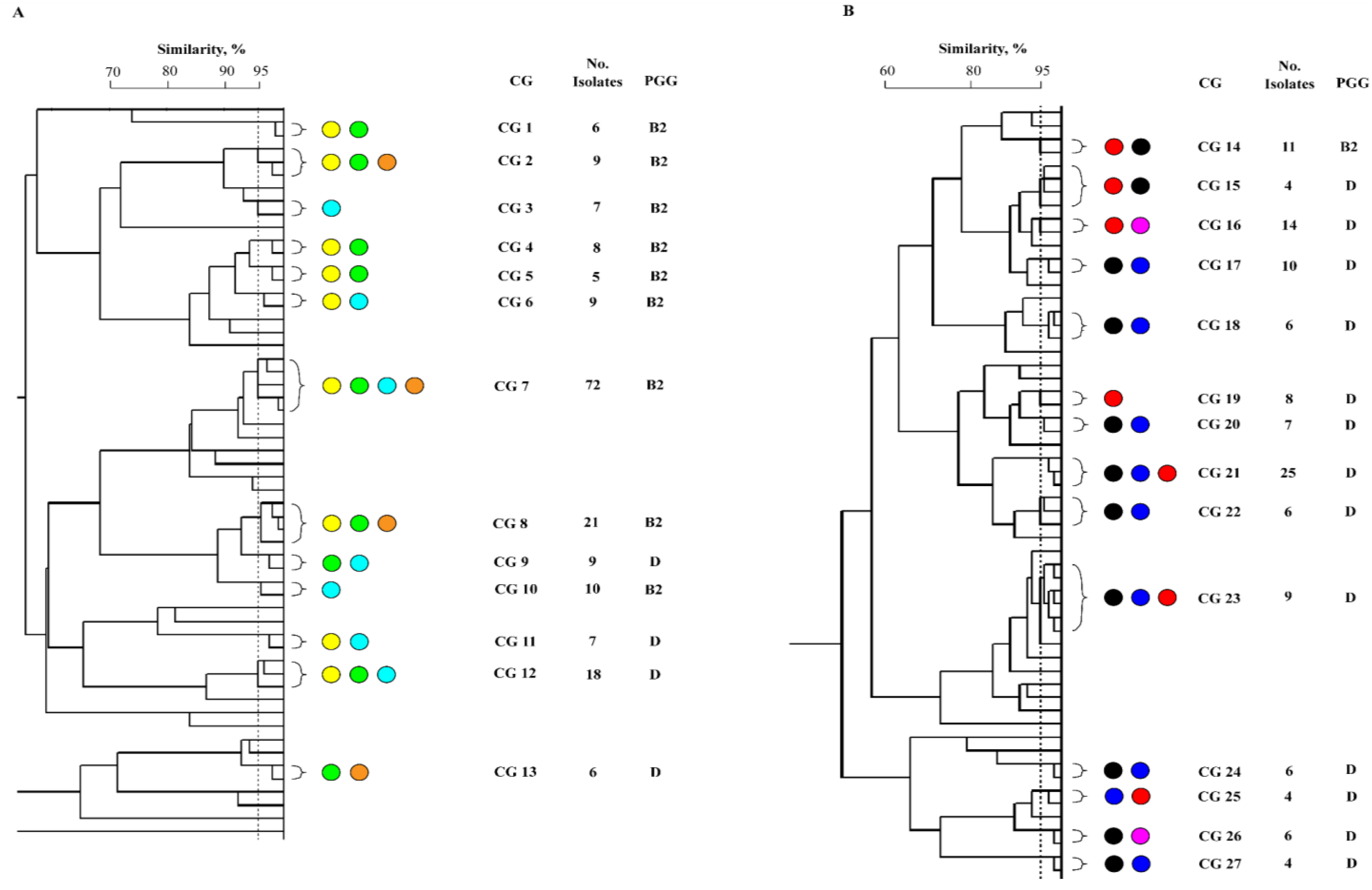
Figure 1. Clonal composition and phylogenetic backgrounds of uropathogenic E. coli (UPEC) clonal groups (CGs) isolated from Australian (A) and Turkish (B) children and adults with hospital- (HA)- or community-acquired (CA)-UTI. The number of isolates represented by each CG are shown. TA-H, Turkish adult with HA-UTI (red circle); TA-C, Turkish adult with CA-UTI (dark blue circle); TC-H, Turkish child with HA-UTI (fuchsia circle); TC-C, Turkish child with CA-UTI (black circle); AA-H, Australian adult with HA-UTI (light blue circle); AA-C, Australian adult with CA- UTI (yellow circle); AC-H, Australian child with HA-UTI (green circle); AC-C, Australian child with CA-UTI (orange circle).
Germs 12 00214 g001
Figure 2. Distribution of clonal clusters (CCs) among a collection of E. coli isolated Australian and Turkish patients with urinary tract infection. CCs detected among ≥2 patient groups from either country are framed. The number of isolates represented by each CC are shown. Turkish adult with HA-UTI (red circle); Turkish adult with CA-UTI (dark blue circle); Turkish child with HA-UTI (fuchsia circle); Turkish child with CA-UTI (black circle); Australian adult with HA-UTI (light blue circle); Australian adult with CA-UTI (yellow circle); Australian child with HA-UTI (green circle); Australian child with CA-UTI (orange circle).
Germs 12 00214 g002
Figure 3. Virulence-associated gene (VAG) scores (mean ± SEM) among clonal groups (CGs) of uropathogenic E. coli (UPEC) isolated from Turkish (white box) and Australian (black box) children and adults with community-acquired (CA-, A) or hospital-acquired (HA- B) urinary tract infections (UTIs). *p≤0.050.
Germs 12 00214 g003
Figure 4. Prevalence (%) of virulence-associated genes (VAGs) among clonal groups (CGs) of uropathogenic E. coli (UPEC) isolated from Turkish (white box) and Australian (black box) children and adults with hospital- (HA) or community-acquired (CA)- urinary tract infections (UTIs). VAGs were assigned to one of five classifications based on their putative functions such as bacterial adhesion (A), iron acquisition (B), toxin production (C), capsule biosynthesis (D) or other functions (E).
Germs 12 00214 g004
Table 1. Distribution of common types (CTs) and single types (STs) of uropathogenic E. coli (UPEC) isolated from Australian and Turkish children and adults with hospital- or community-acquired urinary tract infections. Symbols indicate a significant difference (p<0.05) between the number of isolates within CTs for patient groups in each country.
Germs 12 00214 i001
Table 2. Distribution of phylogenetic groups among uropathogenic E. coli (UPEC) common types (CTs) isolated from Australian (n=56) and Turkish (n=60) children and adults with hospi-tal- (HA)- or community-acquired (CA)- urinary tract infections (UTIs).
Germs 12 00214 i002
Table 3. The prevalence (%) of 18 virulence-associated genes (VAGs) commonly associated with E. coli strains causing extraintestinal infections among a collection of uropathogenic E. coli (UPEC) clonal groups (CGs). Strains were isolated from Turkish and Australian children and adults with hospital-acquired (HA-) or community- acquired urinary tract infections (UTIs).
Germs 12 00214 i003
Table 4. Prevalence of 18 virulence-associated genes (VAGs) among a collection of UPEC belonging to the same clonal clusters (CCs) isolated from Australian (A) and Turkish (T) children and adults with either hospital-acquired (HA-) or community-acquired (CA-) urinary tract infections (UTIs).
Germs 12 00214 i004