Next Article in Journal
Use of High-Dose Ciprofloxacin for Recurrent Biofilm-Forming Multidrug-Resistant Klebsiella pneumoniae Bacteremia
Previous Article in Journal
The Challenges to a Successful COVID-19 Vaccination Programme in Africa
 
 
GERMS is published by MDPI from Volume 15 Issue 4 (2025). Previous articles were published by another publisher in Open Access under a CC-BY (or CC-BY-NC-ND) licence, and they are hosted by MDPI on mdpi.com as a courtesy and upon agreement with the former publisher Infection Science Forum.
Review

Candida auris and Other Phylogenetically Related Species – A Mini-Review of the Literature

by
Cristina Nicoleta Ciurea
1,2,
Anca Delia Mare
1,*,
Irina-Bianca Kosovski
3,
Felicia Toma
1,
Camelia Vintilă
4 and
Adrian Man
1
1
Department of Microbiology, George Emil Palade University of Medicine, Pharmacy, Science and Technology, 38 Gheorghe Marinescu Street, 540149 Târgu Mureș, Romania
2
Doctoral School, George Emil Palade University of Medicine, Pharmacy, Science and Technology of Târgu Mureș, 38 Gheorghe Marinescu Street, 540139 Târgu Mureș, Romania
3
Department of Pathophysiology, George Emil Palade University of Medicine, Pharmacy, Science and Technology of Târgu Mureș, 38 Gheorghe Marinescu Street, 540139 Târgu Mureș, Romania
4
Mureș County Clinical Hospital—Infectious Diseases Laboratory, 6 Bernady Gyorgy Street, 540072 Târgu Mureș, Romania
*
Author to whom correspondence should be addressed.
GERMS 2021, 11(3), 441-448; https://doi.org/10.18683/germs.2021.1281
Submission received: 19 April 2021 / Revised: 11 June 2021 / Accepted: 27 June 2021 / Published: 29 September 2021

Abstract

The lesser-known non-albicans Candida species are often overlooked and difficult to diagnose in most microbiology laboratories. Candida auris, a relatively newly discovered species, is responsible for outbreaks in various geographical regions. Because of its increased resistance to antifungal drugs, C. auris is responsible for hard-to-treat infections and its pathogenicity is still incompletely elucidated. Non- albicans species phylogenetically related to C. auris, like the C. haemulonii complex might also play a role in human pathology. The current summary of the literature presents the emergence, virulence, laboratory identification, and molecular mechanisms responsible for antifungal resistance of emergent rare non-albicans Candida species.

Introduction

Candida genus is polyphyletic and many members of the genus have been reassigned in recent taxonomic studies. The revised taxonomy better reflects the resistance profiles of clinically important species like Nakaseomyces glabrata (previously Candida glabrata), Pichia kudriavzevii (previously Candida krusei) or Debaryomyces hansenii (formerly known as Candida famata) [1].
Until recently, Candida albicans was the most commonly encountered species of the Candida genus. C. albicans is ubiquitarian, it can cause infections mainly in immunocompromised hosts and almost any microbiology laboratory can easily identify it. Non-albicans species, on the other hand, have received scant attention, despite their newly emergent nature. With an increased pool of immunocompromised patients, more prone to fungal infections, non-albicans species have increasing importance and the lack of species identification is often considered inadequate.
Conventional methods of identification for fungi, used by most laboratories, fail to identify the lesser encountered Candida spp., as they are relatively unreactive and insufficiently studied. Although rare, some non-albicans species pose not only diagnostic challenges but are also harder to treat, as they exhibit increased resistance to antifungal drugs [2].
Recent studies have proven that C. auris can be part of the microbiome, raising questions about how this fungus emerged just recently, causing hard-to-treat infections. Various theories aim at explaining the rise of C. auris including the use of antifungal drugs, environmental changes, and human activities that permitted the fungus to escape its specialized niche and to colonize larger areas of skin. It is now known that C. auris can be a colonizer, but screening programs, where needed, are still difficult to draft. A single screening test proved to be imperfectly sensitive and more research is needed for discovering an effective screening strategy [3].

Emergence

In 2009, a novel species of Candida isolated from the external ear canal of a Japanese patient was first described. The strain was named “Candida auris” and it resembles C. ruelliae and C. haemulonii complex at a phylogenetic level [4]. In 2011, C. auris was incidentally found in a stored bloodstream isolate from 1996, in South Korea [5]. In Europe, the first case of an infection with C. auris was imported from India, in 2009 [6].
C. auris belongs to the Clavispora clade of the Metschnikowiaceae family of the order Saccharomycetales [7] and it might have previously existed as a plant saprophyte. Because of its salinity tolerance and thermal tolerance, some authors are considering its emergence to be linked with global warming [8].
Independent different clonal populations (clades separated by thousands of single- nucleotide polymorphisms) have been identified on 3 continents [9]. Although it is commonly accepted that there are four major clades: the South Asian (I), Asian (II), African (III), and South American (IV), a potential fifth clade of C. auris has been reported in Iran, in 2018 [10]. C. auris strains isolated from different geographical areas tend to be phylogenetically distinct, whereas C. haemulonii strains are more interrelated [11]. Analysis of C. auris genome showed a 40% genetic share with C. lusitaniae, especially related to genes that provide multiple antifungal resistance. Moreover, C. auris harbors virulence genes that are present in C. albicans [12].
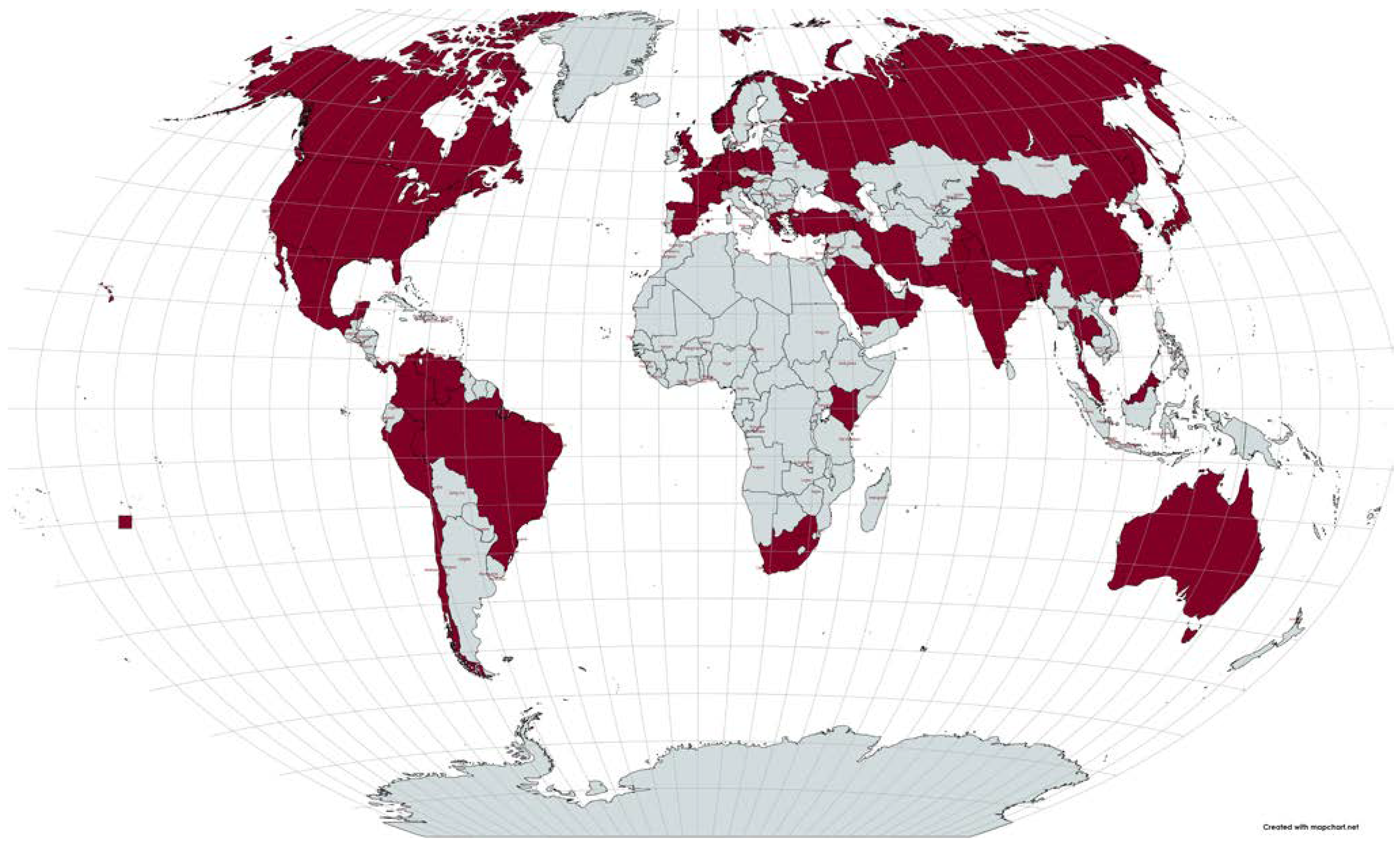
To date, cases of infections have been reported all around the globe (Figure 1) [13,14,15,16,17,18,19,20,21,22,23,24,25,26,27]. However, the real global distribution remains unknown, mainly because the current commercial methods used in the microbiology laboratory are often misidentifying as other non- albicans [28].
C. haemulonii (Torulopsis haemulonii) was first isolated from the gut of a blue-striped grunt (Haemulon sciurus) in 1962, [29] found in the Western Atlantic, Gulf of Mexico, and the Caribbean [7]. The C. haemulonii complex includes C. haemulonii (C. haemulonii group I), duobushaemulonii sp. nov. (C. haemulonii group II), and C. haemulonii var. vulnera var. nov [30]. In 2016, Sugita et al. isolated a strain named Candida pseudohaemulonii sp. nov. from the blood of a patient in Mae Sot Hospital, Thailand [31].
At a genetic level, C. haemulonii resembles Debaryomyces hansenii, C. guilliermondii and C. auris [2,11]. Although rare, its prevalence is increasing, especially from patients with deep- seated infections [32] and it can be transmitted within healthcare facilities [33].
C. haemulonii is an opportunist fungus capable of causing fungemia outbreaks [2,34]. The disease is not clinically distinct from other common nosocomial infections: the patients present high fever and leukocytosis. Risk factors include malignancy, mechanical ventilation, and central venous catheter insertion [35]. In 2021, a case of liver abscess caused by C. haemulonii var. vulnera was reported in Peru [36].
C. duobushaemulonii, first reported in 2016, causes vulvovaginal candidiasis, onychomycosis, fungemia, and chronically infected wounds of lower extremities, mainly in patients with diabetes mellitus. C. haemulonii var. vulnera causes fungemia, ulcers of the feet, and onychomycosis in adult patients with preexisting conditions like sickle-cell anemia, B-cell lymphoma, or other malignancies [37,38].
C. ruelliae has been isolated from the Rouellia sp. (Acanthaceae) flowers in India, in 2008 and it is different from C. haemulonii by a 14% nucleotide sequence divergence [39]. Its role in human pathology is yet to be discovered.

Virulence

Because of the high genetic diversity exerted by C. auris, there are conflicting reports about its virulence. The virulence-associated genes in C. auris encode predominantly hydrolases, mannosyl transferases, hemolysins, oxidoreductases, oligopeptide transporters, aspartyl-secreted proteases (Saps), lipases, phospholipases. The lytic enzymes (lipases and proteinases) are produced in a strain-dependent manner, and the level of Saps is similar to those of C. albicans, and even more, highly expressed even at 42°C, ensuring pathogenicity at high temperatures [12,40].
However, the genes encoding adhesins and integrins seem to be poorly represented. In terms of biofilm formation, C. auris has 686 biofilm- related proteins (ribosomal proteins, transporters, several enzymes, transcription factors). The biofilms are composed mainly of yeast cells, engulfed in a limited amount of extracellular matrix. Mannosyl transferases are known for maintaining the cell wall architecture in C. albicans, and also for influencing the recognition of the fungus by the immune system, and for facilitating the adherence to the host cells. Moreover, because it possesses transporters that act like efflux pumps, this might explain its resistance to different antifungal drugs [7,24,40,41]
C. auris express a greater ability to form biofilms while compared with species from the C. haemulonii complex [42] and the biofilm eradication concentration (MBEC) of antifungal agents proved to be 512-fold higher than the minimal inhibitory concentration (MIC), proving that sessile cells exhibit increased resistance to antifungal drugs compared with planktonic cells [43].

Immune response

Neutrophils, key cells for the control of invasive Candida infections seem to fail to release neutrophil extracellular traps (NETs) or to phagocytose C. auris cells. This impaired innate immune response might explain the high mortality rates associated with C. auris infections, but the underlying mechanisms that can explain this phenomenon are still incompletely elucidated [44]. Also, while compared with other Candida spp., C. auris proved to have a unique stress resistance profile: it has a high tolerance to cationic stress and it is less resistant to the superoxide-generating drug menadione and the organic peroxide tert-butyl hydroperoxide. The stress response is important, as different niches within the human hosts are dynamic microenvironments and the ability of a microorganism to quickly respond and adapt is an important virulence trait [45].
C. auris has a cell wall rich in chitin and is differentially phagocytosed by macrophages, as compared with other Candida spp. and it barely stimulated the production of IL-1β, IL-6, IL-10, and TNFα [46]. In the initial phase (4 h), the immune response induced by C. auris is largely similar to the immune response induced by C. albicans, probably because they have a similar β- glucans structure. At 24 h, the late transcriptomic response induced in peripheral blood mononuclear cells by C. auris is distinctive and is largely mediated by the structural mannoproteins [47].

Laboratory identification

From blood culture bottles, C. auris is usually detected after 33.9 h of incubation. In subcultures, on Sabouraud dextrose agar, it forms white-cream-colored smooth colonies [25]. The growth pattern is similar to C. albicans, as C. auris can reach the stationary phase within approximately 20 h [40]. Some strains grow in clumps, with aggregated daughter-cells after budding, and reduced pathogenicity compared to the non-aggregating variants [12].
Because of the high risk of misdiagnosing these strains, some authors recommend the usage of PCR and electrophoretic karyotyping for etiologically diagnosing fungemia [41]. Vitek 2 YST© (bioMérieux, France) is widely used for the diagnosis of C. auris, but C. auris is frequently misdiagnosed as C. haemulonii, C. duobushaemulonii, C. haemulonii var. vulnera, C. lusitaniae, C. famata, C. albicans or C. tropicalis. When suspecting C. auris, follow-up cultures on cornmeal agar are recommended. If the fungus grows without hyphae or pseudohyphae (lack of candida lysin ECE1 and hyphal cell wall protein HWP1 genes which are highly expressed in C. albicans), the culture is most likely C. auris. This presumptive identification is also flawed, as some strains of C. auris can form hyphae and pseudohyphae [12,28,48] Compared with C. haemulonii, C. auris has a higher thermotolerance (can grow at 42°C), and a higher ATP-dependent drug efflux activity, [11,49] which translates into an increased virulence. Similarly, C. rouelliae can grow at 42°C, and it can assimilate galactose, l- sorbose, cellobiose, ethanol, l-arabinose, salicin, glycerol, and citrate [7].
Kumar et al. described an affordable and rapid method for differentiating C. auris and C. haemulonii by using CHROMagar supplemented with Pal’s agar: C. auris forms white-cream smooth colonies at 37-42°C in 24-48 h of incubation, while C. haemulonii forms light-pink smooth colonies in 24-48 h of incubation and does not grow at 42°C. However, automated identification systems are still necessary to rule out other non-albicans species that can form pink colonies on CHROMagar Candida medium [26,50]. Matrix-assisted laser desorption/ionization time- of-flight mass spectrometry (MALDI-TOF MS) analysis is a quick and accurate method that can be used to identify C. auris, C. haemulonii, C. duobushaemulonii, C. haemulonii var. vulnera and to test their susceptibility to antifungal drugs [28].
Moreover, Arendrup et al. evaluated the susceptibility of 123 isolates of C. auris to eight antifungal drugs and interpreted the results accordingly to EUCAST and CLSI standards, with similar results [51].

Resistance

The significance of C. auris resides in the fact that it can be resistant to multiple classes of antifungal agents, hence the infections are associated with high rates of mortality [40]. The cases of C. auris infections have initially caught the attention because of its high mortality, but in a study for North India, C. auris was not just more commonly isolated than expected (with a prevalence of 5.3%, ranked as the fifth pathogen among the candidemia cases in Indian ICUs), but was also associated with lower mortality [52].
Most C. auris isolates show resistance to the main classes of antifungal drugs. While a reduced susceptibility to fluconazole is often reported, amphotericin B, 5-flucytosine, and even echinocandins might sometimes fail to eradicate the infection [53].
The resistance of C. auris to azole is due to mutations in ERG11, TAC1b, Y132F, K143R, and F126L genes and also, due to the ATB- binding cassette (ABC) and major facilitator superfamily transporters [53,54,55]. While compared with C. albicans, the Erg11 amino acid sequences in C. auris contain nine hot spot amino acid substitutions, which may be responsible for its increased resistance to fluconazole. The mutations in ERG11 are different in each geographic clade, [9] but the high resistance levels cannot be fully explained only by the ERG11 mutations. Rhodamine 6G, a substrate for the ABC type efflux pumps is significantly higher in C. auris strains, as compared with N. glabrata and C. haemulonii [11].
Gain-of-function (GOF) mutations in genes that encode zinc-cluster transcription factors are well-known mechanisms that induce azole resistance in C. albicans strains. A large proportion of the fluconazole-resistant C. auris strains has mutations in TAC1b gene (a gene with a high homology with CaTAC1, described in C. albicans). Mutations that occur in TAC1b gene have been shown to increase the resistance to azoles drugs in C. auris strains from different clades. Some authors showed that these mutations induce resistance by a CDR1- independent mechanism (CDR1 encodes an ABC transporter), [55,56] while others contradicted their research by showing an increase in the CDR1 expression related to the TAC1b mutations [57].
The resistance to flucytosine, a nucleoside analog, might occur due to an F211I amino acid substitution in the FUR1 gene [58]. Also, mutations in MEC3 increase the MIC for polyene, [59] and caspofungin, a drug effective against C. albicans biofilms, is inactive against C. auris biofilms [60].
The mainstay of treatment for C. auris infections includes using an echinocandin such as micafungin and, if no clinical improvement is observed, amphotericin B can be prescribed [48]. Resistance to echinocandins is rare, and when it occurs it is due to mutations in the FKS1 gene that encoded a subunit of the β-D glucan synthase. The mutation occurs at a single amino acid, S639 [33,54]. The incidence of C. auris candidemia might be higher in patients who have been pre-exposed to fluconazole or echinocandin, suggesting that the exposure to antifungal agents exerts a selective pressure that favors the C. auris infections [52].
C. haemulonii has decreased susceptibility to amphotericin B and azoles [2,35]. Echinocandins or newer triazoles may be reliable antifungal choices for invasive infections with C. haemulonii [35]. The molecular mechanisms responsible for antifungal resistance might consist of mutation in the ERG11 gene (e.g., Y132F substitution), efflux pumps, or chromosomal duplications [32,33]. As compared with N. glabrata, C. haemulonii showed an increase rhodamine 6G efflux activity [11]. Candida pseudohaemulonii sp. nov. showed in vitro resistance to fluconazole, miconazole, itraconazole, and amphotericin B [31]. Clinical isolates of C. duobushaemulonii showed increased resistance to fluconazole, voriconazole and amphotericin B [61].

Conclusions

Because of the onerous identification process, the lesser encountered Candida species like C. auris and C. haemulonii complex are often overlooked. Nowadays, we know that the non- albicans group is sufficiently genetically divergent for justifying the laboratory identification at a species level, in selected cases and rare species are emerging in various geographical regions. C. auris outbreaks are particularly worrisome, because of their increased resistance to antifungal drugs, and because of the lack of proper, accessible identification tools, their identification is challenging for numerous laboratories.

Author Contributions

Conceptualization: CNC, ADM, FT, AM; methodology: CNC, ADM, AM; software: CNC, ADM, AM; validation: CNC, ADM, AM, IBK, FT, CV; formal analysis: CNC, ADM, AM, IBK; investigation: CNC, IBK, CV; resources: CNC, ADM, AM, FT; data curation: CNC, IBK, CV; writing—original draft preparation: CNC, ADM, AM; writing—review and editing: CNC, AM; visualization: CNC, ADM, AM, IBK, FT, CV; supervision: AM; project administration: CNC, AM; funding acquisition: CNC. All authors read and approved the final version of the manuscript.

Funding

This research was funded by the University of Medicine, Pharmacy, Science and Technology “George Emil Palade” of Târgu Mureș Research Grant number 10127/2/17.12.2020. The funding source had the following roles in study design; collection, management, analysis, and interpretation of data; writing of the report; and the decision to submit the report for publication.

Conflicts of Interest

none to declare.

References

  1. Borman, A.M.; Johnson, E.M. Name changes for fungi of medical importance, 2018 to 2019. J Clin Microbiol. 2021, 59, e01811–20. [Google Scholar] [CrossRef] [PubMed]
  2. Khan, Z.U.; Al-Sweih, N.A.; Ahmad, S.; et al. Outbreak of fungemia among neonates caused by Candida haemulonii resistant to amphotericin b, itraconazole, and fluconazole. J Clin Microbiol. 2007, 45, 2025–2027. [Google Scholar] [CrossRef]
  3. Eyre, D.W.; Sheppard, A.E.; Madder, H.; et al. A Candida auris outbreak and its control in an intensive care setting. N Engl J Med. 2018, 379, 1322–1331. [Google Scholar] [CrossRef]
  4. Satoh, K.; Makimura, K.; Hasumi, Y.; Nishiyama, Y.; Uchida, K.; Yamaguchi, H. Candida auris sp. nov., a novel ascomycetous yeast isolated from the external ear canal of an inpatient in a Japanese hospital. Microbiol Immunol. 2009, 53, 41–44. [Google Scholar] [CrossRef]
  5. Lee, W.G.; Shin, J.H.; Uh, Y.; et al. First three reported cases of nosocomial fungemia caused by Candida auris. J Clin Microbiol. 2011, 49, 3139–3142. [Google Scholar] [CrossRef] [PubMed]
  6. Desnos-Ollivier, M.; Fekkar, A.; Bretagne, S. Earliest case of Candida auris infection imported in 2007 in Europe from India prior to the 2009 description in Japan. J Mycol Med. 2021, 31, 101139. [Google Scholar] [CrossRef]
  7. Chybowska, A.D.; Childers, D.S.; Farrer, R.A. Nine things genomics can tell us about Candida auris. Front Genet. 2020, 11, 351. [Google Scholar] [CrossRef]
  8. Casadevall, A.; Kontoyiannis, D.P.; Robert, V. On the emergence of Candida auris: Climate change, azoles, swamps, and birds. mBio. 2019, 10, e01397–19. [Google Scholar] [CrossRef]
  9. Lockhart, S.R.; Etienne, K.A.; Vallabhaneni, S.; et al. Simultaneous emergence of multidrug-resistant Candida auris on 3 continents confirmed by whole-genome sequencing and epidemiological analyses. Clin Infect Dis. 2017, 64, 134–140. [Google Scholar] [CrossRef]
  10. Chow, N.A.; de Groot, T.; Badali, H.; Abastabar, M.; Chiller, T.M.; Meis, J.F. Potential fifth clade of Candida auris, Iran, 2018. Emerg Infect Dis. 2019, 25, 1780–1781. [Google Scholar] [CrossRef] [PubMed]
  11. Ben-Ami, R.; Berman, J.; Novikov, A.; et al. Multidrug- resistant Candida haemulonii and C. auris, Tel Aviv, Israel. Emerg Infect Dis. 2017, 23, 195–203. [Google Scholar] [CrossRef]
  12. Rossato, L.; Colombo, A.L. Candida auris: What have we learned about its mechanisms of pathogenicity? Front Microbiol. 2018, 9, 3081. [Google Scholar] [CrossRef] [PubMed]
  13. Allaw, F.; Kara Zahreddine, N.; Ibrahim, A.; et al. First Candida auris outbreak during a COVID-19 pandemic in a tertiary-care center in Lebanon. Pathogens. 2021, 10, 157. [Google Scholar] [CrossRef]
  14. Prestel, C.; Anderson, E.; Forsberg, K.; et al. Candida auris outbreak in a COVID-19 specialty care unit—Florida, July-August 2020. MMWR Morb Mortal Wkly Rep. 2021, 70, 56–57. [Google Scholar] [CrossRef] [PubMed]
  15. de Almeida, J.N., Jr; Francisco, E.C.; Hagen, F.; et al. Emergence of Candida auris in Brazil in a COVID-19 intensive care unit. J Fungi (Basel). 2021, 7, 220. [Google Scholar] [CrossRef] [PubMed]
  16. Magnasco, L.; Mikulska, M.; Giacobbe, D.R.; et al. Spread of carbapenem-resistant gram-negatives and Candida auris during the COVID-19 pandemic in critically ill patients: One step back in antimicrobial stewardship? Microorganisms. 2021, 9, 95. [Google Scholar] [CrossRef]
  17. Schwartz, I.S.; Hammond, G.W. First reported case of multidrug-resistant Candida auris in Canada. Can Commun Dis Rep. 2017, 43, 150–153. [Google Scholar] [CrossRef]
  18. Villanueva-Lozano, H.; Treviño-Rangel, R.J.; González, G.M.; et al. Outbreak of Candida auris infection in a COVID-19 hospital in Mexico. Clin Microbiol Infect. 2021, 27, 813–816. [Google Scholar] [CrossRef]
  19. Paucar-Miranda, C.J.; Sandoval-Ahumada, R.E.; López- Martínez, R.L.; Terrel, *!!! REPLACE !!!*; et al. Primer reporte de Candida auris en Perú. An Fac Med. 2021, 82, 56–61. [Google Scholar]
  20. Barantsevich, N.E.; Orlova, O.E.; Shlyakhto, E.V.; et al. Emergence of Candida auris in Russia. J Hosp Infect. 2019, 102, 445–448. [Google Scholar] [CrossRef]
  21. Plachouras, D.; Lötsch, F.; Kohlenberg, A.; Monnet, D.L. ; Candida auris survey collaborative group. Candida auris: Epidemiological situation, laboratory capacity and preparedness in the European Union and European Economic Area*, January 2018 to May 2019. Euro Surveill. 2020, 25, 2000240. [Google Scholar] [CrossRef] [PubMed]
  22. Kurt, A.F.; Kuskucu, M.A. ; Balkan IiI; et al. Candida auris fungemia and a local spread taken under control with infection control measures: First report from Turkey. Indian J Med Microbiol. 2021, 39, 228–230. [Google Scholar] [CrossRef]
  23. Lone, S.A.; Ahmad, A. Candida auris-the growing menace to global health. Mycoses. 2019, 62, 620–637. [Google Scholar] [CrossRef]
  24. de Cássia Orlandi Sardi, J.; Silva, D.R.; Soares Mendes- Giannini, M.J.; Rosalen, P.L. Candida auris: Epidemiology, risk factors, virulence, resistance, and therapeutic options. Microb Pathog. 2018, 125, 116–121. [Google Scholar] [CrossRef]
  25. Ruiz-Gaitán, A.; Moret, A.M.; Tasias-Pitarch, M.; et al. An outbreak due to Candida auris with prolonged colonisation and candidaemia in a tertiary care European hospital. Mycoses. 2018, 61, 498–505. [Google Scholar] [CrossRef] [PubMed]
  26. Morales-López, S.E.; Parra-Giraldo, C.M.; Ceballos-Garzón, A.; et al. Invasive infections with multidrug-resistant yeast Candida auris, Colombia. Emerg Infect Dis. 2017, 23, 162–164. [Google Scholar] [CrossRef]
  27. Vogelzang, E.H.; Weersink, A.J.L.; van Mansfeld, R. ; The first two cases of Candida auris in the Netherlands. J Fungi (Basel). 2019, 5, 91. [Google Scholar] [CrossRef]
  28. Kathuria, S.; Singh, P.K.; Sharma, C.; et al. Multidrug- resistant Candida auris misidentified as Candida haemulonii: Characterization by matrix-assisted laser desorption ionization-time of flight mass spectrometry and DNA sequencing and its antifungal susceptibility profile variability by Vitek 2, CLSI broth microdilution, and Etest method. J Clin Microbiol. 2015, 53, 1823–1830. [Google Scholar] [CrossRef]
  29. van Uden, N.; Kolipinski, M.C. Torulopsis haemulonii nov. spec. a yeast from the Atlantic ocean. Antonie van Leeuwenhoek. 1962, 28, 78–80. [Google Scholar] [CrossRef]
  30. Cendejas-Bueno, E.; Kolecka, A.; Alastruey-Izquierdo, A.; et al. Reclassification of the Candida haemulonii complex as Candida haemulonii (C. haemulonii Group I), C. duobushaemulonii sp. nov. (C. haemulonii Group II), and C. haemulonii var. vulnera var. nov.: Three multiresistant human pathogenic yeasts. J Clin Microbiol. 2012, 50, 3641–3651. [Google Scholar] [CrossRef] [PubMed]
  31. Sugita, T.; Takashima, M.; Poonwan, N.; Mekha, N. Candida pseudohaemulonii sp. nov., an amphotericin b- and azole- resistant yeast species, isolated from the blood of a patient from Thailand. Microbiol Immunol. 2006, 50, 469–473. [Google Scholar] [CrossRef]
  32. Lima, S.L.; Francisco, E.C.; de Almeida Júnior, J.N.; et al. Increasing prevalence of multidrug-resistant Candida haemulonii species complex among all yeast cultures collected by a reference laboratory over the past 11 years. J Fungi (Basel). 2020, 6, 110. [Google Scholar] [CrossRef] [PubMed]
  33. Gade, L.; Muñoz, J.F.; Sheth, M.; et al. Understanding the emergence of multidrug-resistant Candida: Using whole- genome sequencing to describe the population structure of Candida haemulonii species complex. Front Genet. 2020, 11, 554. [Google Scholar] [CrossRef]
  34. Kim, M.N.; Shin, J.H.; Sung, H.; et al. Candida haemulonii and closely related species at 5 university hospitals in Korea: Identification, antifungal susceptibility, and clinical features. Clin Infect Dis. 2009, 48, e57–61. [Google Scholar] [CrossRef]
  35. Ruan, S.Y.; Kuo, Y.W.; Huang, C.T.; Hsiue, H.C.; Hsueh, P.R. Infections due to Candida haemulonii: Species identification, antifungal susceptibility and outcomes. Int J Antimicrob Agents. 2010, 35, 85–88. [Google Scholar] [CrossRef]
  36. Pérez-Lazo, G.; Morales-Moreno, A.; Soto-Febres, F.; Hidalgo, J.A.; Neyra, E.; Bustamante, B. Liver abscess caused by Candida haemulonii var. vulnera. First case report in Peru. Rev Iberoam Micol. 2021, 38, 138–140. [Google Scholar] [CrossRef] [PubMed]
  37. Frías-De-León, M.G.; Martínez-Herrera, E.; Acosta- Altamirano, G.; Arenas, R.; Rodríguez-Cerdeira, C. Superficial candidosis by Candida duobushaemulonii: An emerging microorganism. Infect Genet Evol. 2019, 75, 103960. [Google Scholar] [CrossRef]
  38. Kumar, A.; Prakash, A.; Singh, A.; et al. Candida haemulonii species complex: An emerging species in India and its genetic diversity assessed with multilocus sequence and amplified fragment-length polymorphism analyses. Emerg Microbes Infect. 2016, 5, e49. [Google Scholar] [CrossRef] [PubMed]
  39. Saluja, P.; Prasad, G.S. Candida ruelliae sp. nov. , a novel yeast species isolated from flowers of Ruellia sp. (Acanthaceae). FEMS Yeast Res. 2008, 8, 660–666. [Google Scholar] [CrossRef]
  40. Larkin, E.; Hager, C.; Chandra, J.; et al. The emerging pathogen Candida auris: Growth phenotype, virulence factors, activity of antifungals, and effect of SCY-078, a novel glucan synthesis inhibitor, on growth morphology and biofilm formation. Antimicrob Agents Chemother. 2017, 61, e02396–16. [Google Scholar] [CrossRef]
  41. Chatterjee, S.; Alampalli, S.V.; Nageshan, R.K.; Chettiar, S.T.; Joshi, S.; Tatu, U.S. Draft genome of a commonly misdiagnosed multidrug resistant pathogen Candida auris. BMC Genomics. 2015, 16, 686. [Google Scholar] [CrossRef]
  42. Muñoz, J.E.; Ramirez, L.M.; Dias, L.D.S.; et al. Pathogenicity levels of Colombian strains of Candida auris and Brazilian strains of Candida haemulonii species complex in both murine and Galleria mellonella experimental models. J Fungi (Basel). 2020, 6, 104. [Google Scholar] [CrossRef]
  43. Romera, D.; Aguilera-Correa, J.J.; Gadea, I.; Viñuela- Sandoval, L.; García-Rodríguez, J.; Esteban, J. Candida auris: A comparison between planktonic and biofilm susceptibility to antifungal drugs. J Med Microbiol. 2019, 68, 1353–1358. [Google Scholar] [CrossRef]
  44. Nett, J.E. Candida auris: An emerging pathogen “incognito”? PLoS Pathog. 2019, 15, e1007638. [Google Scholar] [CrossRef] [PubMed]
  45. Day, A.M.; McNiff, M.M.; da Silva Dantas, A.; Gow, N.A.R.; Quinn, J. Hog1 regulates stress tolerance and virulence in the emerging fungal pathogen Candida auris. mSphere. 2018, 3, e00506–18. [Google Scholar] [CrossRef] [PubMed]
  46. Navarro-Arias, M.J.; Hernández-Chávez, M.J.; García- Carnero, L.C.; et al. Differential recognition of Candida tropicalis, Candida guilliermondii, Candida krusei, and Candida auris by human innate immune cells. Infect Drug Resist. 2019, 12, 783–794. [Google Scholar] [CrossRef]
  47. Bruno, M.; Kersten, S.; Bain, J.M.; et al. Transcriptional and functional insights into the host immune response against the emerging fungal pathogen Candida auris. Nat Microbiol. 2020, 5, 1516–1531. [Google Scholar] [CrossRef] [PubMed]
  48. Oh, M.; Heyl, J.; Babu, B.A. Candida auris in the age of resistance. Cureus. 2020, 12, e10334. [Google Scholar] [CrossRef]
  49. Jackson, B.R.; Chow, N.; Forsberg, K.; et al. On the origins of a species: What might explain the rise of Candida auris? J Fungi (Basel). 2019, 5, 58. [Google Scholar] [CrossRef]
  50. Kumar, A.; Sachu, A.; Mohan, K.; Vinod, V.; Dinesh, K.; Karim, S. Simple low cost differentiation of Candida auris from Candida haemulonii complex using CHROMagar Candida medium supplemented with Pal’s medium. Rev Iberoam Micol. 2017, 34, 109–111. [Google Scholar] [CrossRef]
  51. Arendrup, M.C.; Prakash, A.; Meletiadis, J.; Sharma, C.; Chowdhary, A. Comparison of EUCAST and CLSI reference microdilution MICs of eight antifungal compounds for Candida auris and associated tentative epidemiological cutoff values. Antimicrob Agents Chemother. 2017, 61, e00485–17. [Google Scholar] [CrossRef] [PubMed]
  52. Rudramurthy, S.M.; Chakrabarti, A.; Paul, R.A.; et al. Candida auris candidaemia in Indian ICUs: Analysis of risk factors. J Antimicrob Chemother. 2017, 72, 1794–801. [Google Scholar] [CrossRef] [PubMed]
  53. Černáková, L.; Roudbary, M.; Brás, S.; Tafaj, S.; Rodrigues, C.F. Candida auris: A quick review on identification, current treatments, and challenges. Int J Mol Sci. 2021, 22, 4470. [Google Scholar] [CrossRef]
  54. Lockhart, S.R. Candida auris and multidrug resistance: Defining the new normal. Fungal Genet Biol. 2019, 131, 103243. [Google Scholar] [CrossRef]
  55. Li, J.; Coste, A.T.; Liechti, M.; Bachmann, D.; Sanglard, D.; Lamoth, F. Novel ERG11 and TAC1b mutations associated with azole resistance in Candida auris. Antimicrob Agents Chemother. 2021, 65, e02663–20. [Google Scholar] [CrossRef]
  56. Mayr, E.M.; Ramírez-Zavala, B.; Krüger, I.; Morschhäuser, J. A zinc cluster transcription factor contributes to the intrinsic fluconazole resistance of Candida auris. mSphere. 2020, 5, e00279–20. [Google Scholar] [CrossRef] [PubMed]
  57. Rybak, J.M.; Muñoz, J.F.; Barker, K.S.; et al. Mutations in TAC1B: A novel genetic determinant of clinical fluconazole resistance in Candida auris. mBio. 2020, 11, e00365–20. [Google Scholar] [CrossRef]
  58. Rhodes, J.; Abdolrasouli, A.; Farrer, R.A.; et al. Genomic epidemiology of the UK outbreak of the emerging human fungal pathogen Candida auris. Emerg Microbes Infect. 2018, 7, 43. [Google Scholar] [CrossRef]
  59. Carolus, H.; Pierson, S.; Muñoz, J.F.; et al. genome-wide analysis of experimentally evolved Candida auris reveals multiple novel mechanisms of multidrug resistance. mBio. 2021, 12, e03333–20. [Google Scholar] [CrossRef]
  60. Sherry, L.; Ramage, G.; Kean, R.; et al. Biofilm-forming capability of highly virulent, multidrug-resistant Candida auris. Emerg Infect Dis. 2017, 23, 328–331. [Google Scholar] [CrossRef]
  61. Ramos, R.; Caceres, D.H.; Perez, M.; et al. Emerging multidrug-resistant Candida duobushaemulonii infections in Panama hospitals: Importance of laboratory surveillance and accurate identification. J Clin Microbiol. 2018, 56, e00371–18. [Google Scholar] [CrossRef] [PubMed]
Figure 1. Countries with reported cases of C. auris infections [13,14,15,16,17,18,19,20,21,22,23,24,25,26,27].
Germs 11 00441 g001