
Modelling and Control of Corticotropin Permeation from Hydrogels across a Natural Membrane in the Presence of Albumin


Abstract
:1. Introduction
2. Materials and Methods
2.1. Materials
2.2. Formulation of the Hydrogel Base
Preparation of the Hydrogel Formulations
2.3. pH Testing by Potentiometric Method
2.4. Corticotropin Release Study
2.5. Skin Permeation Study in Simulated In Vivo Conditions
2.5.1. Vertical Franz Diffusion Cells
2.5.2. Skin Preparation
2.6. Rheology
2.7. Statistical Analysis
3. Results
3.1. Measurement of the pH Value
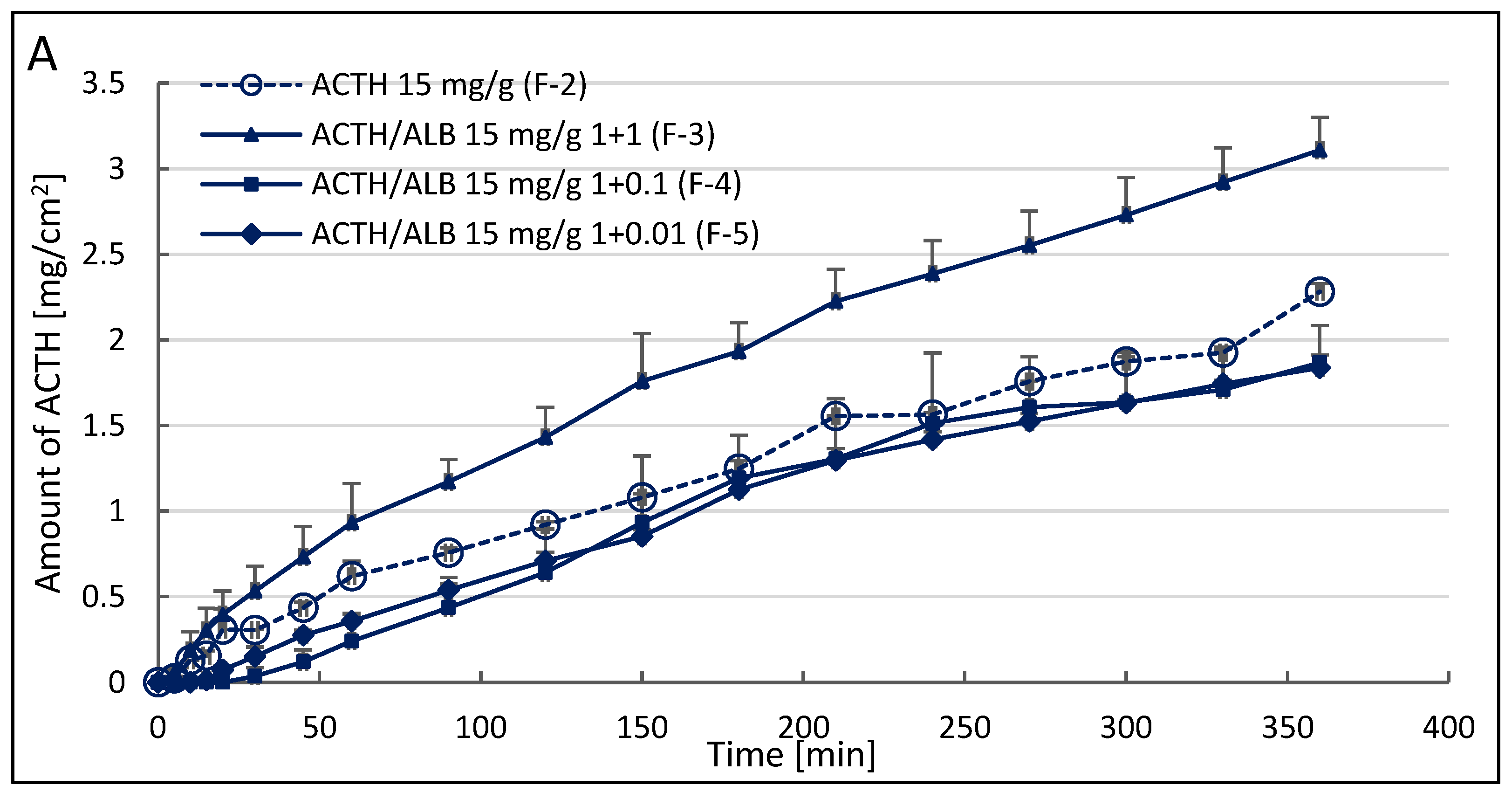
3.2. Corticotropin Release Study from the Hydrogels
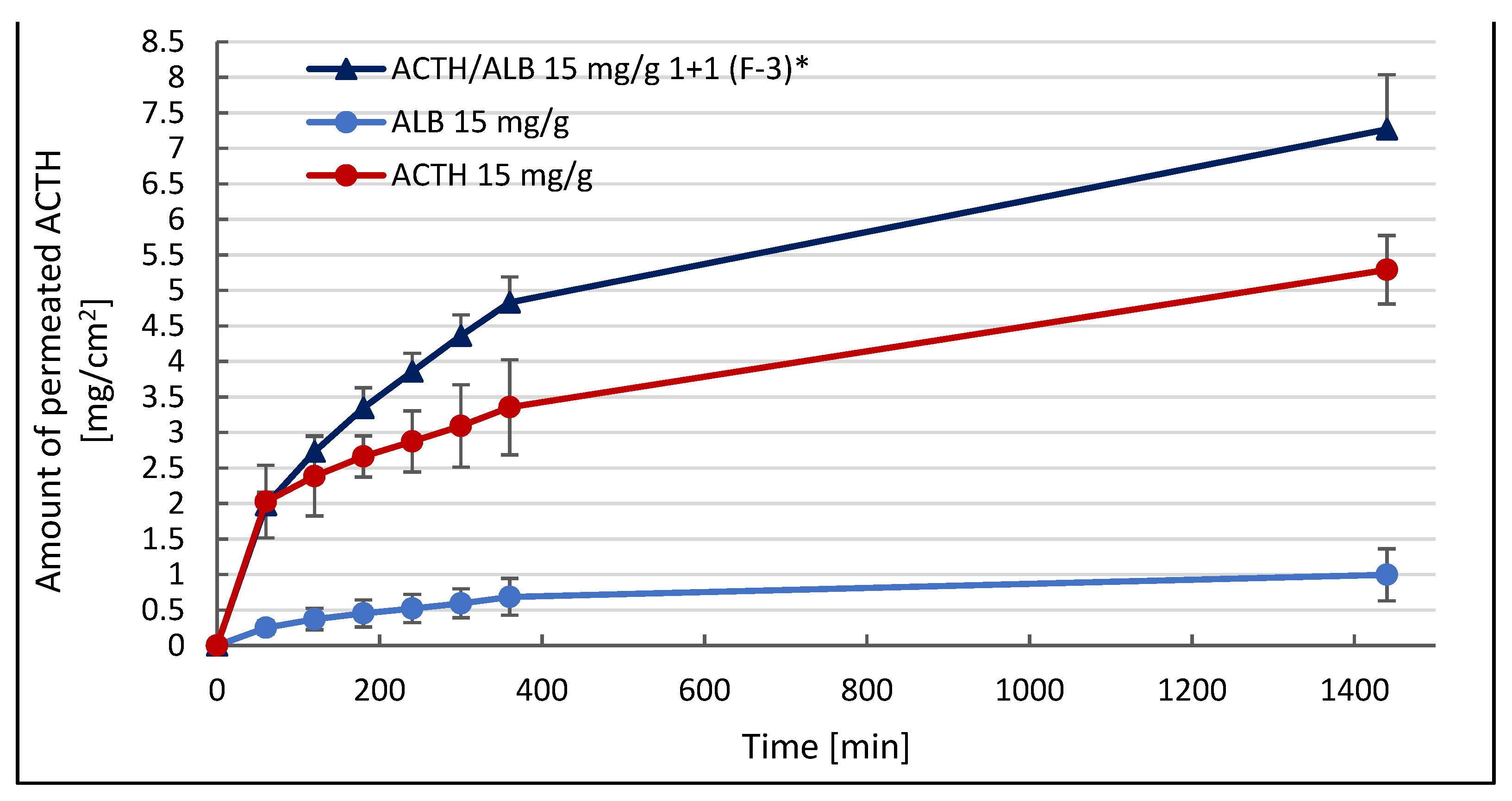
3.3. Corticotropin Permeation Study from the 1.5% Hydrogel with Addition Albumin in Stechiometric Ratio 1:1 through Porcine Skin
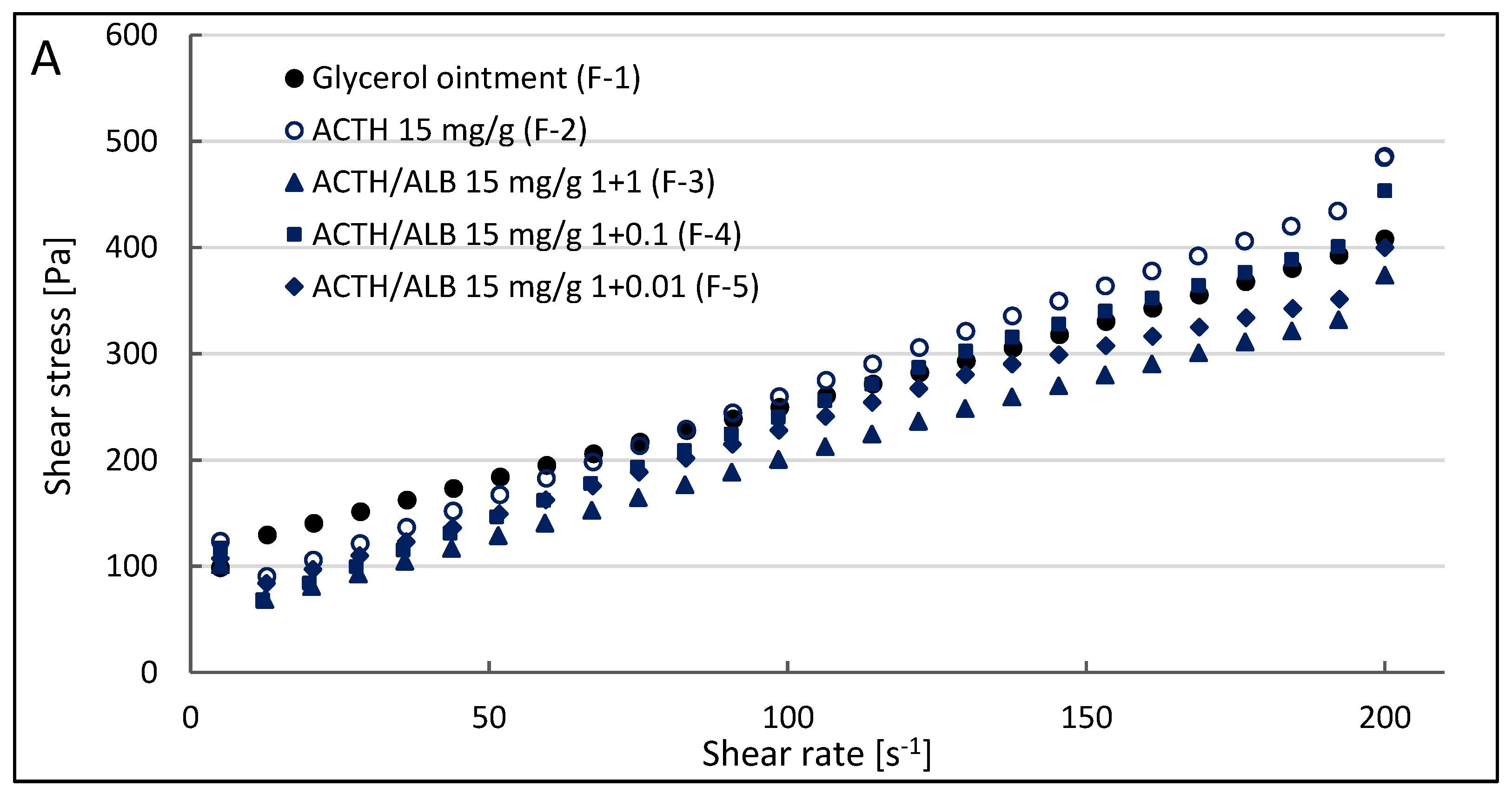
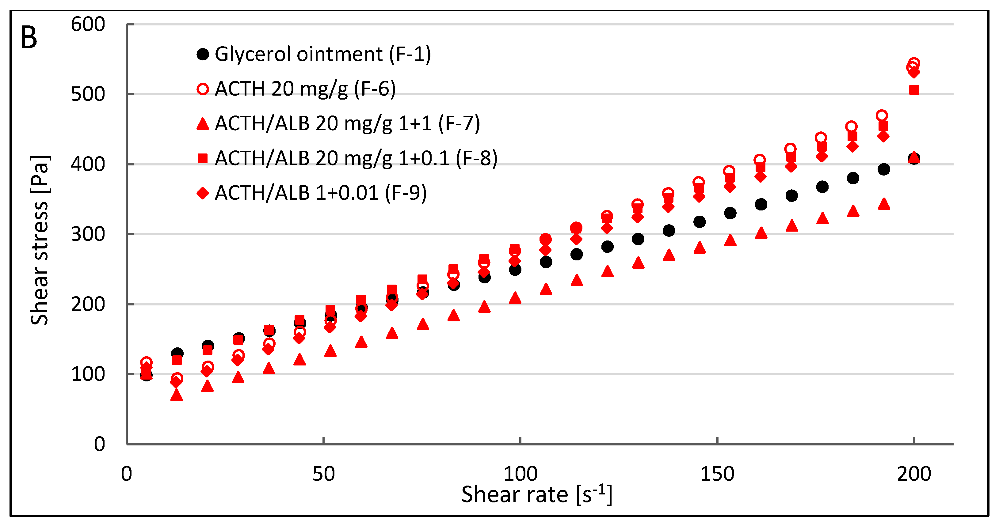
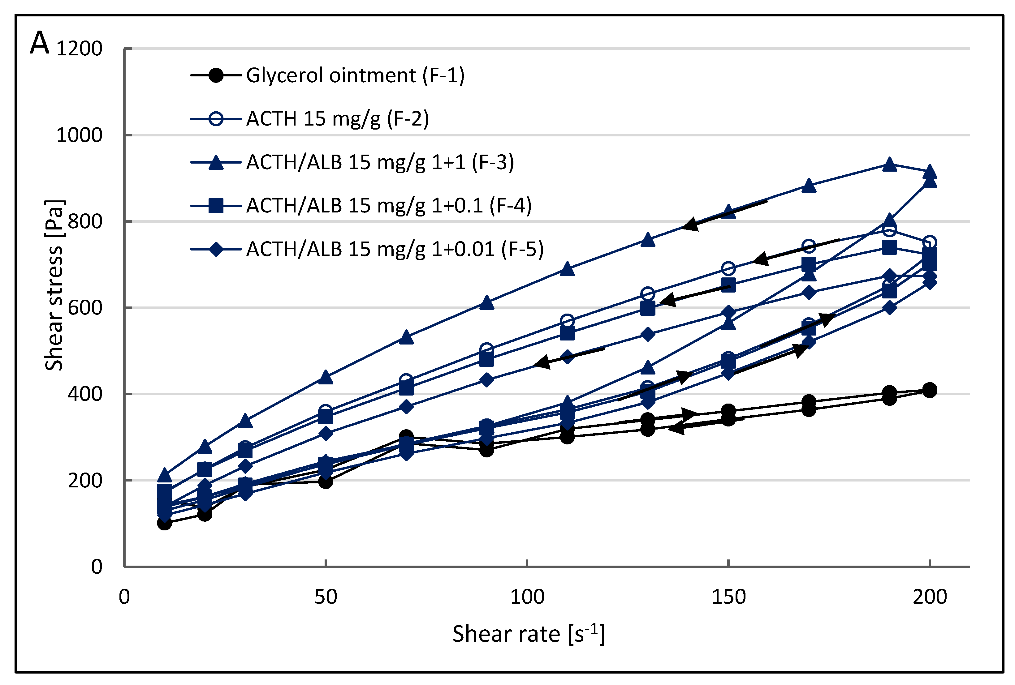
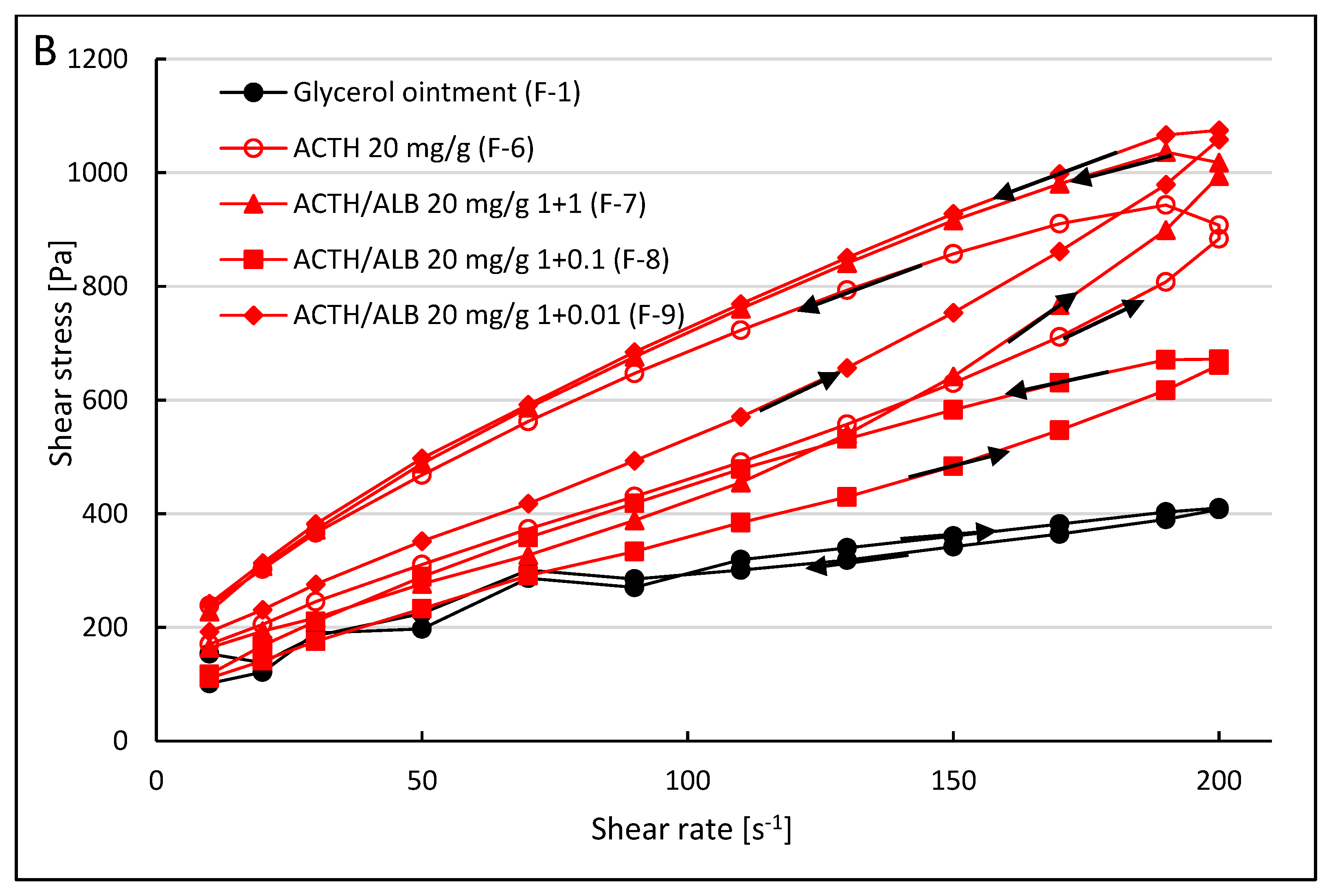
3.4. Effect of Albumin on the Rheological Properties of Prepared Hydrogels with ACTH
4. Discussion
5. Conclusions
Author Contributions
Funding
Institutional Review Board Statement
Informed Consent Statement
Data Availability Statement
Conflicts of Interest
References
- Li, N.; Larin, E.M.; Kerman, K. A Miniaturized impedimetric immunosensor for the competitive detection of adrenocrticotropic hormone. Sensor 2017, 17, 2836. [Google Scholar] [CrossRef] [Green Version]
- Clark, A.J.; Forfar, R.; Hussain, M.; Jerman, J.; McIver, E.; Taylor, D.; Chan, L. ACTH Antagonists. Front. Endocrinol. 2016, 7, 101. [Google Scholar] [CrossRef] [PubMed] [Green Version]
- Gallo-Payet, N. 60 Years of POMC: Adrenal and extra-adrenal functions of ACTH. J. Mol. Endocrinol. 2016, 56, 135–156. [Google Scholar] [CrossRef] [Green Version]
- Fleseriu, M.; Findling, J.W.; Koch, C.A.; Schlaffer, S.M.; Buchfelder, M.; Gross, C. Changes in plasma ACTH levels and corticotroph tumor size in patients with Cushing’s disease during long-term treatment with the glucocorticoid receptor antagonist mifepristone. J. Clin. Endocrinol. Metab. 2014, 99, 3718–3727. [Google Scholar] [CrossRef] [PubMed] [Green Version]
- Montero-Melendez, T. ACTH: The forgotten therapy. Semin. Immunol. 2015, 27, 216–226. [Google Scholar] [CrossRef]
- Philbin, M.; Niewoehner, J.; Wan, G.J. Clinical and Economic Evaluation of Repository Corticotropin Injection: A Narrative Literature Review of Treatment Efficacy and Healthcare Resource Utilization for Seven Key Indications. Adv. Ther. 2017, 34, 1775–1790. [Google Scholar] [CrossRef]
- Vandael, D.; Gounko, N.V. Corticotropin releasing factor-binding protein (CRF-BP) as a potential new therapeutic targetin Alzheimer’s disease and stress disorders. Transl. Psychiatry 2019, 9, 272. [Google Scholar] [CrossRef] [PubMed] [Green Version]
- Berkovich, R.; Agius, M. Mechanisms of action of ACTH in the management of relapsing forms of multiple sclerosis. Ther. Adv. Neurol. Disord. 2014, 7, 83–96. [Google Scholar] [CrossRef] [Green Version]
- Loram, L.; Culp, M.; Connolly-Strong, E. Melanocortin Peptides: Potential Targets in Systemic Lupus Erythematosus. Inflammation 2015, 38, 260–271. [Google Scholar] [CrossRef] [Green Version]
- Berkovich, R.; Bakshi, R.; Amezcua, L.; Axtell, R.C.; Cen, S.Y.; Tauhid, S.; Neema, M.; Steinman, L. Adrenocorticotropic hormone versus methylprednisolone added to interferon β in patients with multiple sclerosis experiencing breakthrough disease: A randomized, rater-blinded trial. Ther. Adv. Neurol. Disord. 2017, 10, 3–17. [Google Scholar] [CrossRef] [Green Version]
- Siemiradzka, W.; Dolińska, B.; Ryszka, F. Development and Study of Semi-Solid Preparations Containing the Model Substance Corticotropin (ACTH): Convenience Application in Neurodegenerative Diseases. Molecules 2020, 25, 1824. [Google Scholar] [CrossRef] [PubMed]
- Siemiradzka, W.; Dolińska, B.; Ryszka, F. Influence of Concentration on Release and Permeation Process of Model Peptide Substance-Corticotropin-From Semisolid Formulations. Molecules 2020, 25, 2767. [Google Scholar] [CrossRef] [PubMed]
- Wunder, A.; Muller-Ladner, U.; Stelzer, E.H.; Funk, J.; Neumann, E.; Stehle, G.; Pap, T.; Sinn, H.; Gay, S.; Fiehn, C. Albumin-based drug delivery as novel therapeutic approach for rheumatoid arthritis. J. Immunol. Methods 2003, 170, 4793–4801. [Google Scholar] [CrossRef] [Green Version]
- Tao, C.; Chuah, Y.J.; Xu, C.J.; Wang, D.A. Albumin conjugates and assemblies as versatile bio-functional additives and carriers for biomedical applications. J. Mater. Chem. B 2019, 7, 357–367. [Google Scholar] [CrossRef] [PubMed]
- Anbouhi, T.S.; Esfidvajani, E.M.; Nemati, F. Albumin binding, anticancer and antibacterial properties of synthesized zero valent iron nanoparticles. Int. J. Nanomed. 2018, 14, 243–256. [Google Scholar] [CrossRef] [Green Version]
- Mou, J.; Liu, Z.; Liu, J.; Lu, J.; Zhu, W.; Pei, D. Hydrogel containing minocycline and zinc oxide-loaded serum albumin nanopartical for periodontitis application: Preparation, characterization and evaluation. Drug Deliv. 2019, 26, 179–187. [Google Scholar] [CrossRef] [Green Version]
- Bernardi, M.; Ricci, C.S.; Zaccherini, G. Role of human albumin in the management of complications of liver cirrhosis. J. Clin. Exp. Hepatol. 2014, 4, 302–311. [Google Scholar] [CrossRef]
- Chytil, P.; Koziolova, E.; Etrych, T.; Ulbrich, K. HPMA Copolymer-Drug Conjugates with Controlled Tumor-Specific Drug Release. Macromol. Biosci. 2018, 18, 1700209. [Google Scholar] [CrossRef]
- Sun, J.H.; Ye, C.; Bai, E.H.; Zhang, L.L.; Huo, S.J.; Yu, H.H.; Xiang, S.Y.; Yu, S.Q. Co-delivery nanoparticles of doxorubicin and chloroquine for improving the anti-cancer effect in vitro. Nanotechnology 2019, 8, 085101. [Google Scholar] [CrossRef]
- Rahimizadeh, P.; Yang, S.; In Lim, S. Albumin: An Emerging Opportunity in Drug Delivery. Biotechnol. Bioprocess Eng. 2020, 25, 985–995. [Google Scholar] [CrossRef]
- Pilati, D.; Howard, K.A. Albumin-based drug designs for pharmacokinetic modulation. Expert Opin. Drug Metab. Toxicol. 2020, 16, 9. [Google Scholar] [CrossRef] [PubMed]
- Sethi, A.; Sher, M.; Akram, M.R.; Karim, S.; Khiljee, S.; Sajjad, A.; Shah, S.N.; Murtaza, G. Albumin as a drug delivery and diagnostic tool and its market approved products. Acta Pol. Pharm. 2013, 70, 597–600. [Google Scholar] [PubMed]
- Kratz, F. A clinical update of using albumin as a drug vehicle—A commentary. J. Control. Release 2014, 190, 331–336. [Google Scholar] [CrossRef] [PubMed]
- Van de Sande, L.; Cosyns, S.; Willaert, W.; Ceelen, W. Albumin-based cancer therapeutics for intraperitoneal drug delivery: A review. Drug Deliv. 2020, 27, 40–53. [Google Scholar] [CrossRef]
- Dolińska, B.; Siemiradzka, W.; Ryszka, F. Penetration of model hormones through the pericardium in simulated conditions in vivo. Biomed. Pharmacother. 2020, 127, 110113. [Google Scholar] [CrossRef]
- Office for Registration of Medicinal Products. Medical Devices and Biocidal Products, Polish Pharmacopoeia, 6th ed.; Polskie Towarzystwo Farmaceutyczne: Warsaw, Poland, 2002; p. 921. [Google Scholar]
- Office for Registration of Medicinal Products. Medical Devices and Biocidal Products, Polish Pharmacopoeia, 11th ed.; Polskie Towarzystwo Farmaceutyczne: Warsaw, Poland, 2017; Volume 3, p. 142. [Google Scholar]
- Caon, T.; Costa, A.C.; de Oliveira, M.A.; Micke, G.A.; Simoes, C.M. Evaluation of the transdermal permeation of di_erent paraben combinations through a pig ear skin model. Int. J. Pharm. 2010, 391, 1–6. [Google Scholar] [CrossRef] [PubMed]
- Arabi, S.H.; Aghelnejad, B.; Schwieger, C.; Meister, A.; Kerth, A.; Hinderberger, D. Serum albumin hydrogels in broad pH andtemperature ranges: Characterization of their self-assembled structures and nanoscopic and macroscopic properties. Biomater. Sci. 2018, 6, 478–492. [Google Scholar] [CrossRef] [PubMed] [Green Version]
- Baler, K.; Michael, R.; Szleifer, I.; Ameer, G.A. Albumin hydrogels formed by electrostatically triggered self-assembly and their drug delivery capability. Biomacromolecules 2014, 15, 3625–3633. [Google Scholar] [CrossRef] [Green Version]
- Tazhbayev, Y.; Mukashev, O.; Burkeev, M.; Kreuter, J. Hydroxyurea-Loaded Albumin Nanoparticles: Preparation, Characterization, and In Vitro Studies. Pharmaceutics 2019, 11, 410. [Google Scholar] [CrossRef] [Green Version]
- Svennebring, A. The connection between plasma protein binding and acute toxicity as determined by the LD50 value. Drug. Dev. Res. 2016, 77, 3–11. [Google Scholar] [CrossRef]
- Zhang, F.; Xue, J.; Shao, J.; Jia, L. Compilation of 222 drugs’ plasma protein binding data and guidance for study designs. Drug Discov. Today 2012, 17, 475–485. [Google Scholar] [CrossRef] [PubMed]
- Tiwari, G.; Tiwari, R.; Singh, R.; Rai, A.K. Ultra-deformable Liposomes as Flexible Nanovesicular Carrier to Penetrate Versatile Drugs Transdermally. Nanosci. Nanotechnol.-Asia 2020, 10, 12–20. [Google Scholar] [CrossRef]
- Tan, Y.L.; Ho, H.K. Navigating albumin-based nanoparticles through various drug delivery routes. Drug Discov. Today 2018, 23, 1108–1114. [Google Scholar] [CrossRef] [PubMed]
- Tomoda, K.; Watanabe, A.; Suzuki, K.; Inagi, T.; Terada, H.; Makino, K. Enhanced transdermal permeability of estradiol using combination of PLGA nanoparticle system and iontophoresis. Colloids Surf. B Biointerfaces 2012, 97, 84–89. [Google Scholar] [CrossRef]
- Tomoda, K.; Tarashima, H.; Suzuki, K.; Inagi, T.; Terada, H.; Makino, K. Enhanced transdermal delivery of indomethacin using combination of PLGA nanoparticles and iontophoresis in vivo. Colloids Surf. B Biointerfaces 2012, 92, 50–54. [Google Scholar] [CrossRef]
- Pal, D.; Nayak, A.K. Nanotechnology for targeted delivery in cancer therapeutics. Int. J. Pharm. Sci. Rev. Res. 2010, 1, 1–7. [Google Scholar]
- Malakar, J.; Ghosh, A.; Basu, A.; Nayak, A.K. Nanotechnology a promising carrier for intracellular drug delivery system. Int. Res. J. Pharm. 2012, 3, 36. [Google Scholar]
- Pawar, H.; Douroumis, D.; Boateng, J.S. Preparation and optimization of PMMA-chitosan-PEG nanoparticles for oral drug delivery. Colloids Surf. B Biointerfaces 2012, 90, 102. [Google Scholar] [CrossRef]
- Jana, S.; Saha, A.; Nayak, A.K.; Sen, K.K.; Basu, S.K. Aceclofenac-loaded chitosan-tamarind seed polysaccharide interpenetrating polymeric network microparticles. Colloids Surf. B Biointerfaces 2013, 105, 303–309. [Google Scholar] [CrossRef]
- Jana, S.; Das, A.; Nayak, A.K.; Sen, K.K.; Basu, S.K. Aceclofenac-loaded unsaturated esterified alginate/gellan gum microspheres: In vitro and in vivo assessment. Int. J. Biol. Macromol. 2013, 57, 129–137. [Google Scholar] [CrossRef]
- Jana, S.; Manna, S.; Nayak, A.K.; Sen, K.K.; Basu, S.K. Carbopol gel containing chitosan-egg albumin nanoparticles for transdermal aceclofenac delivery. Colloids and Surfaces B Biointerfaces 2014, 114, 36–44. [Google Scholar] [CrossRef] [PubMed]
- Shokri, N.; Javar, H.A. Comparison of Calcium Phosphate and Zinc Oxide Nanoparticles as Dermal Penetration Enhancers for Albumin. Indian J. Pharm. Sci. 2015, 77, 694–704. [Google Scholar] [CrossRef] [PubMed]
- Lei, C.; Liu, X.R.; Chen, Q.B.; Li, Y.; Zhou, J.L.; Zhou, L.Y.; Tao Zou, T. Hyaluronic acid and albumin based nanoparticles for drug delivery. J. Control. Release 2021, 331, 416–433. [Google Scholar] [CrossRef]
- Quinlan, G.J.; Martin, G.S.; Evans, T.W. Albumin: Biochemical properties and therapeutic potential. Hepatology 2005, 41, 1211–1219. [Google Scholar] [CrossRef] [PubMed]
- Hobbs, S.K.; Monsky, W.L.; Yuan, F.; Roberts, W.G.; Griffith, L.; Torchilin, V.P. Regulation of transport pathways in tumor vessels: Role of tumor type and microenvironment. Proc. Natl. Acad. Sci. USA 1998, 95, 4607–4612. [Google Scholar] [CrossRef] [Green Version]
- Shen, Z.Y.; Li, Y.; Kohamab, K.; Oneilla, B.; Bi, J.X. Improved drug targeting of cancer cells by utilizing actively targetable folic acid-conjugated albumin nanospheres. Pharmacol. Res. 2011, 63, 51–58. [Google Scholar]
- Chacon, M.; Molpeceres, J.; Berges, L.; Guzman, M.; Aberturas, M.R. Stability and freeze-drying of cyclosporine loaded poly [D, L lactide-glycolide] carriers. Eur. J. Pharm. Sci. 1999, 8, 99–107. [Google Scholar] [CrossRef]
- Martins, M.; Azoia, N.G.; Shimanovich, U.; Matam´a, T.; Gomes, A.C.; Silva, C.; Cavaco-Paulo, A. Design of novel BSA/hyaluronic acid nanodispersions for transdermal pharma purposes. Mol. Pharm. 2014, 11, 1479–1488. [Google Scholar] [CrossRef] [Green Version]
- Dolińska, B.; Siemiradzka, W.; Ryszka, F. Effectiveness of absorption and passage through the small intestine, as a model of oral prolactin administration. Biomed. Pharmacother. 2019, 120, 109515. [Google Scholar] [CrossRef]


| Ingredient | ACTH (mg) | Albumin (mg) | Glycerol Hydrogel |
|---|---|---|---|
| Formulation | |||
| F-1 | - | - | + |
| F-2 | 150.0 | - | + |
| F-3 | 150.0 | 150.0 | + |
| F-4 | 150.0 | 15.0 | + |
| F-5 | 150.0 | 1.5 | + |
| F-6 | 200.0 | - | + |
| F-7 | 200.0 | 200.0 | + |
| F-8 | 200.0 | 20.0 | + |
| F-9 | 200.0 | 2.0 | + |
| F-10 | - | 150.0 | + |
| Formulation | pH ± SD |
|---|---|
| F-1 | 4.955 ± 0.021 |
| F-2 | 5.990 ± 0.028 |
| F-3 | 5.485 ± 0.007 |
| F-4 | 5.425 ± 0.021 |
| F-5 | 5.255 ± 0.035 |
| F-6 | 6.240 ± 0.014 |
| F-7 | 5.605 ± 0.007 |
| F-8 | 5.510 ± 0.014 |
| F-9 | 6.135 ± 0.021 |
| Formulation | First Order | Higuchi Model | Korsmeyer-Peppas Model | Weibull Method |
|---|---|---|---|---|
| Regression Coefficient R2 | ||||
| F-2 (15 mg/g ACTH) | 0.9845 | 0.9838 | 0.9504 | 0.9630 |
| F-3 (15 mg/g ACTH + 15 mg/g ALB) | 0.9780 | 0.9965 | 0.9466 | 0.9710 |
| F-4 (15 mg/g ACTH + 1.5 mg/g ALB) | 0.9782 | 0.9973 | 0.9458 | 0.9610 |
| F-5 (15 mg/g ACTH + 0.15 mg/g ALB) | 0.9886 | 0.9906 | 0.9227 | 0.9400 |
| F-6 (20 mg/g ACTH) | 0.9736 | 0.9922 | 0.9804 | 0.9860 |
| F-7 (20 mg/g ACTH + 20 mg/g ALB) | 0.9923 | 0.9888 | 0.9085 | 0.9710 |
| F-8 (20 mg/g ACTH + 2.0 mg/g ALB) | 0.9898 | 0.9908 | 0.9505 | 0.9580 |
| F-9 (20 mg/g ACTH + 0.2 mg/g ALB) | 0.9857 | 0.9824 | 0.9129 | 0.9580 |
| Formulation | Average Release Rate (mg/cm2/min1/2) ± SD | R2 | Formulation | Average Release Rate (mg/cm2/min1/2) ± SD | R2 |
|---|---|---|---|---|---|
| F-2 (15) | 0.120 ± 0.003 | 0.9838 | F-6 (20) | 0.147 ± 0.008 | 0.9922 |
| F-3 (15 1:1) | 0.164 ± 0.010 * | 0.9965 | F-7 (20 1:1) | 0.168 ± 0.005 ** | 0.9888 |
| F-4 (15 1:0.1) | 0.098 ± 0.005 * | 0.9873 | F-8 (20 1:0.1) | 0.104 ± 0.003 ** | 0.9849 |
| F-5 (15 1: 0.01) | 0.097 ± 0.004 * | 0.9906 | F-9 (20 1:0.01) | 0.093 ± 0.004 ** | 0.9615 |
| Formulation | Zero Order | First Order | Higuchi Model | Korsmeyer-Peppas Model | Average Permeation Rate (mg/cm2/min1/2) ± SD | AUC |
|---|---|---|---|---|---|---|
| Regression Coefficient R2 | ||||||
| F-3 (ACTH + ALB 1:1) | 0.73 | 0.90 | 0.96 | 0.98 | 0.192 ± 0.020 * | 7736.19 |
| ACTH 15 mg/g | 0.73 | 0.76 | 0.94 | 0.99 | 0.139 ± 0.008 | 5623.76 |
| ALB 15 mg/g | 0.73 | 0.74 | 0.89 | 0.97 | 0.026 ± 0.010 * | 1030.35 |
| Formulation | Shear Rate | |||
|---|---|---|---|---|
| 15 s−1 | 30 s−1 | |||
| Shear Stress [Pa] | Viscosity [Pa·s] | Shear Stress [Pa] | Viscosity [Pa·s] | |
| F-1 | 90.10 ± 11.32 | 6.01 ± 0.76 | 182.52 ± 20.81 | 6.08 ± 0.70 |
| F-2 | 113.89 ± 2.77 * | 7.59 ± 0.19 * | 155.80 ± 7.08 * | 5.19 ± 0.24 * |
| F-3 | 73.66 ± 0.47 * | 4.91 ± 0.03 * | 97.93 ± 5.36 * | 3.26 ± 0.18 * |
| F-4 | 113.96 ± 2.31 * | 7.60 ± 0.15 * | 149.40 ± 5.87 * | 4.98 ± 0.20 * |
| F-5 | 94.24 ± 1.91 | 6.28 ± 0.13 | 127.42 ± 5.01 * | 4.25 ± 0.17 * |
| F-6 | 137.71 ± 2.51 * | 9.20 ± 0.17 * | 176.17 ± 6.17 | 5.87 ± 0.21 |
| F-7 | 91.85 ± 1.74 | 6.12 ± 0.12 | 117.52 ± 4.65 * | 3.92 ± 0.16 * |
| F-8 | 94.44 ± 1.76 | 6.30 ± 0.12 | 143.42 ± 7.02 * | 4.78 ± 0.23 * |
| F-9 | 109.82 ± 2.51 * | 7.32 ± 0.17 * | 150.25 ± 4.24 * | 5.02 ± 0.14 * |
Publisher’s Note: MDPI stays neutral with regard to jurisdictional claims in published maps and institutional affiliations. |
© 2021 by the authors. Licensee MDPI, Basel, Switzerland. This article is an open access article distributed under the terms and conditions of the Creative Commons Attribution (CC BY) license (https://creativecommons.org/licenses/by/4.0/).
Share and Cite
Siemiradzka, W.; Dolińska, B.; Ryszka, F. Modelling and Control of Corticotropin Permeation from Hydrogels across a Natural Membrane in the Presence of Albumin. Processes 2021, 9, 1674. https://doi.org/10.3390/pr9091674
Siemiradzka W, Dolińska B, Ryszka F. Modelling and Control of Corticotropin Permeation from Hydrogels across a Natural Membrane in the Presence of Albumin. Processes. 2021; 9(9):1674. https://doi.org/10.3390/pr9091674
Chicago/Turabian StyleSiemiradzka, Wioletta, Barbara Dolińska, and Florian Ryszka. 2021. "Modelling and Control of Corticotropin Permeation from Hydrogels across a Natural Membrane in the Presence of Albumin" Processes 9, no. 9: 1674. https://doi.org/10.3390/pr9091674
APA StyleSiemiradzka, W., Dolińska, B., & Ryszka, F. (2021). Modelling and Control of Corticotropin Permeation from Hydrogels across a Natural Membrane in the Presence of Albumin. Processes, 9(9), 1674. https://doi.org/10.3390/pr9091674