Review

Exploration of Active Site-Directed Plasmin Inhibitors: Beyond Tranexamic Acid

1
Faculty of Pharmaceutical Sciences, Kobe Gakuin University, Chuo-ku, Kobe 650-8586, Japan
2
Cooperative Research Center for Life Sciences, Kobe Gakuin University, Chuo-ku, Kobe 650-8586, Japan
*
Author to whom correspondence should be addressed.
Processes 2021, 9(2), 329; https://doi.org/10.3390/pr9020329
Submission received: 12 January 2021 / Revised: 6 February 2021 / Accepted: 7 February 2021 / Published: 11 February 2021
(This article belongs to the Special Issue Advances of Peptide Engineering)

Abstract

:
Plasmin (Plm), a trypsin-like serine protease, is responsible for fibrinolysis pathway and pathologic events, such as angiogenesis, tumor invasion, and metastasis, and alters the expression of cytokines. A growing body of data indicates that a Plm inhibitor is a potential candidate as an anti-inflammatory and anti-cancer agent. A class of active site-directed plasmin inhibitors containing tranexamic acid residue has been designed. As evidenced by docking studies, the inhibitor binds to the active site not to the lysine binding site (LBS) in plasmin, thus preventing plasmin from digesting the substrate. Further optimization of the series, concerning both activity and selectivity, led to the second generation of inhibitors. This review focuses on the Plm inhibitory activity-structure relationship of Plm inhibitors with the goal of realizing their design and clinical application.

1. Introduction

Plasmin (Plm), a trypsin-like serine protease, plays a critical role in the fibrinolysis pathway, in which Plm removes intra- and extra-vascularly formed thrombi by degrading fibrin clots. Under normal conditions, Plm circulates in the blood in its zymogen form, plasminogen (Plg), which is a single-chain glycoprotein of 92 kDa [1]. The activation process of Plg to Plm takes place on the surface of fibrin. The C-terminal area of fibrin monomers that are rich in lysine residues facilitates binding to the lysine binding sites (LBS) on the kringles domain of both Plg and tissue Plg activator (tPA) to form fibrin-Plg-tPA complex, which promotes the Plg activation process to generate Plm, having two-chains linked by disulfide bonds [1,2,3,4,5].
On the other hand, a growing consensus indicates that Plm participates in a number of physical processes as well as its dominant role in fibrin cleavage. When Plg is activated by urokinase Plg activator (uPA) on a cell surface, Plm facilitates degradation of the extracellular matrix (ECM) to induce tissue remodeling [6,7,8], cell inversion, and metastasis [9,10], and other actions, including the activation of the matrix metalloproteases (MMPs). Plm can also bind to a variety of cells, such as monocytes, induce processing of proinflammatory cytokines, and alter the expression of cytokines [11,12]. In fact, we showed that Plm inhibitor, N-(trans-4-aminomethylcyclohexanecarbonyl: TXA)-L-Tyr (OPicolyl)-NH-octyl (YO-2) [13], suppressed the growth of human tumor xenografts and reduced MMP-9-dependent T-cell lymphoid tumor growth [14]. In order to study the precise role of Plm under a disease state, development of selective Plm inhibitors is required. Plm inhibitors may also be potential candidates not only as anti-fibrinolytic agents but also as anticancer and anti-inflammatory agents.

2. Active Site (AS) and LBS-Directed Inhibitors

Today, in the clinical treatment of hyper-fibrinolysis-associated bleeding events, two kinds of Plm inhibitors are applied: aprotinin [15] and lysine analogues, ε-aminocaproic acid (EACA) [16] and tranexamic acid (TXA) [17]. Aprotinin is a Kunitz-type serine protease inhibitor against Plm; however, its clinical use has been limited due to several side effects. In contrast, EACA and TXA are widely used. The latter is 10-fold more potent than the former, but high doses of TXA are still necessary (typical efficient doses in human: 1–1.5 g given 3–4 times per day). Recent Clinical Randomisation of an Antifibrinolytic in Significant Haemorrhage (CRASH)-2 trials demonstrated that the survival was increased by administration of TXA to a large number of patients with traumatic bleeding [18].
Both inhibitors were discovered by Okamoto et al. from the 1950s to the 1960s. The two agents are surrogates of the amino acid, lysine, and bind to the lysine binding site (LBS) on the kringle domains of Plg, but not to the active site (AS) of Plm. Fibrinolysis is effectively suppressed, as seen in enzyme assays using TXA (IC50 = 50 μM), while fibrinogenolysis and amidolysis are hardly suppressed (IC50 = 10 mM and Ki = 40 mM, respectively) [19]. This means that, despite the presence of TXA, AS is capable of degrading both fibrinogen and a small substrate. The alternative class of Plm inhibitors, which block the digestion of fibrinogen and a small substrate, may also be required in the clinic. Thus, we have focused on the design of AS-directed Plm inhibitors with the objective of obtaining potent and selective inhibitors to prevent not only fibrinolysis but also amidolysis and fibrinogenolysis.
In this review, development of Plm inhibitors, YO-2 and its derivatives, are described. They showed the Plm inhibitory activity against a chromogenic substrate, demonstrating that they interacted to AS not LBS, in spite of having a TXA moiety in the molecule. Other than YO-2 derivatives, cyclohexane-based inhibitors [20,21], CU2010 [22] and its macrocyclic analogues [23,24], nitrile warheaded inhibitors [25,26], peptide aldehyde-based inhibitors [27], etc., are known as AS-directed Plm inhibitors. This review highlights not only functional but also structural aspects of the YO-related compounds.

3. Design and Synthesis of Substrate-Based Inhibitors

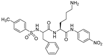
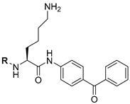
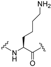
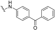
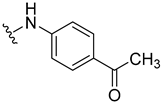
In order to design novel Plm inhibitors, we focused on the structure of the chromogenic substrate, H-D-Ile-Phe-Lys-pNA (1) [28]. We have reported that 1 mildly inhibits Plm fibrinolytic activity (IC50 = 180 μM), although 1 itself is a good substrate for Plm (Km = 20 μM). First of all, we prepared H-Lys-pNA derivatives with substitution at the N-terminus of the Lys residue and screened Plm inhibition; the results are summarized in Table 1 [28]. Interestingly, Tos-Lys-pNA (3) was not cleaved by Plm, but did inhibit amidolytic activity (IC50 = 700 μM against S-2251) as well as fibrinolytic activity (IC50 = 780 μM). For the purpose of obtaining lysine derivatives that are not susceptible to Plm, we replaced the p-nitroanilide group with more hydrophobic amides such as 4-benzylpiperidineamide (BPP), 4-benzoylanilide (BZA), or 4-acethylanilide (ACA). Their effects on Plm are summarized in Table 2 [28]; replacement increased the Plm inhibition not only of amidolytic activity by some 3- to 5-fold but also of fibrinolytic activity by some 5-fold.
Tos-Lys-BZA (7: IC50 = 140 and 150 μM for S-2251 and fibrin, respectively) hydrolyzed a small chromogenic substrate, S-2251, more strongly compared with TXA (IC50 = 75,000 and 60 μM for S-2251 and fibrin, respectively), demonstrating that 7 was an AS-directed Plm inhibitor rather than an LBS-directed inhibitor.

4. Substitution of P2, P1 and P1′ Residues

4.1. Incorporation of TXA

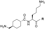
On referring to the structure of 7, each part of the Tos, Lys, and BZA moieties was replaced with various moieties: Lys, EACA, TXA, and cis-4-aminocyclohexycarboxyric acid instead of Tos (Table 3); D-Lys and ornithine instead of Lys (Table 4); and ACA, 4-benzoylphenyl ester, BPP instead of BZA (Table 5). In this series (Table 3, Table 4 and Table 5) [29], H-TXA-Lys-BZA (13: IC50 = 15 and 6.1 μM for S-2251 and fibrin, respectively) and H-TXA-Lys-ACA (17: IC50 = 39 and 9.3 μM for S-2251 and fibrin, respectively) exhibited strong Plm inhibition. The main question that arises from this result is: which amino group (TXA or Lys in 13 and 17) interacts with Asp residue at the S1 site of Plm? To clarify this point, an amino group of TXA and Lys in H-TXA-Lys-4-benzoylphenoxymethyl ketone (20: IC50 = 660 and 200 μM for S-2251 and fibrin, respectively) molecule were individually masked by a benzyloxycarbonyl (Cbz) to give Cbz-TXA-Lys-benzoylphenoxymethyl ketone (21) and H-TXA-Lys(Cbz)-benzoylphenoxymethyl ketone (22) (Figure 1). The former derivative was significantly less active than the parent molecule, while the latter derivative retained inhibitory activity (IC50 = 200 and 60 μM for S-2251 and fibrin, respectively). Those findings [29] implied that the free amine of TXA, not Lys, interacted with the Asp at the S1 site, and the new series of Plm inhibitors, having a sequence of (P1-P1′-P2′), in which P1 was a TXA residue, was proposed.

4.2. Switch from Lys to Phe/Tyr

Starting from H-TXA-Lys-ACA (17), Lys and ACA moieties were converted to various moieties; Phe and Tyr (2-BrZ) were used as an alternative to Lys and 4-carboxymethylanilide and octylamide were used as an alternative to ACA. The structure-inhibitory activity relationships are summarized in Table 6 [30]. H-TXA-Phe-ACA (23: IC50 = 36 and 21 μM for S-2251 and fibrin, respectively) shows Plm inhibition corresponding to that of 17. This means that the Lys residue is replaceable by a Phe residue. The enhanced inhibition on compounds 24, 25, and 26 indicated that an additional phenyl ring of Bzl, Z, and Br-Z may be acceptable at the S1’ site of Plm. In the new series, H-TXA-Tyr (2-BrZ)-ACA (26) was the most potent Plm inhibitor; however, it inhibited not only Plm (IC50 = 0.23 μM for S-2251), but also plasma kallikrein (PK: IC50 = 0.37 μM), uPA (IC50 = 43 μM), and thrombin (Thr: IC50 = 63 μM).

4.3. Discovery of H-TXA-Tyr(OPic)-NH-Octyl (36, YO-2)

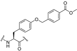
To improve selectivity for Plm, the ACA moiety was modified with branched and unbranched alkylamides, p-alkylanilides, pyridineanilides, and pyridinealkylanilides [31]. As far as the P2’ substituent is concerned, increasing the alkyl chain length resulted in enhanced inhibitory activity against Plm but reduced inhibition for PK and uPA (compounds 27–29: Table 7). Compounds 27–29, bearing unbranched alkylamides, strongly and selectively inhibited Plm, but compound 30, having a branched alkylamide, strongly inhibited Plm as well as PK. Furthermore, the introduction of an aromatic ring into the P2′ enhanced the PK inhibition rather than the Plm inhibition (compounds 31–34: Table 7), meaning that the bulkiness of S2′ sub-sites of Plm and PK was quite different. The above compounds had low solubility in an aqueous solution due to the hydrophobic property of the 2-BrZ moiety. To overcome this disadvantage, the 2-BrZ moiety was substituted with 4-methoxycarboylbenzl, 4-pyridylmethyl (Pic) and 4-pyridineacethyloxycarbonyl to yield compounds 35–37 (see Table 8) [13]. All things considered, we decided to use compound 36 (YO-2) as a lead of Plm specific inhibitor; 36 inhibited Plm (IC50 = 0.53 μM) but hardly affected PK (IC50 = 30 μM) and Thr (IC50 > 400 μM). However, 36 also retained uPA inhibition (IC50 = 5.3 μM), meaning that 36 lost specificity (Plm/uPA) to a limited but small degree.

5. Improvement of Specificity (Plm/uPA)

For the further development of specific inhibitors, docking experiments with the μPlm-36 (YO-2) complexes were performed, and the results are as follows [32]: the TXA moiety was located in the S1 site where its amino group formed polar interaction with the carboxyl group of Asp; the Tyr(OPic) laid along the hydrophobic S2 wall and the extra S2 site in an extend conformation; the octylamide moiety was oriented toward the hydrophobic region in the S1′ pocket [32]. The docking positions suggested that 36 (YO-2) with the μPlm acquired a tripod shape (P2-P1-P1′), in which polar interactions between the amino moiety of TXA and the side chain of Asp in the S1 pocket and with additional hydrophobic contacts at the S2, extra S2, and S1′ pockets produced the high inhibitory activity of 36 (YO-2).
A number of trypsin-like serine proteases have a common Asp residue in the bottom of the S1 pocket; however, there are also other secondary sites located from the S2 to S3/S4 of the sub-sites. Structural differences at the secondary sites among those enzymes do exist. In Plm, the secondary binding pocket, S2/S3, is widely open due to the absence of the loop segment 95–100 (corresponding to the chymotrypsin-numbering). On the other hand, in uPA, the size of S2/S4 is greatly reduced because of an insertion of the loop segment 95–100, which obstructs both sites. Regarding the differences within the secondary sites, S2/S3 sub-site, Saupe and Steinmetzer designed the macrocyclic CU2010 analogues, which occupies the larger binding groove apart from the primary binding pocket to specifically inhibit Plm [23,24]. Similarly, extension of the hydrophobic substituents in the Tyr residue of 36 may generate Plm specific inhibitors.
Based on the above concept, the P2 residues were explored [33]. All compounds (38–41) having the hydrophobic extension on P2 residue inhibited Plm with IC50 values in the μM range (0.22–0.75 μM), while inhibiting uPA more moderately (IC50 values 77 to >200 μM) (Table 9). Compound 39, containing an O-(quinolin-2-yl)methyl moiety, exhibited the highest and most Plm selective inhibition (IC50 = 0.22 and 77 μM for Plm and uPA,
Further, our docking studies with the μPlm-39 complexes revealed that 39 bound with μPlm in a similar manner to 36 (YO-2): the TXA inserted into the bottom of the S1 site; the quinoline ring lay on the hydrophobic region of the S2 and extra S2 groove; the octyl residue contributed to the additional hydrophobic interactions [33].

6. X-ray Crystal Structures of 35 and 39 in the Complex with μPlm

The co-crystal structures of 39 in the complex with μPlm [34] revealed an unexpected binding mode in which the TXA residue goes straight into the primary binding site (S1 site). In addition, the double aromatic rings occupy other secondary binding sites (the S1′ and S3′ sites) in particular to make extensive interactions at the S3′subsite where the pyridine ring of the quinolin moiety takes a perfect face-to-edge CH- stacking with Phe587. A larger P3′ residue would be acceptable by the S3′ site of Plm. Additionally, a number of different possible binding forms between 39 and Lys607 are observed by the electron density map. Surprisingly, the hydrophobic octyl residue has no interaction with the S2′ pocket; on the contrary, it point away from the surface of the protease. Although 36 (YO-2) with μPlm reveals a similar binding mode to 39 at the primary binding site, the pyridine ring of the picolyl moiety forms an imperfect face-to-face-stacking with Phe587 [34]. Compared with amino acid sequence of Plm and uPA, first, Phe587 is replaced with Val181 in uPA and, second, the equivalent position to Lys607 is Asp208 in uPA. Ruby et al. ague that those replacements may interfere with the binding of 39 to reduce the uPA inhibitory activity (Figure 2).

7. Challenge for Application beyond TXA

The data that 36 suppresses matrix metalloproteinase 9-mediated cytokine release to protect colitis in mice [35] and prevents macrophage activation syndrome in a murine model [36] are discussed relative to the various functions of Plm. However, the biological properties of 39 have not been determined.
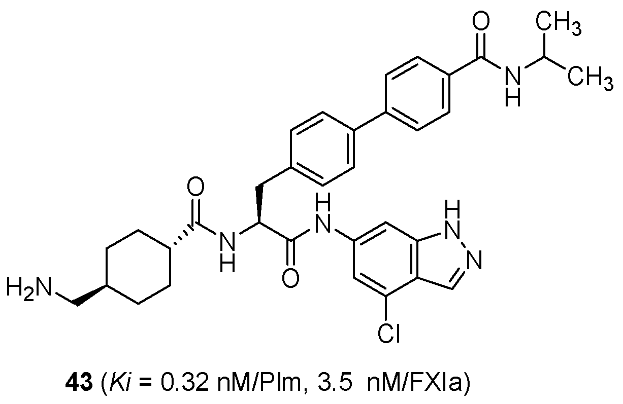
Independently, Bristol-Myers Squibb (BMS) reported the discovery of compound 40 [37], which contains TXA and phenylalanine (Phe) residues, comparable to that of 36 and 39. The dual inhibition of Plm and FXa was shown for 40 (Ki = 8.4 and 0.0003 μM for Plm and FXa, respectively) [37,38]. Based on the structure of 40, the development of orally bioavailable FXIa inhibitors (41, 42) is progressing at BMS [39,40] (Figure 3). Bayer AG investigated other analogues of Plm inhibitors containing both TXA and side-chain extended Phe residues. For example, compound 43 elongated at the para position of Phe and at the C-terminal heterocycle moiety and exhibited dual inhibition (IC50 = 0.32 and 3.5 nM for Plm and FXIa, respectively) [41] (Figure 4). Steinmetzer et al. discussed the similar molecular alteration in their review [42]. Bayer AG derivatives are currently being evaluated for their pharmacokinetics and pharmacodynamics properties, and for safety for eventual application in antithrombotic therapy.

8. Conclusions

To seek active site-directed Plm inhibitors, substrate H-D-Ile-Phe-Lys-pNA (1) was engineered into H-TXA-Tyr(OPic)-NH-Octyl (36, YO-2), which inhibited Plm (IC50 = 0.53 μM) and uPA (IC50 = 5.3 μM), but only weakly inhibited PK (IC50 = 30 μM) and Thr (IC50 > 400 μM). Following various explorative studies, the TXA residue (as a primary contacting moiety) and Tyr(OPic) residue (as a secondary contacting moiety) led to the development of 36. Considering the difference of the secondary binding site between Plm and uPA, 36 was further engineered to H-TXA-Tyr[O-(quinolin-2-yl)methyl]-NH-Octyl (39), which showed similar inhibitory activity to that of 36 with a significant improvement in selectivity (Plm/uPA) by 35-fold. The crystal structures of 36 and 39 disclose that the TXA residue in both 36 and 39 interacts to the primary site, the S1 site, as well as to the secondary sites, S1′, S2′, and S3′. Those crystal structures might provide comprehension for a rational approach to developing new compounds.

Author Contributions

Conceptualization, Y.T. and Y.O.; analysis and visualization of co-crystal structure, K.H. (Koushi Hidaka); writing—review and editing, Y.T., with the assistance of K.H. (Koushi Hidaka); review and editing, Y.O.; K.H. (Koushi Hidaka) and K.H. (Keiko Hojo). All authors have read and agreed to the published version of the manuscript.

Funding

This research was funded by the Ministry of Education, Science, Sports, and Culture of Japan, Grant-in-Aid of Scientific Research ”C, 25460164” and Grant-in-Aid of Scientific Research “C, 16K08333”.

Institutional Review Board Statement

Not applicable.

Informed Consent Statement

Not applicable.

Acknowledgments

The work was supported by Grant-in-Aid of Scientific Research (C, 25460164/2013-2015) and Grant-in-Aid of Scientific Research (C, 16K08333/2016-2018) from the Ministry of Education, Science, Sports, and Culture of Japan. The authors express deep thanks to Lawrence H. Lazarus for his kind help in the preparation of the manuscript.

Conflicts of Interest

The authors declare no conflict of interest.

References

  1. Bachmann, F. The Plasminogen-Plasmin Enzyme System (Chapter 84). Hemostasis and Thrombosis: Basic Principles and Clinical Practice, 3rd ed.; Colman, R.W., Hirsh, J., Marder, V.J., Zalzman, E.W., Eds.; Lippincott Company: Philadelphia, PA, USA, 1994; pp. 1592–1600. [Google Scholar]
  2. Angles-Cano, E. Overview on fibrinolysis: Plasminogen activation pathway on fibrin and cell surface. Chem. Phys. Lipids 1994, 68, 353–362. [Google Scholar] [CrossRef]
  3. Hoylaerts, M.; Rijken, D.C.; Lijinen, H.R.; Collen, D. Kinetics of the activation of plasminogen by human tissue plasminogen activator. Role of fibrin. J. Biol. Chem. 1982, 257, 2912–2919. [Google Scholar] [CrossRef]
  4. Lerch, P.G.; Rickli, E.E.; Lergier, W.; Gillessen, D. Localization of individual lysine-binding regions in human plasminogen and investigation on their complex-forming properties. Eur. J. Biochem. 1980, 107, 7–13. [Google Scholar] [CrossRef]
  5. Lijnen, H.R.; Van Hoef, B.; Collen, D. On the molecular interactions between fibrin, tissue-plasminogen activator and plasminogen. Thromb. Res. Suppl. 1990, 10, 45–54. [Google Scholar] [CrossRef]
  6. Deryugina, E.I.; Quigley, J.P. Cell surface remodeling by plasmin: A new function for an old enzyme. J. Biomed. Biotechnol. 2012, 2012, 564259. [Google Scholar] [CrossRef] [Green Version]
  7. Schaller, J.; Gerber, S.S. The plasmin-antiplasmin system, Structural and functional aspects. Cell Mol. Life Sci. 2011, 68, 785–801. [Google Scholar] [CrossRef] [PubMed] [Green Version]
  8. Lijnen, H.R. Plasmin and matrix metalloproteases in vascular remodeling. Thromb. Haemost. 2001, 86, 324–333. [Google Scholar]
  9. Tang, L.; Han, X. The urokinase plasminogen activator system in breast cancer invasion and metastasis. Biomed. Pharmacother. 2013, 67, 179–182. [Google Scholar] [CrossRef]
  10. Li, X.; Syrovent, T.; Ganze, F.; Pitterle, K.; Oberhunber, A.; Orend, K.H.; Simmet, T. Plasmin triggers chemotaxis of monocyte-derived dendritic cells through an Akt2-dependent pathway and promotes a T-helper type-1 response. Arterioscler. Thromb. Vasc. Biol. 2010, 30, 582–590. [Google Scholar] [CrossRef] [PubMed] [Green Version]
  11. Syrovets, T.; Jendrach, M.; Rohwedder, A.; Scheule, A.; Simmer, T. Plasmin-induced expression of cytokines and tissue factor in human monocytes involves AP-1 and IKKbeta-mediated NF-kappaB activation. Blood 2001, 97, 3941–3950. [Google Scholar] [CrossRef]
  12. Li, Q.; Laumonnier, Y.; Simmet, T. Plasmin triggers cytokine induction in human monocyte-derived macrophages. Arterioscler Thromb. Vasc. Biol. 2007, 27, 1383–1389. [Google Scholar] [CrossRef]
  13. Okada, Y.; Tsuda, Y.; Tada, M.; Wanaka, K.; Okamoto, U.; Hijikata-Okunomiya, A.; Okamoto, S. Development of potent and selective plasmin and plasma kallikrein inhibitors and studies on the structure-activity relationship. Chem. Pharm. Bull. 2000, 48, 1964–1972. [Google Scholar] [CrossRef] [PubMed] [Green Version]
  14. Szende, B.; Okada, Y.; Tsuda, Y.; Horvath, A.; Bökönyi, G.; Okamoto, S.; Wanaka, K.; Kéri, G. A novel plasmin-inhibitor inhibits the growth of human tumor xenografts and decreases metastasis number. In Vivo 2002, 16, 281–286. [Google Scholar] [PubMed]
  15. Dietrich, W.; Barankay, A.; Dilthey, G.; Henze, R.; Niekau, E.; Sebening, F.; Richter, J.A. Reduction of homologous blood requirement in cardiac surgery by intraoperative aprotinin application--clinical experience in 152 cardiac surgical patients. Thorac. Cardiovasc. Surg. 1989, 37, 92–98. [Google Scholar] [CrossRef]
  16. Okamoto, S.; Nakajima, T.; Okamoto, U.; Watanabe, H.; Iguchi, Y.; Igawa, T.; Chien, C.; Hayashi, T. A suppressing effect of epsilon-amino-N-caproic acid on the bleeding of dogs, produced with the activation of plasmin in the circulatory blood. Keio J. Med. 1959, 8, 247–266. [Google Scholar] [CrossRef] [Green Version]
  17. Okamoto, S.; Okamoto, U. Amino-methyl-cyclohexane-carboxylic Acid: AMCHA. Keio J. Med. 1962, 11, 105–115. [Google Scholar] [CrossRef]
  18. Roberts, I.; Perel, P.; Prieto-Merino, D.; Shakur, H.; Coate, T.; Hunt, B.J.; Lecky, F.; Brohi, K.; Willett, K. Effect of tranexamic acid on mortality in patients with traumatic bleeding: Prespecified analysis of data from randomised controlled trial. Brit. Med. J. 2012, 345, 5839–5847. [Google Scholar] [CrossRef] [Green Version]
  19. Wanaka, K.; Okamoto, S.; Horie, N.; Hijikata-Okunomiya, A.; Okamoto, U.; Naito, T.; Ohno, N.; Bohgaki, M.; Tsuda, Y.; Okada, Y. Use of an active center-directed plasmin inhibitor elucidates the multiplicity of plasmin actions. Thromb. Res. 1996, 82, 79–86. [Google Scholar] [CrossRef]
  20. Sander, T.C.; Seto, C.T. 4-Heterocyclohexanone-based inhibitors of the serine protease plasmin. J. Med. Chem. 1999, 42, 2969–2976. [Google Scholar] [CrossRef] [PubMed]
  21. Abato, P.; Yuen, C.M.; Cubanski, J.K.; Seto, C.T. Inhibitors of plasmin that extend into both the S and S’ binding sites: Cooperative interactions between S1 and S2. J. Org. Chem. 2002, 67, 1184–1191. [Google Scholar] [CrossRef]
  22. Dietrich, W.; Nicklish, S.; Koster, A.; Spannabl, M.; Giersiefen, H.; Van de Locht, A. CU-2010-a novel small molecule protease inhibitor with antifibrinolytic and anticoagulant properties. Anesthesiology 2009, 110, 123–130. [Google Scholar] [CrossRef] [Green Version]
  23. Saupe, S.M.; Leubner, S.; Betz, M.; Klebe, G.; Steinmetzer, T. Development of new cyclic plasmin inhibitors with excellent potency and selectivity. J. Med. Chem. 2013, 56, 820–831. [Google Scholar] [CrossRef] [PubMed]
  24. Hinkes, S.; Wuttke, A.; Saupe, S.M.; Ivanova, T.; Wagner, S.; Knörlein, A.; Heine, A.; Klebe, G.; Steinmetzer, T. Optimization of cyclic plasmin inhibitors: From benzamidines to benzylamines. J. Med. Chem. 2016, 59, 6370–6386. [Google Scholar] [CrossRef] [PubMed]
  25. Teno, N.; Gohda, K.; Wanaka, K.; Sueda, T.; Tsuda, Y. Identification of novel plasmin inhibitors possessing nitrile moiety as warhead. Bioorg. Med. Chem. Lett. 2011, 21, 6305–6309. [Google Scholar] [CrossRef]
  26. Teno, N.; Gohda, K.; Yamashita, Y.; Otsubo, T.; Yamaguchi, M.; Wanaka, K.; Tsuda, Y. Plasmin inhibitors with hydrophobic amino acid-based linker between hydantoin moiety and benzimidazole scaffold enhance inhibitory activity. Bioorg. Med. Chem. Lett. 2016, 26, 2259–2261. [Google Scholar] [CrossRef]
  27. Swedberg, J.E.; Harris, J.M. Plasmin substrate binding site cooperativity guides the design of potent peptide aldehyde inhibitors. Biochemistry 2011, 50, 8454–8462. [Google Scholar] [CrossRef]
  28. Okada, Y.; Tsuda, Y.; Teno, N.; Wanaka, K.; Bohgaki, M.; Hijikata-Okunomiya, A.; Naito, T.; Okamoto, S. Synthesis of active center-directed peptide inhibitors of plasmin. Chem. Pharm. Bull. 1988, 36, 1289–1297. [Google Scholar] [CrossRef] [Green Version]
  29. Teno, N.; Wanaka, K.; Okada, Y.; Tsuda, Y.; Okamoto, U.; Hijikata-Okunomiya, A.; Naito, T.; Okamoto, S. Development of active center-directed inhibitors against plasmin. Chem. Pharm. Bull. 1991, 39, 2340–2346. [Google Scholar] [CrossRef] [PubMed] [Green Version]
  30. Teno, N.; Wanaka, K.; Okada, Y.; Taguchi, H.; Okamoto, U.; Hijikata-Okunomiya, A.; Okamoto, S. Development of active center-directed plasmin and plasma kallikrein inhibitors and studies on the structure-inhibitory activity relationship. Chem. Pharm. Bull. 1993, 41, 1079–1090. [Google Scholar] [CrossRef] [Green Version]
  31. Okada, Y.; Matsumoto, Y.; Tsuda, Y.; Tada, M.; Wanaka, K.; Hijikata-Okunomiya, A.; Okamoto, S. Development of plasmin-selective inhibitors and studies of their structure-activity relationship. Chem. Pharm. Bull. 2000, 48, 184–193. [Google Scholar] [CrossRef] [Green Version]
  32. Gohda, K.; Teno, N.; Wanaka, K.; Tsuda, Y. Predicting subsite interactions of plasmin with substrates and inhibitors through computational docking analysis. J. Enzym. Inhib. Med. Chem. 2012, 27, 571–577. [Google Scholar] [CrossRef]
  33. Hidaka, K.; Gohda, K.; Teno, N.; Wanaka, K.; Tsuda, Y. Active site-directed plasmin inhibitors: Extension on the P2 residue. Bioorg. Med. Chem. 2016, 24, 545–553. [Google Scholar] [CrossRef] [PubMed]
  34. Law, R.H.P.; Wu, G.; Leung, E.W.W.; Hidaka, K.; Quek, A.J.; Caradoc-Davies, T.T.; Jeevarajah, D.; Conroy, P.J.; Kirby, N.M.; Norton, R.S.; et al. X-ray crystal structure of plasmin with tranexamic acid-derived active site inhibitors. Blood Adv. 2017, 1, 766–771. [Google Scholar] [CrossRef] [Green Version]
  35. Munakata, S.; Tashiro, Y.; Nishida, C.; Sato, A.; Komiyama, H.; Shimazu, H.; Dhahri, D.; Salama, Y.; Eiamboonsert, S.; Takeda, K.; et al. Inhibition of plasmin protects against colitis in mice by suppressing matrix metalloproteinase 9-mediated cytokine release from myeloid cells. Gastroenterology 2015, 148, 565–578.e4. [Google Scholar] [CrossRef] [PubMed]
  36. Shimazu, H.; Munakata, S.; Tashiro, Y.; Salama, Y.; Dhahri, D.; Eiamboonsert, S.; Ota, Y.; Onoda, H.; Tsuda, Y.; Okada, Y.; et al. Pharmacological targeting of plasmin prevents lethality in a murine model of macrophage activation syndrome. Blood 2017, 130, 59–72. [Google Scholar] [CrossRef] [PubMed] [Green Version]
  37. Hangeland, J.J.; Friend, T.J.; Rossi, K.A.; Smallheer, J.M.; Wang, C.; Sun, Z.; Corte, J.R.; Fang, T.; Wong, P.C.; Rendina, A.R.; et al. Phenylimidazoles as potent and selective inhibitors of coagulation factor XIa with in vivo antithrombotic activity. J. Med. Chem. 2014, 57, 9915–9932. [Google Scholar] [CrossRef]
  38. Corte, J.R.; Fang, T.; Hangeland, J.J.; Friend, T.J.; Rendina, A.R.; Rossi, K.A.; Wei, A.; Ramamurthy, V.; Morin, P.E.; Seiffert, D.A.; et al. Pyridine and pyridinone-based factor XIa inhibitors. Bioorg. Med. Chem. Lett. 2015, 25, 925–930. [Google Scholar] [CrossRef]
  39. Clark, C.G.; Rossi, K.A.; Corte, J.R.; Fang, J.M.; Smallheer, J.M.; De Lucca, I.; Nirschl, D.S.; Orwat, M.J.; Pinto, D.J.P.; Hu, Z.; et al. Structure based design of macrocyclic factor XIa inhibitors: Discovery of cyclic P1 linker moieties with improved oral bioavailability. Bioorg. Med. Chem. Lett. 2019, 29, 126604. [Google Scholar] [CrossRef]
  40. Corte, J.R.; Pinto, D.J.P.; Fang, T.; Osuna, H.; Yang, W.; Wang, Y.; Lai, A.; Clark, C.G.; Sun, J.H.; Rampulla, R.; et al. Potent, orally bioavailable, and efficacious macrocyclic inhibitors of factor XIa. Discovery of pyridine-based macrocycles possessing phenylazole carboxamide P1 groups. J. Med. Chem. 2020, 63, 784–803. [Google Scholar] [CrossRef]
  41. Roehn, U.; Ellermann, M.; Strassburger, J.; Wendt, A.; Roehrig, S.; Webster, R.A.; Schmidt, M.V.; Tersteegen, A.; Bayer, K.; Schaefer, M.; et al. Preparation of substituted phenylalanine derivatives as FXIa modulators for treating thrombotic and thromboembolic diseases. WO2015044163A1, 2015. [Google Scholar]
  42. Steinmetzer, T.; Pilgram, O.; Wenzel, B.M.; Wiedemeyer, S.J.A. Fibrinolysis inhibitors: Potential drugs for the treatment and prevention of bleeding. J. Med. Chem. 2020, 63, 1445–1472. [Google Scholar] [CrossRef]
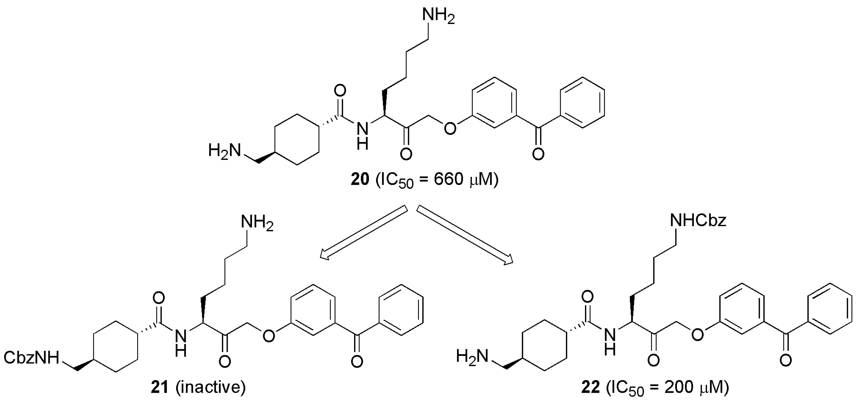
Figure 1. Confirmation of which amino acid interacts with Asp at the S1 site by using two derivatives (21 [29] and 22 [29]) from 20 [29].
Processes 09 00329 g001
Figure 2. Tranexamic acid (TXA) derivatives bond with μPlm. (A) Binding orientation of 36 (green stick, pdb 5ugg) and 39 (orange stick, pdb 5ugg) at the sub-site S1 to S1′, S2′, and S3′; (B) Picolyl and quinolinyl moieties extended from Tyr side chain in the S3′ pocket; (C) Superimposition of the active sites of μPlm (white molecular surface, pdb 5ugd) and uPA (magenta molecular surface, pdb 1fv9); (D) Close up view of the S3′ pockets; The quinoline moiety collides with both Tyr209 and Arg178 residues of at the S3′ site of uPA. *The numbers of amino acid residues correspond to full-length Plm or uPA.
Processes 09 00329 g002
Figure 3. Structure of compounds 40–42 [37,38,39,40] developed by Bristol-Myers Squibb (BMS).
Processes 09 00329 g003
Figure 4. Structure of compound 43 [41] developed by Bayer AG.
Processes 09 00329 g004
Table 1. Plasmin (Plm) inhibitory activity of compounds 13 [28].
Comp. IC50 (μM)
S-2251Fibrin
1 Processes 09 00329 i001(Km = 20 μM)180
2 Processes 09 00329 i002(Km = 200 μM)250
3 Processes 09 00329 i003700780
S-2251: H-D-Val-Leu-Lys-pNA.
Table 2. Plm inhibitory activity of compounds 410 [28].
Comp. IC50 (μM)
S-2251Fibrin
4 Processes 09 00329 i004300150
5 Processes 09 00329 i005210100
6 Processes 09 00329 i0063,0002,100
7 Processes 09 00329 i007140150
8 Processes 09 00329 i008>100>25
9 Processes 09 00329 i00969180
10 Processes 09 00329 i010210250
S-2251: H-D-Val-Leu-Lys-pNA.
Table 3. Plm inhibitory activity of compounds 11–14 [29].
Comp.R:IC50 (μM)
S-2251Fibrin
Processes 09 00329 i01111 Processes 09 00329 i0121617
12 Processes 09 00329 i01312>10 (18%)
13 Processes 09 00329 i014156.1
14 Processes 09 00329 i015400260
S-2251: H-D-Val-Leu-Lys-pNA.
Table 4. Plm inhibitory activity of compounds 13 [29], 15 [29], and 16 [29].
Comp.R:IC50 (μM)
S-2251FibrinFibrinogen
Processes 09 00329 i016TXA Processes 09 00329 i01775,000609,500
13 Processes 09 00329 i018156.113
15 Processes 09 00329 i019>200 (0%)>200 (0%)ND
16 Processes 09 00329 i020>300 (33%) >300 (43%)ND
S-2251: H-D-Val-Leu-Lys-pNA; ND: Not determined.
Table 5. Plm inhibitory activity of compounds 17–19 [29].
Comp.R:IC50 (μM)
S-2251Fibrin
Processes 09 00329 i02113 Processes 09 00329 i022156.1
17 Processes 09 00329 i023399.3
18 Processes 09 00329 i02424170
19 Processes 09 00329 i025>500 (7%)>500 (16%)
S-2251: H-D-Val-Leu-Lys-pNA.
Table 6. Inhibitory activity of compounds 2326 [30] against various proteases.
Comp.R:IC50 (μM)
PlmPKuPAThr
S-2251FibrinS-2302S-2444S-2238
Processes 09 00329 i02623 Processes 09 00329 i02736210.8558>1000
24 Processes 09 00329 i0281.80.400.6331>200 (10%)
25 Processes 09 00329 i0290.640.290.5845>100 (11%)
26 Processes 09 00329 i0300.230.0510.374363
S-2251: H-D-Val-Leu-Lys-pNA; S-2302: H-D-Pro-Phe-Arg-pNA, S-2444: pyroGlu-Gly-Arg-pNA, S-2238: H-D-Phe-Pip-Arg-pNA.
Table 7. Inhibitory activity of compounds 27–34 [31] against various proteases.
Comp.R:IC50 (μM)
PlmPKuPAThr
S-2251FibrinS-2302S-2444S-2238
Processes 09 00329 i03127 Processes 09 00329 i0321.10.4310>50>50
28 Processes 09 00329 i0330.800.231650>50
29 Processes 09 00329 i0340.500.1022>10100
30 Processes 09 00329 i0350.460.0562.126070
31 Processes 09 00329 i0360.630.0980.71>5089
32 Processes 09 00329 i0371.10.307.0>50>50
33 Processes 09 00329 i0380.980.170.291991
34 Processes 09 00329 i0395.31.4014.072240
S-2251: H-D-Val-Leu-Lys-pNA; S-2302: H-D-Pro-Phe-Arg-pNA, S-2444: pyroGlu-Gly-Arg-pNA, S-2238: H-D-Phe-Pip-Arg-pNA.
Table 8. Inhibitory activity of compounds 28 [31] and 35–37 [13] against various proteases.
Comp.R:IC50 (μM)
PlmPKuPA
S-2251S-2302S-2444
Processes 09 00329 i04028 Processes 09 00329 i0410.8016>50
35 Processes 09 00329 i0420.6538>50
36 Processes 09 00329 i0430.53305.3
37 Processes 09 00329 i0441.01231
S-2251: H-D-Val-Leu-Lys-pNA; S-2302: H-D-Pro-Phe-Arg-pNA; S-2444: pyroGlu-Gly-Arg-pNA.
Table 9. Inhibitory activity of compounds 38–41 [33] against various proteases.
Comp.R:IC50 (μM)
PlmuPAtrypsin
S-2251S-2444S-2238
Processes 09 00329 i04538 Processes 09 00329 i0460.34>10093
39 Processes 09 00329 i0470.22771.4
40 Processes 09 00329 i0480.97>501.3
41 Processes 09 00329 i0490.75>1006.7
S-2251: H-D-Val-Leu-Lys-pNA; S-2444: pyroGlu-Gly-Arg-pNA; S-2238: H-D-Phe-Pip-Arg-pNA.
Publisher’s Note: MDPI stays neutral with regard to jurisdictional claims in published maps and institutional affiliations.