Article

Socheongryongtang Modulates Asthma-Related Changes via Modulation of TNF-α and T-bet as well as IFN-γ in an Asthma Murine Model

1
College of Korean Medicine, Dongshin University, Naju, Jeonnam 58245, Korea
2
Department of Biomedicine, Health & Life Convergence Sciences, BK21 Four, College of Pharmacy, Mokpo National University, Muan, Jeonnam 58554, Korea
3
College of Veterinary Medicine, Chonnam National University, Gwangju 61186, Korea
4
College of Medicine, Hanyang University, Guri, Gyunggi 11923, Korea
*
Authors to whom correspondence should be addressed.
These authors equally contributed.
Processes 2020, 8(9), 1167; https://doi.org/10.3390/pr8091167
Submission received: 4 August 2020 / Revised: 9 September 2020 / Accepted: 15 September 2020 / Published: 17 September 2020
(This article belongs to the Section Pharmaceutical Processes)

Abstract

:
In 2017 the World Health Organization (WHO) reported that 235 million people suffered from asthma, and that 383,000 deaths were due to asthma in 2015. Asthma cannot be completely eradicated and the medications for asthma are associated with many adverse effects. Socheongryongtang is one of the prescriptions which has traditionally been used for the treatment of pulmonary disease, but the anti-asthmatic mechanism is unclear. To investigate the anti-asthmatic mechanism of socheongryongtang, BALB/c mice were divided into five groups: control, asthma-induced control, dexamethasone treatment, and 150 mg/kg or 1500 mg/kg socheongryongtang treatment and several biomarkers were analyzed, such as white blood cell (WBC) and differential counts in broncheoalveolar fluid (BALF), immunoglobulin E (IgE) in serum, and morphological changes/helper T cell-related cytokines/transcription factor in the lung. The therapeutic ingredients were also analyzed. Socheongryongtang inhibited the neutrophils differentiation in BALF, controlled interleukin (IL)-12p40 releasing, down-regulated not only GATA-3 and helper 2 T (Th2) cell transcription factors but also IL-4, and also decreased the level of tumor necrosis factor (TNF)-α in the lung. In addition, through high-performance liquid chromatography (HPLC) analysis, we confirmed that the therapeutic ingredients in socheongryongtang were paeoniflorin, liquiritin, and glycyrrhizin. The oral intake of 7.3 g of socheongryongtang is beneficial for suppressing the possibility of the occurrence of asthma via modulation of TNF-α and T-bet as well as IFN-γ.

Graphical Abstract

1. Introduction

In 2017 the World Health Organization (WHO) released statistics regarding asthma worldwide [1]. According to this report, there were 235 million asthma patients in 2017, many of them children, and in 2015 383,000 died from this disease. The symptoms vary widely from just cough to apnea, and apnea in this case is caused by the lack of a gas change that stems from respiratory system obstruction due to mucous hypersecretion, epithelial cell hyperplasia, base membrane enlargement, etc. [2,3]. There are so many stimulators to provoke asthma in the environment they can be divided two items such as indoor allergens [4] and outdoor allergens [5]. Several medications are currently in use for the treatment of asthma: relievers, such as short-acting β2-agonists and anticholinergics; controllers, such as long-acting β2-agonists, combinations of these two; and preventers, such as inhaled corticosteroids and leukotriene receptor antagonists. In recognition of the fact that asthma is currently one of the major public health problems faced worldwide, the WHO published their 17th Model List of Essential Medications, including a standardized asthma management strategy that suggested inhaled corticosteroids (e.g., beclometasone 100 μg hydrofluoroalkane) and bronchodilators (e.g., salbutamol 100 μg hydrofluoroalkane) [6]. However, the remedies presently used cannot completely eradicate asthma, as they can only conceal the symptoms. Beyond that, they can cause many adverse effects such as retardation of height [7], high blood suppression, ulceration of stomach, decline in immunity, high lipid blood [8], etc.
Therefore, for several decades there have been many trials to identify anti-asthmatic drug candidates which are more effective and less toxic [9,10,11,12]. In particular, many have attempted to develop medications for asthma based on the traditional prescriptions/drugs through the investigation of the mechanisms and/or modes of action. Approximately 600 years ago in Korea, Jun Huh compiled Donguibogam, which was a medical encyclopedia [13]. It was an encyclopedia related to medicine and remedies. Socheongryongtang is one of the prescriptions listed for pulmonary disease treatment in the Donguibogam.
Asthma is caused by the malfunction of the immune system, and one of the many theories regarding asthma occurrence involves the imbalance of sub-helper T cells [2]. Generally, in asthma patients, the levels of Th1-related cytokines such as interferon gamma (IFN-γ) and interleukin 12 (IL-12) are decreased [14,15] while those of Th2-related cytokines are upregulated [16,17,18]. T-bet is a Th1 cell transcription factor and it has a positive feedback relation with IFN-γ, which is related to asthma severity [14,19,20]. IL-12 regulates the functions of Th1-related cytokines [15] and controls the level of helper 2 T (Th2) cells [21,22]. GATA-3 is a Th2 cell transcription factor and the levels of GATA-3 and IL-4 are positively controlled by feedback phenomenon [23]. IL-4 induces eosinophil and neutrophil’s differentiations from mast cells, B cell activation, IgE production, mucous hypersecretion, etc. [24,25] IL-13 stimulates the activation of B cells as well as change in the pulmonary system [26,27,28,29]. Tumor necrosis factor (TNF)-α activates T cells [30] and induces respiratory organ allergy [31]. IL-6 increases IL-4 levels and induces the differentiation of Th17 cells [32].
Although socheongryongtang is listed in Donguibogam as the prescription for pulmonary diseases, there is no evidence for its anti-asthmatic mechanism, we analyzed the biological effect against asthma, investigated the treatment mechanism, and analyzed the therapeutic ingredients in socheongryongtang.

2. Materials and Methods

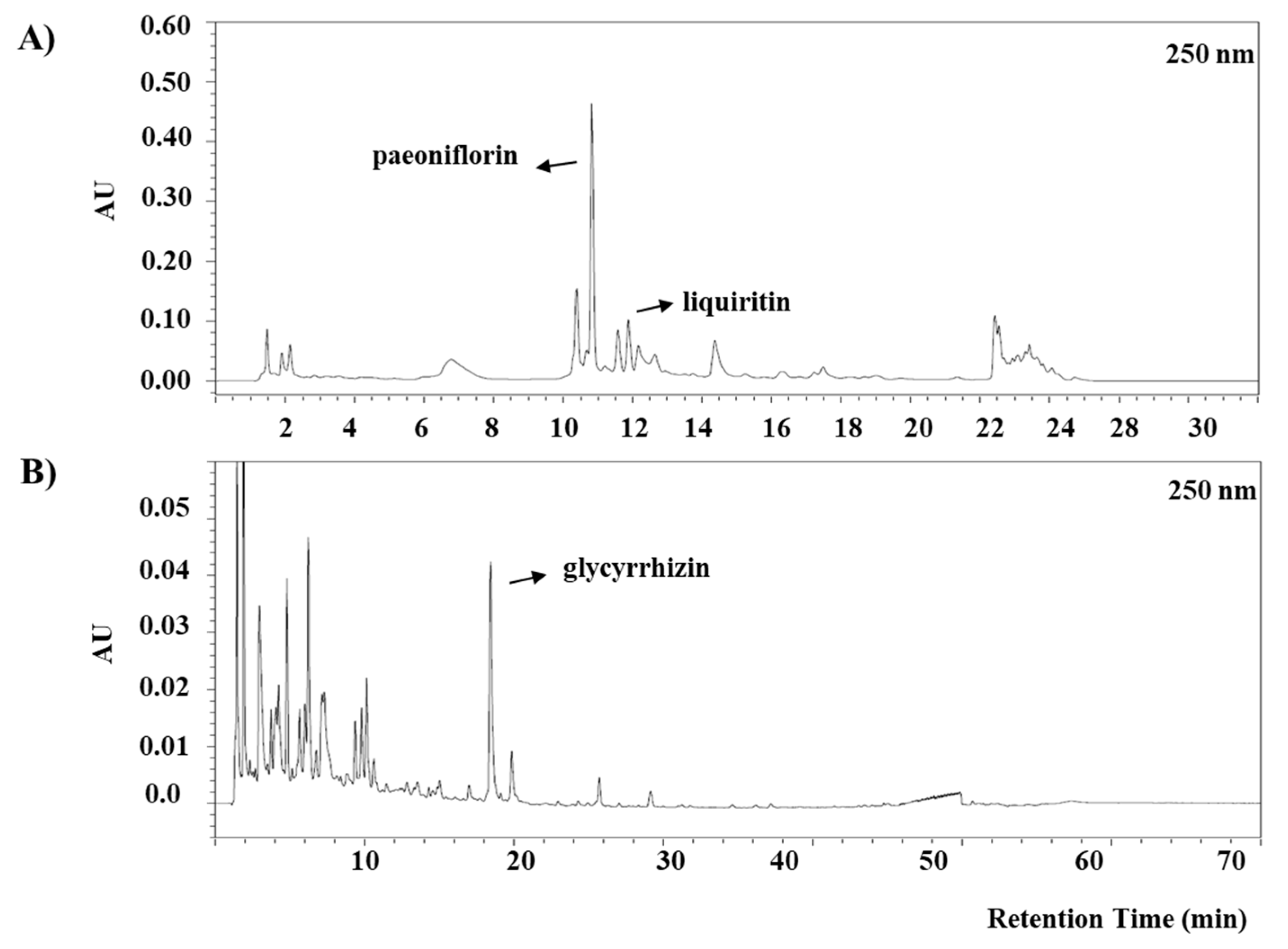
2.1. Socheongryongtang and Identification of Anti-Asthmatic Compounds

According to the Donguibogam [13], socheongryongtang has long been used as a prescription for pulmonary disease and consists of 1.5 × Pinellia ternate, 1.5 × Ephedra sinica, 1.5 × Paeonia lactiflora, 1.5 × Schisandra chinensis BAALL, 1.0 × Zingiberis rhizome, 1.0 × Glycyrrhizae radix, 1 × Cinnamomi ramulus and 1.0 × Asarum sieboldii. However, the socheongryongtang (Batch No. 14007, hot water extracted-powder form) used in this study was made by Hankuk Inspharm, Ltd. (Jeonnam, Korea) according to Donguibogam [13].
Three markers (paeoniflorin, glycyrrhizin, and liquiritin) were used for high-performance liquid chromatography (HPLC) analysis. Briefly, analyses were conducted by Alliance 2695 HPLC system (Waters, Milford, MA, USA). A reverse phase column (C18, 5 μm, 150 mm × 5 mm) was used with a mobile phase consisting of a mixture of solvent A (acetonitrile) and B (0.2% phosphoric acid). A gradient elution (from 10/90 to 100/0 v/v) at a flow rate of 1.0 mL/min was used for paeoniflorin and liquiritin. A gradient elution (from 15/85 to 100/0 v/v) at a flow rate of 1.0 mL/min was used for glycyrrhizin. The analytical conditions are described in Table 1.

2.2. Animal Experiments

Animal experiments were done based on our previous study [9]. Using the same method, two animal studies were conducted at different times. Seventy female BALB/c mice were purchased from Samtako Korea (Osan, Korea) and divided into five groups according to treatment: (1) vehicle control (sterilized tap water), (2) ovalbumin (OVA)-induced asthma model, (3) 1 mg/kg/day dexamethasone with OVA induction, (4) 150 mg/kg/day socheongryongtang with OVA induction, and (5) 1500 mg/kg/day socheongryongtang with OVA induction. On days 1 and 8, all mice except for those in the vehicle control group were sensitized via intraperitoneal injections of 20 µg OVA (Sigma-Aldrich, St. Louis, MO, USA) and 1 mg of aluminum hydroxide hydrate (Sigma-Aldrich) in 500 µL saline. From days 21 to 25, mice were inhaled with 5% ovalbumin one time per a day for 30 min (3 mL/min, NE-U17, OMRON Co. Ltd., Kyoto, Japan). During the same five-day period, treatment groups were treated once daily 1 h prior to OVA inhalation with tap water, dexamethasone, 150 mg/kg/day socheongryongtang, or 1500 mg/kg/day socheongryongtang. The mice in tap water-treated group were treated with OVA as same as the other groups (20 µg OVA and 1 mg aluminum hydroxide hydrate in 500 µL saline), after which they were inhaled saline or aluminum hydroxide hydrate by a nebulizer for five days.

2.3. Ethic Statement

After the approval of the Institute of Animal Care and Use Committee (IACUC) of Dongshin University (Animal Study Approval No. 2014-08-02, date of approval; 2 August 2014) the animal study was conducted.

2.4. Broncheoalveolar Fluid (BALF) and Serum Analysis

Broncheoalveolar fluid (BALF) and serum analyses were conducted as previously described [9,33]. Briefly, one day after the final treatment, mice were intraperitoneally anesthetized using with 50 mg/kg Zoletin (Virbac, Fort Worth, TX, USA) and were cannulated on tracheas with flexible needles. Using with 0.4 mL phosphate-buffered saline (PBS) lavages were collected. Whole collected fluid was centrifuged at 3000 rpm for 5 min at 4 °C (Sorvall Legend Micro 17R, Thermo Fisher Scientific, Inc. Marietta, OH, USA). In order to measure the differential cell count cell pellets were suspended again and using the Hemavet Multispecies Hematology System (Drew Scientific Inc, Waterbury, CT, USA) the numbers of white blood cells (WBCs) and each cell portion were analyzed.
Using immunoglobulin E (IgE) enzyme-linked immunosorbent assay (ELISA) kit (BD Bioscience, 555248, San Jose, CA, USA) the levels of serum IgE were measured according to the manufacturer’s protocols.

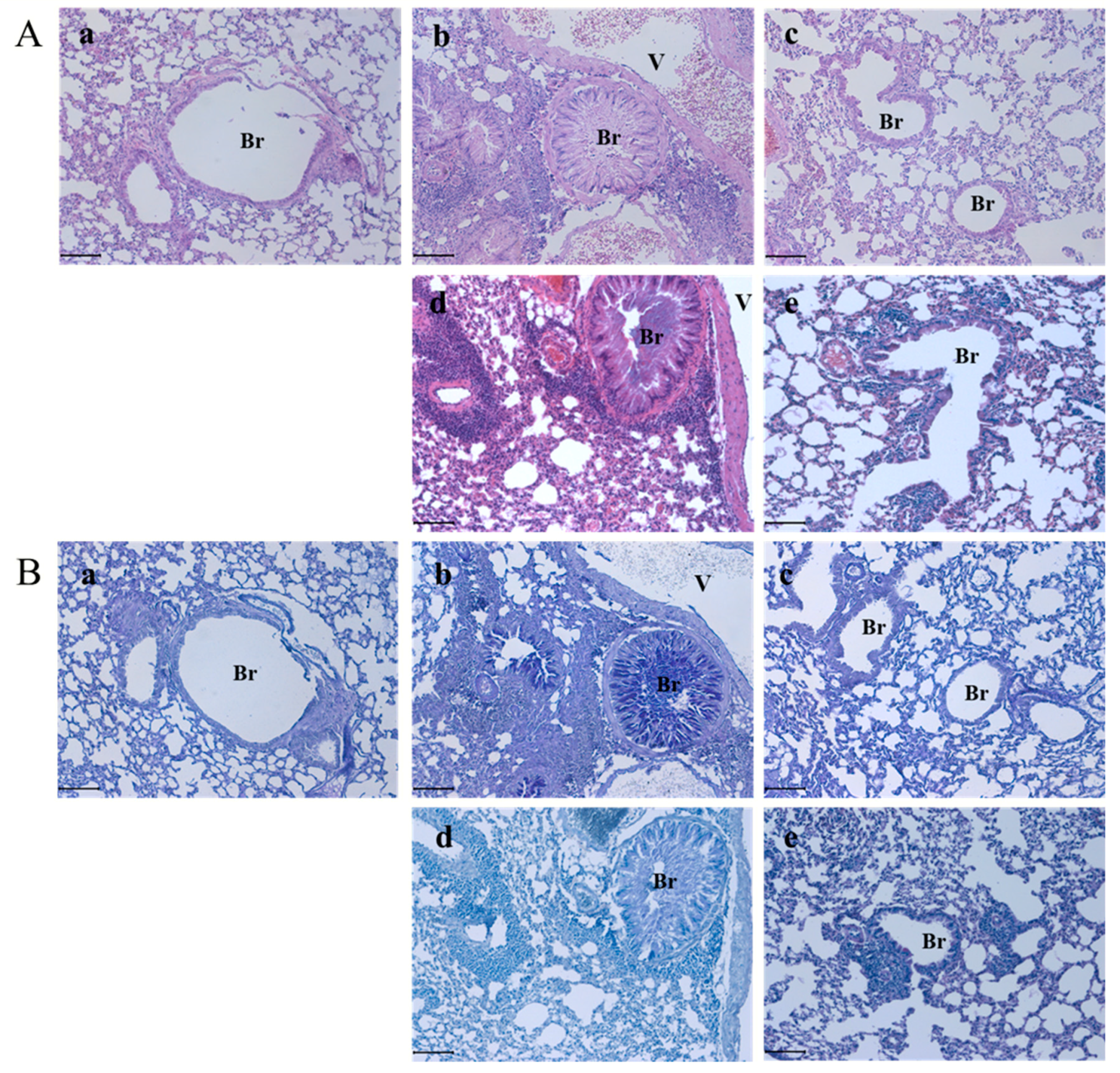
2.5. Histopathological Analysis

According to the previous study, a histopathological analysis was undertaken [9]. Briefly, collected pulmonary systems were fixed in formaldehyde solution, dehydrated in a graded ethanol series, and embedded in paraffin. After the paraffinized lung tissues were sectioned (4 µm in thickness) longitudinally and in order to observe the morphological changes they were stained with hematoxylin and eosin (H&E). Additionally, to analyze the glycoprotein’s change periodic acid schiff (PAS) stains were conducted. The results were obtained by an Axioscope A1 (Carl Zeiss, Gottingen, Germany).

2.6. Immunofluorescent Analysis

To confirm the intracellular distribution of Th1 cell transcription factor, T-bet, and Th2 cell transcription factor, GATA-3 immunofluorescent analysis was conducted for four groups: control, OVA, dexamethasone with OVA induction, and 1500 mg/kg socheongryongtang with OVA induction. T-bet (Biorbyt, orb7075, Cambridge, UK) or GATA-3 (OriGene, TA305795, Rockville, MD, USA) was used as a primary antibody. All samples were then incubated with fluorescein isothiocyanate (FITC)-conjugated IgG (Jackson Immunoresearch, 315-095-003, West Grove, PA, USA) or AlexaFlur 555-conjugated IgG (ThermoFisher Scientific, A-21127, Waltham, MA, USA) for 2 h. In order to counterstain, 4′,6-diamidino-2-phenylindol (DAPI) was used (ThermoFisher Scientific, 62249, Waltham, MA, USA). The results were obtained by a K1-Fluo confocal microscope (Nanoscope System, Daejeon, Korea).

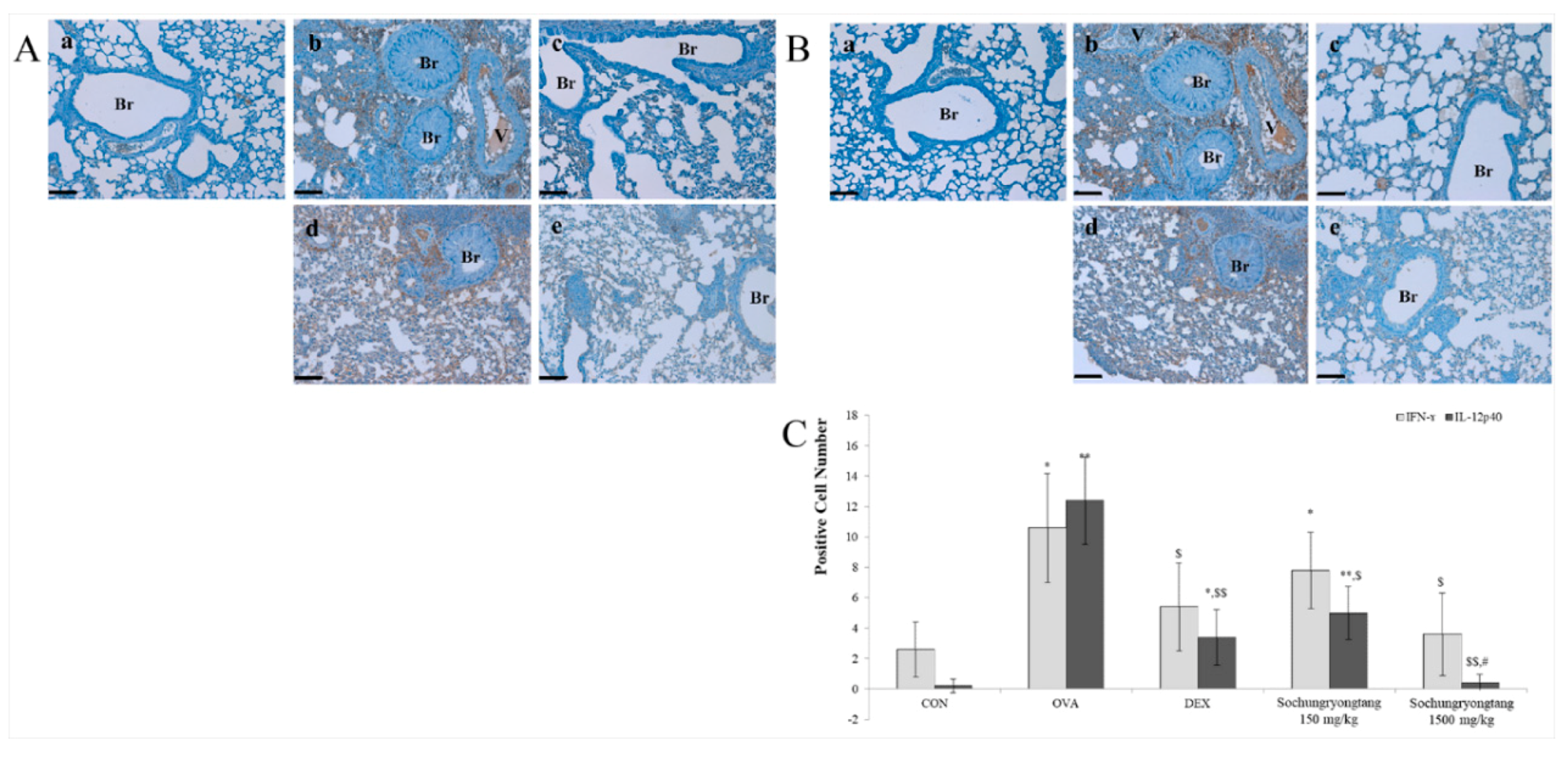
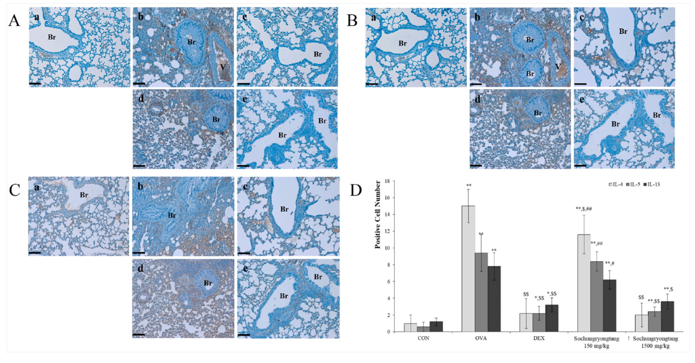
2.7. Immunohistochemical (IHC) Analysis

Immunohistochemical analysis was conducted as described in our previous study [9]. Briefly, after getting rid of paraffin from tissues on the slides in order to eliminate endogenous peroxidase they were immersed in 3% hydrogen peroxide in methanol for 10 min. Sodium citrate buffer (0.1 M) was used for the antigen retrieval. In order to prevent non-specific binding normal horse serum was used, and then they were bounded with primary antibodies (diluted 1:100 to 1:200) including IFN-γ (Santa Cruz, sc-74104), IL-12p40 (Santa Cruz, sc-57258), IL-4 (Santa Cruz, sc-73318), IL-5 (Santa Cruz, sc-7887), IL-13 (Santa Cruz, sc-1776), TNF-α (BioVision, 3053R-100, Milpitas, CA, USA), and IL-6 (Santa Cruz, sc-1265) antibodies at 4 °C for 1 h. Using biotinylated secondary antibody, all slides were incubated for 10 min and reacted with streptavidin peroxidase complex for 5 min (Vector Laboratories Universal Quick Kit, Burlingame, Canada). Signals were obtained using 3,3-diaminobenzidine tetrahydrochloride substrate chromogen solution and they were stained with Mayer’s hematoxylin. The results were obtained by an Axioscope A1 (Carl Zeiss) and to measure the positive cells we counted them in five randomly selected non-overlapping fields (×200 magnification) of three separately.

2.8. Statistical Analysis

Results are expressed as mean ± standard deviation (SD). Group differences were evaluated through one-way analysis of variance followed by Dunnett’s multiple comparison test. Significance was considered at p < 0.01 or p < 0.05.

3. Results

3.1. Identification of Anti-Asthmatic Markers in Socheongryongtang

HPLC analyses were performed in order to identify any anti-asthmatic markers in socheongryongtang. Typical HPLC chromatograms of phytochemical contents and their retention times are shown in Figure 1. Three compounds [namely, paeoniflorin (0.49%), glycyrrhizin (0.38%), and liquiritin (0.05%)] were identified by HPLC analysis.

3.2. Socheongryongtang Effectively Inhibited the Proliferation of WBC and Neutrophil and IgE Overexpression

In asthma patients, the level of WBC in BALF is usually higher than normal, and eosinophil population is up-regulated [34]. Recently, it has been shown to be important to measure not only eosinophil increment but also the increase of neutrophil levels in asthma [35]. In the ovalbumin treatment group, the populations of WBC and neutrophil increased, but dexamethasone significantly decreased these populations, and socheongryongtang suppressed the populations of WBC and neutrophil (Figure 2A,B & Supplementary Materials). Based on the results, we found that socheongryongtang could not modulate the eosinophil level but could control the change of neutrophil in the asthma model. IgE is one of the important biomarkers for hyperresponsiveness criteria which significantly increases in asthma patients [36] and in the ovalbumin-induced asthma animal model [9,10]. Like the reported results in the ovalbumin treatment group, the expression level of IgE significantly increased compared to that in control (Figure 2C). Dexamethasone controlled the overexpression of IgE and down-regulated IgE expression (Figure 2C).

3.3. Socheongryongtang Effectively Prevented Typical Asthmatic Morphological Changes in the Pulmonary System

In order to evaluate the preventive effect of socheongryongtang, H&E staining and PAS staining were conducted (Figure 3). In asthma patients’ lungs several typical changes were observed such as mucous hypersecretion in the bronchioalveola, epithelial cell overgrowth, inflammatory cells such as eosinophil or/and neutrophil, mast cells infiltration near the brochioalveola and vessel, goblet cells hyperplasia, airway smooth muscle proliferation, etc. [37,38]. Ovalbumin treatment induced the typical asthma morphology in the lung (Figure 3A(b)) but dexamethasone prevented the morphological changes caused by ovalbumin (Figure 3A(c)). This was not different between the lung in 150 mg/kg socheongryongtang treatment group or the ovalbumin treatment group (Figure 3A(c,d)). However, 1500 mg/kg socheongryongtang treatment prevented ovalbumin-induced pulmonary changes: mucous hypersecretion, epithelial hyperplasia, inflammatory cell infiltration, etc. (Figure 3A(c)). When asthma occurs, one of the most important criteria to observe is mucous hypersecretion, and in order to evaluate the suppression effect of socheongryongtang against mucous secretion, PA staining was conducted (Figure 3B). Both in the ovalbumin treatment group and in the 150 mg/kg socheongryongtang treatment group, the mucous was filled in the bronchioalveola (Figure 3B(b,d)) but in the dexamethasone treatment group and in the 1500 mg/kg socheongryongtang treatment group a decrease of mucous secretion was observed (Figure 3B(c,e). From the results, we could find that socheongryongtang dose-dependently and effectively reduced mucous secretion.

3.4. Socheongryongtang Inhibited Both Activations of Th1 Cell Transcription Factor, T-bet and Th2 Cell Transcription Factor, GATA-3

In the control group both T-bet and GATA-3 were not activated based on their existence in the cytoplasm (Figure 4D(a)) but ovalbumin treatment induced them to activate as both of them are located in the nucleus (Figure 4D(b)). We obtained the same results in the dexamethasone treatment group and in the 1500 mg/kg socheongryongtang treatment group in that the distribution of them, T-bet and GATA-3 was mainly in the cytoplasm (Figure 4D(c,d)). This meant that dexamethasone and socheongryongtang could prevent the translocation of T-bet and GATA-3, which should be activated as transcription factors.

3.5. Socheongryongtang Suppressed Th1-Related Cytokine Expression

In order to analyze the change of Th1-related cytokine such as IFN-γ and IL-12p40, the IHC method was conducted (Figure 5). IFN-γ is one of the very important cytokines as it can stimulate T-bet, Th1 cell transcription factor to activate with the positive feedback loop [20,39]. Similar to the change pattern of T-bet (Figure 4), the expression level of IFN-γ by ovalbumin treatment significantly increased compared to that in control but dexamethasone treatment controlled the level of that (Figure 5A,C). Socheongryongtang dose-dependently reduced IFN-γ and in particular the level of IFN-γ expression in the 1500 mg/kg socheongryongtang treatment group was very similar to that in dexamethasone treatment (Figure 5A,C). IL-12p40 is one of the asthma-concerned cytokines which stimulates IFN-γ production [15], as shown in Figure 5B,C. IL-12p40 was increased by ovalbumin treatment and was dose-dependently controlled by socheongryongtang, and particularly in the 1500 mg/kg socheongryongtang treatment group, the level of IL-12p40 was lower than that in the dexamethasone treatment group. These results meant that socheongryongtang was more effective on IL-12p40 than on IFN-γ.

3.6. Socheongryongtang Dose-Dependently Modulated Th2-Related Cytokine: Interleukin 4 (IL-4), IL-5, and IL-13

Asthma is caused by the imbalance of Th1/Th2-related factors [40] and the role of IL-4 against Th2 cell transcription factor, GATA-3 is similar to the part of IFN-γ against Th1 cell transcription factor, T-bet. Therefore, although GATA-3 stimulates IL-4 production, IL-4 activates GATA-3 with a positive feedback loop. The change of IL-4 (Figure 6A,D) was very similar to that of GATA-3 (Figure 4). Ovalbumin treatment significantly increased the expression level of IL-4 but dexamethasone effectively suppressed this increase. Socheongryongtang controlled the IL-4 expression not only in the 1500 mg/kg treatment group but also in the 150 mg/kg treatment group and, therefore, IL-4 expression was effectively down-regulated by socheongryongtang treatment. The change of IL-5 expression (Figure 6B,D) caused by socheongryongtang was very similar to that of IL-13 (Figure 6C,D). Socheongryongtang dose-dependently modulated both the expressions of IL-5 and IL-6, which were significantly increased by ovalbumin treatment.

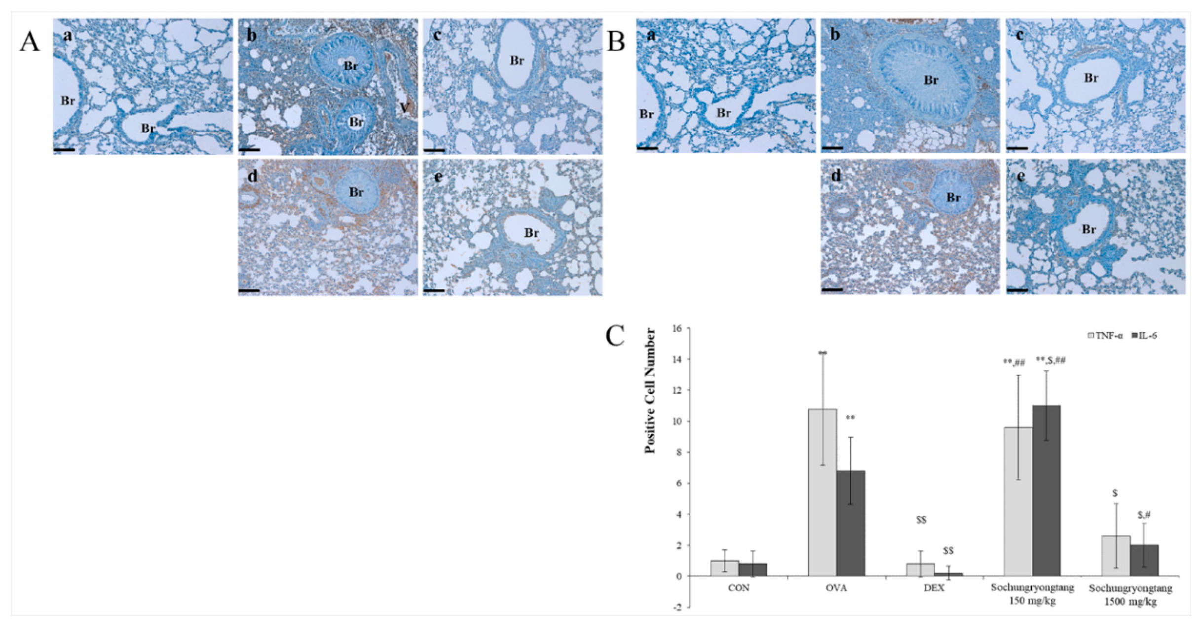
3.7. Socheongryongtang Dose-Dependently Controlled the Expression Levels of TNF-α but not IL-6

Although we obtained different results of socheongryongtang’s modulation aspect against TFN-α and IL-6, it commonly decreased the expressions of both of them in 1500 mg/kg treatment (Figure 7). Ovalbumin induced the expression increment of TFN-α and IL-6 and 1500 m/kg socheongryongtang treatment decreased the expressions of both of them. However, 150 mg/kg socheongryongtang treatment controlled TNF-α expression (Figure 7A) while the same dose treatment of socheongryongtang increased IL-6 expression (Figure 7B,C).

4. Discussion

Asthma is type I hyperresponsiveness, and is deeply related with the sub-helper T cells’ balance, and the symptoms related to that are very diverse from just a cough to apnea which is caused by mucous hypersecretion, epithelial cell hyperplasia, airway smooth muscle proliferation, and inflammatory cell infiltration, and can finally induce death in patients [2]. According to a WHO report, in 2015 383,000 deaths were caused by asthma, and the worldwide number of patients might be 235 million [1]. As there are a lot of causes of asthma everywhere around us [41], it is impossible to perfectly cure it [42], and most anti-asthmatic drugs which have been used have several adverse effects such as growth control in the young [7], hypertension, hyperlipidemia, peptic ulcers, immunological suppression, etc. [8], so there are many trials aimed at developing new anti-asthmatic drugs from natural products [9,10]. Asthma inducers are called allergens and are classified into two groups: indoor allergens and outdoor allergens. The former include house dust mites, pet dander, tobacco smoke, etc. [4] and the latter include flower pollen, pollutant, extreme temperature, excessive exercise, etc. [5].
Although it is impossible to explain the whole mechanism of asthma occurrence, the imbalance of helper T cells is one of the theories of asthma onset [40]. In asthma patients, Th2-related factors increase such as IL-4, IL-5, IL-13, etc. [15,17,18] and as it belongs to allergy type I, when it onsets, the level of IgE significantly increases [43]. IL-4 is a particularly important player in asthma occurrence as it activates GATA-3, the Th2 cell transcription factor to produce Th2-related cytokine, stimulates mast cell to differentiate eosinophil and neutrophil in blood and BALF, activates B cell to produce IgE, and secretes excessive mucous in the pulmonary system, etc. [24,25]. As shown in Figure 4 and Figure 6, socheongryongtang significantly prevented the translocation of GATA-3 into the nucleus to transcript Th2-related cytokine and decreased the level of IL-4. In particular, the level of IL-4 in the 1500 mg/kg socheongryongtang treatment group was similar to that in the dexamethasone treatment group (Figure 6D). One of the functions of IL-4 is to reduce neutrophil differentiation [24,25] and in asthma patients it has been observed in not only the surge of eosinophil but also in that of neutrophil [35]. Socheongryongtang might control the level of neutrophil rather than eosinophil (Figure 2B). Although GATA-3 is a Th2 cell transcription factor, IL-4 regulates it with a positive feedback loop [23]. The significant decrease of the IL-4 level meant that socheongryongtang inactivated Th2 cell transcription factor, GATA-3, and finally prevented the release of Th2-related cytokines. In asthma occurrence, IL-13 plays many important roles, such as increasing the level of IgE, secretion of respiratory mucous [24,44], and induction of airway hyperresponsiveness (AHR) and airway remodeling [29]. Socheongryongtang dose-dependently controlled not only the level of IL-4 but also that of IL-13 (Figure 6).
Like the role of IL-4 against the Th1 cell transcription factor, IFN-γ is regulated by T-bet, the Th1 cell transcription factor with positive feedback [19,20]. Although the standard deviation is too large, the level of IFN-γ was controlled by socheongryongtang similar to that by dexamethasone (Figure 5A,C) and T-bet activation was inhibited by socheongryongtang (Figure 4). IL-12p40 plays a role in the Th1-related factor modulation such as macrophage chemoattractant [45], the TNF-α inducer [46], and in children with severe asthma the IL-12 serum level significantly decreases [47]. In addition, TNF-α plays an important role in the interaction of mast cells and smooth muscle and then induces pulmonary system hyperresponsiveness [31]. In the present study, we found a relation between IL-12p40 (Figure 5B,C) and TNF-α (Figure 7A,C), that is, socheongryongtang decreased the IL-12p40 level and then the decrease of IL-12p40 level down-regulated the TNF-α induction. Finally, according to the results, ovalumin-induced respiratory hyperresponsiveness was controlled.
Socheongryongtang is a traditional prescription for asthma [13]. These plants are known to contain anti-asthmatic markers such as cinnamic acid, cinnamaldehyde, glycyrrhizin, schizandrin, paeoniflorin, and anti-inflammatory markers such as liquiritin. In the present study, we investigated whether there were anti-asthmatic compounds in socheongryongtang using HPLC. Three such compounds, paeoniflorin (0.49%), glycyrrhizin (0.38%), and liquiritin (0.05%), were identified.
Paeoniflorin is derived from Paeonia lactiflora and known to have anti-inflammatory [48] and anti-asthmatic activity [49]. Zhang et al. (2015) reported that paeoniflorin showed anti-asthmatic effects in an ovalbumin-induced mice model [49]. Paeoniflorin (10 and 20 mg/kg) treatment group controls the eosinophil, IL-4 and IgE levels in in vivo and regulate the Th1/Th2 balance. Wang et al. (2014) described the anti-inflammatory effect of paeoniflorin in lipopolysaccharides (LPS) induced RAW 264.7 cells [48]. Paeoniflorin inhibited the inflammation mediators such as inducible nitric oxide synthase, cyclooxygenase-2 (COX-2), IL-6, and TNF-α.
Glycyrrhizin is one of the major compounds of Glycyrrhiza uralensis and Glycyrrhiza glabra. The pharmacological effect of glycyrrhizin is also closely related to an immune response. Ram et al. (2006) evaluated the anti-asthmatic effect of glycyrrhizin in a mouse model of asthma [50]. The mice were orally treated with glycyrrhizin (2.5, to 20 mg/kg) during or after OVA-sensitization and OVA-challenge to evaluate its protective or reversal effect, respectively on the above asthmatic features. Parameters associated with asthma such as IL-4, IL-5, IFN-γ, OVA-specific IgE, total IgG(2a) and cortisol were affected by glycyrrhzin in the range of tested oral administration. The results revealed that glycyrrhizin alleviates asthmatic features in vivo and it gives beneficial advantages towards developing a better therapeutic molecule [51].
In the present study, liquiritin was also identified as a key marker of socheongryongtang. Liquiritin is flavonoid purified from Glycyrrhiza uralensis [51]. Liquiritin is known to have anti-inflammatory activity. Wang et al. reported that α-liquiritin inhibited the pro-inflammatory mediators, such as inducible nitric oxide synthase, COX-2, TNF-α, IL-1β, and IL-6 in LPS-induced inflammation in vitro [52].
Zhang et al. (2015) reported that the minimum efficacy level of paeoniflorin started at 10 mg/kg for an anti-asthmatic effect in vivo [49]. Ram et al. (2006) also calculated the initial oral administration dose as 5 mg/kg in mice [50]. In the present study, we found that 1500 mg/kg dosage clearly showed anti-asthmatic effects in our animal model. The dose of 1500 mg/kg as socheongryongtang is equally calculated as 7.5 mg/kg and 5.7 mg/kg when converted to paeoniflorin and glycyrrhizin. Thus, the optimal dose of our results was consistent with those of previous reports and it reveals that paeoniflorin and glycyrrhizin are key compounds of socheongryongtang for the treatment of asthmatic disease.

5. Conclusions

Although we obtained the cytokines’ level which socheongryongtang modulated based on the immunohistochemistry analysis in order to confirm the results, we need to obtain quantitative results such as ELISA, quantitative polymerase chain reaction (q-PCR), etc. in a further study. In general, we considered oral intake of socheongryongtang of 7.3 g daily for the treatment of asthma. The oral dose of a mouse is 1500 mg/kg/day and the conversion factor between human and mouse is 12.33 [53]. Therefore, if the effective dose for mice is 1500 mg/kg/day, the human equivalent dose is 7300 mg/60 kg/day as socheongryongtang. The official daily intake of socheongryongtang for humans is 9 g. Thus, the optimal dose for asthma treatment is less than the daily intake. We concluded that oral intake of 7.3 g of socheongryongtang is beneficial for suppressing the possibility of the occurrence of asthma.

Supplementary Materials

The following are available online at https://www.mdpi.com/2227-9717/8/9/1167/s1: S1 Dataset. This is the raw data for the Figure 2, Figure 5, Figure 6 and Figure 7.

Author Contributions

Conceptualization, H.-S.S. and D.-H.P.; Data curation, S.-H.B., C.-S.B. and B.K.; Formal analysis, S.-H.B.; Funding acquisition, S.S.C. and D.-H.P.; Investigation, S.-H.B., S.S.C., B.K. and D.-H.P.; Methodology, S.-H.B., B.K. and C.-S.B.; Project administration, S.S.C. and D.-H.P.; Resources, S.S.C. and D.H.; Supervision, H.-S.S. and D.-H.P.; Visualization, S.S.C. and D.-H.P.; Writing—original draft, S.-H.B.; Writing—review and editing, H.-S.S. and D.-H.P. All authors have read and agreed to the published version of the manuscript.

Funding

This research was supported by Basic Science Research Program through the National Research Foundation of Korea (NRF) funded by the Ministry of Education (2015R1D1A1A01059523) and by Funds of MNU Innovative Programs for National University in 2020.

Conflicts of Interest

The Authors declare that there is no conflict of interest.

References

  1. World Health Organization. Asthma Fact Sheet N°307; World Health Organization: Geneva, Switzerland, 2017. [Google Scholar]
  2. Kay, A.B. Allergy and allergic diseases. First of two parts. N. Engl. J. Med. 2001, 344, 30–37. [Google Scholar] [CrossRef] [PubMed]
  3. National Asthma Education and Prevention Program. Expert panel report: Guidelines for the diagnosis and management of asthma update on selected topics. J. Allergy Clin. Immunol. 2002, 110, S141–S219. [Google Scholar]
  4. Platts-Mills, T.A.; Vervloet, D.; Thomas, W.R.; Aalberse, R.C.; Chapman, M.D. Indoor allergens and asthma: Report of the third international workshop. J. Allergy Clin. Immunol. 1997, 100, S1–S24. [Google Scholar]
  5. Burge, H.A.; Rogers, C.A. Outdoor allergens. Environ. Health Perspect. 2000, 108, 653–659. [Google Scholar] [PubMed] [Green Version]
  6. World Health Organization. The selection and used of essential medicines (2011: Geneva, Switzerland) & World health organization. In World Model List of Essential Medicines: Essential Medicines 17th List; World Health Organization: Geneva, Switzerland, 2011. [Google Scholar]
  7. Wise, J. Corticosteroids for asthma may suppress growth in children in first year of treatment, researchers say. BMJ 2014, 349, g4623. [Google Scholar] [CrossRef]
  8. Ciriaco, M.; Ventrice, P.; Russo, G.; Scicchitano, M.; Mazzitello, G.; Scicchitano, F.; Russo, E. Corticosteroid-related central nervous system side effects. J. Pharmacol. Pharmacother. 2013, 4, s94–s98. [Google Scholar]
  9. Lee, S.Y.; Bae, C.S.; Choi, Y.H.; Seo, N.S.; Na, C.S.; Yoo, J.C.; Cho, S.S.; Park, D.H. Opuntia humifusa modulates morphological changes characteristic of asthma via IL-4 and IL-13 in an asthma murine model. Food Nutr. Res. 2017, 61, 1–12. [Google Scholar] [CrossRef] [Green Version]
  10. Lee, S.Y.; Bae, C.S.; Seo, J.H.; Cho, S.S.; Oh, D.S.; Park, D.H. Mycoleptodonoides aitchisoii suppresses asthma via IL-6 and IL-13 in ovalbumin-induced asthma mouse model. Mol. Med. Rep. 2018, 17, 11–20. [Google Scholar]
  11. Seo, J.W.; Cho, S.C.; Park, S.J.; Lee, E.J.; Lee, J.H.; Han, S.S.; Pyo, B.S.; Park, D.H.; Kim, B.H. 1′-Acetoxychavicol acetate isolated from Alpinia galanga ameliorates ovalbumin-induced asthma in mice. PLoS ONE 2013, 8, e56447. [Google Scholar] [CrossRef]
  12. Seo, J.H.; Bang, M.A.; Cho, S.S.; Park, D.H. Erythronium japonicum significantly suppresses OVA-induced asthma via upregulation the IFN-γ expression and downregulation the expression of TNF-α and IL-4. Int. J. Mol. Med. 2016, 37, 1221–1228. [Google Scholar] [CrossRef] [Green Version]
  13. Huh, J. Donguibogam; Bubinmunhwasa: Seoul, Korea, 2002. [Google Scholar]
  14. Kumar, R.K.; Webb, C.C.; Herbert, C.; Foster, P.S. Interferon-gamma as a possible target in chronic asthma. Inflamm. Allergy Drug Targets 2006, 5, 253–256. [Google Scholar] [CrossRef] [PubMed]
  15. Hamaza, T.; Barnett, J.B.; Li, B. Interleukin 12 a key immunoregulatory cytokine in infection applications. Int. J. Mol. Sci. 2010, 11, 789–806. [Google Scholar] [CrossRef] [PubMed] [Green Version]
  16. Busse, W.; Banks-Schlegel, S.P.; Larsen, G.L. Childhood- versus Adult-onset asthma. Am. J. Respir. Crit. Care Med. 1995, 151, 1635–1639. [Google Scholar] [CrossRef] [PubMed]
  17. Irvin, C.; Zafar, I.; Good, J.; Rollins, D.; Christianson, C.; Gorska, M.M.; Martin, R.J.; Alam, R. Increased frequency of dual-positive Th2/Th17 cells in bronchoalveolar lavage fluid characterizes a population of patients with severe asthma. J. Allergy Clin. Immunol. 2014, 134, 1175–1186. [Google Scholar] [CrossRef] [Green Version]
  18. Mosmann, T.R.; Coffiman, R.L. Th1 and Th2 cells: Different patterns of lymphokine secretion lead to different functional properties. Annu. Rev. Immunol. 1989, 7, 145–173. [Google Scholar] [CrossRef]
  19. Lazarevic, V.; Glimcher, L.H. T-bet in disease. Nat. Immunol. 2011, 12, 597–606. [Google Scholar] [CrossRef]
  20. Zhu, J.; Jankovic, D.; Oler, A.J.; Wei, G.; Sharma, S.; Hu, G.; Guo, L.; Yagi, R.; Yamane, H.; Punkosdy, G.; et al. The transcription factor T-bet is induced by multiple pathways and prevents an endogenous Th2 cell program during Th1 cell responses. Immunity 2012, 37, 660–673. [Google Scholar] [CrossRef] [Green Version]
  21. Mattes, J.; Yang, M.; Mahalingam, S.; Kuehr, J.; Webb, D.C.; Simson, L.; Hogan, S.P.; Koskinen, A.; Mckenzie, A.N.J.; Dent, L.A.; et al. Intrinsic defect in T cell production of Interleukin (IL)-13 in the absence of both IL-5 and eotaxin precludes the development of eosinophilia and airways hyperreactivity in experimental asthma. J. Exp. Med. 2002, 195, 1433–1444. [Google Scholar] [CrossRef]
  22. Manetti, R.; Parronchi, P.; Giudizi, M.G.; Piccinni, M.P.; Maggi, E.; Trinchieri, G.; Romagnani, S. Natural killer cell stimulatory factor (interleukin 12 (IL-12)) induces T helper type 1 (Th1)-specific immune responses and inhibits the development of IL-4 producing Th cells. J. Exp. Med. 1993, 177, 1199–1204. [Google Scholar] [CrossRef] [Green Version]
  23. Yagi, R.; Zhu, J.; Paul, W.E. An updated view on transcription factor GATA3-mediated regulation on Th1 and Th2 cell differentiation. Int. Immunol. 2011, 23, 415–420. [Google Scholar] [CrossRef] [Green Version]
  24. Larche, M.; Robinson, D.S.; Kay, A.B. The role of T lymphocytes in the pathogenesis of asthma. J. Allergy Clin. Immunol. 2003, 111, 450–463. [Google Scholar] [CrossRef] [PubMed]
  25. Lee, S.Y.; In, K.H. Immunopathogenesis of asthma. Tuberc. Respir. Dis. 2006, 60, 379–390. [Google Scholar] [CrossRef]
  26. Hershey, G.K. IL-13 receptors and signaling pathways: An evolving web. J. Allergy Clin. Immunol. 2003, 111, 677–690. [Google Scholar] [CrossRef] [PubMed]
  27. Rankin, J.A.; Picarella, D.E.; Geba, G.P.; Temann, U.A.; Prasad, B.; DiCosmo, B.; Tarallo, A.; Stripp, B.; Whitsett, J.; Flavell, R.A. Phenotypic and physiologic characterization of transgenic mice expressing interleukin 4 in the lung: Lymphocytic and eosinophilic inflammation without airway hyperreactivity. Proc. Natl. Acad. Sci. USA 1996, 93, 7821–7825. [Google Scholar] [CrossRef] [Green Version]
  28. Wills-Karp, M.; Luyimbazi, J.; Xu, X.; Schofield, B.; Neben, T.Y.; Karp, C.L.; Donaldson, D.D. Interleukin-13: Central mediator of allergic asthma. Science 1998, 282, 2258–2261. [Google Scholar] [CrossRef] [Green Version]
  29. Zhu, Z.; Homer, R.J.; Wang, Z.; Chen, Q.; Geba, G.P.; Wang, J.; Zhang, Y.; Elias, J.A. Pulmonary expression of interleukin-13 causes inflammation, mucus hypersecretion, subepithelial fibrosis, physiologic abnormalities, and eotaxin production. J. Clin. Investig. 1999, 103, 779–788. [Google Scholar] [CrossRef] [Green Version]
  30. Scheurich, P.; Thoma, B.; Ucer, U.; Pfizenmaier, K. Immunoregulatory activity of recombinant human tumor necrosis factor (TNF)-alpha: Induction of TNF receptors on human T cells and TNF-alpha-mediated enhancement of T cell responses. J. Immunol. 1987, 138, 1786–1790. [Google Scholar]
  31. Berry, M.; Brightling, C.; Pavord, I.; Wardlaw, A. TNF-alpha in asthma. Curr. Opin. Pharmacol. 2007, 7, 279–282. [Google Scholar] [CrossRef] [Green Version]
  32. Rincon, M.; Irvin, C.G. Role of IL-6 in asthma and other inflammatory pulmonary diseases. Int. J. Biol. Sci. 2012, 8, 1281–1290. [Google Scholar] [CrossRef] [Green Version]
  33. Bang, M.A.; Seo, J.H.; Seo, J.W.; Jo, G.H.; Jung, S.K.; Yu, R.; Park, D.H.; Park, S.J. Bacillus subtilis KCTC 11782BP-produced alginate oligosaccharide effectively suppresses asthma via T-helper cell type 2-related cytokines. PLoS ONE 2015, 10, e0117524. [Google Scholar] [CrossRef]
  34. Bousquet, J.; Chanez, P.; Lacoste, J.Y.; Barneon, G.; Ghavanian, N.; Enander, I.; Venge, P.; Ahlstedt, S.; Simony-Lafontaine, J.; Godard, P. Eosinophilic inflammation in asthma. N. Engl. J. Med. 1990, 323, 1033–1039. [Google Scholar] [CrossRef] [PubMed]
  35. Monteseirin, J. Neutrophils and asthma. J. Investig. Allergol. Clin. Immunol. 2009, 19, 340–354. [Google Scholar] [PubMed]
  36. Burrows, B.; Martinez, F.D.; Halonen, M.; Barbee, R.A.; Cline, M.G. Association of asthma with serum IgE levels and skin-test reactivity to allergens. N. Engl. J. Med. 1989, 320, 271–277. [Google Scholar] [CrossRef] [PubMed]
  37. Brightling, C.E.; Bradding, P.; Symon, F.A.; Holgate, S.T.; Wardlaw, A.J.; Pavord, I.D. Mast-cell infiltration of airway smooth muscle in asthma. N. Engl. J. Med. 2002, 346, 1699–1705. [Google Scholar] [CrossRef]
  38. Bentley, J.K.; Hershenson, M.B. Airway smooth muscle growth in asthma: Proliferation, hypertrophy, and migration. Proc. Am. Thorac. Soc. 2008, 5, 89–96. [Google Scholar] [CrossRef]
  39. Szabo, S.J.; Kim, S.T.; Costa, G.L.; Zhang, X.; Fathman, C.G.; Glimcher, L.H. A novel transcription factor, T-bet, directs Th1 lineage commitment. Cell 2000, 100, 655–669. [Google Scholar] [CrossRef] [Green Version]
  40. Kidd, P. Th1/Th2 balance: The hypothesis, its limitations, and implications for health and disease. Altern. Med. Rev. 2003, 8, 223–246. [Google Scholar]
  41. Galli, S.J.; Tsai, M.; Piliponsky, A.M. The development of allergic inflammation. Nature 2008, 454, 445–454. [Google Scholar] [CrossRef] [Green Version]
  42. Slejko, J.F.; Ghushchyan, V.H.; Sucher, B.; Globe, D.R.; Lin, S.L.; Globe, G.; Sullivan, P.W. Asthma control in the United States, 20082–010: Indicators of poor asthma control. J. Allergy Clin. Immunol. 2014, 133, 1579–1587. [Google Scholar] [CrossRef]
  43. Ariza, A.; Fernandez, T.D.; Dona, I.; Aranda, A.; Blanca-Lopez, N.; Melendez, L.; Canto, G.; Blanca, M.; Torres, M.J.; Mayorga, C. Basophil activation after nonsteroidal anti-inflammatory drugs stimulation in patients with immediate hypersensitivity reactions to these drugs. Cytometry A 2014, 85, 400–407. [Google Scholar] [CrossRef]
  44. Platts-Mills, T.A. The role of immunoglobulin E in allergy and asthma. Am. J. Respir. Crit. Care Med. 2001, 164, S1–S5. [Google Scholar] [CrossRef] [PubMed]
  45. Russell, T.D.; Yan, Q.; Fan, G.; Khalifah, A.P.; Bishop, D.K.; Brody, S.L.; Walter, M.J. IL-12p40 homodimer-dependent macrophages chemotaxis and respiratory viral inflammation are mediated through IL-12 receptor beta 1. J. Immunol. 2003, 171, 6866–6874. [Google Scholar] [CrossRef] [PubMed] [Green Version]
  46. Jana, M.; Dasgupata, S.; Saha, R.N.; Liu, X.; Pahan, K. Induction of tumor necrosis factor-alpha (TNF-alpha) by interleukin-12 p40 monomer and homodimer in microglia and macrophages. J. Neurochem. 2003, 86, 519–528. [Google Scholar] [CrossRef] [Green Version]
  47. Zedan, M.M.; El-Chennawi, F.A.; Fouda, A.E. Interleukin-12 and peripheral blood invariant natural killer T cells as an axis in childhood asthma pathogenesis. Iran. J. Allergy Asthma Immunol. 2010, 9, 43–48. [Google Scholar]
  48. Wang, J.; Wang, D.; Yu, J.; Liu, C.; Li, L.; Zhang, Y. Isolation of liquiritigenin-4’-apiosylglucoside and liquiritin form the root of Glycyrrhiza uralensis by high-performance centrifugal partition chromatography. J. Chromatogr. Sci. 2014, 52, 310–314. [Google Scholar] [CrossRef] [PubMed] [Green Version]
  49. Zhang, T.; Yang, Z.; Yang, S.; Du, J.; Wang, S. Immunoregulatory effects of Paeoniflorin exerts anti-asthmatic effects via modulation of the Th1/Th2 equilibrium. Inflammation 2015, 38, 2017–2025. [Google Scholar] [CrossRef]
  50. Ram, A.; Mabalirajan, U.; Das, M.; Bhattacharya, I.; Dinda, A.K.; Gangal, S.V.; Ghosh, B. Glycyrrhizin alleviates experimental allergic asthma in mice. Int. Immunopharmacol. 2006, 6, 1468–1477. [Google Scholar] [CrossRef]
  51. Wang, Q.S.; Gao, T.; Cui, Y.L.; Gao, L.N.; Jiang, H.L. Comparative studies of paeoniflorin and albiflorin from Paeonia lactiflora on anti-inflammatory activities. Pharm. Biol. 2014, 52, 1189–1195. [Google Scholar] [CrossRef]
  52. Yu, J.Y.; Ha, J.Y.; Kim, K.M.; Jung, Y.S.; Jung, J.C.; Oh, S. Anti-inflammatory activities of licorice extract and its active compounds, glycyrrhizic acid, liquiritin and liquiritigenin, in BV2 cells and mice liver. Molecules 2015, 20, 13041–13054. [Google Scholar] [CrossRef]
  53. Shin, J.W.; Seol, I.C.; Son, C.G. Interpretation of animal dose and human equivalent dose for drug development. J. Korean Orient. Med. 2010, 31, 1–7. [Google Scholar]
Figure 1. High-performance liquid chromatography (HPLC) chromatogram of socheongryongtang. (A) The peak of paeoniflorin was shown at about retention time 11 min and that of liquiritin was at about retention time 12 min. (B) At retention time 19 min glycyrrhizin had a peak.
Processes 08 01167 g001
Figure 2. Socheongryongtang dose-dependently inhibited both immune cells’ proliferation in broncheoalveolar fluid (BALF) and IgE overexpression in serum. Socheongryongtang decreased both (A) WBC and (B) neutrophil in BALF which was up-regulated by ovalbumin treatment. (C) Socheongryongtang dose-dependently controlled ovalbumin-induced IgE overexpression. N = 6 per each group. * p < 0.05 vs. control; ** p < 0.001 vs. control; $ p < 0.05 vs. asthma induction; $$ p < 0.001 vs. asthma induction; # p < 0.05 vs. dexamethasone; ## p < 0.001 vs. dexamethasone.
Processes 08 01167 g002
Figure 3. Socheongryongtang prevented ovalbumin-induced morphological change in the lung. (A) In the H&E stain, ovalbumin induced typical asthmatic change in the pulmonary system: mucous hypersecretion, epithelial cell hyperplasia, inflammatory cell infiltration, etc. more so than the control group. Socheongryongtang effectively and significantly prevented ovalbumin-induced morphological alterations such as secretion of mucous, overgrowth of epithelial cells, infiltration of inflammatory cells, etc. (B) In the PAS stain, socheongryongtang down-regulated the mucous secretion which caused by ovalbumin treatment. Scale Bar = 50 µm. Magnification, ×200. N = 8 per each group. Br, bronchiole; V, vessel. a, vehicle control; b, asthma induction; c, dexamethasone; d, 150 mg/kg/day socheongryongtang treatment; e, 1500 mg/kg/day socheongryongtang treatment.
Processes 08 01167 g003
Figure 4. Socheongryongtang significantly inhibited both the activations of T-bet, Th1 cell transcription factor and GATA-3, Th2 cell transcription factor. (A) Blue dots were nuclei which were stained DAPI. (B) T-bet, Th1 cell transcription factor was stained green color by FITC. (C) GATA-3, Th2 cell transcription factor was done in a red color by Alexa Flur 555. (D) Merged photos of nuclei, T-bet, and GATA-3. In ovalbumin treatment group T-bet and GATA-3 translocated from cytoplasm to nuclei and meant that they were under activation. However, dexamethasone and socheongryongtang perfectly blocked the translocation of T-bet and GATA-3 from cytoplasm to nuclei and then both of them existed in the cytoplasm. Scale Bar = 100 µm. Magnification, ×400. N = 8 per each group. a, vehicle control; b, asthma induction; c, dexamethasone; d, 1500 mg/kg/day socheongryongtang treatment.
Processes 08 01167 g004
Figure 5. Socheongryongtang dose-dependently reduced the expressions of IFN-γ and IL-12p40 in the pulmonary system. (A) Ovalbumin significantly increased the level of IFN-γ but dexamethasone and socheongryongtang decreased that. And in dose-dependent manner socheongryongtang down-regulated the IFN-γ expression. (B) Socheongryongtang controlled the expression level of IL-12p40 which was increased by ovalbumin treatment. (C) Socheongryongtang effectively controlled Th1-related cytokine expression, such as IFN-γ and IL-12p40, and especially in 1500 mg/kg socheongryongtang treatment group the level of IL-12p40 was similar to that in control group and was lower than that in dexamethasone group. Scale Bar = 50 µm. Magnification, ×200. N = 8 per each group. Br, bronchiole. a, vehicle control; b, asthma induction; c, dexamethasone; d, 150 mg/kg/day socheongryongtang treatment; e, 1500 mg/kg/day socheongryongtang treatment; * p < 0.05 vs. control; ** p < 0.001 vs. control; $ p < 0.05 vs. asthma induction; $$ p < 0.001 vs. asthma induction; # p < 0.05 vs. dexamethasone.
Processes 08 01167 g005
Figure 6. Socheongryongtang dose-dependently controlled the Th2-related cytokine expression such as interleukin 4 (IL-4), IL-5, and IL-13 in the respiratory system. (A) Based on the immunohistochemical observation the expression of IL-4, which was dark brown in color, was increased by ovalbumin treatment compared to that in control but socheongryongtang significantly reduced the expression of that. Socheongryongtang effectively controlled ovalbumin-induced (B) IL-5 expression and (C) IL-13. (D) Socheongryongtang dose-dependently controlled the expression of Th2-related cytokine: IL-4, IL-5, and IL-13. Especially 1500 mg/kg socheongryongtang treatment down-regulated the expression level of IL-4 similar to the level in control. Scale Bar = 50 µm. Magnification, ×200. N = 8 per each group. Br, bronchiole. a, vehicle control; b, asthma induction; c, dexamethasone; d, 150 mg/kg/day socheongryongtang treatment; e, 1500 mg/kg/day socheongryongtang treatment. * p < 0.05 vs. control; ** p < 0.001 vs. control; $ p < 0.05 vs. asthma induction; $$ p < 0.001 vs. asthma induction; # p < 0.05 vs. dexamethasone; ## p < 0.01 vs. dexamethasone.
Processes 08 01167 g006
Figure 7. Socheongryongtang reduced the expressions of TNF-α and IL-6. Socheongryongtang dose-dependently down-regulated (A) TNF-α expression and (B) IL-6 in the lung. (C) Socheongryongtang dose-dependently inhibited the expression of TNF-α, and although 150 mg/kg socheongryongtang slightly increased the expression 1500 mg/kg, socheongryongtang controlled the expression level of TNF-α like that in control. Scale Bar = 50 µm. Magnification, ×200. N = 8 per each group. Br, bronchiole. a, vehicle control; b, asthma induction; c, dexamethasone; d, 150 mg/kg/day socheongryongtangtreatment; e, 1500 mg/kg/day socheongryongtang treatment. ** p < 0.001 vs. control; $ p < 0.05 vs. asthma induction; $$ p < 0.001 vs. asthma induction; # p < 0.05 vs. dexamethasone; ## p < 0.01 vs. dexamethasone.
Processes 08 01167 g007
Table 1. Analysis Conditions for socheongryongtang. A, acetonitrile; B, 0.2% phosphoric acid. A, acetonitrile; B, 0.2% phosphoric acid.
ParametersConditions
(Paeoniflorin, Liquiritin)
Conditions
(Glycyrrhizin)
ColumnZorbax Extended-C18 (C18, 4.6 mm × 150 mm, 5 µm)
Flow Rate1 mL/min
Injection volume10 μL
Ultraviolet (UV) detection250 nm
Run time70 min
GradientTime (min)% A% BTime (min)% A% B
0109001585
71090356535
82080451000
202575501000
211000551585
251090
301090

