Article

Identification of Fungi in the Debitterizing Water of Apricot Kernels and Their Preliminary Evaluation on Degrading Amygdalin

1
Institute of Food & Physical Field Processing, School of Food Engineering and Nutrition Sciences, Shaanxi Normal University, Xi’an 710062, China
2
Departamento de Ingeniería Química, Facultad de Química, Universidad de Sevilla, 41012 Seville, Spain
*
Author to whom correspondence should be addressed.
Processes 2020, 8(9), 1061; https://doi.org/10.3390/pr8091061
Submission received: 24 June 2020 / Revised: 15 August 2020 / Accepted: 18 August 2020 / Published: 1 September 2020
(This article belongs to the Special Issue Wastewater Treatment Processes)

Abstract

:
Debitterizing water contains a great amount of amygdalin, a potential toxic compound, so it is mandatory the degradation of amygdalin to reduce the water’s toxicity and environmental pollution. In this paper, the suspended mycelia in debitterizing water were firstly investigated by digital microscope, cold field emission scanning electron microscope, and internal transcribed spacers (ITS) high-throughput sequencing. Thereafter, the degradation of commercial amygdalin by the identified species was assessed by determining the changes of amygdalin content and the β-glucosidase activity. The results indicate that the mycelia matched with genus of lower fungi, mainly including Irpex, Trichoderma and white rot fungus. Among them, Irpex lacteus had a definite promoting effect on the degradation of amygdalin, which was not caused by producing β-glucosidase, and the suitable degrading colony numbers ranged from 6.4 × 10⁶ CFU/mL to 6.4 × 10⁷ CFU/mL. In conclusion, this research might provide a potential novel approach to treat debitterizing water, so as to reduce the water pollution in the processing industry of apricot kernels.

1. Introduction

Apricot kernels contain abundant nutritional components such as carbohydrates, proteins, polyphenols, flavonoids, oils, and amygdalin [1,2], which are beneficial for human body. For instance, carbohydrates could provide the body energy demand [3], and polyphenols, because of their antioxidant capacity, could exert a beneficial bio-function of [4]. The main characteristic compound of apricot kernels, the amygdalin, on the one hand, could cure or prevent some diseases such as asthma, cardiovascular diseases, bronchitis, and cancer [5,6]. On the other hand, it is also regarded as a potentially toxic compound, since it can be hydrolyzed to benzoic acid and hydrogen cyanide (HCN), which has shown the toxicity at high intake [7], with its lethal dose for humans being 0.5–3.5 mg/kg body weight [8]. Therefore, the occurring of amygdalin has been regarded as an obstacle to the utilization and commercialization of the apricot kernels in industry, and the debitterizing or removal of bitterness has been an essential step for the processing of apricot kernels. To this end, a number of methods have been reported, such as cold water debitterizing, acid debitterizing, hot water debitterizing, ultrasonic debitterizing and acid-base alternate debitterizing [9,10,11], but all these methods have the problem of wastewater discharging, since some nutrients are transferred into the water during the debitterizing operation, resulting in a serious issue of water pollution [12]. Among these lost components, amygdalin is one of the most important pollutants in the discarded debitterizing water [13]. Considering the potential toxicity of the debitterizing water, it is prohibited to discard it until a treatment is conducted on the water to comply with environmental regulations, so the selection of the treating methods on amygdalin has been a key issue to clean the debitterizing water in the industry of apricot kernels processing.
Several methods have been reported to treat the debitterizing water, including chemical, physical, and biological techniques in situ and ex situ [14,15,16]. However, these methods mainly focused on the degradation of amygdalin in the debitterizing water, and each method has certain shortcomings. For example, the chemical degradation method has the problems of increasing contaminants, difficultly operating and producing by-products [17], and physical degradation by ultrasound irradiation has the disadvantages of the excessive energy consumption and complicated operation at the pilot scale [18]. Although the conditions of biodegradation by enzymolysis are mild and the degradation efficiency is higher [19,20], the price of the enzymes is high. In recent years, much attention has been paid to microbial degradation by researchers, which has been employed for degrading substances in soil, cellulose, marine oil pollutants, and organophosphorus pesticides in the fermented food [21,22,23,24,25]. Due to the lower damage to the environment, microorganisms are given more and more emphasis to degrade organic substances [26]. Enlightened by the application of microorganisms in treating the polluted water, one could think that some types of microorganisms could be identified possessing the capacity of degrading amygdalin. In this sense, some cotton-like substances were unexpectedly found floating in the debitterizing water during storage in our labs, when we were doing other experiments related to the aroma beverage development of the debitterizing water. Excitingly, these cotton-like substances were tentatively described as microorganisms, so their responses to amygdalin are very expected. However, no information is available on the species, names, and functions of these microorganisms.
With the development of molecular bio-technology, high-throughput sequencing has emerged, and the ITS encodes ribosome subunits and is highly conserved in structure and function, which can be used to identify the differences between the bacteria and fungi [27]. In the biological genome, multiple copies exist, and the conservative and variable regions are arranged alternately [28]. Through the ITS sequence diversity, the microbial diversity can be directly investigated at the molecular level, and the limitation of microbial culturability could also be broken through [29]. High throughput sequencing, a novel technique, has been widely used to study the microbial community diversity, due to its advantages of high reading length, high precision, high throughput, and the unbiased results [30]. Compared with the culture methods to isolate and identify the microorganisms, the high-throughput sequencing method can not only detect the common culturable species, but also the species that are difficult to be cultivated or low abundance, and those that are difficult to be obtained in living beings or difficult to be isolated from samples [31]. It has been successfully used to detect the relatively few microorganisms in some fermented foods, such as cheese, kefir grains, and fermented seafood [32,33,34]. The analysis based on large-scale data has a stronger statistical power and more accurate reflection of the sample situation, which provides a new development for the ITS diversity research.
In this paper, the unknown microorganisms in the debitterizing water were tentatively identified by the high-throughput sequencing and other related techniques. Then, the effects of the identified microorganisms on the degradation of amygdalin were assessed, so as to explore the feasibility of the amygdalin degradation by microorganisms isolated from the debitterizing water. The ultimate objective was to clarify the debitterizing water by the screened microorganisms and reduce the environmental pollution in the processing of apricot kernels.

2. Materials and Methods

2.1. Materials and Reagents

Microorganisms were isolated from the debitterizing water of apricot kernels, which was collected from the pilot scale test plant attached to School of Food Engineering and Nutrition Sciences of Shaanxi Normal University, Shaanxi, China.
Standard amygdalin was purchased from Chengdu Perferred Bio-Technology Co., Ltd. (Chengdu, China). Methanol and acetonitrile were of HPLC-grade and supplied by Fisher Scientific Co., Ltd. (Walsham, MA, USA). Sodium chloride (NaCl), dipotassium hydrogen phosphate (K2HPO4), magnesium sulphate (MgSO4) were purchased from Tianjin Tianli Reagent Co., Ltd. (Tianjin, China). Potato dextrose agar (PDA) medium, beef extract, and yeast extract were purchased from Beijing Aoboxing Biotechnology Co., Ltd. (Beijing, China). VB1 was supplied by Shanghai Lanji biology Co., Ltd. (Shanghai, China). Lactose was purchased from Tianjin Shengao Chemical Reagent Co., Ltd. (Tianjin, China). All other chemicals were of analytical grade. Ultra-pure water was prepared using a Millipore Milli-Q purification system, which was purchased from Chengdu YouPu Biotechnology Co., Ltd. (Chengdu, China).

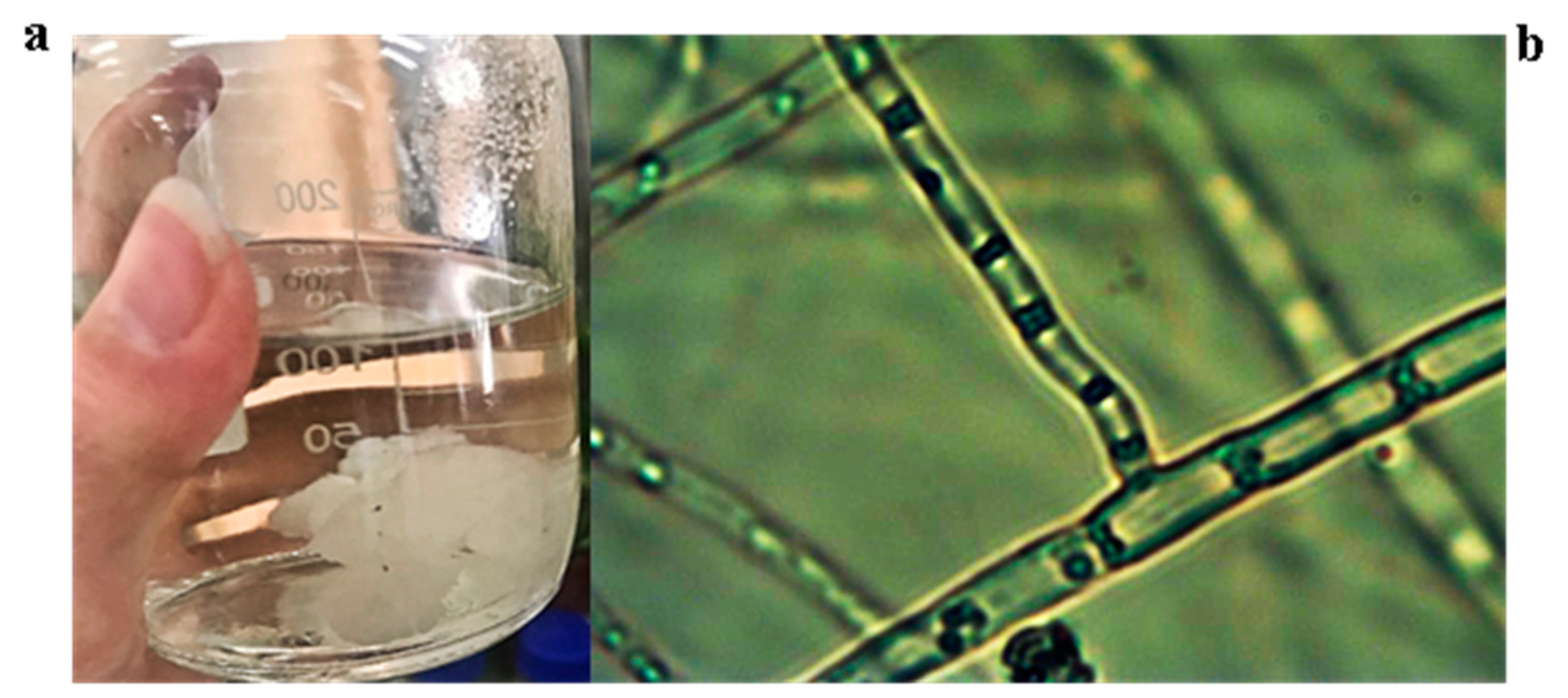
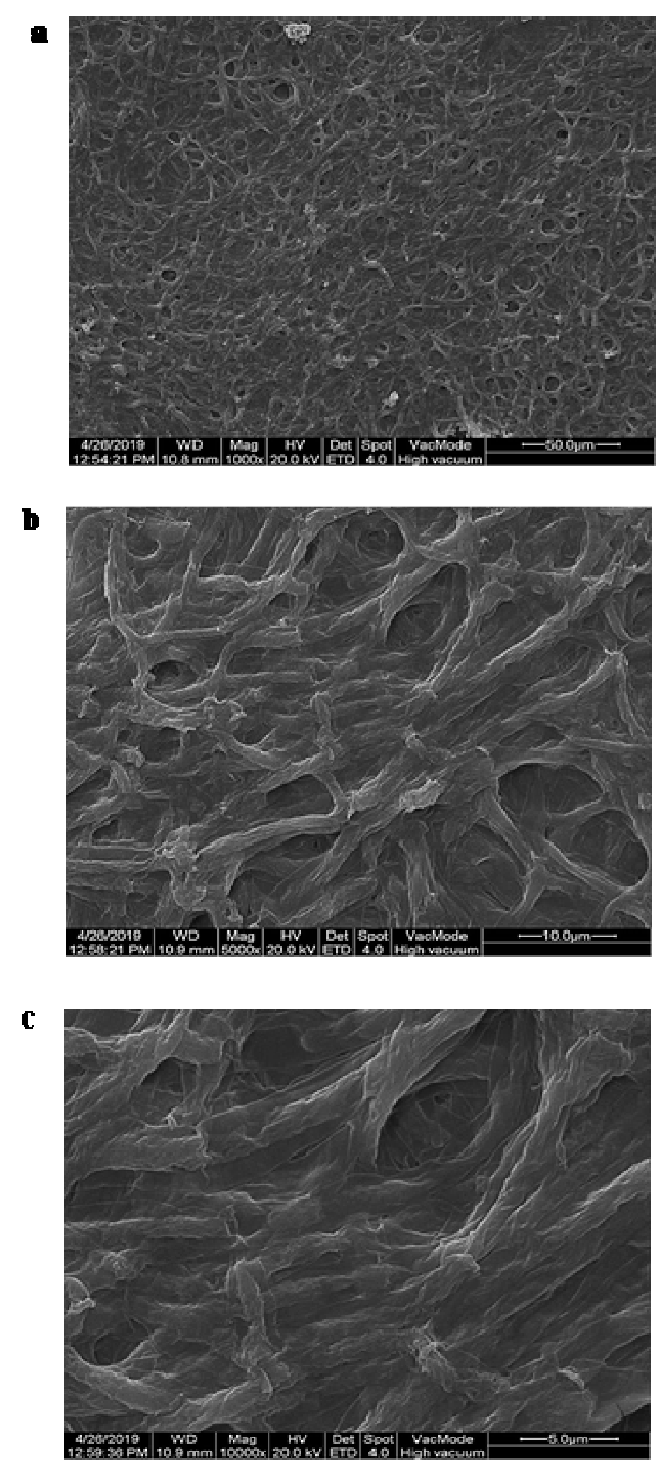
2.2. Morphology Observation of the Isolated Strains

An amount of the isolated strains was made into micro-slices, the morphology of color and hyphae was observed by a digital biomicroscope (Angel Precision Instrument Co., Ltd., Zhenjiang, China), and the data was recorded. Then the strains were coated and the ultrastructure was observed using a cold field emission scanning electron microscope (HITACHI S-4800, Hitachi Co., Ltd., Tokyo, Japan). The species of the strains were identified according to the morphological characteristics and consulting the relevant strain manual [35,36,37,38].

2.3. Pretreatment of the Samples and Extraction of Genomic DNA

The unknown bacteria were numbered and immediately stored in sterile reagent bottles. Total DNA extraction from the microorganisms was selected, and the quality of DNA extraction was detected by 0.8% agarose gel electrophoresis [39] and quantified by a TU-1810 spectrophotometer (PERSEE Analytical Instrument Ltd., Beijing, China).

2.4. PCR Amplification, Recovery and Purification of the PCR Products

Targeting ribosomal RNA sequences can reflect the composition and diversity of the microbial flora. The primers were designed according to the conserved region in the sequence, and the sample of the specific sequence was added to amplify the variable region (single or continuous multiple) or specific gene fragment of rRNA gene by PCR [40]. The first primer sequence of this sample was GGAAGTAAAAGTCGTAACAAGG, and the second primer sequence was GCTGCGTTCTTCATCGATGC. The amplified fragment size was 280 bp. The Q5 high-fidelity DNA polymerase (NEB Company, Ipswich, MA, USA) was used for 2720 PCR amplification instrument (ABI, Walsham, MA, USA) [41], which ensures that the same batch of samples has the same amplification conditions by strictly controlling the number of amplification cycles as low as possible. The amplified products of PCR were detected by 2% agarose gel electrophoresis, and the target fragments were recovered by AXYGEN gel recovery kit (Axygen Scientific Co., Ltd., Union City, CA, USA).
The PCR amplification system (25 μL) was as follows: 5× reaction buffer 5 μL, 5× GC buffer 5 μL, dNTP (2.5 mM) 2 μL, Forwardprimer (10 uM) 1 μL, Reverseprimer (10 uM) 1 μL, DNA Template 2 μL, ddH2O 8.75 μL, Q5 DNA Polymerase 0.25 μL.
The PCR amplification parameters were as follows: initial denaturation 98 °C 2 min, denaturation 98 °C 15 s, annealing 55 °C 30 s, extension 72 °C 30 s, final extension 72 °C 5 min, 10 °C hold. 25–30 cycles.

2.5. Preparation of the Medium and Strain Culture

The dominant species were tentatively identified as Irpex lacteus and Trichoderma through the ITS technique. The morphology of the white rot fungi was similar to that of the unknown fungus through the microscopic observation and referring the relevant literature, so three kinds of culture media were used to culture them for further research. The mycelium was extracted from the apricot kernels debitterizing water by washing with the deionized water, and 1 g of mycelium was cultured on PDA medium for 4 days. The strains preserved on the slant medium were selected, placed along with proper amount of fermentation medium (100 mL) into shaking flasks (250 mL) and cultured for 2 days in the incubator shaker (150 r/min, 26 °C). The optimum medium for treating Irpex lacteus was based on the research of Li et al. [42]. The specific nutrition formulas were as follows: 2.94 g lactose/100 mL, 1.5 g yeast extract/100 mL, 1.5 g beef extract/100 mL, 0.065 g KH2PO4·3H2O/100 mL, 0.06 g MgSO4·7H2O/100 mL, 0.0008 g NaCl/100 mL, and 0.0289 g VB1/100 mL.
The optimum medium for treating the white rot fungi was as follows [43]: 1 g glucose/100 mL, 0.07 g ammonium tartrate/100 mL, 0.1 g KH2PO4/mL, 0.02 g NaHPO4/mL, 0.04 g MgSO4/mL, 0.0005 g VB1/mL, 0.01 g CaCl2/mL, 0.01 g FeSO4/mL, 0.003 g ZnSO4/mL, and 0.0005 g MnSO4/mL.
PDB was chosen as the culture medium for Trichoderma, according to the literature [44].
Gradients of the cultured strains were diluted to different concentrations from 10−1 to 10−6, and repeated in triplicate. One hundred μL liquids of fermentation medium were removed by a pipette and cultured in the PDA solid medium. Plate counts were carried out at 27 °C at appropriate times, and were used to investigate the effect of the microorganisms on the degradation of amygdalin. The total number of colonies was calculated as follows:
Colony   count   ( g / mL ) = C a / D × V
where Ca is the average colony number, D is the dilution applied, and V is the volume of diluent.

2.6. Determination of Amygdalin by HPLC

The HPLC system (Dalian Elite Analytical Instruments Co., Ltd., Dalian, China) consisted of two P230II pumps, a UV230II detector and a ZW230II column oven. Chromatograms were recorded by the EC2006 software (Dalian Elite Analytical Instruments Co., Ltd., Dalian, China). The mobile phases were ultrasonically degassed for 25 min and filtered through a membrane of 0.45 μm before used. An isocratic elution was applied for the separation of amygdalin on a column of TC-C18 (5.0 μm, 4.6 mm × 250 mm, Agilent, Santa Clara, CA, USA) with methanol-H2O (28:72; v/v) as the mobile phase, being 1.0 mL/min of the flow rate, 35 °C as the column temperature, 214 nm as the detection wavelength, and 20 μL as the injection volume.

2.7. Degradation Investigation of the Amygdalin Caused by the Three Strains

A total of 100 mL of amygdalin standard solution (0.200 mg/mL) was taken and added in three 250 mL flasks (A, B, and C), respectively. Then, 1 mL of sterile water was added into the flask A and shaken as the control sample, 1 mL of the liquid culture medium of Irpex lacteus was added into the flask B, and 1 mL of the liquid culture medium of Irpex lacteus with 10−1 dilution was added into the flask C. Similarly, the other two strains were also conducted according to the aforementioned methods. Afterwards, the effects of different concentration of fungi were investigated on the degradation of amygdalin, and the degradation rate of amygdalin was calculated as follows:
Y ( % ) = ( A 0 A 1 ) / A 0
where Y is the degradation rate of amygdalin, A0 is the HPLC peak area for amygdalin in the solution without addition of fungi, and A1 is the HPLC peak area of amygdalin in the solution with fungi addition.

2.8. Measurement of the Beta-Glucosidase (β-GC) Activity

A total of 0.1 mL of fermentation medium and amygdalin standard solution with fungi addition were added into 1 mL of citric acid-sodium citrate extraction solution, then mixed in ice bath, and centrifuged at 4 °C, 12000 g for 10 min. The supernatant was separated as the crude β-GC sample to be tested. 0.05 mg/mL of β-GC standard were diluted to a certain concentration to make the standard curve.
p-Nitrophenyl-β-d-galactopyranoside (pNPG) was used as the reaction substrate, and p-nitrophenol was regarded as the standard to determine the enzyme activity. The measurement of the p-nitrophenol standard solution (1 μmol/mL) with dilutions of 6.25, 12.5, 25, 50, and 100 nmol/mL was as shown in Table 1. The calibration curve for p-nitrophenol was y = 0.007x + 0.003, R2 = 0.9986.
The samples were prepared according to Table 1, and placed at room temperature for 2 min. The corresponding control tube was used to calibrate the zero point of the UV spectrophotometer, and the absorbance at 400 nm of the p-nitrophenol was measured. The specific calculation method of the β-GC activity was as follows:
β GC   enzyme   activity   ( U / 10 4   cell ) = ( y × V 1 ) ÷ ( 1000 × V 2 ÷ V 3 ) ÷ T = 0.02 × y
where y stands for the concentration of p-nitrophenol generated, T is the reaction time (0.5 h), V1 is total volume of reaction system (1 mL), and V2 and V3 are the volumes of sample (0.1 mL) and citric acid-sodium citrate extraction solution (1 mL).

2.9. Statistical Analysis

The OTU (operational taxonomic units) abundance information and taxonomic composition data of each sample were mapped to the microbial taxonomy tree provided by NCBI Taxonomy using the software MEGAN [45], so that the specific composition of all samples at each taxonomic level can be presented uniformly in a standard taxonomy system.
All the results were statistically analyzed by calculating the mean and the standard deviation, and were presented as mean ± standard deviations of three determinations. Analysis of variance (ANOVA) was conducted using the SPSS statistics software, version 11.0 (SPSS Inc., Chicago, IL, USA).

3. Results and Discussion

3.1. Morphology Characterization of Strains

The micro-morphology of the suspended cotton-like mycelium in the debitterizing water of apricot kernels was characterized by the digital biomicroscopy and cold field emission scanning electron microscopy, respectively. As shown in Figure 1, the structure of the mycelium is transparent and branched, and there are many nuclei and no segregations in the mycelium, which could be preliminarily identified for the germination and reproduction. The mycelium is densely distributed and has many branches as observed by the cold field emission scanning electron microscope (Figure 2). Through the morphological observations and referring to the literature [36], the colony could tentatively be identified as a lower fungal organism, and its further identification as fungal species was subsequently investigated by the high-throughput ITS sequencing technique.

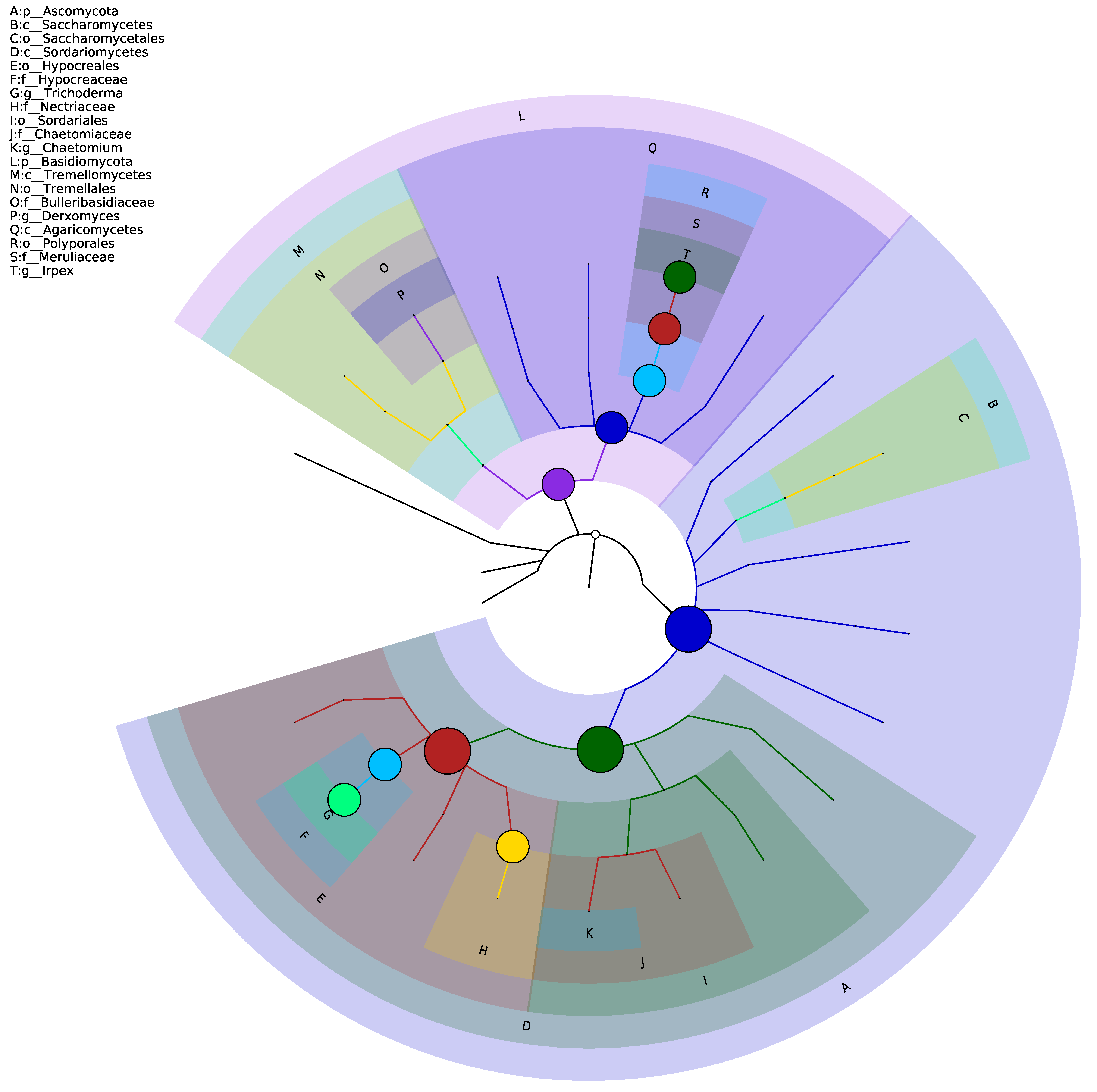
3.2. Diversity Analysis of the Fungal in the Samples

Both Chao 1 and ACE represent the estimation index of richness, which is usually used to estimate the actual number of the species in the community. Generally, ACE has a wider detection range than Chao 1, the larger the Chao 1 or the ACE index is, the higher the community richness is. Furthermore, the higher the Shannon diversity index is, the higher the community diversity is. The results in Table 2 demonstrated that, at the level of OUT, the Chao 1 indexes of the three samples were 26, 35, and 161.54 at different times, the ACE indexes were 26, 31.44, and 168.31, the Shannon indexes were 0.19, 0.02, 2.19, and the Simpson indexes were 0.0386, 0.0029, and 0.485, respectively. An increasing trend is shown by the richness index of the three samples, while the diversity showed an initial decrease, followed by an increase. The results indicate that the longer the community is stored, the richer the community will be, and the more diverse the community will be. The reason remains unknown, and it will be further studied.

3.3. Distribution of the Fungal Colonies at Different Levels

Table 3 shows the results of the compositions and abundance distribution of the three colonies at five taxonomic levels of phylum, class, order, family, and genus, and Figure 3 illustrates the mapped tree for classification of hierarchy of the three colonies, which is usually used to reflect the grade relationship of the sample classification. The average of the relative abundance in the total determined samples is represented by the circle size, and the larger the circle is, the greater the sample richness is. Combined with Table 3 and Figure 3, it could be seen that 98% of the dominant fungi in the first fungus colony level are Basidiomycota, Agaricomycetes, Polyporales, Meporidae. Ruliaceae and Irpex, and about 1.8% of the dominant fungi are Ascomycota, Sordariomycetes, Hypocreales, Hypocreaceae and Trichoderma. The second fungus had 99.6% of the dominant fungi, such as Ascomycota, Sordariomycetes, Hypocreales and Nectriaceae, while the third fungus had 99.9% of dominant fungi, Ascomycota, Sordariomycetes, Hypocreales, Hypocreaceae and Trichoderma. The second was consistent with the third with the 0.1% dominant fungi. The reason might be attributed to the quick growth rate of Trichoderma fungi, which is much faster than that of the Irpex lacteus, which is also in agreement with the existing research that the bacterial diversity of Maotai flavor liquor increased with the extension of storage time [46]. The richness of the community increases with the increase of time, and the Trichoderma species multiply in large quantities in the debitterizing water of apricot kernels.

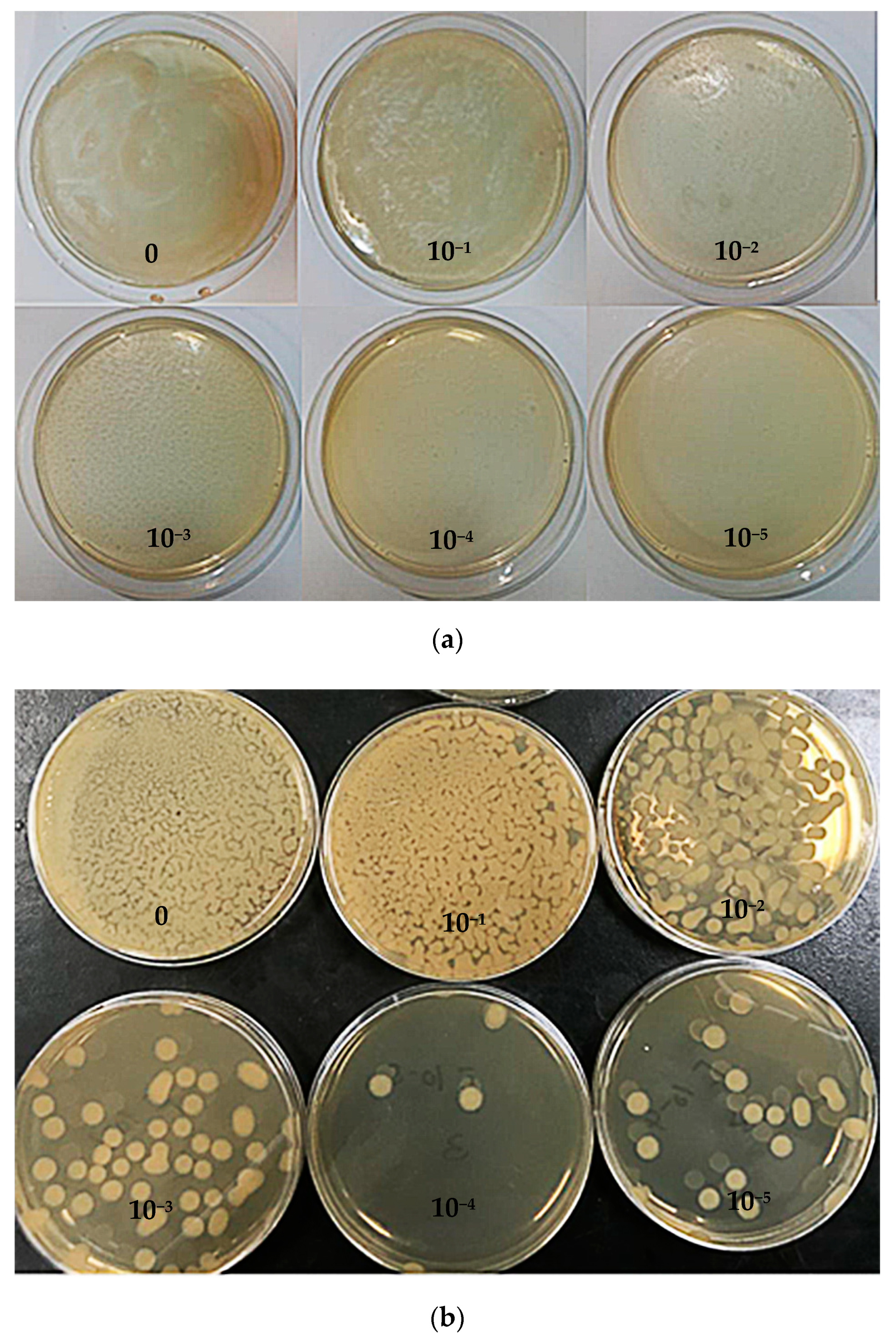
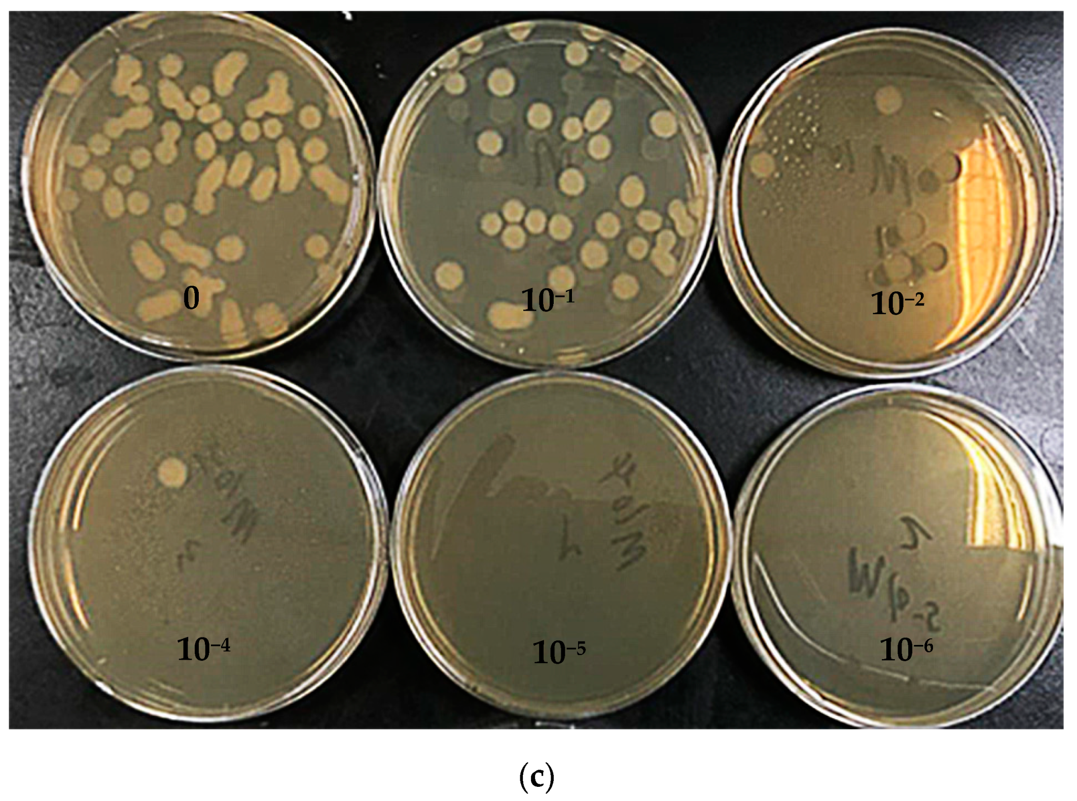
3.4. Colony Count in the Culture Medium

According to the medium configuration of the dominant species, the colony numbers were determined, and the results are shown in Figure 4 and Table 4. The total colony numbers of Irpex lacteus and white rot fungi were 6.4 × 10⁷ CFU/mL and 5.4 × 10⁷ CFU/mL after two days culture, respectively. The total colony numbers of Trichoderma were 6.8 × 10⁴ CFU/mL after one day culture. The total colony numbers of Irpex lacteus in the dilutions of 10−1 and 10−2 were 6.4 × 10⁶ CFU/mL and 6.4 × 10⁵ CFU/mL, respectively. Similarly, the total colony numbers of the white rot fungi in the dilutions of 10−1 and 10−2 were 5.4 × 10⁷ CFU/mL and 5.4 × 10⁶ CFU/mL, respectively. For Trichoderma, the total colony numbers were 6.8 × 10⁷ CFU/mL and 6.8 × 10⁶ CFU/mL in the dilutions of 10−1 and 10−2, respectively. All the results indicate that Trichoderma had the strongest reproduction ability during the culture, followed by the white rot fungus and Irpex lacteus, which is consistent with the results from the ITS analysis. It could be deduced that the dominant species in the original community should be the Irpex lacteus, although Trichoderma became the dominant genus, due to its strong reproduction ability.

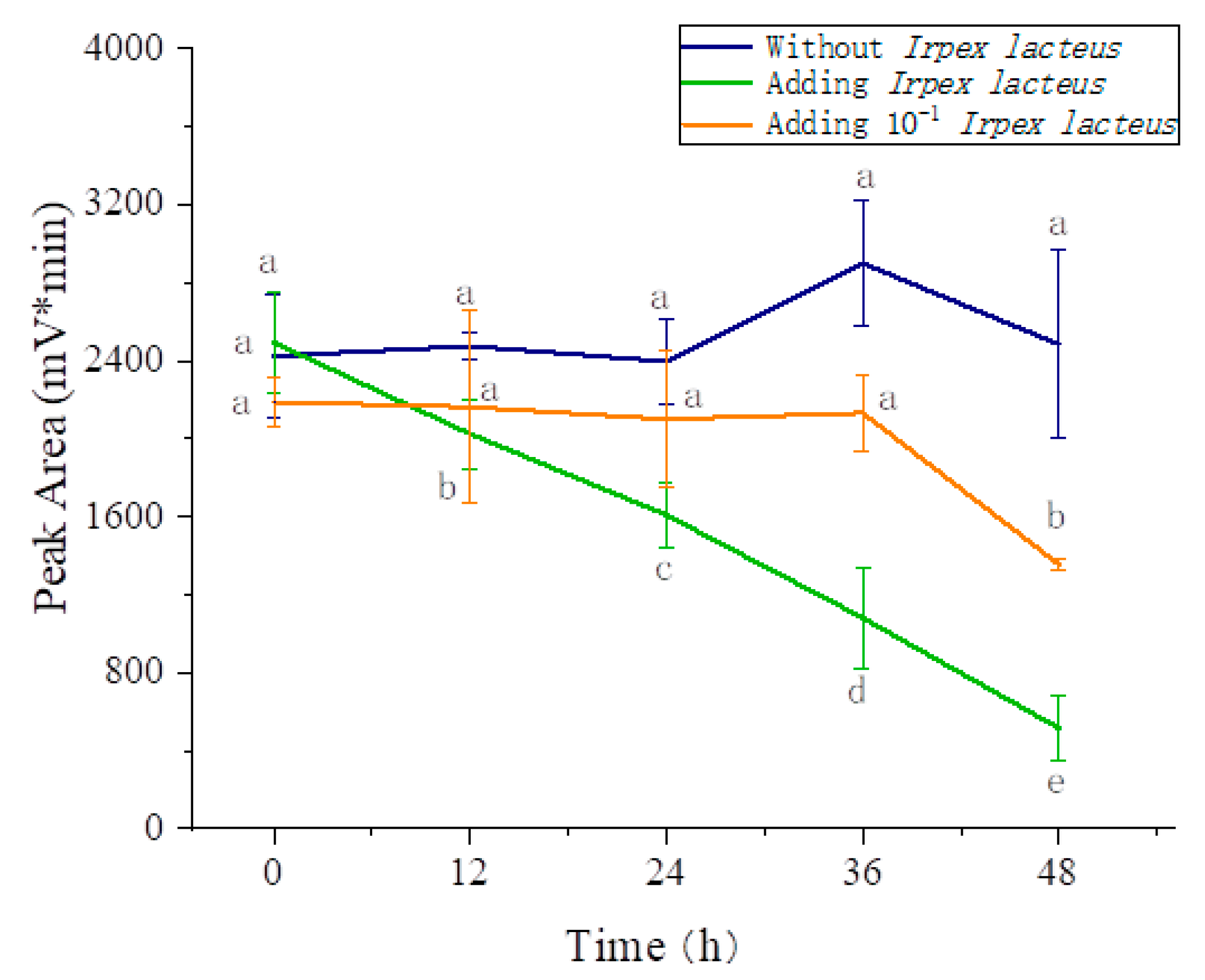
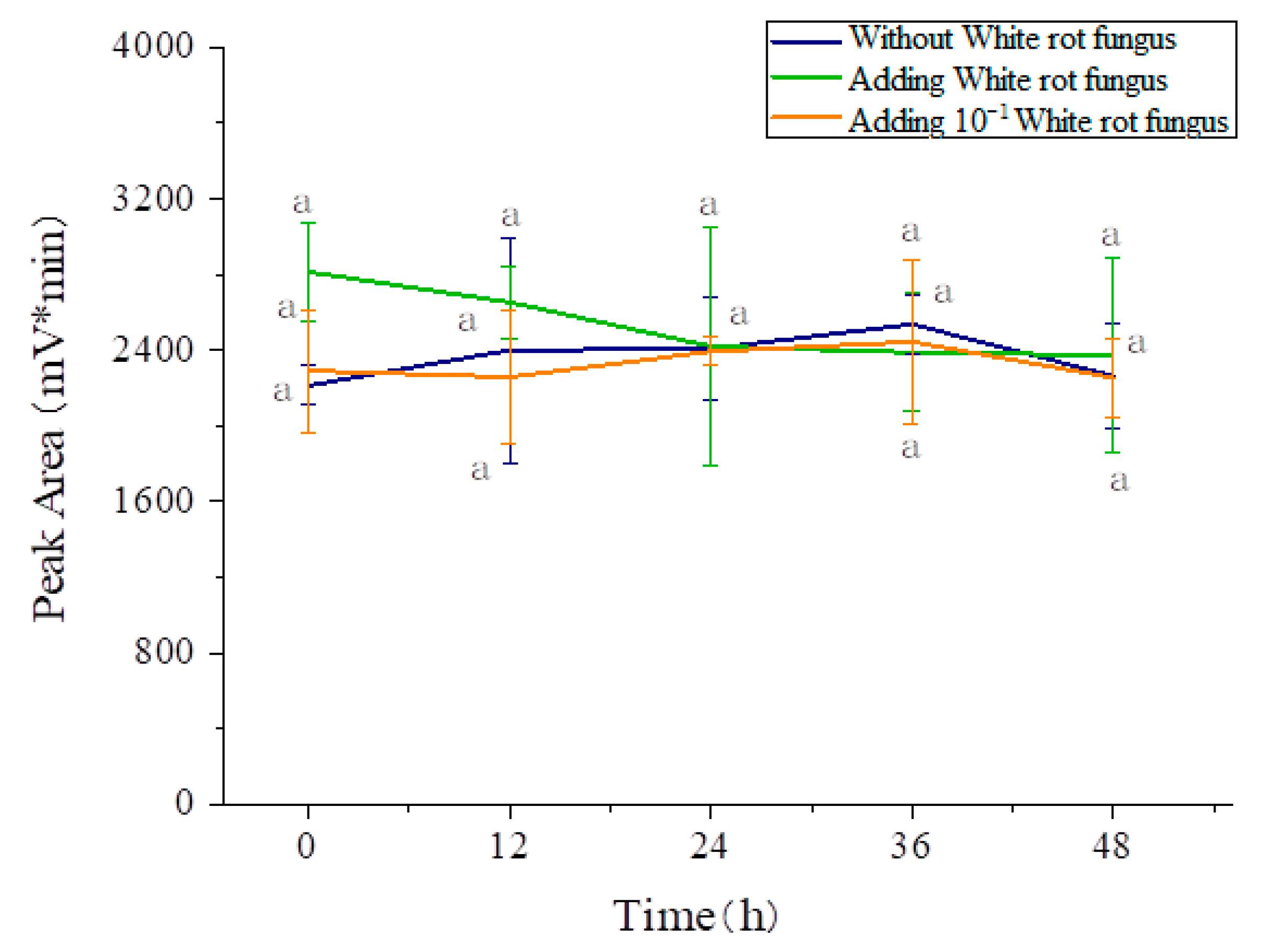
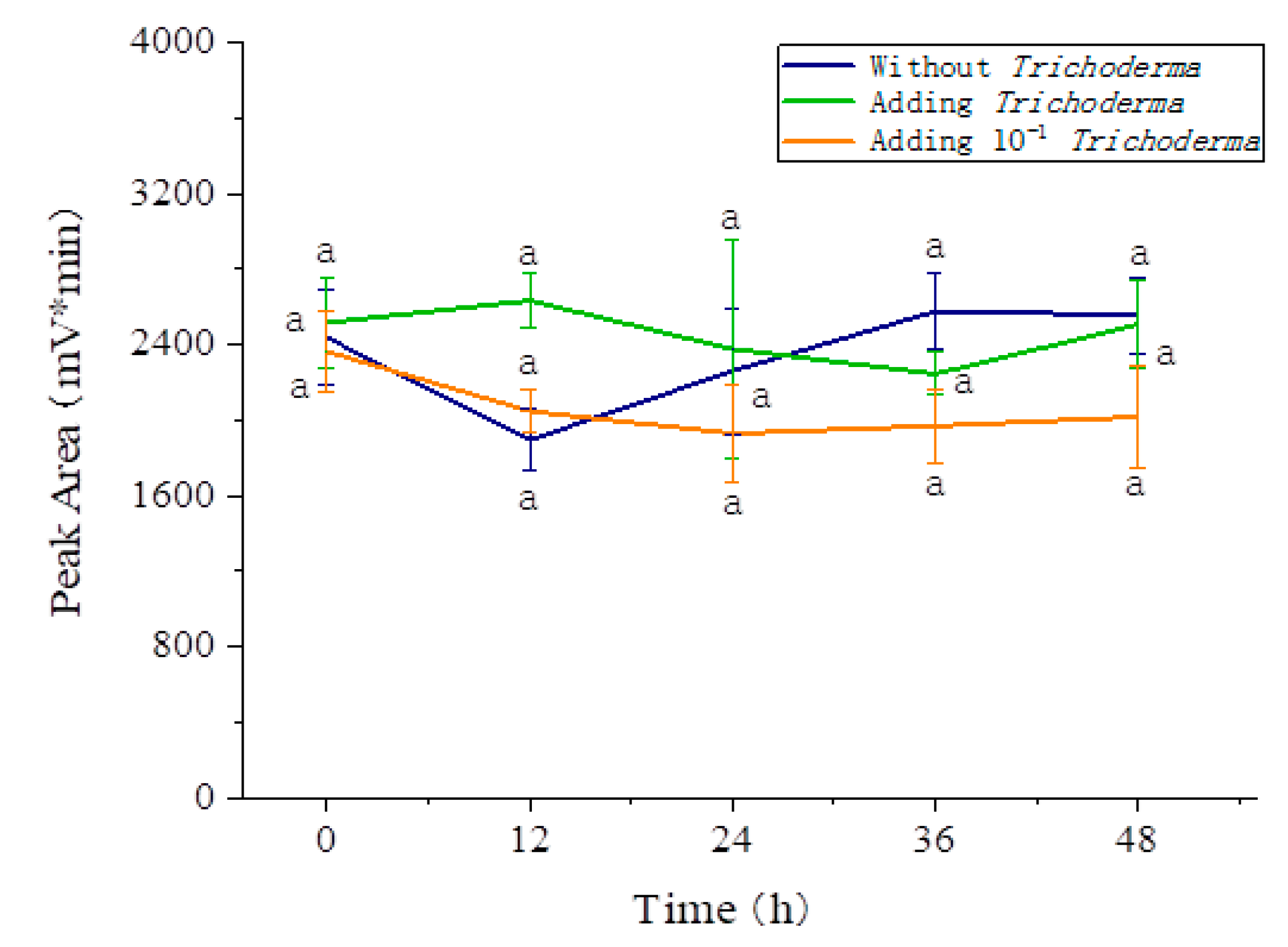
3.5. Effects of the Three Fungi on the Degradation of Amygdalin

The effects of the three fungi on the degradation of amygdalin were investigated and the results are shown in Figure 5 Figure 6 Figure 7, respectively. It can be seen that the addition of Irpex lacteus had a noticeable effect on amygdalin degradation, while the contents of the amygdalin were not influenced by the addition of white rot fungus and Trichoderma. In comparison, the amygdalin in the solution without Irpex lacteus addition was hardly degraded during the storage, while 79.31% of the amygdalin in the solution with Irpex lacteus was degraded after 48 h storage. The degradation of the amygdalin in the 10−1 diluted solution could not be detected within the first 36 h; however, with the increase of time, the degradation rate reached 38.06% at 48 h. Moreover, the colony numbers of Irpex lacteus, which could degrade the amygdalin, were in the range of 6.4 × 10⁶ CFU/mL and 6.4 × 10⁷ CFU/mL, and the more the colonies of Irpex lacteus, the better the degradation of amygdalin, which is consistent with the available literature [47]. Compared with the lactic acid bacteria, the yeast and filamentous fungi had a stronger degradation rate of amygdalin, and the colony numbers had a certain impact on the degradation. The Irpex lacteus screened from the debitterizing water might be considered for further deep study on its degrading of amygdalin in the debitterizing wastewater of apricot kernels, so as to clarify the water in future, while Trichoderma and white rot fungus can be regarded as not suitable for treating the amygdalin in the wastewater.

3.6. Effects of the Three Fungi on the Activity of β-Glucosidase

As is well known, β-glucosidase is the most important endogenous enzyme in the apricot kernels. Once the kernels are damaged and in contact with water, some amygdalin would be degraded by the β-glucosidase, resulting in the debitterizing of apricot kernels. As mentioned above, the three fungi isolated from the debitterizing had a certain influence on the amygdalin degradation; however, the reason for this is not clear. In order to well understand the mechanism, the β-glucosidase activity in solutions with addition of the three fungi was determined, respectively. As shown in Table 5, the activities of β-glucosidase were all negative and close to zero, which might suggest that no β-glucosidase was produced over the growth of the strains and the degradation of amygdalin was not caused by the β-glucosidase. Considering the effect of Irpex lacteus on the degradation of amygdalin, its mechanism is not clear, and further study would need to be conducted in the future. Generally, the degradation of fungus such as white rot fungus could be influenced by the medium conditions, and the optimal medium condition can stimulate the fungus by stress reaction to produce some enzymes such as laccases and peroxidases for degrading some organic pollutants [48], which might suggest that the amygdalin degradation by fungi (especially Irpex lacteus) could be improved by optimizing the medium conditions, and this work would need to be investigated in future.

4. Conclusions

The cotton-like suspended mycelia in the debitterizing water were tentatively identified for the genus of lower fungi by observing the microscopic morphology. Three species of microorganisms were further confirmed through the ITS high-throughput sequencing analysis, including Irpex, Trichoderma, and white rot fungus. The results indicate that the species Trichoderma and white rot fungus had no effects on the amygdalin degradation, while Irpex lacteus had a definite promoting effect on the degradation of amygdalin, being the suitable degrading colony number from 6.4 × 10⁶ CFU/mL to 6.4 × 10⁷ CFU/mL. This might be attributed to the other metabolites of Irpex lacteus apart from the β-glucosidase such as laccases and peroxidases, and the specific mechanisms need to be further studied. This notwithstanding, this research might provide a novel promising approach to treat the debitterizing water so as to reduce the water pollution from the processing industry of apricot kernels.

Author Contributions

In this research, all authors contributed effectively. N.Z. designed, achieved experiments, performed research and wrote the paper; J.-L.Y. analyzed the data, performed data interpretation; Q.-A.Z. supervised the project and revised the manuscript; J.F.G.-M. proposed the design, the frame work of the manuscript, and modified the revised manuscript. All authors have read and agreed to the published version of the manuscript.

Funding

This study was funded by National Natural Science Foundation of China [No. 31101324, 31972206], Key Research Development Program of Shaanxi Province, China [2018ZDXM-NY-086], and the Innovation Talents of Science and Technology Serving Enterprise Project of Xi’an, Shaanxi Province, China [2020KJRC0011].

Conflicts of Interest

Author of Qing-An Zhang has received research grants from the above-mentioned organizations, and declares that he has no conflict of interest. Ning Zhang declares that she has no conflict of interest. Jian-Li Yao declares that she has no conflict of interest, and Juan F. García-Martín also declares that he has no conflict of interest.

References

  1. Bolarinwa, I.F.; Orfila, C.; Morgan, M.R.A. Amygdalin content of seeds, kernels and food products commercially-available in the UK. Food Chem. 2014, 152, 133–139. [Google Scholar] [CrossRef] [Green Version]
  2. Dixit, A.K. Drying characteristics of wild apricot (Prunus armeniaca) fruit bar and economic evaluation of market potential of the enterprise. Indian J. Agric. Sci. 2018, 83. [Google Scholar] [CrossRef]
  3. Zacharias, E. Carbohydrates; Springer: New York, NY, USA, 2012; pp. 127–139. [Google Scholar] [CrossRef]
  4. Zhang, Q.-A.; Wei, C.-X.; Fan, X.-H.; Shi, F.-F. Chemical compositions and antioxidant capacity of by-products generated during the apricot kernels processing. CyTA J. Food 2018, 16, 422–428. [Google Scholar] [CrossRef] [Green Version]
  5. Abboud, M.M.; Awaida, W.A.; Alkhateeb, H.H.; Abu-Ayyad, A.N. Antitumor action of amygdalin on human breast cancer cells by selective sensitization to oxidative stress. Nutr. Cancer 2019, 71, 483–490. [Google Scholar] [CrossRef] [PubMed]
  6. Zhang, A.; Pan, W.; Lv, J.; Wu, H. Protective effect of amygdalin on LPS-induced acute lung injury by inhibiting NF-κB and NLRP3 signaling pathways. Inflammation 2017, 40, 745–751. [Google Scholar] [CrossRef] [PubMed]
  7. Bolarinwa, I.F.; Orfila, C.; Morgan, M.R.A. Determination of amygdalin in apple seeds, fresh apples and processed apple juices. Food Chem. 2015, 170, 437–442. [Google Scholar] [CrossRef] [PubMed] [Green Version]
  8. Zhang, N.; Zhang, Q.-A.; Yao, J.-L.; Zhang, X.-Y. Changes of amygdalin and volatile components of apricot kernels during the ultrasonically-accelerated debitterizing. Ultrason. Sonochem. 2019, 58, 104614. [Google Scholar] [CrossRef] [PubMed]
  9. Lv, Z.; Jiao, Z.; Liu, J.; Liu, H.; Zhang, C.; Yang, W. Response surface optimization of ultrasonic-assisted extraction and HPCPC purification of amygdalin from bitter apricot kernel meal (Prunus armeniaca). Food Sci. 2016, 37, 81–87. [Google Scholar] [CrossRef]
  10. Silem, A.; Günter, H.O.; Einfeldt, J.; Boualia, A. The occurrence of mass transport processes during the leaching of amygdalin from bitter apricot kernels: Detoxification and flavour improvement. Int. J. Food Sci. Technol. 2006, 41, 201–213. [Google Scholar] [CrossRef]
  11. Zhang, N.; Zhang, X.-Y.; Fan, X.-H.; Zhang, Q.-A. Optimization on fast debitterizing technologies of apricot seed by ultrasound with response surface methodology. Food Mach. 2018, 34, 189–194. (In Chinese) [Google Scholar] [CrossRef]
  12. Zhang, N.; Zhang, Q.-A.; Yao, J.-L. Consecutive membrane filtration and re-utilization of the debitterizing wastewater of apricot kernels for a flavor beverage-making. J. Clean. Prod. 2020, 262, 121360. [Google Scholar] [CrossRef]
  13. Song, Y.; Zhang, Q.-A.; Fan, X.-H.; Zhang, X.-Y. Effect of debitterizing treatment on the quality of the apricot kernels in the industrial processing. J. Food Proc. Preser. 2017, 42, 1–8. [Google Scholar] [CrossRef]
  14. Jin, Y.N.; Wu, M.F.; Zhao, G.H.; Li, M.F. Photocatalysis-enhanced electrosorption process for degradation of high-concentration dye wastewater on TiO2/carbon aerogel. Chem. Eng. J. 2011, 168, 1248–1255. [Google Scholar] [CrossRef]
  15. Nout, M.J.; Tunçel, G.; Brimer, L. Microbial degradation of amygdalin of bitter apricot seeds (Prunus armeniaca). Int. J. Food Microbiol. 1995, 24, 407–412. [Google Scholar] [CrossRef]
  16. Zhu, J.; Sanidad, K.Z.; Sukamtoh, E.; Zhang, G. Potential roles of chemical degradation in the biological activities of curcumin. Food Funct. 2017, 8, 907–914. [Google Scholar] [CrossRef]
  17. Chen, L.; Wu, H. Research advances in degradation of polysaccharides. Chin. Arch. Tradit. Chin. Med. 2008, 26, 133–135. (In Chinese) [Google Scholar] [CrossRef]
  18. Pan, Z. Research development of the degradation methods of lignin. Guangdong Chem. Indus. 2015, 42, 100–101. (In Chinese) [Google Scholar] [CrossRef]
  19. Hu, Y.; Catchmark, J.M. Integration of cellulases into bacterial cellulose: Toward bioabsorbable cellulose composites. J. Biomed. Mater. Res. Part B 2011, 97, 114–123. [Google Scholar] [CrossRef]
  20. Purschke, B.; Meinlschmidt, P.; Horn, C.; Rieder, O.; Jäger, H. Improvement of techno-functional properties of edible insect protein from migratory locust by enzymatic hydrolysis. Eur. Food Res. Technol. 2018, 244, 1–15. [Google Scholar] [CrossRef] [Green Version]
  21. Hao, T.; Yan, J.M.; Wan, Y.L.; Jian, W.W. Efficient degradation of triclosan by an endophytic fungus Penicillium oxalicum B4. Environ. Sci. Pollut. Res. 2018, 25, 8963–8975. [Google Scholar] [CrossRef]
  22. Feng, Y.M.; Fan, X.H.; Zhan, H.; Teng, S.Y.; Yang, F.; Chen, S.H. Research progress on ecotoxicity and microbial degradation of strobilurin fungicides. Biotechnol. Bull. 2017, 33, 52–58. [Google Scholar] [CrossRef]
  23. Meng, Y.; Fu, Y.B.; Liang, S.K.; Chen, W.; Liu, Z.H. Effects of oil pollutants on the performance of marine benthonic microbial fuel cells and its acceleration of degradation. Environ. Sci. 2015, 36, 3080–3085. [Google Scholar] [CrossRef]
  24. Hechmi, N.; Bosso, L.; El-Bassi, L.; Scelza, R.; Testa, A.; Jedidi, N.; Rao, M.A. Depletion of pentachlorophenol in soil microcosms with byssochlamys nivea and scopulariopsis brumptii as detoxification agents. Chemosphere 2016, 165, 547–554. [Google Scholar] [CrossRef] [PubMed]
  25. Tian, H.; Yan, M.; Treu, L.; Angelidaki, I.; Fotidis, I.A. Hydrogenotrophic methanogens are the key for a successful bioaugmentation to alleviate ammonia inhibition in thermophilic anaerobic digesters. Bioresour. Technol. 2019, 293, 122070. [Google Scholar] [CrossRef]
  26. Zhang, X.; Cai, T.; Xu, X. Isolation and identification of a tetracycline-degrading bacterium and optimizing condition for tetracycline degradation. Biotechnol. Bull. 2015, 1, 173–180. (In Chinese) [Google Scholar] [CrossRef]
  27. Yang, T.; Yan, H.L. Comparative analysis of bacteria associated with different mosses by 16S rRNA and 16S rDNA sequencing. J. Basic Microbiol. 2016, 57, 57–67. [Google Scholar] [CrossRef] [Green Version]
  28. Cao, B.Y.; Wang, M.; Liu, L.; Zhou, Z.M.; Wen, S.P.; Rozalskif, A.; Wang, L. 16S-23S rDNA internal transcribed spacer regions in four Proteus species. J. Microbiol. Methods 2009, 77, 109–118. [Google Scholar] [CrossRef]
  29. Wang, P.; Cui, Z.L.; Liu, B.; Sun, B.; Cao, H. Effect of cultivation methods on bacterial diversity by PCR-rflp analysis. Acta Pedol. Sin. 2009, 46, 1096–1101. [Google Scholar] [CrossRef]
  30. Wang, L.; Li, B.; Zhu, J. Review of high-throughput sequencing techniques on constructed wetland microbial diversity. Chin. Agric. Sci. Bull. 2016, 32, 10–15. [Google Scholar]
  31. Xiong, W.; Li, H.T.; Zhan, A.B. Early detection of invasive species in marine ecosystems using high-throughput sequencing: Technical challenges and possible solutions. Mar. Biol. 2016, 163, 139. [Google Scholar] [CrossRef]
  32. Dalmassoa, A.; Civeraa, T.; Pattonoa, D.; Cardazzob, B.; Botteroa, M.T. Characterization of microbiota in Plaisentif cheese by high-throughput sequencing. LWT Food Sci. Technol. 2016, 69, 490–496. [Google Scholar] [CrossRef]
  33. He, G.Q.; Liu, T.J.; Sadiq, F.A.; Gu, J.S.; Zhang, G.H. Insights into the microbial diversity and community dynamics of Chinese traditional fermented foods from using high throughput sequencing approaches. J. Zhejiang Univer. Sci. B 2017, 18, 289–302. [Google Scholar] [CrossRef] [PubMed]
  34. Lu, Y.Z.; Ding, Z.W.; Ding, J.; Fu, L.; Zeng, R.J. Design and evaluation of universal 16S rRNA gene primers for high-throughput sequencing to simultaneously detect DAMO microbes and anammox bacteria. Water Res. 2015, 87, 385–394. [Google Scholar] [CrossRef]
  35. Bary, A.D.; Balfour, I.B. Comparative Morphology and Biology of the Fungi, Mycetozoa and Bacteria; Clarendon Press: Oxford, UK, 2015; p. 554. [Google Scholar]
  36. Pazouki, M.; Panda, T. Understanding the morphology of fungi. Bioprocess Eng. 2000, 22, 127–143. [Google Scholar] [CrossRef]
  37. Bosso, L.; Lacatena, F.; Varlese, R.; Nocerino, S.; Cristinzio, G.; Russo, D. Plant pathogens but not antagonists change in soil fungal communities across a land abandonment gradient in a Mediterranean landscape. Acta Oecol. 2017, 78, 1–6. [Google Scholar] [CrossRef]
  38. Mohanty, S.; Ghosh, S.; Nayak, S.; Das, A.P. Isolation, identification and screening of manganese solubilizing fungi from low-grade manganese ore deposits. Geomicrobiol. J. 2017, 34, 309–316. [Google Scholar] [CrossRef]
  39. Anandan, R.; Deenathayalan, T.; Prakash, M.; Sunilkumar, B.; Narayanan, G.S. Sassessment of genetic diversity among sesame (Sesamum indicum L.) germplasm as revealed by RAPD and SSR markers. Indian J. Biochem. Biophys. 2018, 55, 143–150. [Google Scholar]
  40. Sun, K.; Wei, S.; Liu, Y.L.; Xu, R.S.; Liu, Z.Y.; Yin, J.P.; Yuan, J.J.; Wu, X.Y. Real-time PCR for the detection of Lactobacillus plantarum based on dual priming oligonucleotide system. Food Fermn. Ind. 2016, 42, 178–183. (In Chinese) [Google Scholar] [CrossRef]
  41. De, A.; Jorgensen, A.N.; Beatty, W.L.; Lemos, J.; Wen, Z.T. Deficiency of MecA in streptococcus mutans causes major defects in cell envelope biogenesis, cell division, and biofilm formation. Front. Microbiol. 2018, 9, 2130. [Google Scholar] [CrossRef]
  42. Li, M.; Du, L.N.; Zhang, Y.; Zhang, N.; Teng, L.R. Improvement of medium for the fermentation of Irpex lacteus Fr.by respond surface methodology coupled with desirability function. Chin. J. Pharm. 2011, 42, 17–20. (In Chinese) [Google Scholar] [CrossRef]
  43. Wang, M.L. Study on optimization of wastewater with DDNP of high concentration by medium of white rot fungus. Shanxi Chem. Ind. 2015, 35, 78–81. (In Chinese) [Google Scholar] [CrossRef]
  44. Lv, S.X.; Yu, X.D.; Zhang, C.X.; Chen, J. Influence of fermentation conditions on hyphae growth and chitinase activity of Trichoderma strain of T23. J. Shenyang Agric. Univ. 2005, 36, 332–335. (In Chinese) [Google Scholar] [CrossRef]
  45. Huson, D.H.; Weber, N. Microbial community analysis using MEGAN. Methods Enzymol. 2013, 531, 465–485. [Google Scholar] [CrossRef] [PubMed]
  46. Dai, Y.J.; Li, Z.J.; Tian, Z.Q. Analysis of bacterial diversity in Daqu and fermented grains for Maotai-flavor liquor. Food Sci. 2019, 40, 152–159. [Google Scholar] [CrossRef]
  47. Brimer, L.; Tuncel, G.; Nout, M.J.R. Simple screening procedure for microorganisms to degrade amygdalin. Biotechnol. Tech. 1993, 7, 683–687. [Google Scholar] [CrossRef]
  48. Xu, C.Y. Advance in decolorization and biodegradation of dye by white rot fungus. Prog. Biotechnol. 2002, 22, 57–60. [Google Scholar] [CrossRef]
Figure 1. Macromorphological (a) and micromorphological (b) characteristics of the cotton-like mycelium (100×).
Processes 08 01061 g001
Figure 2. Field emission scanning electron microscope pictures of the cotton-like mycelium (with magnifications of (a): 1000×, (b): 5000×, (c): 10,000×).
Processes 08 01061 g002
Figure 3. GraPhlAn-based classification hierarchical tree map chart of dominant fungi communities in the debitterizing water of apricot kernels.
Processes 08 01061 g003
Figure 4. Plate counting of (a) Irpex lacteus, (b) white rot fungus, and (c) Trichoderma.
Processes 08 01061 g004aProcesses 08 01061 g004b
Figure 5. Effect of colony numbers of Irpex lacteus on the degradation of amygdalin.
Processes 08 01061 g005
Figure 6. Effect of colony numbers of white rot fungus on the degradation of amygdalin.
Processes 08 01061 g006
Figure 7. Effect of colony numbers of Trichoderma on the degradation of amygdalin.
Processes 08 01061 g007
Table 1. Measurement of the β-glucosidase activity.
ReagentsControlMeasurementStandard Tube
pNPG (μL) 400
Disodium hydrogen phosphate-citric acid buffer (μL)500500
β-glucosidase crude enzyme solution (μL)100100
Operation step 1
pNPG (μL)400
Operation step 2
Supernatant (μL)500500
p-Nitrophenol standard solution (μL) 500
Na2CO3 (μL)100010001000
Step 1: The mixture was quickly mixed and placed in a 37 °C water bath for 30 min. The heated mixture was immediately put into a boiling water bath for 5 min to inactivate the enzymes and terminate the reaction. Finally, the boiled solution was cooled to room temperature. Step 2: After pNPG addition and mixing, the samples were centrifuged at 8000× g at 4 °C for 5 min, and the supernatant was taken for the following operation.
Table 2. Diversity index of fungi communities.
SamplesSimpsonChao1ACEShannon
10.038626.0026.000.19
20.00293531.440.02
30.485161.54168.312.19
Table 3. Distribution of dominant fungi communities at different levels in the debitterizing water of apricot kernels.
LevelOUT ID123
PhylumAscomycota1.9%99.9%99.8%
Basidiomycota98.1%0.1%0.1%
Other 0.1%
ClassSordariomycetes1.9%99.9%99.7%
Agaricomycetes98.1%0.1%0.1%
Other 0.1%
Tremellomycetes 0.1%
OrderHypocreales1.9%99.9%99.7%
Polyporales98.0%0.1%0.1%
Other 0.1%
Tremellales 0.1%
Sordariales0.0%
FamilyHypocreaceae1.8% 99.6%
Nectriaceae 99.9%
Meruliaceae98.0%
Other 0.1%
GenusTrichoderma1.8% 99.6%
Unidentified 99.9%
Irpex98.0%0.1%0.1%
Other 0.1%
Table 4. Colony counting results.
SamplesAverage of Colony (pcs)Colony Count (CFU/mL)
Iprex lacteus64 ± 76.4 × 10⁷
White rot fungi54 ± 55.4 × 10⁸
Trichoderma68 ± 116.8 × 10⁸
Table 5. Contents of β-glucosidase in the standard amygdalin solution with the three identified species.
Timeβ-Glucosidase Activity (U/104 cell)
Irpex lacteusTrichodermaWhite Rot Fungus
0 h−0.10 ± 0.16−0.41 ± 0.26−0.26 ± 0.15
12 h−0.22 ± 0.19−0.33 ± 0.16−0.10 ± 0.33
24 h−0.45 ± 0.17−0.88 ± 0.19−0.26 ± 0.30
36 h−0.28 ± 0.09−0.52 ± 0.19−0.11 ± 0.23
48 h−0.30 ± 0.03−0.32 ± 0.24−0.05 ± 0.09

