Review

Downstream Processes in a Microalgae Biorefinery: Cascaded Enzymatic Hydrolysis and Pulsed Electric Field as Green Solution

by
Gianpiero Pataro
*,
Elham Eslami
,
Francesco Pignataro
and
Alessandra Procentese
*
Department of Industrial Engineering, University of Salerno, Via Giovanni Paolo II, 132, 84084 Fisciano, SA, Italy
*
Authors to whom correspondence should be addressed.
Processes 2025, 13(6), 1629; https://doi.org/10.3390/pr13061629
Submission received: 14 March 2025 / Revised: 19 April 2025 / Accepted: 20 May 2025 / Published: 22 May 2025
(This article belongs to the Special Issue Process Intensification towards Sustainable Biorefineries)

Abstract

:
Microalgae are a promising source of valuable compounds, including proteins, pigments, lipids, vitamins, and ingredients for cosmetics and animal feed. Despite their potential, downstream processing remains a major bottleneck in microalgae biorefineries, particularly in achieving high extraction efficiency with low energy and chemical input. While several extraction methods exist, few balance efficiency with selectivity and sustainability. Recently, mild and selective techniques such as Pulsed Electric Field (PEF) and Enzymatic Hydrolysis (EH) have gained attention, both individually and in combination. This review provides the first comprehensive comparative analysis of PEF and EH, emphasizing their mechanisms of action, specific cellular targets, and potential for integration into a cascaded, wet-route biorefinery process. Studies involving PEF, EH, and their sequential application (PEF-EH and EH-PEF) are analyzed, focusing on microalgae species, operational conditions, and extraction yields. The advantages and challenges of each method, including compound selectivity, environmental impact, and economic feasibility, are critically evaluated. The goal is to gain insight into whether the synergistic use of PEF and EH can enhance the recovery of intracellular compounds while improving the overall sustainability and efficiency of microalgae-based bioprocessing.

1. Introduction

Microalgae comprise a highly diverse group of microorganisms with a wide range of physiological and biochemical characteristics. It has been estimated that about 200,000–800,000 microalgae species exist, of which about 30,000 species are described [1].
Some of the most biotechnologically relevant strains are the green algae (Chlorophyceae), such as Chlorella vulgaris, Nannochloropsis oceanica, Haematococcus pluvialis, and Dunaliella salina; the red algae (Rhodophyta), such as Rodophythe Porphyridium cruentum; and the cyanobacterium, such as Arthrospira platensis [2].
Thanks to their unicellular or simple multicellular structures, microalgae can grow rapidly in both freshwater and marine environments, thriving even in extreme conditions while demonstrating a remarkable ability to capture CO2 [3].
Through photosynthesis, microalgae efficiently convert sunlight, CO2, and water into organic matter, with a light-to-chemical energy conversion efficiency up to five times greater than that of conventional plants. This extraordinary efficiency has fueled growing interest in microalgae as a sustainable feedstock for biofuels, biochemicals, and other high-value products [4].
Specifically, under optimized cultivation conditions, microalgae can produce significant amounts of valuable compounds, including proteins, lipids, carbohydrates, carotenoids, pigments, and vitamins, making them highly valuable across industries such as food, feed, pharmaceuticals, cosmetics, and renewable energy [5,6,7,8,9]. For instance, microalgal lipids hold potential as a biofuel source, as chemical industry precursors, and as high-value edible oils for functional foods and health supplements [10,11]. Additionally, microalgal proteins, carbohydrates, and pigments have applications in food, feed, health, and bulk chemical markets, as well as in ethanol and chemical production [12,13,14,15,16].
The valuable compounds within microalgae are enclosed within the cells, either freely suspended in the cytoplasm, contained within internal organelles, or bound to the cell structure. Their extraction is challenging due to the complex cell envelope, consisting of membranes and a cell wall, which acts as a significant barrier to the efficient mass transfer of solvents and solubilized intracellular compounds [17]. This challenge is further explored in the following section.
Therefore, the effective recovery of valuable compounds from microalgae necessitates suitable extraction methods tailored to the biological diversity and specific intracellular localization of these bioactive substances.
The conventional extraction of bioactive compounds from microalgae typically relies on dry biomass and the use of either aqueous or organic solvents, depending on the compound’s polarity [17]. Common solvent-based extraction techniques include maceration, percolation, counter-current extraction, thermal or pressurized liquid extraction, and Soxhlet extraction [18].
Despite its widespread application, conventional solvent extraction faces significant challenges due to the presence of a rigid cell wall and membrane system that encapsulates the cytoplasm and internal organelles, where valuable compounds are stored [6,18,19]. These structural barriers greatly hinder mass transfer, requiring large volumes of potentially harmful and environmentally polluting solvents, as well as high temperatures and extended maceration times [18,20]. Nevertheless, this method often yields low recovery rates of target compounds while also co-extracting unintended substances, thereby increasing process complexity and escalating downstream purification costs.
In light of these drawbacks, before extraction, cell disruption is crucial to reduce mass transfer resistances from the cellular structures housing the target compounds [2,21].
Conventional techniques rely on highly effective mechanical disruption methods, such as high-pressure homogenization (HPH), bead milling (BM), and ultrasonication (US). These methods break down the cell wall and membrane system, leading to the indiscriminate release of intracellular compounds from both the cytoplasm and internal organelles [5,14,17,22,23]. Despite their efficiency in cell rupture, these methods are typically energy-intensive and generate large amounts of finely sized cell debris, complicating downstream separation processes. Additionally, they can cause irreversible damage to sensitive compounds, partly due to challenges in temperature control during processing, ultimately resulting in a significant reduction in both the quality and purity of the extracted compounds [2,18,24].
Other conventional cell disruption techniques include chemical treatments, which utilize alkali solutions (e.g., NaOH) to facilitate cell wall lysis and enhance the solubilization of target compounds such as proteins. However, this method can also degrade sensitive compounds, including proteins. Additionally, it generates high-pH wastewater and carries the risk of chemical residues in the extracts, raising concerns regarding environmental impact and food safety [21,22].
To address these challenges, innovative and selective approaches for microalgae cell permeabilization have been explored, leveraging physical (e.g., pulsed electric field) and (bio)chemical (e.g., enzymatic cell disruption) methods. These strategies aim to enhance the extraction efficiency of valuable compounds while preserving their functionality, optimizing biomass utilization with lower energy consumption, and minimizing cell debris formation to streamline downstream separation [2,10,25].
A key challenge in microalgae processing lies in developing a wet-route biorefinery approach to maximize the economic feasibility of large-scale production. In this context, a progressive cascaded permeabilization strategy, adaptable to specific microalgal strains, has emerged as a fundamental step toward the complete and sustainable valorization of microalgal biomass within an efficient biorefinery framework [7,19,26].
This review represents the first comprehensive comparative analysis of two mild and selective cell disruption technologies, namely pulsed electric field (PEF) and enzymatic hydrolysis (EH), with a particular focus on their complementary roles and synergistic potential in microalgae biorefinery. While previous reviews have explored PEF or EH individually or within the broader context of extraction methods, this work stands out by emphasizing the mechanistic interplay between these technologies, their specific targets within the cellular architecture, and their integration into a cascaded, wet-route biorefinery process. Through this approach, the review outlines a promising pathway to enhance extraction efficiency, preserve the functionality of bioactive compounds, and improve the overall sustainability of microalgae-based processing.
The review begins with an overview of microalgae, highlighting their morphology, structural complexity, and the intracellular distribution of key bioactive compounds. It then critically examines the potential of PEF and EH as mild cell disruption techniques targeting different cellular structures, namely the cell membrane and cell wall, to enhance the extraction of valuable intracellular compounds with minimal degradation. The study explores their application both individually and in an integrated biorefinery cascade strategy for microalgae processing. Finally, it offers a detailed discussion on the mechanisms of action, benefits, and challenges associated with each technique, with particular attention to extraction yield, selectivity, and overall biomass valorization.

2. Microalgae and Bioactive Intracellular Compounds

Microalgae are microorganisms that can be either eukaryotic or prokaryotic, with cell sizes ranging from 0.1 to 40 µm [27]. The structure and composition of the cell envelope in microalgae and cyanobacteria vary significantly between species, ranging from simple membranes to complex, multilayered cell walls [28,29]. A typical cell structure is depicted in Figure 1.
The plasma membrane, a phospholipid bilayer that surrounds the cytoplasm, serves as a selective barrier between the cell and its environment. Its structure primarily consists of a lipid bilayer interspersed with proteins that play essential roles in transport, signaling, and energy transfer. The presence of both saturated and unsaturated fatty acids plays a crucial role in maintaining membrane fluidity and structural integrity, ensuring proper cellular function and adaptability to environmental changes [28].
Like terrestrial plants, most algae possess a cell wall that provides structural support, protection, and shape to the cell [28]. The composition of the cell wall varies significantly across microalgal groups (see Table 1).
In green algae (Chlorophyta), the cell wall is primarily made of cellulose, a polysaccharide, with other components such as hemicellulose and pectins. Some species also contain glycoproteins or uronic acids. For example, the green alga Chlorella vulgaris and the eustigmatophyte Nannochloropsis oculata have cell walls mainly composed of cellulose and hemicelluloses [22]. In contrast, another Chlorophycean, Haematococcus pluvialis, has a thick, trilaminar cell wall made of cellulose and sporopollenin [22]. This specialized wall composition, similar to that of bacterial spores, makes Haematococcus pluvialis highly resistant to mechanical treatments [22].
In red algae (Rhodophyta), the cell wall is made up of cellulose, agar, carrageenan, and other sulfated polysaccharides. For example, Porphyridium cruentum, a red alga, lacks a true cell wall and is instead encapsulated by a layer of sulfurized polysaccharides [22]. Cyanobacteria (blue-green algae), in contrast, lack a true cell wall but have a peptidoglycan layer between two membranes, similar to Gram-negative bacteria. Some species also have an additional polysaccharide sheath [22].
In terms of cellular organization, prokaryotic algae have DNA that is uniformly distributed throughout the cell, whereas eukaryotic microalgae have a membrane-bound nucleus, plastids, endoplasmic reticulum, mitochondria, Golgi apparatus, pyrenoids, and chloroplasts. The chloroplasts, which contain photosynthetic lamellae, discs, or thylakoids confined within membranes, may exhibit varying structures. In many eukaryotic microalgae, the pyrenoids are present within the plastids that are the centers for enzymatic condensation of glucose into starch. These chloroplasts house membrane-bound photosynthetic pigments, including chlorophylls, carotenoids, and phycobiliproteins. Chloroplasts also contain soluble proteins and a central pyrenoid composed of RuBisCO. Additionally, microalgae cells often accumulate lipids in droplets within the cytoplasm [22,28].
Inside microalgae cells, the distribution of biomolecules and compounds such as proteins, carbohydrates, fatty acids, chlorophyll, carotenoids, pigments, and vitamins occurs in specific structures and regions. Table 2 reports the composition of the main microalgae used in biorefinery processes.
Proteins are primarily located in the cytoplasm, where they play key roles in metabolic processes, enzymatic reactions, and structural functions. They are also found in various organelles, such as chloroplasts, where they function as photosynthetic proteins, and ribosomes, which house the protein synthesis machinery. Additionally, proteins are present in the plasma membrane, serving as transport proteins, receptors, and structural components. The cell wall also contains proteins, mainly contributing to its integrity and structural support.
Carbohydrates are stored as polysaccharides, like starch or glycogen, within plastids (e.g., starch granules in chloroplasts). Additionally, carbohydrates are present in the cell wall as structural components, such as cellulose or sulfated polysaccharides.
Microalgae also produces a range of saturated and unsaturated fatty acids, which are key components of lipids in cellular membranes (plasma membrane, chloroplast membrane, mitochondrial membrane). Polyunsaturated fatty acids (PUFAs), including ω-3 eicosapentaenoic acid (EPA), docosahexaenoic acid (DHA), and ω-6 γ-linolenic acid, are often found in triacylglycerols (TAGs) stored in lipid bodies (lipid droplets) within the cytoplasm. PUFAs are considered to play a vital role in cardiovascular health, helping prevent coronary heart disease, atherosclerosis, hypertension, and cancer [3,17,43].
Carotenoids, lipid-soluble pigments ranging from vibrant yellow to red, are another important group of compounds produced by microalgae [17,18]. These pigments are categorized into primary carotenoids, such as α-carotene, β-carotene, fucoxanthin, lutein, neoxanthin, violaxanthin, and zeaxanthin, which are integral to the photosynthetic system. Secondary carotenoids, like astaxanthin, canthaxanthin, and echinenone, accumulate in large quantities under specific environmental conditions [17,18]. Carotenoids in microalgae are typically found in the thylakoid membranes of chloroplasts, but they may also be stored in lipid bodies dispersed throughout the cytoplasm, attached to membranes, or bound to proteins and other macromolecules. Species like Dunaliella salina, Chlorella vulgaris, Haematococcus pluvialis, and Nannochloropsis oculata are known for their high carotenoid content with notable biological activities, including antioxidant effects and potential protection against cardiovascular diseases, cancer, diabetes, and obesity [3,17,18].
Similarly, chlorophylls are lipid-soluble pigments with low polarity and a distinctive green coloration, making them commercially significant. These pigments provide various health benefits and are widely used in nutraceutical products for their antioxidant, anti-inflammatory, antimutagenic, and antimicrobial properties [17,44]. Chlorophylls are primarily located within the thylakoid membranes of chloroplasts, where they play a crucial role in photosynthesis.
Another class of pigments, phycobiliproteins (especially C-phycocyanin), are water-soluble proteins found in the thylakoid membranes of chloroplasts. Phycobiliproteins are commercially extracted from cyanobacteria like A. platensis (spirulina) and red algae such as Porphyridium [6,17]. They are primarily used as natural dyes and are gaining attention for their potential health benefits, such as reducing the risk of degenerative, neural, and renal diseases, as well as their use as fluorescent biomarkers [17,45,46].
Microalgae are also rich in polyphenolic compounds, including simple phenolic acids and more complex structures like phlorotannins. These polyphenols are known for their strong antioxidant activity and provide additional health benefits, such as chemopreventive, UV-protective, and antiproliferative effects [17,47,48].
Vitamins are also abundant in microalgae and are distributed throughout the cell. Fat-soluble vitamins (e.g., vitamin E and vitamin A precursors like beta-carotene) are found in lipid membranes or stored in lipid droplets, while water-soluble vitamins (e.g., B vitamins and vitamin C) are dissolved in the cytoplasm or associated with enzymes and metabolic pathways [17].
Given the wide range of bioactive compounds in microalgae, it is essential to develop and optimize efficient extraction methods based on a biorefinery approach that ensures selective, high-yield recovery of these compounds. Such methods must take into account appropriate cell disruption techniques tailored to the structural properties of microalgae cells, as well as the biological diversity and the specific localization of bioactive substances within the cells.

3. Pulsed Electric Field (PEF) Technology

Pulsed Electric Field (PEF) technology has emerged as a promising non-thermal processing method in the context of sustainable biorefinery applications. Particularly suited for the treatment of wet biomass such as microalgae, PEF enables efficient cell disruption without extensive mechanical damage or thermal degradation, thereby preserving the integrity of sensitive bioactive compounds. The following sections provide an in-depth overview of the principles behind PEF technology, the mechanisms of electroporation it induces, and its practical relevance in enhancing the extraction of intracellular biomolecules from microalgal cells. Additionally, the critical PEF factors influencing the extractability of valuable compounds from microalgae biomass, as well as the main limitations and challenges of the PEF process, are also described.

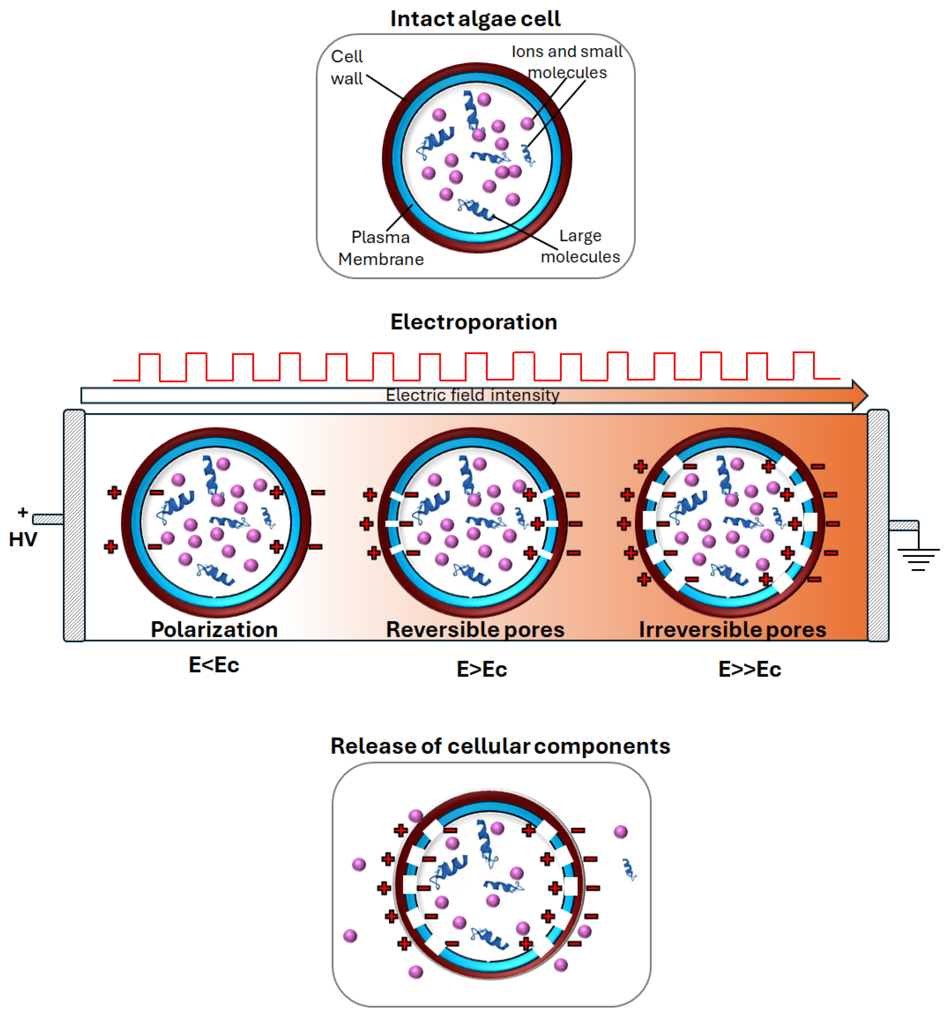
3.1. Overview Pulsed Electric Fields (PEF) Technology

In recent decades, pulsed electric fields (PEF) technology has gained increasing attention as a mild and scalable cell disruption technique for enhancing the extraction efficiency of valuable compounds from various wet biomaterials, including algae suspensions [49]. This approach eliminates the need for energy-intensive biomass drying, resulting in significant energy savings and preserving sensitive compounds that might otherwise be lost [5,17].
During the PEF processing, the algae suspension is placed in contact with the electrodes of either a batch or continuous flow treatment chamber and exposed to intermittent short bursts of electric field pulses (ranging from several nanoseconds to several milliseconds), with varying intensity levels (10–80 kV/cm) and total specific energy input (20–150 kJ/kg) [18]. The electric pulses used are typically unipolar or bipolar and have an exponential or square-wave form [49].
As depicted in Figure 2, exposing biological cells to an external electric field causes an increase in transmembrane potential, which leads to the permeabilization of cell membranes (including cytoplasmic and intracellular membranes), known as electroporation or electropermeabilization [50]. Depending on the treatment intensity, the cell size, and morphological traits, electroporation can be either reversible or irreversible [50].
In reversible electroporation, which occurs when an external field strength comparable to the critical value (Ec) is applied, the pores reseal after the field is removed, allowing cells to recover. In irreversible electroporation, occurring when a field strength above the critical value is applied, the pore formation on the cell membrane is permanent, leading to cell death and reducing mass transfer resistance of intracellular contents and solvent through the cell envelope. This improves the efficiency of the conventional extraction process of valuable compounds from algae biomass, facilitating the penetration of the solvent into the cells and the selective release of intracellular matter [18].
Critical PEF factors influencing the degree of electroporation include the electric field strength, duration of the treatment, total specific energy input, and processing temperature. Generally, increasing these parameters’ intensity boosts the degree of electroporation [49]. To enhance mass transfer processes, conditions must be optimized to achieve irreversible electroporation of the cell membranes.
One of the key advantages of PEF application to microalgae biorefinery is that it can be tailored to the specific morphological characteristics of different microalgae species, optimizing extraction processes and preserving valuable biomolecules [17].
Furthermore, measurements of electrical conductivity, particle size distribution, and scanning electron microscopy analyses confirm that PEF treatment primarily induces membrane permeabilization in microalgae and cyanobacteria, such as Chlorella vulgaris [5,7,19], Arthrospira platensis [6,51], Arthrospira maxima [52], Chlamydomonas reinhardtii [53], and Chlorella pyrenoidosa [54]. This results in increased surface roughness and the formation of cracks and depressions on the cell surface. The extent of these effects depends on treatment intensity and cell morphology, but the overall cell structure remains intact, preventing cell debris formation [5,51,55,56] while selectively promoting the release of ions and small biomolecules. In contrast, highly intensive cell disruption methods such as BM and HPH lead to complete cell fragmentation, resulting in debris formation and the indiscriminate release of intracellular contents [5,25,55].
This confirms that PEF technology can be classified as a relatively mild cell disruption method with minimal impact on microalgal morphology. By facilitating the selective release of target intracellular compounds, PEF enhances the efficiency of downstream processes such as biomass fractionation, extract separation, and purification [5,6,7,57]. As a result, PEF plays a crucial role in optimizing biomass valorization in microalgae-based biorefineries.

3.2. Effect of PEF Processing Conditions on the Extractability of Valuable Compounds

The potential benefits of PEF technology as a pre-treatment for microalgae and cyanobacterial biomass to enhance the extraction of intracellular compounds, such as proteins, carbohydrates, lipids, pigments, and antioxidants, have been widely documented. An overview of studies on PEF-assisted extraction for the enhanced recovery of valuable intracellular compounds from microalgae is reported in Table 3.
While variations in PEF equipment, experimental conditions, algal strains, and cultivation techniques make direct comparisons between literature results challenging, PEF pre-treatment has generally been shown to enhance the selective extraction of both water-soluble and hydrophobic compounds when appropriate solvents are used [28].
Furthermore, the literature findings suggest that several key factors critically influence the effectiveness of PEF treatment. Among these, electric field strength, treatment duration, specific energy input, and pulse polarity consistently emerge as the most impactful factors.
Notably, multiple studies have reported a progressive increase in the release of intracellular compounds into the extracellular medium with increasing PEF intensity (field strength and energy input). These studies predominantly employed monopolar pulses, highlighting their common use in optimizing extraction efficiency.
For example, Carullo et al. [5] observed that the extractability of carbohydrates and low molecular weight proteins from C. vulgaris cell suspensions increased with increasing the field strengths (10–30 kV/cm) and energy inputs (20–100 kJ/kg) of the PEF treatment, leading to extraction yields of 36% for carbohydrates and 5.2% for water-soluble proteins. In contrast, HPH caused a non-selective release of the entire intracellular content, yielding 1.1- and 10.3-fold higher amounts of carbohydrates and protein, respectively, compared to PEF.
Similarly, other researchers have reported that increasingly intense PEF treatments promote the progressive permeabilization of microalgal and cyanobacterial cell membranes, such as those of C. vulgaris, C. reinhardtii, Auxenochlorella protothecoides, and A. platensis, thereby enhancing the extractability of valuable compounds, including carbohydrates, water-soluble proteins, and phenolic compounds [9,51,53,61,70].
Interestingly, across all these studies, the energy input, along with the applied electric field strength, has been identified as a key factor in determining the extent of cell membrane permeabilization required to enhance the extractability of target intracellular compounds. This is particularly evident when the applied field strength is sufficiently high (>10 kV/cm) to induce irreversible electroporation of algal cells.
In only a few studies were controversial results observed, especially when the potential of PEF technology was explored to improve pigment recovery from microalgae. For example, Pataro et al. [9] found that energy input had a greater impact than field strength on pigment extraction from PEF-treated Nannochloropsis oceanica, achieving maximum recoveries of 41.8 mg/gDW for total carotenes and 60.2 mg/gDW for chlorophyll a. In contrast, Grimi et al. [65] detected no pigments in water extracts from Nannochloropsis sp. (20 kV/cm, 13.3–53.1 kJ/kg). Luengo et al. [59] reported significantly higher carotenoid (42%) and chlorophyll a (54%) yields from PEF-treated Chlorella vulgaris (20 kV/cm, 75 µs) using 96% ethanol.
These discrepancies can be partly explained by the hydrophobic nature of these pigments, which are soluble only in organic solvents or mixtures of polar and nonpolar solvents, but not in water. Additionally, the relatively low efficiency of PEF treatments may be due to the fact that PEF primarily electroporates the cytoplasmic membrane of algal cells, causing minimal damage to the membrane of smaller intracellular organelles like chloroplasts, where pigments are stored. Since the electric field required for electroporation is inversely related to cell size [18], the increased pigment yield observed with PEF pretreatment may result from subsequent plasmolysis of chloroplasts during maceration, caused by osmotic imbalance in the cytoplasm after cytoplasmic membrane electroporation [20].
Besides treatment intensity, pulse polarity may also play an important role in determining the effectiveness of the PEF process as it affects the polarization of cell membranes [51]. However, only a few studies have compared the effects of monopolar and bipolar pulses on cell membrane permeabilization, primarily focusing on microbial inactivation, with controversial results. In this field, some authors found bipolar pulses more effective due to membrane fatigue [71,72], while others reported no advantage [73]. Evrendilek and Zhang [74] reported no significant difference in microbial inactivation in apple juice; however, bipolar pulses proved more effective in skim milk, likely due to electrode fouling that impaired the performance of monopolar pulses.
To date, only one work compared the effects of monopolar and bipolar pulses with varying delay times (1–20 µs) at a fixed treatment intensity (20 kV/cm, 100 kJ/kg) on the extractability of intracellular compounds (e.g., water-soluble proteins, C-phycocyanin, and carbohydrates) from A. platensis cells [51]. The results showed that bipolar pulses significantly increased water-soluble molecules in extracts compared to untreated samples, regardless of delay time. However, varying the delay time led to different extraction yields, with a threshold delay time above which no further improvements were observed. Additionally, bipolar pulses were less effective than monopolar ones applied at the same intensity, likely due to slightly lower efficacy in pore formation at the cell membrane.
The lower extractability of water-soluble molecules from A. platensis cells with bipolar pulses, compared to monopolar ones, may stem from their reduced effectiveness in opening membrane pores. This can be attributed to the polarization behavior of cell membranes, where monopolar pulses promote charge accumulation and electroporation [73]. In contrast, bipolar pulses may cause residual polarization, leading to partial charge cancellation and decreased membrane permeabilization [73]. This effect, which limits both pore formation and size, could potentially be mitigated by increasing the applied voltage or the delay time between pulses of opposite polarity. However, further research is needed to better differentiate the effects of monopolar and bipolar pulses, as well as to clarify the underlying membrane charging mechanisms that lead to pore formation. Understanding how these processes are influenced by delay time in bipolar pulses could help optimize processing conditions to achieve the desired level of permeabilization for the selective extraction of target intracellular compounds.
Another crucial factor influencing the electroporation of microalgae is temperature during PEF processing. For example, Postma et al. [19] studied the effect of PEF treatment (17.1 kV/cm, 100 kJ/kg) combined with mild heating (25–65 °C) on the extraction of valuable compounds from C. vulgaris suspensions. They observed a slight positive interaction between PEF and temperature on water-soluble protein recovery, with a maximum yield of 4.4% at 45 °C. However, a clear synergistic effect on carbohydrate extraction appeared when the temperature increased from 45 °C to 55 °C, raising the yield from 25% to 39% of the total biomass carbohydrate content. Similarly, Carullo et al. [51] demonstrated a synergistic effect when combining PEF (20 kV/cm, 100 kJ/kg) with moderate heating on the aqueous extraction of water-soluble compounds from A. platensis. Raising the processing temperature from 25 °C to 35 °C increased protein extraction from 25.4% to 37.4%, carbohydrate yield from 64.1% to 73.1%, and C-phycocyanin yield from 37.4% to 72.5%. In line with these results, Luengo et al. [59] found that an increase in the temperature of PEF treatment (25 kV/cm, 150 µs) of C. vulgaris from 10 to 40 °C increased the concentration of lutein in the extracts from 451 to 753 µg/gdw [55,60].
Interestingly, all these studies highlight that mild heating of algae suspensions not only enhances the diffusivity and solubility of intracellular compounds but also makes the lipid bilayer of the cell membrane more susceptible to breakdown under an external electric field [51,55,60]. This likely explains the enhanced extractability of valuable compounds when PEF is combined with moderate heating. Furthermore, rising temperature within levels that do not cause degradation by heat of the molecules permits a reduction of the required electric field strength and treatment time to achieve a certain extraction yield and, as a result, a reduction in the total specific energy supplied by the treatment [55,60].
In addition to the PEF processing conditions, the incubation time following PEF treatment plays a critical role in improving extraction yields from the treated biomass. Generally, longer incubation periods following PEF treatment are linked to higher extraction yields. For instance, in the case of Nannochloropsis oceanica, a one-hour incubation at room temperature after PEF treatment at 10 kV/cm and 100 kJ/kg significantly enhanced the recovery of hydrophobic compounds, such as carotenes and chlorophyll a, with yield increases of 1.6- and 1.4-fold, respectively [9]. Similarly, for C. vulgaris, incubation for one hour after PEF treatment (20 kV/cm, 75 µs) resulted in increased yields of carotenoids and chlorophylls compared to immediate extraction. This indicates that delayed extraction enhances pigment release efficiency [20]. The observed improvement is likely attributed to ongoing chloroplast plasmolysis driven by osmotic imbalances within the cytoplasmic space [9,20].
Extended incubation times were also necessary to facilitate the release of high molecular weight intracellular compounds. For example, Martínez et al. [55] found that in Arthrospira platensis, a 150 min delay was required before extracting water-soluble proteins like phycocyanin. This delay was attributed to the gradual enlargement of PEF-induced pores, which facilitates the release of larger biomolecules.
A similar phenomenon was reported by Martínez et al. [66] for Porphyridium cruentum, where the red protein-pigment complex phycoerythrin was not detectable immediately after PEF treatment. However, after 24 h of aqueous extraction, the full protein content was recovered. The release was delayed by more than six hours, suggesting that it depends not only on protein diffusion through the membrane but also on the breakdown of molecular associations within the cell. It has been proposed that PEF treatment may trigger the release of hydrolytic enzymes from P. cruentum organelles, which then cleave the bonds between pigments and other cellular components, enabling diffusion of the water–protein complex along a concentration gradient. Nonetheless, further studies are needed to elucidate this mechanism and identify the enzymes involved, particularly those responsible for PEF-induced autolysis, to enhance its potential for industrial-scale applications.

3.3. Limitations and Challenges of Electroporation Process

Published studies highlight the capability of PEF to efficiently unlock small intracellular components, such as lipids, pigments, carbohydrates, and proteins of relatively low molecular weight. However, larger molecules, or those tightly bound to cellular structures, often remain trapped within microalgal cells, especially in species with “hard-structured” cell walls and membranes, where the rigid architecture significantly hinders mass transfer processes [5,75].
Notably, PEF has been found to be inefficient for extracting large proteins, especially when compared to more disruptive techniques such as BM [19] or HPH [5,7,25,76]. For instance, Carullo et al. [5] demonstrated that PEF treatment (20 kV/cm, 100 kJ/kg) selectively released 36% of total carbohydrates and only 5.2% of total proteins from C. vulgaris suspensions. In contrast, HPH (150 MPa) caused a non-selective release of intracellular components, reaching 41.9% of total carbohydrates and 54.1% of total proteins after five passes. Although combining PEF with moderate heat improved extraction efficiency, the yields remained lower than those obtained through mechanical disruption methods [51]. For example, when comparing water-soluble compound extraction using PEF-heat treatment versus complete cell disruption by BM, Postma et al. [19] reported that more than 95% of total water-soluble proteins remained inside the C. vulgaris microalgal cells after PEF.
This can be explained by the fact that, unlike highly disruptive mechanical treatments, PEF is a mild technology that selectively permeabilizes cell membranes [5] while leaving the rigid outer wall of most algae cells intact [25,75]. Consequently, the mass transfer of certain intracellular compounds remains limited.
It is therefore hypothesized that, despite the application of sufficiently high field strength or the combined effects of the electric field and moderate temperature, the pores formed in the microalgal cell membrane during PEF treatment are not large enough to facilitate the release of high-molecular-weight proteins. These proteins likely remain trapped within the cell or bound to the unaltered cell wall [7].
Several studies have also identified the microalgal cell wall as a key barrier that hinders PEF efficiency and restricts the extraction of large molecules [28,53,75]. Consistently, the efficiency of PEF-assisted extraction has been shown to vary across microalgal species due to differences in cell wall composition. A. platensis, which has a fragile, peptidoglycan-based cell wall lacking cellulose [22], exhibited higher protein extraction yields (17.1% DW) [51] compared to species with more robust, cellulose-rich cell walls, such as C. vulgaris, Auxenochlorella protothecoides, Neochloris oleoabundans, and C. reinhardtii [5,19,25,53,61,70], where extraction yields ranged from 1% to 13% DW. The robustness of these cell walls limits protein release, underscoring the need for optimized processing conditions and pretreatments designed to weaken or degrade the cell wall.
In line with this, ‘t Lam et al. [53] investigated the role of the cell wall in PEF treatment by comparing cell-wall-containing C. reinhardtii with cell-wall-free mutants. As will be discussed in more detail in a subsequent section, the mutants served as a model for enzymatic pre-treatment strategies aimed at weakening the cell wall before PEF application in the wild-type strain. Their findings demonstrated that PEF treatment enabled the complete release of hydrophilic proteins from the cell-wall-free mutants, whereas protein yields were significantly lower in species with an intact cell wall.
These results provide valuable insights into future strategies for enhancing PEF as innovative technology for selective release with high yield of cytoplasmic molecules in a multistage biorefinery process.

4. Enzymatic Hydrolysis (EH) Technology

Efficient extraction of intracellular compounds from microalgae remains a key challenge in biomass valorization, primarily due to the presence of complex and robust cell wall and membrane structures. These physical barriers can significantly hinder mass transfer, especially when mild cell disruption techniques like PEF are used. While PEF is effective in permeabilizing the cytoplasmic membrane, it often leaves the rigid outer cell wall intact, particularly in species with “hard-structured” walls, necessitating additional strategies to enhance compound recovery. One such strategy is the application of specific enzymes capable of degrading the various components of the cell wall and membrane systems.

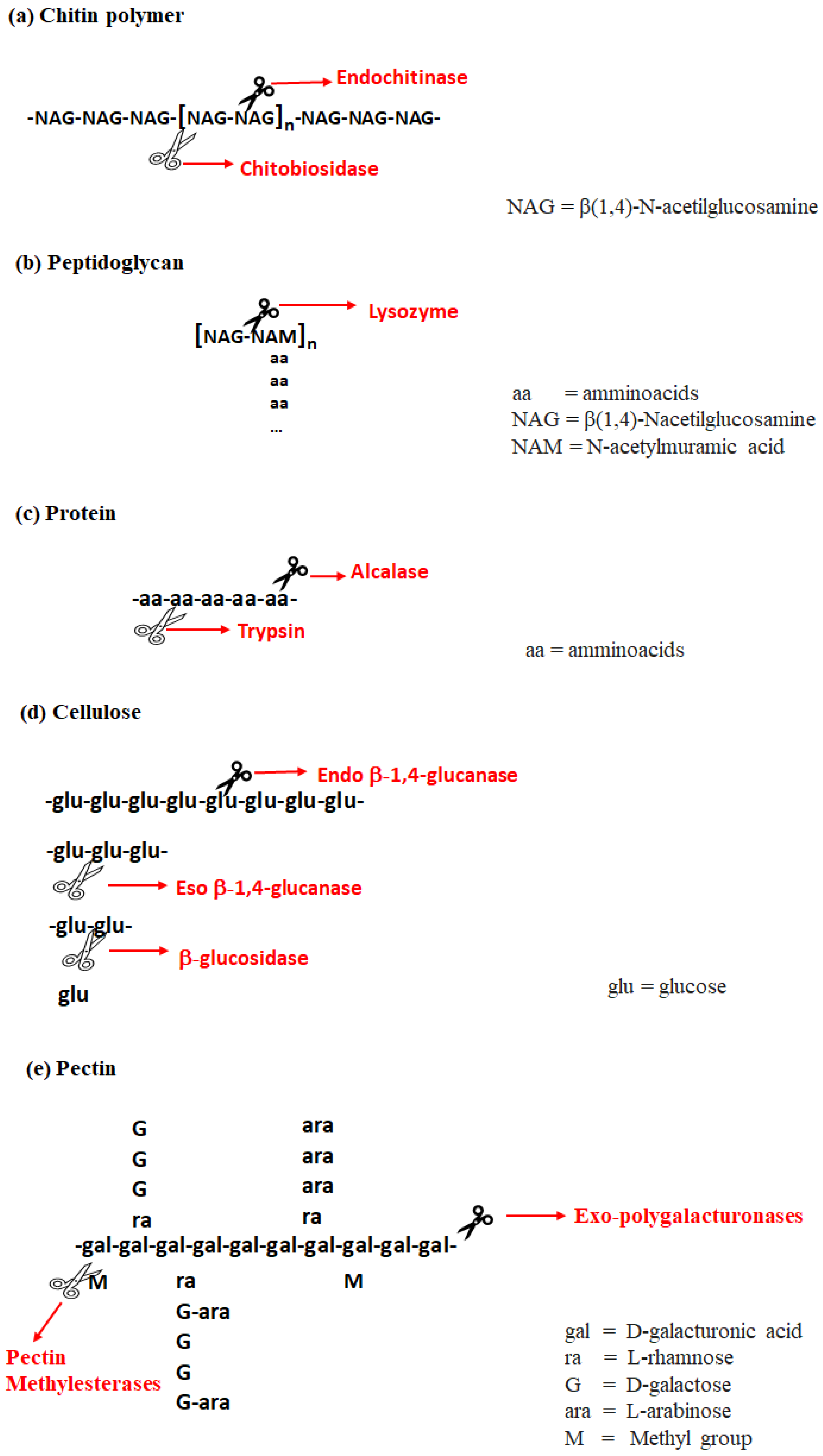
4.1. Overview of Enzymatic Treatment

To understand the rationale behind enzymatic treatments, it is essential to first consider the biochemical nature and functional properties of enzymes. Enzymes are specialized proteins that catalyze a wide range of biochemical reactions, including the hydrolysis of complex macromolecules such as polysaccharides and proteins. Structurally, enzymes are composed of amino acid chains linked by peptide bonds and organized into a complex hierarchy. This includes a primary sequence (the linear chain of amino acids), secondary structures (such as α-helices and β-sheets formed through hydrogen bonding), a tertiary structure (the overall three-dimensional folding driven by side-chain interactions), and, in some cases, a quaternary structure (the assembly of multiple polypeptide chains into a functional unit). This hierarchical structure is essential for determining the enzyme’s specificity and catalytic function [77]. In many cases, however, enzymatic activity also depends on the presence of cofactors or coenzymes, namely non-protein components such as metal ions or organic molecules, that assist in facilitating the reaction.
Recent advancements in biotechnology have made it possible to produce highly pure and selective enzymes, enabling targeted degradation of specific microalgal cell wall components in a controlled and efficient manner [77,78].
Microalgal cell wall composition varies widely depending on the species, class, and cultivation conditions (as outlined in Table 1). The primary components typically include polysaccharides (e.g., cellulose, chitin, pectin, and peptidoglycan) and proteins, each of which requires specific enzymatic treatments for effective breakdown. Figure 3 illustrates the major structural components of microalgae cell walls and the corresponding enzymes that can degrade them.
Chitin is the primary structural component of the cell wall in red algae (Rhodophytaceae), providing rigidity and protection. It is a long-chain polymer composed of N-acetylglucosamine (NAG), an amide derivative of glucose. Chitin can be enzymatically degraded by chitinases, which hydrolyze the glycosidic bonds within the polymer. There are two main types of chitinases: endochitinases and exochitinases. Endochitinases act by randomly cleaving internal bonds within the chitin chain, producing soluble, low-molecular-weight oligomers such as chitotetraose, chitotriose, and predominantly di-acetylchitobiose. In contrast, exochitinases, such as chitobiosidases, progressively cleave chitin from the ends of the polymer, releasing di-acetylchitobiose as the sole product, without generating mono- or other oligosaccharides [79].
Peptidoglycan is a key structural polysaccharide found in the cell walls of green algae, composed of alternating units of N-acetylglucosamine (NAG) and N-acetylmuramic acid (NAM) linked by β-(1→4) glycosidic bonds. Attached to each NAM unit is a short peptide chain consisting of three to five amino acids. This complex matrix provides mechanical strength and helps resist osmotic pressure within the cell. The enzyme lysozyme facilitates the hydrolysis of the β-(1→4) linkages between NAG and NAM, effectively degrading the peptidoglycan network and weakening the cell wall structure [80].
Proteins are found throughout the cytoplasm, organelles (e.g., chloroplasts), and plasma membrane, and serve as essential components of the cell wall in Cryptophyceae. They play crucial roles in cellular metabolism, signaling, transport, and other fundamental biological processes. However, proteins in their native form, particularly in high-molecular-weight or bound states, are not as bioavailable or functional as free amino acids. To enhance their usability, proteins are often hydrolyzed into smaller peptides or individual amino acids [57]. While both chemical and enzymatic hydrolysis methods exist, enzymatic hydrolysis (EH) is preferred due to its milder conditions, which preserve sensitive amino acids and eliminate the need for neutralization steps required in chemical hydrolysis, thus avoiding unwanted byproducts like excessive ash content [57]. Commercial proteases such as trypsin and alcalase are commonly used in EH. Trypsin cleaves peptide bonds specifically at the C-terminal side of lysine and arginine residues, whereas alcalase has broader specificity, preferentially targeting bonds near hydrophobic amino acids [77].
Cellulose is a major structural component of the rigid cell wall in green microalgae, contributing significantly to its mechanical strength and integrity. It is a polysaccharide composed of linear chains of several hundred to thousands of β(1→4)-linked D-glucose units. The enzymatic degradation of cellulose typically involves a synergistic action of three main enzymes: endo-β-1,4-glucanase, which cleaves internal bonds within the cellulose chain to produce oligosaccharides with free chain ends; exo-β-1,4-glucanase, which acts on these chain ends to release cellobiose; and β-glucosidase, which hydrolyzes cellobiose into glucose monomers.
Pectin, another key polysaccharide in green algae cell walls, forms part of the matrix structure and plays a vital role in regulating growth, wall porosity, and the ionic environment of cells. It is composed primarily of D-galacturonic acid units, methoxy groups, and various sugar side chains. Pectin can be enzymatically degraded by pectinases such as polygalacturonase, which breaks down the galacturonic acid backbone into monomers and dimers, and pectinesterase, which removes methoxy groups to facilitate pectin demethylation [81].
In summary, enzymatic treatment represents a highly selective and complementary approach to mechanical or physical cell disruption methods. By targeting specific structural components of the cell wall, enzymes facilitate improved mass transfer and compound recovery, making them essential tools in the development of efficient and sustainable microalgal biorefinery processes.

4.2. Effect of EH Processing Conditions on the Extractability of Valuable Compounds

Several key parameters influence the efficiency of enzymatic hydrolysis in degrading microalgae cell walls. These include the type and concentration of the enzyme used, as well as reaction conditions such as temperature, pH, and duration. While optimal temperature, pH, and reaction time are largely dependent on the specific enzyme employed, the selection of enzyme type and its appropriate concentration must be tailored to the characteristics of the microalgal species under investigation. Table 4 summarizes findings from the literature regarding enzymatic hydrolysis applied to both microalgae and macroalgae cell wall disruption.
However, drawing direct comparisons across these studies remains challenging due to variations in algal strains, cultivation conditions, and processing methodologies. For instance, Steinbruch et al. [75] achieved a 9.7% protein yield using 2% cellulase on Ulva species. In contrast, Safi et al. [76] reported a significantly higher protein yield (24.8%) from Nannochloropsis gaditana by combining alcalase pretreatment with an ultrafiltration recovery step, highlighting the impact of process integration and enzyme selection.
Similarly, lipid recovery outcomes differ widely across studies. Zhang et al. [89] reached an 86.4% lipid yield using enzymatic hydrolysis followed by chloroform-methanol solvent extraction on Scenedesmus sp. Conversely, Papachristou et al. [26] obtained a lower lipid yield (16%) from Scenedesmus almeriensis using alcalase in combination with ethanol-hexane extraction. These discrepancies further underscore the difficulty of benchmarking results across studies with different enzymes, species, and operating protocols.
To enhance recovery yields, several researchers have investigated the combination of enzymatic hydrolysis with mechanical or chemical cell disruption techniques. Koruyucu et al. [86], for instance, evaluated various pretreatment methods, including US, BM, and HPH, prior to enzymatic processing. Among these, BM with 2 mm stainless-steel beads was the most effective, achieving 87–97% cell disruption within 40 min. This was followed by enzymatic hydrolysis using cellulases and mannanases, which resulted in a saccharification efficiency of up to 25% after 72 h.
Further insights were provided by Rojo et al. [88], who compared the effects of enzymatic hydrolysis on pure microalgae versus a microalgae–bacteria consortium. They found higher carbohydrate solubilization from the consortium (38.5%) compared to microalgae alone (27%) using Celluclast over a 5 h treatment. Moreover, alcalase enabled the highest peptide recovery (≈34%) from both biomass types, though peptide sizes were generally <10 kDa. In contrast, Protamex yielded lower peptide recoveries (<20%) but with significantly larger molecular weights (up to 135 kDa).
As shown in Table 4, most studies focus on protein hydrolysis using proteases [75,76,82], largely due to the versatility and value of the resulting amino acid concentrates. These products have diverse applications in agriculture (as biofertilizers), the food industry (as additives), and the pharmaceutical sector [87,88]. Compared to traditional acid hydrolysis, enzymatic hydrolysis offers several significant advantages: it avoids the formation of harmful by-products, eliminates the need for a neutralization step, and preserves the integrity of chemically sensitive amino acids. These characteristics make enzymatic hydrolysis an attractive, safe, and economically viable strategy for protein extraction and valorization from microalgae biomass [88].

4.3. Limitations and Challenges of Enzymatic Hydrolysis

The use of enzymes in microalgae envelope disruption is characterized by several advantages and disadvantages, which have been summarized in Table 5.
Enzymatic hydrolysis offers several advantages over conventional chemical pretreatments. One of the key benefits is its high specificity and selectivity, allowing enzymes to target specific bonds or substrates and thereby minimizing undesired reactions. In contrast, chemical pretreatments typically result in non-selective cell membrane disruption, producing a variety of products that require further identification and separation [56].
Another important advantage of enzymatic hydrolysis is its environmental friendliness. Being a green process, it avoids the use of harsh chemicals, making it safer and more sustainable. Additionally, it does not generate by-products, typically yielding clean, well-defined products and reducing the need for extensive downstream purification. On the other hand, chemical methods often produce waste streams containing acids, alkalis, or organic solvents that must be properly treated or disposed of [91].
Finally, enzymatic hydrolysis operates under mild conditions, usually around 50 °C and atmospheric pressure, which significantly reduces energy consumption compared to conventional processes [91,92].
Despite the numerous advantages of enzymatic hydrolysis, several challenges still limit its competitiveness in large-scale applications. One of the primary concerns is the high cost of certain enzymes, which can be expensive to produce or purchase, particularly for industrial use.
Another limitation is the longer reaction time compared to traditional chemical methods. Enzymatic hydrolysis typically requires up to 24 h, whereas conventional chemical treatments may be completed within minutes, significantly affecting overall process throughput.
In addition, separating the enzymes from the final product can be technically demanding. To address this, enzymes are often immobilized and reused across multiple cycles, a strategy that helps to reduce operational costs and improve process sustainability [56,93].
However, it is important to note that while chemical pretreatments may be faster, they often involve additional time for product separation and purification due to the formation of by-products. This “dead time” must be taken into account when performing a comprehensive process analysis [56].

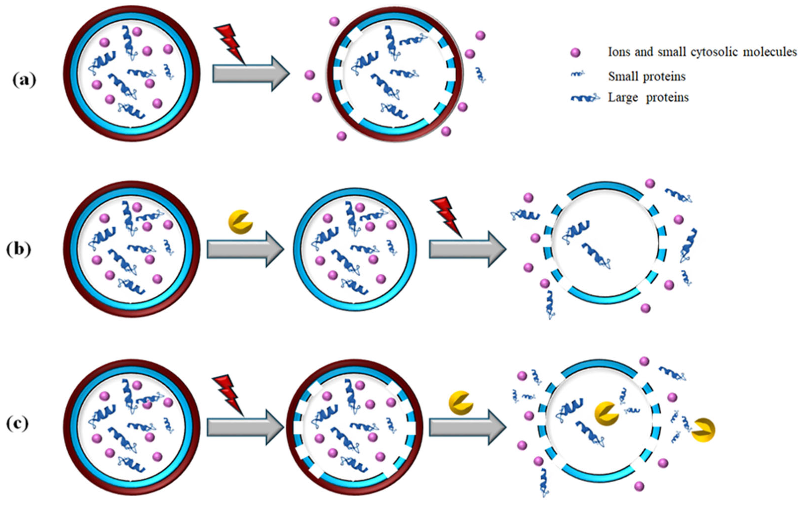
5. Cascaded PEF and Enzymatic Hydrolysis (EH): Mechanism, Synergies, and Benefits

The integration of cascaded PEF treatment with enzymatic hydrolysis offers a promising, green, and efficient strategy for microalgae biorefineries. This combined approach significantly reduces energy consumption, chemical usage, and environmental impact compared to conventional methods [28].
Both PEF and enzymatic hydrolysis are selective and scalable processes that preserve valuable intracellular compounds without causing thermal or chemical degradation. Unlike conventional acidic or alkaline extraction, which requires chemical waste treatment, or highly disruptive mechanical methods that lead to undifferentiated release of intracellular compounds while generating large amounts of cell debris, this approach maintains the integrity of the biomass and enhances product quality [75]. Additionally, because both techniques preserve cell structure and improve biomass separability, they enable cascade processing, making them suitable alternatives for downstream production of valuable compounds from microalgae biomass [57].
The synergistic effect of PEF and enzymatic hydrolysis is particularly advantageous. PEF facilitates the extraction of intracellular metabolites while enhancing enzymatic hydrolysis, improving the conversion of polysaccharides into fermentable sugars and proteins into bioactive peptides. This optimization ultimately increases the yield of biofuels and biochemicals [57]. Furthermore, this combined process aligns with the biorefinery concept, enabling the simultaneous production of multiple high-value products from the same biomass. By supporting a circular and sustainable economy, this strategy enhances the efficiency and viability of microalgae-based bioprocessing. Nevertheless, as reported in Table 6, only a few studies have explored the combination of PEF treatment with enzymatic hydrolysis in a cascade biorefinery approach to selectively enhance the recovery of intracellular compounds, particularly large or bound proteins.
Figure 4 illustrates two potential sequences for combining PEF and enzymatic treatment in a cascaded approach: (1) enzymatic cell wall degradation followed by PEF-induced membrane permeabilization (E → PEF) and (2) PEF treatment applied before enzymatic hydrolysis (PEF → EH). For the sake of comparison, the schematics of the single PEF treatment have also been included.
As discussed in previous sections, the effectiveness of PEF treatment in microalgae with rigid cell walls is significantly limited by the presence of this outer barrier [53]. PEF primarily permeabilizes the cell membrane while leaving the cell wall intact, allowing only small molecules and ions to migrate, whereas larger cytosolic molecules, such as proteins, remain trapped within the cytoplasm or bound to cell structure [5,75].
To overcome this limitation, integrating a suitable enzymatic pre-treatment to weaken or degrade the cell wall before PEF application could significantly enhance the extraction of proteins and other large intracellular molecules (Figure 4b). By first breaking down the outer cell wall, enzymatic treatment facilitates the release of valuable compounds that would otherwise remain inaccessible.
Some researchers have investigated the synergistic effect of combining enzymatic cell wall degradation with PEF-induced membrane permeabilization, demonstrating improved yields of water-soluble proteins from microalgae cells (Table 6).
The hypothesis that PEF treatment is hindered by the rigid outer cell wall in microalgae was first investigated by ‘t Lam et al. [53]. To simulate pre-treated microalgae with a removed cell wall, the authors used a cell wall-free mutant of C. reinhardtii to assess the feasibility of PEF (7.5 kV/cm, 2 kWh/kgdw) for protein release. Their findings showed that PEF treatment of the cell wall-deficient mutant yielded 31% protein, three times higher than in microalgae with an intact cell wall and comparable to mechanical disruption. Interestingly, PEF achieved high protein yields with minimal cell disruption, only 27% compared to 99% with bead beating, while chlorophyll levels in the supernatant remained low. This suggests that PEF selectively released hydrophilic proteins, leaving hydrophobic chlorophyll entrapped. Such selective extraction could simplify downstream fractionation in biorefinery processes. These results strongly support the effectiveness of enzymatic pre-treatment in degrading the cell wall before PEF application. By weakening or removing this structural barrier, enzymatic pre-treatment significantly improves the subsequent release of target intracellular compounds from electroporated microalgae cells.
Building on this preliminary research, Steinbruch et al. [75] explored the combined approach of enzymatic cell wall degradation followed by PEF for the efficient and sustainable extraction of proteins from the green marine macroalga Ulva sp. Their findings demonstrated a significantly higher protein extraction yield using the combined process (19.6%) compared to PEF alone (10.87%) and enzymatic pre-treatment alone (9.7%). Notably, the protein yield in the enzyme + PEF treatment was 182% higher than that achieved with PEF alone, underscoring the strong synergistic effect of these combined techniques. Additionally, the water-soluble protein extract obtained through PEF following enzymatic degradation exhibited notable antioxidant activity, further highlighting the potential benefits of this method. These results suggest that integrating enzymatic pre-treatment with PEF could significantly enhance protein extraction from Ulva sp., contributing to the development of a more sustainable seaweed biorefinery.
Conversely, as summarized in Table 6, it has been reported that applying PEF as a pre-treatment before enzymatic hydrolysis (EH) may enhance protein release and, consequently, improve the efficiency of the hydrolysis process [57]. As illustrated in Figure 4c, the electroporation induced by PEF is hypothesized to serve two key functions. First, it facilitates enzyme penetration into the cells, allowing intracellular proteins to be cleaved into smaller peptides that can diffuse more easily. Second, proteins released by PEF are expected to be more accessible for cleavage by extracellular enzymes. To maximize enzymatic hydrolysis yields, pre-treating microalgae biomass with PEF is recommended, as it enhances enzyme access to intracellular proteins.
The effect of PEF on enzymatic hydrolysis yield has been investigated to evaluate its potential for producing bioactive peptides, a valuable product known as an amino acid concentrate. This concentrate has potential applications in agriculture, providing plants with a rich source of free amino acids. For instance, in the study conducted by Akaberi et al. [57], fresh and concentrated biomass (50 g/kg to 80 g/kg) of Scenedesmus almeriensis microalgae was pre-treated with PEF at 40 kV/cm, applying treatment energies of 75 kJ/kg and 150 kJ/kg. Subsequent enzymatic hydrolysis was conducted using the commercial enzymes Alcalase 2.5 L and Flavourzyme 1000 L for 180 min. PEF pre-treatment at both energy levels resulted in identical hydrolysis kinetics and a final degree of hydrolysis (DH) of 50%. The incomplete protein hydrolysis was attributed to the presence of hydrophobic protein fractions and protein aggregation, which hindered complete degradation by the applied enzyme cocktail.
Protein extraction efficiency is highly influenced by biomass concentration. For this reason, Scherer et al. [82] investigated the effect of biomass concentration on protein recovery efficiency from C. vulgaris. In particular, PEF (40 kV/cm, 150 kJ/kg suspension) at different biomass concentrations (between 5 mg/mL and 25 mg/mL) was investigated. Half of the proteins in the cells were recovered at dilute concentrations (5 mg/mL), but this efficiency decreased with increasing biomass concentration, indicating that diffusion gradients play a role in the protein release. Furthermore, the protease inhibitor effect was evaluated, demonstrating that the protease inhibition impairs the release of proteins. Indeed, in Western blots, it was observed that some proteins are not released when the activity of proteases is blocked.
The crucial role of proteases in enhancing protein extraction efficiency after PEF treatment was demonstrated by Scherer et al. [82]. Their study primarily investigated the impact of biomass concentration (2.5–12.5 mg/mL), temperature (4–50 °C), and protease inhibition on protein recovery efficiency from C. vulgaris following PEF treatment (40 kV/cm, 150 kJ/kg) and incubation for up to 24 h. The results revealed that at dilute biomass concentrations (2.5 mg/mL), nearly half of the total cellular proteins were recovered, but extraction efficiency declined as biomass concentration increased, indicating that diffusion gradients influence protein release. Moreover, protein extraction efficiency was highest at intermediate temperatures (30 °C), rather than at the extremes. This temperature-dependent pattern suggested that protein release is not solely governed by diffusion but also involves an enzyme-driven process, as it was most effective within a physiological temperature range. Further supporting this hypothesis, when protease inhibitors were introduced, protein release decreased significantly, confirming that enzymatic activity plays a key role in the extraction process.
In another study, Maribu et al. [94] explored the potential of combining PEF treatment with enzymatic hydrolysis (EH) for the extraction of sugars and minerals, while simultaneously obtaining a protein-rich pellet as an ingredient for animal feed. Specifically, P. palmata microalgae were subjected to single PEF (E = 0.5 kV/cm, 134.6 kJ/kg), single EH using a polysaccharide-degrading enzyme mixture (β-glucanase, pectin lyase, and cellulase), and a combined PEF+EH approach. The results indicated that PEF alone was not effective for protein extraction under the tested conditions, as minimal protein was released into the supernatant. However, when PEF was combined with enzymatic treatment, the supernatant became enriched with sugars and minerals, while the pellet retained a high protein content, making it a valuable protein-rich ingredient for animal feed.
Papachristou et al. [26] explored the use of PEF treatment in a biorefinery process for S. almeriensis, incorporating water fraction extraction, EH, and lipid extraction. The study compared this approach with HPH as a benchmark. Specifically, following PEF treatment (1.5 MJ/kgDW) and an incubation step, EH and lipid extraction were performed to assess carbohydrate release in the water fraction, protein extraction efficiency, and lipid recovery. The results revealed that PEF-treated samples exhibited low concentrations of carbohydrates in the extracts, and they showed slightly improved enzymatic protein hydrolysis kinetics compared to untreated or HPH-treated biomass. Although similar lipid yields were obtained for EH and PEF-EH treatments, indicating that PEF had little impact on lipid extraction, a key finding was that the highest degree of hydrolysis was achieved when PEF was combined with EH in a sequential approach (PEF-EH), outperforming both control-EH and HPH-EH treatments.
Unfortunately, only a limited number of studies in the scientific literature have explored so far the potential of cascade combination of PEF and EH for the selective and efficient extraction of valuable compounds from microalgae. Moreover, the existing studies often focus on different microalgae species and operative conditions, making direct comparisons challenging.
This limited number of studies on the integration of PEF and EH can be attributed to several technical and practical challenges. Integrating two distinct technologies like PEF and EH requires precise coordination, as each operates under its own set of optimal conditions. PEF disrupts cell membranes through electrical pulses, while EH depends on specific enzymatic activity under tightly controlled pH and temperature. Achieving synergy between these steps without compromising efficiency is a complex task. Additionally, if not carefully managed, PEF parameters may negatively impact enzyme stability and activity.
Optimizing this cascade approach is further complicated by the interdependence of numerous process variables. The inherent structural variability among microalgae species also limits the development of a universal method, as each strain may respond differently to both treatments, requiring individual optimization. The lack of standardized protocols, along with the high costs associated with enzymes and PEF equipment, presents additional barriers to broader implementation.
Nevertheless, this integrated approach holds significant promise for enhancing the selective and sustainable recovery of intracellular compounds. To realize its full potential, further interdisciplinary and collaborative research is essential to evaluate its feasibility and optimize its application in industrial-scale microalgae biorefineries.

6. Conclusions and Future Direction

The use of Pulsed Electric Field (PEF) and enzymatic hydrolysis (EH) as individual treatments for algae cell disruption has demonstrated their effectiveness in enhancing intracellular compound extraction. PEF facilitates membrane permeabilization through electroporation, promoting the release of valuable biomolecules, while enzymatic hydrolysis degrades the cell wall, improving the extraction yield. When applied in sequence, these techniques exhibit a synergistic effect, leading to a more efficient and sustainable bioprocess. In particular, when PEF-EH treatment is carried out, enzymes increase the final yields of bioproducts released by PEF pretreatment. When EH-PEF treatment is applied, enzymatic hydrolysis facilitates the releasing of bioproducts by PEF pretreatment.
Despite the limited number of publications on the subject, findings suggest that the cascaded integration of PEF and enzymatic hydrolysis presents a promising pathway for sustainable and efficient downstream processing in microalgae biorefineries. The synergistic effects of these technologies can improve biomass extraction, enhance the selective extraction of target intracellular compounds, including those with large molecular weights or bound to cell structure, and support the production of high-value bio-fuels and biochemicals.
While the initial investment in PEF equipment and enzyme costs must be carefully assessed, the integration of cascaded PEF and enzymatic hydrolysis has the potential to reduce operational expenses by improving extraction efficiency and product yields, ultimately enhancing the profitability of microalgae-based biorefineries. Despite its promising potential, further research is necessary to optimize scalability, cost-effectiveness, and industrial application. Future studies should explore the impact of this cascade approach across a broader range of microalgae species and conduct comprehensive techno-economic and life cycle assessments to fully evaluate its benefits.

Author Contributions

Conceptualization, G.P. and A.P.; Data curation and Figure design, E.E. and F.P.; Writing—original draft preparation, E.E. and F.P.; Writing—review and editing, G.P. and A.P.; Supervision, G.P. and A.P. All authors have read and agreed to the published version of the manuscript.

Funding

This research received no external funding.

Data Availability Statement

Data will be made available on request.

Conflicts of Interest

The authors declare no conflicts of interest.

References

  1. Starckx, S. A Place in the Sun—Algae is the Crop of the Future, According to Researchers in Geel, Flanders Today. 2012. Available online: https://www.flanderstoday.eu/current-affairs/place-sun (accessed on 27 October 2024).
  2. Günerken, E.; D’Hondt, E.; Eppink, M.H.M.; Garcia-Gonzales, L.; Elst, K.; Wijffels, R.H. Cell disruption for microalgae biorefineries. Biotechnol. Adv. 2015, 33, 243–260. [Google Scholar] [CrossRef] [PubMed]
  3. Fu, W.; Nelson, D.R.; Yi, Z.; Xu, M.; Khraiwesh, B.; Jijakli, K.; Chaiboonchoe, A.; Alzahmi, A.; Al-Khairy, D.; Brynjolfsson, S.; et al. Bioactive Compounds From Microalgae: Current Development and Prospects. Stud. Nat. Prod. Chem. 2017, 54, 199–225. [Google Scholar]
  4. Moshood, T.D.; Nawanir, G.; Mahmud, F. Microalgae biofuels production: A systematic review on socioeconomic prospects of microalgae biofuels and policy implications. Environ. Chall 2021, 5, 100207. [Google Scholar] [CrossRef]
  5. Carullo, D.; Abera, B.D.; Casazza, A.A.; Donsì, F.; Perego, P.; Ferrari, G.; Pataro, G. Effect of Pulsed Electric Fields and High-Pressure Homogenization on the aqueous extraction of intracellular compounds from the microalgae Chlorella vulgaris. Algal Res. 2018, 31, 60–69. [Google Scholar] [CrossRef]
  6. Carullo, D.; Donsì, F.; Ferrari, G.; Pataro, G. Extraction improvement of water-soluble compounds from Arthrospira platensis through the combination of high-shear homogenization and pulsed electric fields. Algal Res. 2021, 57, 102341. [Google Scholar] [CrossRef]
  7. Carullo, D.; Abera, B.D.; Scognamiglio, M.; Donsì, F.; Ferrari, G.; Pataro, G. Application of pulsed electric fields and high-pressure homogenization in biorefinery cascade of C. vulgaris microalgae. Foods 2022, 11, 471. [Google Scholar] [CrossRef]
  8. Garcia, E.S.; van Leeuwen, J.; Safi, C.; Sijtsma, L.; Eppink, M.H.M.; Wijffels, R.H.; van den Berg, C. Selective and energy efficient extraction of functional proteins from microalgae for food applications. Bioresour. Technol. 2018, 268, 197–203. [Google Scholar] [CrossRef]
  9. Pataro, G.; Carullo, D.; Ferrari, G. PEF-assisted Supercritical CO2 Extraction of Pigments from Microalgae Nannochloropsis oceanica in a Continuous Flow System. Chem. Eng. Trans. 2019, 74, 97–102. [Google Scholar] [CrossRef]
  10. Vanthoor-Koopmans, M.; Wijffels, R.H.; Barbosa, M.J.; Eppink, M.H.H. Biorefinery of microalgae for food and fuel. Bioresour. Technol. 2013, 135, 142–149. [Google Scholar] [CrossRef]
  11. Zhang, R.; Gu, X.; Xu, G.; Fu, X. Improving the lipid extraction yield from Chlorella based on the controllable electroporation of cell membrane by pulsed electric field. Bioresour. Technol 2021, 330, 124933. [Google Scholar] [CrossRef]
  12. Kokkali, M.; Martí-Quijal, F.J.; Taroncher, M.; Ruiz, M.J.; Kousoulaki, K.; Barba, F.J. Improved Extraction Efficiency of An-tioxidant Bioactive Compounds from Tetraselmis chuii and Phaedoactylum tricornutum Using Pulsed Electric Fields. Molecules 2020, 25, 3921. [Google Scholar] [CrossRef] [PubMed]
  13. Radakovits, R.; Jinkerson, R.E.; Darzins, A.; Posewitz, M.C. Genetic Engineering of Algae for Enhanced Biofuel Production. Eukaryot. Cell 2010, 9, 486–501. [Google Scholar] [CrossRef] [PubMed]
  14. Postma, P.R.; ‘t Lam, G.P.; Barbosa, M.J.; Wijffels, R.H.; Eppink, M.H.M.; Olivieri, G. Microalgal Biorefinery for Bulk and High-Value Products: Product Extraction Within Cell Disintegration. In Handbook of Electroporation, 1st ed.; Miklavcic, D., Ed.; Springer International Publishing AG: Cham, Switzerland, 2017; pp. 2205–2224. [Google Scholar]
  15. de Jesus Raposo, M.F.; de Morais, R.M.S.C.; de Morais, A.M.M.B. Health applications of bioactive compounds from marine microalgae. Life Sci. 2013, 15, 479–486. [Google Scholar] [CrossRef] [PubMed]
  16. Carbone, D.A.; Pellone, P.; Lubritto, C.; Ciniglia, C. Evaluation of Microalgae Antiviral Activity and Their Bioactive Compounds. Antibiotics 2021, 10, 746. [Google Scholar] [CrossRef]
  17. Donsì, F.; Ferrari, G.; Pataro, G. Emerging Technologies for the Clean Recovery of Antioxidants from Microalgae. In Microalgae; Galanakis, C.M., Ed.; Academic Press: Cambridge, MA, USA, 2021; pp. 173–205. [Google Scholar]
  18. Poojary, M.M.; Barba, F.J.; Aliakbarian, B.; Donsì, F.; Pataro, G.; Dias, D.A.; Juliano, P. Innovative Alternative Technologies to Extract Carotenoids from Microalgae and Seaweeds. Mar. Drugs 2016, 14, 214. [Google Scholar] [CrossRef] [PubMed]
  19. Postma, P.R.; Pataro, G.; Capitoli, M.; Barbosa, M.J.; Wijffels, R.H.; Eppink, M.H.M.; Olivieri, G.; Ferrari, G. Selective extraction of intracellular components from the microalga Chlorella vulgaris by combined pulsed electric field–temperature treatment. Bioresour. Technol. 2016, 203, 80–88. [Google Scholar] [CrossRef]
  20. Luengo, E.; Condón-Abanto, S.; Álvarez, I.; Raso, J. Effect of pulsed electric field treatments on permeabilization and extraction of pigments from Chlorella vulgaris. J. Membr. Biol. 2014, 247, 1269–1277. [Google Scholar] [CrossRef]
  21. Rahman, M.M.; Hosano, N.; Hosano, H. Recovering Microalgal Bioresources: A Review of Cell Disruption Methods and Extraction Technologies. Molecules 2022, 27, 2786. [Google Scholar] [CrossRef]
  22. Safi, C.; Ursu, A.V.; Laroche, C.; Zebib, B.; Merah, O.; Pontalier, P.Y.; Vaca-Garcia, C. Aqueous extraction of proteins from microalgae: Effect of different cell disruption methods. Algal Res. 2014, 3, 61–65. [Google Scholar] [CrossRef]
  23. Parniakov, O.; Apicella, E.; Koubaa, M.; Barba, F.J.; Grimi, N.; Lebovka, N.; Pataro, G.; Ferrari, G.; Vorobiev, E. Ultrasound-assisted green solvent extraction of high-added value compounds from microalgae Nannochloropsis spp. Bioresour. Technol. 2015, 198, 262–267. [Google Scholar] [CrossRef]
  24. Golberg, A.; Sack, M.; Teissie, J.; Pataro, G.; Pliquett, U.; Saulis, G.; Stefan, T.; Miklavcic, D.; Vorobiev, E.; Frey, W. Energy-efficient biomass processing with pulsed electric fields for bioeconomy and sustainable development. Biotechnol. Biofuels 2016, 9, 94. [Google Scholar] [CrossRef] [PubMed]
  25. ‘t Lam, G.P.; Postma, P.R.; Fernandes, D.A.; Timmermans, R.A.H.; Vermue, M.H.; Barbosa, M.J.; Eppink, M.H.M.; Wijffels, R.H.; Olivieri, G. Pulsed electric field for protein release of the microalgae Chlorella vulgaris and Neochloris oleoabundans. Algal Res. 2017, 24, 181–187. [Google Scholar] [CrossRef]
  26. Papachristou, I.; Akaberi, S.; Silve, A.; Navarro-Lopez, R.; Wustner, R.; Leber, K.; Nazarova, N.; Muller, G.; Frey, W. Analysis of the lipid extraction performance in a cascade process for Scenedesmus almeriensis biorefinery. Biotechnol. Biofuels 2021, 14, 20. [Google Scholar] [CrossRef] [PubMed]
  27. Mata, T.M.; Martins, A.A.; Caetano, N.S. Microalgae for Biodiesel Production and Other Applications: A Review. Ren. Sust. Energy Rev. 2010, 14, 217–232. [Google Scholar] [CrossRef]
  28. Martínez, J.M.; Delso, C.; Álvarez, I.; Raso, J. Pulsed electric field-assisted extraction of valuable compounds from microorganisms. Comp. Rev. Food Sci. Food Saf. 2020, 19, 530–552. [Google Scholar] [CrossRef] [PubMed]
  29. D’Hondt, E.; Martin-Juarez, J.; Bolado, S.; Kasperoviciene, J.; Koreiviene, J.; Sulcius, S.; Bastiaens, L. Cell disruption technologies. In Microalgae-Based Biofuels and Bioproducts; Muñoz, R., Gonzalez-Fernandez, C., Eds.; Woodhead Publishing: Cambridge, UK, 2017; pp. 133–154. [Google Scholar]
  30. Chan, W.S.; Kwok, A.C.M.; Wong, J.T.Y. Knockdown of Dinoflagellate Cellulose Synthase CesA1 Resulted in Malformed Intracellular Cellulosic Thecal Plates and Severely Impeded Cyst-to-Swarmer Transition. Front. Microbiol. 2019, 10, 546. [Google Scholar] [CrossRef]
  31. Hoef-Emden, K.; Melkonian, M. Revision of the Genus Cryptomonas (Cryptophyceae): A combination of molecular phylogeny and morphology provides insights into a long-hidden dimorphism. Protist 2003, 154, 371–409. [Google Scholar] [CrossRef]
  32. Walker, J.M.; Marzec, B.; Lee, R.B.Y.; Vodrazkova, K.; Day, S.J.; Tang, C.C.; Rickaby, R.E.M.; Nudelman, F. Polymorph Selectivity of Coccolith-Associated Polysaccharides from Gephyrocapsa Oceanica on Calcium Carbonate Formation in Vitro. Adv. Funct. Mater. 2018, 29, 1807168. [Google Scholar] [CrossRef]
  33. De Tommasi, E.; Gielis, J.; Rogato, A. Diatom Frustule Morphogenesis and Function: A Multidisciplinary Survey. Mar. Genom. 2017, 35, 1–18. [Google Scholar] [CrossRef]
  34. Okuda, K. Structure and phylogeny of cell coverings. J. Plant Res. 2002, 115, 283–288. [Google Scholar] [CrossRef]
  35. Rawindran, H.; Khoo, K.S.; Satpati, G.G.; Maity, S.; Chandran, K.; Lim, J.W.; Tong, W.Y.; Setiabudi, H.D.; Yunus, N.M. Composition of carbohydrate, protein and lipid derived from microalgae using thermally pretreated solid waste. J. Sci. Food Agric. 2024. [Google Scholar] [CrossRef] [PubMed]
  36. Chini Zittelli, G.; Lauceri, R.; Faraloni, C.; Silva Benavides, A.M.; Torzillo, G. Valuable pigments from microalgae: Phycobiliproteins, primary carotenoids, and fucoxanthin. Photochem. Photobiol. Sci. 2020, 22, 1733–1789. [Google Scholar] [CrossRef] [PubMed]
  37. Demirbas, M.F. Biofuels from algae for sustainable development. Appl. Energy 2011, 88, 3473–3480. [Google Scholar] [CrossRef]
  38. Di Lena, G.; Casini, I.; Lucarini, M.; Lombardi-Boccia, G. Carotenoid profiling of five microalgae species from large-scale production. Food Res. Int. 2019, 120, 810–818. [Google Scholar] [CrossRef] [PubMed]
  39. Russo, G.L.; Langellotti, A.L.; Oliviero, M.; Baselice, M.; Sacchi, R.; Masi, P. Valorization of second cheese whey through cultivation of extremophile microalga Galdieria sulphuraria. AIMS Environ. Sci. 2021, 8, 435–448. [Google Scholar] [CrossRef]
  40. Tsvetanova, F.; Yankov, D. Bioactive compounds from red microalgae with therapeutic and nutritional value. Microorganisms 2022, 10, 2290. [Google Scholar] [CrossRef]
  41. Natesungnoen, M.; Pongrakhananon, V.; Lindblad, P.; Jantaro, S. Overexpressing carotenoid biosynthetic genes in Synechocystis sp. PCC 6803 improved intracellular pigments and antioxidant activity, which can decrease the viability and proliferation of lung cancer cells in vitro. Int. J. Mol. Sci. 2023, 24, 9370. [Google Scholar] [CrossRef]
  42. Gentscheva, G.; Nikolova, K.; Panayotova, V.; Peycheva, K.; Makedonski, L.; Slavov, P.; Radusheva, P.; Petrova, P.; Yotkovska, I. Application of Arthrospira platensis for Medicinal Purposes and the Food Industry: A Review of the Literature. Life 2023, 13, 845. [Google Scholar] [CrossRef]
  43. Geada, P.; Rodrigues, R.; Loureiro, L.; Pereira, R.; Fernandes, B.; Teixeira, J.A.; Vasconcelos, V.; Vicente, A.A. Electrotechnologies applied to microalgal biotechnology–Applications, techniques and future trends. Renew. Sustain. Energy Rev. 2018, 94, 656–668. [Google Scholar] [CrossRef]
  44. da Silva Ferreira, V.; Sant’Anna, C. Impact of culture conditions on the chlorophyll content of microalgae for biotechnological applications. World J. Microbiol. Biotechnol. 2017, 33, 20. [Google Scholar] [CrossRef]
  45. Memije-Lazaro, I.N.; Blas-Valdivia, V.; Franco-Colín, M.; Cano-Europa, E. Arthrospira maxima (Spirulina) and C-phycocyanin prevent the progression of chronic kidney disease and its cardiovascular complications. J. Funct. Foods 2018, 43, 37–43. [Google Scholar] [CrossRef]
  46. Fernández-Rojas, B.; Hernández-Juárez, J.; Pedraza-Chaverri, J. Nutraceutical properties of phycocyanin. J. Funct. Foods. 2014, 11, 375–392. [Google Scholar] [CrossRef]
  47. Kang, K.; Park, Y.; Hye, J.H.; Seong, H.K.; Jeong, G.L.; Shin, H.C. Antioxidative properties of brown algae polyphenolics and their perspectives as chemopreventive agents against vascular risk factors. Arch. Pharm. Res. 2003, 26, 286–293. [Google Scholar] [CrossRef]
  48. Ibañez, E.; Herrero, M.; Mendiola, J.A.; Castro-Puyana, M. Extraction and characterization of bioactive compounds with health benefits from marine resources: Macro and micro algae, cyanobacteria, and invertebrates. In Marine Bioactive Compounds: Sources, Characterization and Applications; Springer: Boston, MA, USA, 2012; pp. 55–98. [Google Scholar]
  49. Raso, J.; Frey, W.; Ferrari, G.; Pataro, G.; Knorr, D.; Teissie, J.; Miklavčič, D. Recommendations guidelines on the key information to be reported in studies of application of PEF technology in food and biotechnological processes. Innov. Food Sci. Emer. Technol. 2016, 37, 312–321. [Google Scholar] [CrossRef]
  50. Kotnik, T.; Kramar, P.; Pucihar, G.; Miklavčič, D.; Tarek, M. Cell membrane electroporation-Part 1: The phenomenon. IEEE Electr. Insul. 2012, 28, 14–23. [Google Scholar] [CrossRef]
  51. Carullo, D.; Pataro, G.; Donsì, F.; Ferrari, G. Pulsed Electric Fields-Assisted Extraction of Valuable Compounds From Arthrospira Platensis: Effect of Pulse Polarity and Mild Heating. Front. Bioeng. Biotechnol. 2020, 8, 551272. [Google Scholar] [CrossRef]
  52. Käferböck, A.; Smetana, S.; de Vos, R.; Schwarz, C.; Toepfl, S.; Parniakov, O. Sustainable extraction of valuable components from Spirulina assisted by pulsed electric fields technology. Algal Res. 2020, 48, 101914. [Google Scholar] [CrossRef]
  53. ‘t Lam, G.P.; van der Kolk, J.A.; Chordia, A.; Vermue, M.H.; Olivieri, G.; Eppink, M.H.M.; Wijffels, R.H. Mild and Selective Protein Release of Cell Wall Deficient Microalgae with Pulsed Electric Field. ACS Sustain. Chem. Eng. 2017, 5, 6046–6053. [Google Scholar] [CrossRef]
  54. Han, S.F.; Jin, W.; Yang, Q.; Abomohra, A.E.F.; Zhou, X.; Tu, R.; Chen, C.; Xie, G.Y.; Wang, Q. Application of pulsed electric field pretreatment for enhancing lipid extraction from Chlorella pyrenoidosa grown in wastewater. Renew. Energy 2019, 133, 233–239. [Google Scholar] [CrossRef]
  55. Martínez, J.M.; Luengo, E.; Saldaña, G.; Álvarez, I.; Raso, J. C-phycocyanin extraction assisted by pulsed electric field from Artrosphira platensis. Food Res. Int. 2017, 99, 1042–1047. [Google Scholar] [CrossRef]
  56. Phong, W.N.; Show, P.L.; Ling, T.C.; Juan, J.C.; Ng, E.-P.; Chang, J.-S. Mild cell disruption methods for bio-functional proteins recovery from microalgae—Recent developments and future perspectives. Algal Res. 2018, 31, 506–516. [Google Scholar] [CrossRef]
  57. Akaberi, S.; Gusbeth, C.; Silve, A.; Senthilnathan, D.S.; Navarro-López, E.; Molina-Grima, E.; Müller, G.; Frey, W. Effect of pulsed electric field treatment on enzymatic hydrolysis of proteins of Scenedesmus almeriensis. Algal Res. 2019, 43, 101656. [Google Scholar] [CrossRef]
  58. Töpfl, S. Pulsed Electric Fields PEF for Permeabilization of Cell Membranes in Food and Bioprocessing—Applications, Process and Equipment Design and Cost Analysis. Ph.D. Thesis, University of Technology, Berlin, Germany, 2006. [Google Scholar]
  59. Luengo, E.; Martínez, J.M.; Coustets, M.; Álvarez, I.; Teissié, J.; Rols, M.P.; Raso, J. A comparative study on the effects of millisecond-and microsecond-pulsed electric field treatments on the permeabilization and extraction of pigments from Chlorella vulgaris. J. Membr. Biol. 2015, 248, 883–891. [Google Scholar] [CrossRef]
  60. Luengo, E.; Martínez, J.M.; Bordetas, A.; Álvarez, I.; Raso, J. Influence of the treatment medium temperature on lutein extraction assisted by pulsed electric fields from Chlorella vulgaris. Innov. Food Sci. Emerg. Technol. 2015, 29, 15–22. [Google Scholar] [CrossRef]
  61. Pataro, G.; Goettel, M.; Straessner, R.; Gusbeth, C.; Ferrari, G.; Frey, W. Effect of PEF treatment on extraction of valuable compounds from microalgae C. vulgaris. Chem. Eng. Trans. 2017, 57, 67–72. [Google Scholar] [CrossRef]
  62. Jaeschke, D.P.; Mercali, G.D.; Marczak, L.D.F.; Müller, G.; Frey, W.; Gusbeth, C. Extraction of valuable compounds from Arthrospira platensis using pulsed electric field treatment. Bioresour. Technol. 2019, 283, 207–212. [Google Scholar] [CrossRef] [PubMed]
  63. Parniakov, O.; Barba, F.J.; Grimi, N.; Marchal, L.; Jubeau, S.; Lebovka, N.; Vorobiev, E. Pulsed electric field assisted extraction of nutritionally valuable compounds from microalgae Nannochloropsis spp. using the binary mixture of organic solvents and water. Innov. Food Sci. Emerg. Technol. 2015, 27, 79–85. [Google Scholar] [CrossRef]
  64. Parniakov, O.; Barba, F.J.; Grimi, N.; Marchal, L.; Jubeau, S.; Lebovka, N.; Vorobiev, E. Pulsed electric field and pH assisted selective extraction of intracellular components from microalgae nannochloropsis. Algal Res. 2015, 8, 128–134. [Google Scholar] [CrossRef]
  65. Grimi, N.; Dubois, A.; Marchal, L.; Jubeau, S.; Lebovka, N.I.; Vorobiev, E. Selective extraction from microalgae Nannochloropsis sp. using different methods of cell disruption. Bioresour. Technol. 2014, 153, 254–259. [Google Scholar] [CrossRef]
  66. Martínez, J.M.; Delso, C.; Álvarez, I.; Raso, J. Pulsed electric field permeabilization and extraction of phycoerythrin from Porphyridium cruentum. Algal Res. 2019, 37, 51–56. [Google Scholar] [CrossRef]
  67. Coustets, M.; Joubert-Durigneux, V.; Hérault, J.; Schoefs, B.; Blanckaert, V.; Garnier, J.P.; Teissié, J. Optimization of protein electroextraction from microalgae by a flow process. Bioelectrochemistry 2015, 103, 74–81. [Google Scholar] [CrossRef] [PubMed]
  68. Zbinden, M.D.A.; Sturm, B.S.; Nord, R.D.; Carey, W.J.; Moore, D.; Shinogle, H.; Stagg-Williams, S.M. Pulsed electric field (PEF) as an intensification pretreatment for greener solvent lipid extraction from microalgae. Biotechnol. Bioeng. 2013, 110, 1605–1615. [Google Scholar] [CrossRef] [PubMed]
  69. Lai, Y.S.; Parameswaran, P.; Li, A.; Baez, M.; Rittmann, B.E. Effects of pulsed electric field treatment on enhancing lipid recovery from the microalga, Scenedesmus. Bioresour. Technol. 2014, 173, 457–461. [Google Scholar] [CrossRef]
  70. Goettel, M.; Eing, C.; Gusbeth, C.; Straessner, R.; Frey, W. Pulsed electric field assisted extraction of intracellular valuables from microalgae. Algal Res. 2013, 2, 401–408. [Google Scholar] [CrossRef]
  71. Chang, D.C. Cell fusion and cell poration by pulsed radio-frequency electric fields. In Electroporation and Electrofusion in Cell Biology; Springer: Boston, MA, USA, 1989; pp. 215–227. [Google Scholar]
  72. Qin, B.L.; Zhang, Q.; Barbosa-Canovas, G.V.; Swanson, B.G.; Pedrow, P.D. Inactivation of microorganisms by pulsed electric fields of different voltage waveforms. IEEE Trans. Dielectr. Elect. Insul. 1994, 1, 1047–1057. [Google Scholar]
  73. Beveridge, J.R.; MacGregor, S.J.; Marsili, L.; Anderson, J.G.; Rowan, N.J.; Farish, O. Comparison of the effectiveness of biphase and monophase rectangular pulses for the inactivation of micro-organisms using pulsed electric fields. IEEE Trans. Plasma Sci. 2002, 30, 1525–1531. [Google Scholar] [CrossRef]
  74. Evrendilek, G.A.; Zhang, Q.H. Effects of pulse polarity and pulse delaying time on pulsed electric fields-induced pasteurization of E. coli O157: H7. J. Food Eng. 2005, 68, 271–276. [Google Scholar] [CrossRef]
  75. Steinbruch, E.; Wise, J.; Levkov, K.; Chemodanov, A.; Israel, Á.; Livney, Y.D.; Golberg, A. Enzymatic cell wall degradation combined with pulsed electric fields increases yields of water-soluble-protein extraction from the green marine macroalga Ulva sp. Innov. Food Sci. Emerg. Technol. 2023, 84, 103231. [Google Scholar] [CrossRef]
  76. Safi, C.; Olivieri, G.; Campos, R.P.; Engelen-Smit, N.; Mulder, W.J.; Van den Broek, L.A.M.; Sijtsma, L. Biorefinery of microalgal soluble proteins by sequential processing and membrane filtration. Bioresour. Technol. 2017, 225, 151–158. [Google Scholar] [CrossRef]
  77. Nelson, D.L.; Cox, M.M. Lehninger Principles of Biochemistry, 4th ed.; Recording for the Blind & Dyslexic: New York, NY, USA, 2004. [Google Scholar]
  78. Demuez, M.; Mahdy, A.; Tomás-Pejó, E.; González-Fernández, C.; Ballesteros, M. Enzymatic cell disruption of microalgae biomass in biorefinery processes. Biotechnol. Bioeng. 2015, 112, 1955–1966. [Google Scholar] [CrossRef]
  79. Lau, N.S.; Furusawa, G. Polysaccharide degradation in Cellvibrionaceae: Genomic insights of the novel chitin-degrading marine bacterium, strain KSP-S5-2, and its chitinolytic activity. Sci. Total Environ. 2024, 912, 169134. [Google Scholar] [CrossRef] [PubMed]
  80. Astafyeva, Y.; Gurschke, M.; Streit, W.R.; Krohn, I. Interplay between the microalgae Micrasterias radians and its symbiont Dyadobacter sp. HH091. Front Microbiol. 2022, 13, 1006609. [Google Scholar] [CrossRef]
  81. Flores-Fernández, C.N.; Cárdenas-Fernández, M.; Lye, G.J.; Ward, J.M. Synergistic action of thermophilic pectinases for pectin bioconversion into D-galacturonic acid. Enzyme Microb. Technol. 2022, 160, 110071. [Google Scholar] [CrossRef]
  82. Scherer, D.; Krust, D.; Frey, W.; Mueller, G.; Nick, P.; Gusbeth, C. Pulsed electric field (PEF)-assisted protein recovery from Chlorella vulgaris is mediated by an enzymatic process after cell death. Algal Res. 2019, 41, 101536. [Google Scholar] [CrossRef]
  83. Harun, R.; Danquah, M.K. Enzymatic hydrolysis of microalgal biomass for bioethanol production. Chem. Eng. J. 2011, 168, 1079–1084. [Google Scholar] [CrossRef]
  84. Al-Zuhair, S.; Ashraf, S.; Hisaindee, S.; Darmaki, N.A.; Battah, S.; Svistunenko, D.; Reeder, B.; Stanway, G.; Chaudhary, A. Enzymatic pre-treatment of microalgae cells for enhanced extraction of proteins. Eng. Life Sci. 2017, 17, 175–185. [Google Scholar] [CrossRef]
  85. Coelho, D.; Lopes, P.A.; Cardoso, V.; Ponte, P.; Brás, J.; Madeira, M.S.; Alfaia, C.M.; Bandarra, N.M.; Gerken, H.G.; Fontes, C.M.G.A.; et al. Novel combination of feed enzymes to improve the degradation of Chlorella vulgaris recalcitrant cell wall. Sci. Rep. 2019, 9, 5382. [Google Scholar] [CrossRef]
  86. Koruyucu, A.; Peest, T.; Korzin, E.; Gröninger, L.; Patricia; Brück, T.; Weuster-Botz, D. Cell Disruption and Hydrolysis of Microchloropsis salina Biomass as a Feedstock for Fermentation. Appl. Sci. 2024, 14, 9667. [Google Scholar] [CrossRef]
  87. Zhang, J.; He, Y.; Luo, M.; Chen, F. Utilization of enzymatic cell disruption hydrolysate of Chlorella pyrenoidosa as potential carbon source in algae mixotrophic cultivation. Algal Res. 2020, 45, 101730. [Google Scholar] [CrossRef]
  88. Rojo, E.M.; Filipigh, A.A.; Bolado, S. Assisted-enzymatic hydrolysis vs chemical hydrolysis for fractional valorization of microalgae biomass. Process Saf. Environ. Prot. 2023, 174, 276–285. [Google Scholar] [CrossRef]
  89. Zhang, Y.; Kong, X.; Wang, Z.; Sun, Y.; Zhu, S.; Li, L.; Lv, P. Optimization of enzymatic hydrolysis for effective lipid extraction from microalgae Scenedesmus sp. Renew. Energy 2018, 125, 1049–1057. [Google Scholar] [CrossRef]
  90. Cho, H.S.; Oh, Y.K.; Park, S.C.; Lee, J.W.; Park, J.Y. Effects of enzymatic hydrolysis on lipid extraction from Chlorella vulgaris. Renew. Energy 2013, 54, 156–160. [Google Scholar] [CrossRef]
  91. Shokrkar, H.; Keighobadi, A. Effect of fluid hydrodynamic situations on enzymatic hydrolysis of mixed microalgae: Experimental study and simulation. Energy 2022, 241, 122804. [Google Scholar] [CrossRef]
  92. de Farias Silva, C.E.; Meneghello, D.; de Souza Ab-ud, A.K.; Bertucco, A. Pretreatment of microalgal biomass to improve the enzymatic hydrolysis of carbohydrates by ultrasonication: Yield vs. energy consumption. J. King Saud Univ. Sci. 2020, 32, 606–613. [Google Scholar] [CrossRef]
  93. Lyu, X.; Gonzalez, R.; Horton, A.; Li, T. Immobilization of Enzymes by Polymeric Materials. Catalysts 2021, 11, 1211. [Google Scholar] [CrossRef]
  94. Maribu, I.; Blikra, M.J.; Eilertsen, K.E.; Elvevold, K. Protein enrichment of the red macroalga Palmaria palmata using pulsed electric field and enzymatic processing. J. Appl. Phycol. 2024, 36, 3665–3673. [Google Scholar] [CrossRef]
Figure 1. Schematic representation of the cell envelope and internal organelles in a typical eukaryotic microalgal cell.
Processes 13 01629 g001
Figure 2. Schematic illustration of the electroporation mechanism in a microalgae cell membrane under an applied electric field. The electroporated region is indicated by a dashed line, with Ec representing the critical electric field strength.
Processes 13 01629 g002
Figure 3. Schematic representation of the enzymatic degradation mechanisms for key structural components in microalgal cell walls. (a) Chitin degradation by endochitinase and chitobiosidase; (b) Peptidoglycan hydrolysis by lysozyme; (c) Protein hydrolysis by alcalase and trypsin; (d) Cellulose breakdown by endo-β-1,4-glucanase, exo-β-1,4-glucanase, and β-glucosidase; (e) Pectin depolymerization by pectin methylesterases and exo-polygalacturonases. Enzyme action sites are indicated by scissors.
Processes 13 01629 g003
Figure 4. Schematic illustration of the mechanism of action of (a) single PEF treatment and combined treatment (b) Enzyme + PEF and (c) PEF + Enzyme.
Processes 13 01629 g004
Table 1. Composition of cell wall structure in different microalgae.
ClassMajor CompoundsReference
DinophyceaeCellulose[30]
CryptophyceaeProteins[31]
HaptophyceaeCalcium carbonate[32]
BacillariophyceaeSilica[33]
ChlorophyceaeCellulose[34]
CharophyceaeCellulose[34]
RhodophyceaeCellulose agar sulphated polysaccharides[22]
Table 2. Biomass composition of the main cyanobacteria, green and red microalgae species used in biorefinery processes.
MicroalgaeBiochemical Composition
(% Dry Cell Weight)
Pigments (mg g−1 DW)References
CarbohydrateProteinLipidViolaxanthinZeaxanthinLuteinβ-Carotene
Nannochloropsi oceanica12.436.427.8 30.2 [35]
[36]
Chlorella vulgaris1455183.7 [37]
[36]
Scenedesmus sp.10–528–562–40 7.4–42.0 [37]
[36]
Dunaliella salina32576 30–130[37]
[36]
Porphyridium Cruentum493312 1 0.5[37]
[38]
Galdieria sulphuraria45308 0.4 [39]
[40]
Synechococcus sp.35350.5 0.4 * 0.8 *[35]
[41]
Arthrospira platensis11566 0.02–2.3[37]
[42]
(* µg/OD730).
Table 3. Summary of PEF-assisted extraction studies enhancing the recovery of valuable compounds from microalgae.
MicroalgaeOperating ConditionsExtraction ConditionsImproved Recovery of Target ProductsReference
Chlorella vulgaris15 kV/cm, 100 kJ/kg dwNAEnhanced carotenoid recovery (+525% compared to conventional ball milling)[58]
10–25 kV/cm, 0.6–93 kJ/L of culture96% ethanol, 20 °C, 1 hEnhanced carotenoid recovery (up to 1.04 mg/g dw)[20]
10–25 kV/cm, 9–150 kJ/L of culture96% ethanol, 20 °C, 1 hEnhanced carotenoid recovery (up to 1.58 mg/L)[59]
25 kV/cm, 61 kJ/kg,
10–40 °C,
96% ethanol, 20 °C, 1 hEnhanced lutein recovery (up to 0.753 mg/g dw)[60]
27 kV/cm, 100 kJ/kgWater, 25 °C for 1 hEnhanced recovery of proteins (20 times) and carbohydrates (2.7 times) in comparison to untreated[61]
17 kV/cm, 100 kJ/kg,
25–55 °C
Recovery:
25–39% carbohydrates and 3–5% proteins
[19]
20 kV/cm, 100 kJ/kg, 25 °CWater, 25 °C for 1 hRecovery: 35.8% of
total carbohydrates, and 5.2%
of total proteins
[5]
20 kV/cm and 100 kJ/kg-mono/bipolar pulses, temperature (35 °C)Water, 1 h at 25 °CRecovery: 74% of
total carbohydrates, 37% of total water-soluble proteins, and 74% of total C-Phycocyanin,
Bipolar pulses
less effective than monopolar pulses
[51]
10 kV/cm,
100 kJ/kg, 25 °C
10% ethanol, h at 25 °CTotal carotenes and chlorophyll a were 1.6 and 1.4 times greater than untreated[9]
20 kV/cm, 100 kJ/kg, 25 °CI step: Carbohydrates and protein extraction in water, 1 h at 25 °C
II step: Lipid extraction in ethyl acetate, 3 h, 25 °C
Recovery: 4.9% of total proteins, 24.3% of total carbohydrates, 46.7% of total lipids[7]
25 kV/cm 93 kJ/kgDistilled water,
at 20 °C for up to 420 min
Total C-phycocyanin content extraction after 6 h[55]
Arthrospiraplatensis20 kV/cm and 100 kJ/kg-mono/bipolar pulses, pulse delay (1–20 µs), 35 °CWater, 25 °C, 1 hRecovery: 73.8% of
total carbohydrates, 37.4% of total water-soluble proteins, and 73.7% of total C-Phycocyanin.
Bipolar pulses were less effective than monopolar pulses
[6]
20 kV/cm, 100 kJ/kg, 25 °CWater, 25 °C, 3 hEnhanced recovery of proteins, (14.1 times), carbohydrates (20 times), and C-Phycocyanin (130 times) in comparison to untreated[6]
40 kV/cm, 56 J/mL, 25 °CSodium-phosphate buffer (pH 7.2), 25 °C, 6 hEnhanced recovery of phycocyanin (up to 85.2 mg/g dw) and proteins (48.4 mg/g dw)[62]
Arthrospira
maxima
25 kV/cm, 100 kJ/kgWater, 21 °C, 2 hEnhanced recovery of phycocyanin (2.5 times) in comparison to untreated[52]
Rhodotorula
glutinous
15 kV/cm, 150 µsI step: Citrate
phosphate buffer (pH 8), 24 h, 25 °C
II step: ethanol, 1 h, 25 °C
Enhanced recovery of carotenoids (up to 375 µg/g dw)[55]
Nannochloropsis spp.20 kV/cm 96 kJ/kgI step: water, 10 min 20 °C
II step: pure water and binary mixtures with 30%, 50%, and 100% DMSO or ethanol in water, 240 min, 20 °C
Efficient extraction of proteins in water in first step and improved extraction in dimethyl sulfoxide/ethanol of pigments in the second step[63]
20 kV/cm, 0.01–6 ms treatment time, 1–600 pulses.1% (w/w) in distilled water, 3 h, 50 °C, pH = 8.5–11Enhanced carotenoid recovery (up to 0.2 mg/g dw)[64]
20 kV/cm 13.3–53 kJ/kgWaterWater-soluble
proteins
Recovery: 5%
Protein extraction: 5% after PEF versus 91% after HPH (150 MPa, 6 passes).
Pigment Extraction: Negligible after PEF treatment
[65]
Porphyridium cruentum8 kV/cm 15 kJ/kgCitrate-phosphate McIlvaine buffer, 25 °C.Total content of β-phycoerythrin (32 mg/g dw) extracted after
24 h
[66]
Chlamydomonas reinhardtii15 kV/cm 12 kJ/kgWater, 1 hProteins Recovery 70%[53]
Haematococcus pluvialis3 kV/cm 8 kJ/kg,
bipolar electric pulses of 2 ms
Phosphate buffer (pH = 7), 20 °C for 24 hProteins Release 8 times greater than
from untreated
[67]
Ankistrodesmus falcatus45 kV/cm 42 kJ/kgEthyl acetate, for 24 hLipids 130% extraction with respect
to untreated
[68]
Scenedesmus spp.30 kV/cm 216 kJ/kgchloroform:methanol:water = 1:2:0.8, room temperature for 3 hEnhanced recovery of crude lipid and
fatty acid methyl ester (3.1 times) in comparison to untreated
[69]
Scenedesmus almeriensis40 kV/cm, 1.5 MJ/kg dwEthanol:hexane (1:0.41 vol/vol), 24 hPEF-treatment promoted extraction with almost 70% of total lipids extracted against 43% from untreated
biomass.
[26]
NA: Not Available.
Table 4. Enzymatic hydrolysis treatments for microalgae cell wall degradation and product recovery.
Microalgae/MacroalgaeEnzymatic HydrolysisResultsReference
Ulva sp.2% cellulase Onozuka R-10 Protein extraction yield (%) = 9.72[75]
Chlorella vulgarisProtease inhibitor (PI) and 0.1 M NaOH for 24 h.Protein extraction efficiency (%dbm) = 13.5[82]
Chlorococcum sp.Cellulase obtained from Trichoderma reesei, ATCC 26921
40 °C, and a substrate concentration of 10 g/L of microalgal biomass
Glucose yield (%) 64.2 [83]
ChlorellaCellulase from Trichoderma longibrachiatum 1 U/mgProtein yield (mg/mg) = 0.7[84]
Chlorella vulgarisCarbohydrate-Active enzymes (CAZymes) 20 mg/L
37 °C
Oligosaccharides
2 mmol/microalgae
[85]
Microchloropsis salina5.9% cellulase mixture, and 0.12% mannanaseSaccharification efficiency (%) = 25[86]
Chlorella pyrenoidosaCellulase derived from Trichoderma viride 15,000 U/g,protein (%) = 32.30–42.16 (dry cell weight)
lipid (%) = 16.9–23.7 DCW
[87]
Scenedesmus almeriensisProtamex (endo-protease, consisting of a mixture of Alcalase and Neutrase) 1:100 w/wdry biomass of ProtamexProtein solubilization yield (%) = 20
Carbohydrate solubilization yield (%) = 40
[88]
Protamex + Celluclast 1:100 w/dry biomass of Protamex and 10 FPU/carbohydrate of Celluclast 1.5 LProtein solubilization yield (%) = 30
Carbohydrate solubilization yield (%) = 40
MicroalgaeEnzymatic hydrolysis and extraction procedureResultsReference
Scenedesmus almeriensisEnzymatic Hydrolysis
3 h at 50 °C
Alcalase
2.5 L and
Flavorzyme 1000 L 3% (vol/w)
Organic solvent
24 h ethanol:hexane (1:0.41 vol/vol)
Lipid yield (% CDW) = 16[26]
Nannochloropsis gaditanaEnzymatic hydrolysis
4 h 5% (v/w) Alcalase per dry matter.
pH = 8
T = 50 °C
Ultrafiltration/Diafiltration
Membrane cutoff = 300 kDa
Trans membrane pressure = 2.07 bar
Filtration area = 50 cm2
Protein yield (%) = 24.8[76]
Scenedesmus sp.Enzymatic hydrolysis
cellulase, lysozyme, protease, and pectinase 200 IU/g of each enzyme
solvent extraction
chloroform: methanol = 1:1 v/v
Lipid recovery from dry biomass (%) = 86.4%[89]
Chlorella vulgarisEnzymatic hydrolysis
Cellulases pH 4.8 and 50 °C
solvent extraction
hexane, methanol, and chloroform
Lipid extraction yield increased from 29.2% to 73.1%, depending on the organic solvents used, compared to extraction without hydrolysis[90]
Table 5. Advantages and challenges of enzymatic hydrolysis.
AdvantagesChallenges
High specificity and selectivityHigh enzyme cost
No by-products formationReaction time
Green process (no chemicals)Enzyme recovery and separation
Reusability of enzymesLoss of enzymatic activity
Low energy demand
Table 6. Combined Pulsed Electric Field and Enzymatic Hydrolysis Treatments.
Microalgae/MacroalgaeCascaded CombinationProcedureResultsReference
C. reinharditiiE + PEFWild type C. reinharditii (cc-124)
Cell wall deficient C. reinharditii (cc-400)
PEF
E = 7.5 kV/cm; 2 kWh/kgdw
Protein yield from C. reinharditii (cc-400): 31% (3 times higher in comparison to wild type, and similar to bead beating)[53]
Ulva sp.E + PEFEH
2% cellulase Onozuka R-10, precooled (4 °C) deionized water with 1% NaCl, 2 mM 2-[N-Morpholino] ethanesulfonic acid (MES), 0.5% dextran sulfate, pH 6,
25 °C for 120 min
PEF
E = 1 kV/cm, 30 pulses of 30 µs pulse width duration;
Aqueous extraction
30 °C, 120 rpm, 60 min
Recovery yield of protein:
E: 9.7%
PEF: 10.8%
E+PEF: 19.6% (+182% higher
than single PEF)
[75]
Chlorella vulgarisPEF + EHPEF
E = 40 kV/cm; WT = 150 kJ/kg; Biomass concentration: 2.5–12.5 mg/mL;
Tinitial = 21 °C; Tmax = 38 °C;
Incubation after PEF: for up to 24 h
EH
Protease inhibitor (PI) and 0.1 M NaOH for 24 h.
Enhanced proteins release at the lowest biomass concentration and 30 °C. Protease inhibition impaired protein release.[82]
Palmaria palmataPEF + EHPEF
E = 0.5 kV/cm; 134.6 kJ/kg; 20 °C
EH
Enzyme: Depol 793 (mixture of β-glucanase, pectin lyase, and cellulase). Incubation at 40 °C for 1 h at 50 rpm.
Protein content in supernatant on dry basis: 11% (after EH)
10% (after PEF)
9% (after PEF+EH)
Protein content in pellet on dry basis: 42% (after EH)
22% (after PEF)
40% (after PEF+EH)
[94]
Scenedesmus almeriensisPEF + EHPEF
E = 40 kV/cm; WT = 75–150 kJ/kg; Pulses duration = 1 ms
EH
3 h incubation at 50 °C using 3% enzymes (vol/w). Alcalase and Flavourzyme pH = 8, 0.1 M NaOH.
Degree of
Hydrolysis (%) = 50.6
[57]
PEF + EHPEF
E = 40 kV/cm; WT = 1.5 MJ/kgdw
EH
3 h incubation at 50 °C, 3% enzymes
Alcalase and Flavourzyme. pH = 8 0.1 M NaOH
Extraction
24 h extraction with ethanol:hexane (1:0.41 vol/vol)
Lipid yield:
16% (EH)
17% (PEF+EH)
11% (HPH+EH).
PEF-EH had the highest degree of hydrolysis
[26]
PEF + EHPEF
E = 40 kV/cm; WT = 1.5 MJ/kgdw; Pulses duration = 1 ms
EH
3 h incubation at 50 °C using 3% enzymes (vol/w).
Alcalase and Flavourzyme pH = 8 0.1 M NaOH.
Degree of
Hydrolysis (%) = 57
Disclaimer/Publisher’s Note: The statements, opinions and data contained in all publications are solely those of the individual author(s) and contributor(s) and not of MDPI and/or the editor(s). MDPI and/or the editor(s) disclaim responsibility for any injury to people or property resulting from any ideas, methods, instructions or products referred to in the content.

