Review

Fe3+ Reducing Power as the Most Common Assay for Understanding the Biological Functions of Antioxidants

1
Department of Chemistry, Faculty of Sciences, Atatürk University, Erzurum 25240, Turkey
2
Department of Zoology, College of Science, King Saud University, Riyadh 11451, Saudi Arabia
*
Author to whom correspondence should be addressed.
Processes 2025, 13(5), 1296; https://doi.org/10.3390/pr13051296
Submission received: 17 February 2025 / Revised: 13 March 2025 / Accepted: 22 April 2025 / Published: 24 April 2025
(This article belongs to the Section Chemical Processes and Systems)

Abstract

:
Antioxidants counteract the harmful effects of free radicals on metabolism and prevent fatty food degradation during processing and storage. The Fe3+-reducing assay, based on reduction of ferric ions (Fe3+) to ferrous ions (Fe2+) in the presence of antioxidants acting as reducing agents, is widely recognized and used to evaluate the antioxidant capacity of various biological samples, including plant extracts, food, beverages, and pharmaceuticals. Reduction of Fe3+ to Fe2+ is also crucial in biogeochemical cycling, microbial metabolism, and industrial applications. This review comprehensively describes the Fe3+-reducing assay, its adaptation to different analytes, identification of the most potent antioxidants, and optimization of measurement techniques. It outlines the chemical and fundamental principles of Fe3+ reducing ability, along with an in-depth analysis of Fe3+-reducing activity, covering biochemical mechanisms, microbial contributions, analytical methods, and practical applications along with recent advances and future perspectives in Fe3+ reduction research. The assay is straightforward, testing compounds or plant extracts are mixed with an Fe3+ solution, and their absorbance is measured after a specific incubation period. Despite significant advancements in analytical instrumentation and techniques, this method remains largely unchanged.

1. Introduction

1.1. Reactive Oxygen Species (ROS)

Oxidation processes are crucial for metabolism and cell survival. In aerobic cellular respiration, organisms generate energy from organic molecules like glucose, also leading to the production of free radicals that can cause cellular damage via metabolic activity [1]. In aerobic organisms, the process of breaking down organic molecules like glucose to release energy involves a series of reactions occurring in the mitochondria. These reactions are important for synthesizing adenosine triphosphate (ATP), the primary energy currency of the cell. Although this process is necessary for cellular energy metabolism, it generates byproducts known as free radicals and reactive oxygen species (ROS). These highly reactive molecules cause significant damage to cellular structures and contribute to various degenerative diseases [2]. ROS are highly reactive molecules derived from molecular oxygen (Table 1). They include various oxygen-containing molecules that are chemically reactive owing to the presence of unpaired electrons. ROS are by-products of normal cellular metabolism; however, their levels can increase under stress and other conditions [3]. Although ROS play essential roles in cellular signaling and defense, their excessive accumulation can cause oxidative damage to cells, tissues, and DNA, leading to disease and aging. ROS, including superoxide (O2) and hydroxyl radicals (∙OH), contribute to Fe3+ reduction. Superoxide, often produced by photochemical reactions involving dissolved organic matter, enhances Fe3+ reduction rates and alters iron solubility [4].
Reactive oxygen species (ROS) include free radicals such as superoxide anion (O2•−) and hydroxyl radical (·OH) and non-radical molecules like hydrogen peroxide (H2O2) and singlet oxygen (1O2) [5]. ROS play a dual role in biological systems, acting both as signaling molecules for physiological processes and as agents of oxidative damage, leading to various diseases. ROS are generated from both endogenous and exogenous sources. Endogenous sources of ROS include the mitochondrial electron transport chain (ETC), enzymatic reactions, and peroxisomes. Of these, the primary source of ROS in aerobic organisms is the mitochondrial ETC, where incomplete reduction of oxygen generates O2•− [6]. Furthermore, enzymes, such as NADPH oxidase and xanthine oxidase, also contribute to ROS production [7]. Oxidative reactions in peroxisomes generate H2O2 [8]. Among exogenous sources of ROS, the most important are ultraviolet (UV) radiation, ionizing radiation, pollutants, toxins, drugs, and chemicals. UV and ionizing radiation cause water radiolysis and production of OH radicals [9]. Furthermore, pollutants and toxins, including heavy metals, cigarette smoke, and environmental pollutants, enhance ROS production [10]. Drugs, chemicals, and some chemotherapeutics induce ROS production via cytotoxic mechanisms [11]. Notably, ROS play important roles in various biological processes including cell signaling, homeostasis, immune defense, cell proliferation, and cell differentiation. Moderate ROS levels regulate kinase pathways, transcription factors, and immune responses [12]. Furthermore, phagocytic cells produce ROS to destroy pathogens during infection [13]. ROS also modulate growth signals and differentiation in various cell types [14].
Excessive ROS production or insufficient antioxidant defense leads to oxidative stress, which can cause lipid peroxidation, protein oxidation, and DNA damage. ROS react with membrane lipids, leading to loss of membrane integrity [15,16]. Furthermore, oxidative modification of proteins can impair their function [17]. ROS-induced DNA damage contributes to mutations and carcinogenesis [18]. Overall, ROS are important in both physiological and pathological processes. Although they contribute to essential signaling pathways, excessive ROS can cause oxidative stress and damage biomolecules, leading to various diseases, such as cancer, neurodegeneration, and cardiovascular disorders. Thus, maintaining a balance between ROS production and antioxidant defense is the key to cellular health [19,20].
A free radical has an unpaired electron, which exhibits a quantum-mechanical property known as spin. Free radicals are generally highly reactive because of their open-shell structure [21]. However, numerous free radicals remain stable under laboratory conditions, even in the presence of air and at room temperature [22]. Free radicals are often linked with oxidative stress [23], a relatively modern concept that has gained significant attention in medical research [24]. Oxidative stress arises when excessive ROS are produced within cellular mitochondria. Free radicals are known to be involved in various degenerative diseases, including cancer, acute inflammation, hypertension, diabetes, preeclampsia, acute kidney failure, atherosclerosis, Alzheimer’s disease, Parkinson’s disease, mutagenesis, aging, and cardiovascular disorders [25]. Multiple environmental factors, such as UV radiation and pollutants, contribute to oxidative stress and continuously impact human health [26]. Cellular oxygen metabolism leads to the formation of potentially harmful ROS. Under normal physiological conditions, oxidant production is counterbalanced by their elimination. However, oxidative stress occurs when the equilibrium between antioxidants and pro-oxidants is disrupted [27].
Recently, extensive scientific research has classified reactive species and free radicals into three primary categories: reactive nitrogen species (RNS), ROS, and reactive sulfur species (RSS). These groups comprise nitrogen-, oxygen-, and sulfur-containing molecules, respectively [28,29]. Among these, hydroxyl (HO·), superoxide anion (O2·), nitric oxide (NO·), alkoxyl (RO·), and peroxyl (ROO·) radicals are classified as free radicals. Furthermore, nitrogen monoxide (NO), singlet oxygen (1O2), hydrogen peroxide (H2O2), ozone (O3), nitrous acid (HNO2), nitrous oxide (N2O), lipid hydroperoxide (LOOH), and hypochlorous acid (HOCl) are classified as non-radical reactive oxygen species [30].
Reactive species are naturally present in living organisms and are of great importance to the immune system. Phagocytic cells, including monocytes, macrophages, and neutrophils, produce large amounts of O2· or NO· to eliminate foreign pathogens as part of their cellular defense mechanisms [31]. Antioxidants mitigate oxidative reactions and reduce the harmful effects of reactive species, thus playing a crucial role in maintaining health [32]. Excessive accumulation of free radicals in the human body can cause severe damage to various tissues [33,34]. Lipid peroxidation in the plasma membrane is one of the most critical consequences that promotes the formation of RNS and ROS. Additionally, transition metals such as iron and copper catalyze the Fenton and Haber–Weiss reactions, leading to the generation of highly reactive OH· [35,36,37]. In the presence of metal ions and oxygen, H2O2 undergoes the Fenton reaction to produce OH radicals. Similarly, the Haber–Weiss reaction, first proposed by Fritz Haber, generates OH· from O2· and H2O2 with an iron catalyst [38,39]. Subsequent studies have confirmed that both reactions serve as major sources of free radicals and are primarily responsible for cellular damage [40,41].
       F e 2 + + H 2 O 2 F e 3 + + O H + O H              ( F e n t o n   r e a c t i o n )
        O 2 + H 2 O 2 O 2 + H 2 O + O H        ( H a b e r W e i s s   r e a c t i o n )

1.2. Oxidative Stress

Oxidative stress is characterized by an imbalance between ROS production and the ability of biological systems to detoxify reactive intermediates or repair the resulting damage. This phenomenon plays a dual role in cellular biology, acting both as a signaling mechanism for physiological functions and as a pathological factor contributing to various diseases, including cancer, aging, and neurodegenerative and cardiovascular diseases [42,43]. Oxidative stress is induced when ROS levels overwhelm the cellular antioxidant defenses. This imbalance causes cellular damage and disrupts normal cellular function. Oxidative stress is implicated in numerous diseases, including cancer, neurodegenerative diseases (e.g., Alzheimer’s and Parkinson’s diseases), cardiovascular diseases, and diabetes [44].
The term “oxidative stress” was first introduced by Helmut Sies in 1985, who defined it as a disturbance in the pro-oxidant–antioxidant balance that leads to cellular and tissue damage [45]. Under normal physiological conditions, cells maintain a delicate balance between ROS production and antioxidant defense. ROS, which include O2·, H2O2, ·OH, and ROO· radicals, are primarily generated as by-products of mitochondrial respiration and some enzymatic reactions [46,47]. Low ROS levels act as crucial signaling molecules in processes such as cell proliferation, differentiation, and immune responses, excessive ROS accumulation leads to oxidative stress, resulting in damage to lipids, proteins, and DNA [48,49].
Endogenous ROS sources include mitochondrial oxidative phosphorylation, peroxisomal metabolism, and enzymatic reactions involving NADPH oxidases, xanthine oxidase, and cytochrome P450 enzymes [50,51]. External factors such as UV radiation, pollution, smoking, heavy metals, and certain drugs also contribute to ROS production and oxidative stress [52,53]. The persistent oxidative burden can overwhelm cellular antioxidant defense mechanisms, leading to cellular dysfunction and the initiation of pathological conditions. Numerous studies have linked oxidative stress with the pathogenesis of various chronic diseases. Oxidative damage contributes to neuronal dysfunction and cell death in neurodegenerative disorders such as Alzheimer’s disease and Parkinson’s disease [54,55]. In cardiovascular diseases, ROS mediates endothelial dysfunction, inflammation, and atherosclerosis [56]. Furthermore, oxidative stress plays a significant role in cancer progression by inducing DNA mutations and promoting tumor growth [57,58]. Thus, understanding oxidative stress mechanisms provides opportunities for therapeutic interventions aimed at enhancing antioxidant defenses and reducing disease risks. Overall, oxidative stress is a fundamental biological process with physiological and pathological implications. Although ROS perform essential cellular functions, their excessive accumulation leads to oxidative damage and contributes to numerous chronic diseases [59,60]. Future research should thus focus on the development of targeted antioxidant therapies and lifestyle modifications to mitigate oxidative stress and improve health outcomes.

1.3. The Importance of Iron in Biological Systems
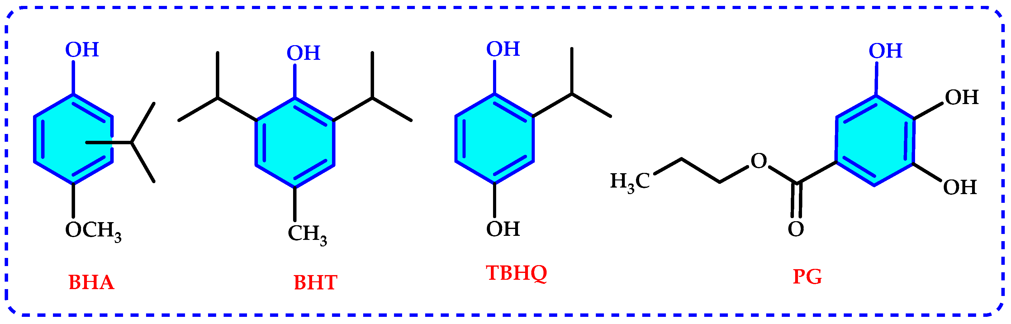
Iron is an essential element in biological and geochemical processes, participating in numerous redox reactions that influence both microbial metabolism and environmental transformations [61,62]. In nature, iron predominantly exists in two oxidation states: ferric iron (Fe3+) and ferrous iron (Fe2+). The reduction of Fe3+ to Fe2+ is a crucial step in the biogeochemical cycling of iron, with significant implications in microbial respiration, metal detoxification, and industrial applications [63].
Fe3+ reduction is a key process in anaerobic environments, where it serves as a terminal electron acceptor for various iron-reducing bacteria, such as Geobacter and Shewanella species [64]. These microorganisms utilize Fe3+ ions in respiration, playing a vital role in carbon and nutrient cycling [65]. The electron transfer processes facilitating Fe3+ reduction is mediated by cytochromes, electron shuttles, and conductive pili, allowing efficient energy generation even in oxygen-limited conditions [66,67]. Beyond microbial systems, Fe3+ reduction is influenced by abiotic factors, including natural organic matter, pH variations, and the presence of reducing agents such as flavins and quinones [68]. Photochemical reactions, particularly in sunlit aquatic systems, also contribute to Fe3+ reduction, affecting the bioavailability of iron for phytoplankton growth [69]. These redox transformations are essential for maintaining iron homeostasis in both terrestrial and marine environments. From an industrial perspective, Fe3+ reduction has applications in bioremediation, wastewater treatment, and corrosion control [70,71]. The ability of Fe3+-reducing bacteria to immobilize heavy metals and degrade organic pollutants makes them valuable tools for environmental sustainability [72]. Additionally, Fe3+-reducing ability plays an important role in medical applications, particularly in iron supplementation therapies and drug formulations [73,74]. Thus, understanding Fe3+ reduction mechanisms will facilitate studies on antioxidant and an understanding of the relevant mechanisms. Despite its importance, Fe3+ reduction is a complex and multifaceted process influenced by various biological and physicochemical factors. Recent advances in molecular biology, spectroscopy, and electrochemical techniques have provided deeper insights into the mechanisms governing Fe3+-reducing activity [66,75]. However, many aspects of Fe3+ reduction, particularly its interactions with emerging contaminants and nanomaterials, remain poorly understood, warranting further investigation.
Thousands of review articles that evaluate antioxidants can be found in a general literature search. This review is the first to focus only on reduction power. This review aims to provide a comprehensive overview of Fe3+-reducing ability, detailing its mechanisms, influencing factors, and applications in diverse fields. By synthesizing the current knowledge and identifying future research directions, this study seeks to enhance our understanding of iron redox chemistry and its broader implications.

2. Antioxidants

Antioxidants can be categorized in various ways depending on their environment and functional roles [1,76]. An antioxidant is defined as a compound that can significantly slow down or completely inhibit the oxidation of substrates, even when present in low concentrations [77]. These molecules neutralize free radicals by donating electrons, thereby reducing oxidative damage to biological systems [78,79,80]. In addition, they prevent free radical formation and interrupt oxidative processes at any of the three key stages: initiation, propagation, and termination [81,82].
Several factors influence antioxidant efficacy. The key parameters include the physical state of the system, temperature, molecular structure, characteristics of the oxidation-sensitive substrate, concentration, synergistic interactions, and the presence of prooxidant molecules [83]. The chemical composition of an antioxidant determines its reactivity and efficiency in neutralizing free radicals and ROS, its reducing potential, singlet oxygen quenching, and hydrogen peroxide decomposition [84]. Moreover, its effectiveness depends on its concentration and localization in the system, such as its distribution at the phase interfaces. The kinetics of the reaction also play a crucial role in the long- and short-term protective functions of antioxidants. These include the thermodynamic feasibility of reactions with oxidants, reaction rates, and overall antioxidant capacity [85,86].
By regulating the balance of oxidants, antioxidant compounds contribute to metabolic homeostasis. Additionally, antioxidants slow down lipid peroxidation in stored and processed foods, prevent spoilage, preserve pharmaceuticals, and extend the shelf life of products. Both synthetic and natural antioxidants are commonly used in food and pharmaceutical industries. The pharmaceutical industry has primarily focused on the use of synthetic antioxidants to decrease intracellular ROS and RNS levels. Synthetic antioxidants are widely utilized because of their high purity, low cost, and strong activity even at low concentrations. However, some studies have reported potential adverse effects associated with their use [87].
Consequently, natural antioxidants are increasingly favored over synthetic antioxidants, leading to the development of a growing number of methods for assessing antioxidant effectiveness. Among these, Fe3+ reduction ability is one of the most widely employed techniques. These assay results are generally compared to commonly used synthetic antioxidants which include butylated hydroxytoluene (BHT), propyl gallate (PG), butylated hydroxyanisole (BHA), and tert-butylhydroquinone (TBHQ), which are frequently added to food and pharmaceutical products to prevent oxidative degradation [88]. Despite their known adverse effects, manufacturers continue to use synthetic antioxidants, leaving consumers with limited options. The chemical structures of these synthetic antioxidants are shown in Figure 1. Notably, recent studies have raised concerns regarding the safety of these synthetic additives because they have been found to inhibit various enzymes, leading to potential health risks [85,86]. Owing to these toxicological concerns, researchers are actively searching for alternative natural antioxidant compounds that offer similar protective benefits with fewer side effects. In this regard, there has been a significant shift toward replacing synthetic antioxidants with natural alternatives that are characterized by lower toxicity, higher biodegradability, and safer mechanisms of action [1,87].
Prolonged use of synthetic antioxidants has been linked with several health problems, including carcinogenesis, skin allergies, fatty liver, and gastrointestinal discomfort. Consequently, informed consumers are increasingly wary of these negative effects and tend to opt for natural antioxidants. Fruits, vegetables, herbs, and spices are the most readily available and abundant sources of antioxidants. Plants such as tea, linden, cinnamon, cloves, fennel, anise, and rosemary are widely used because of their high concentrations of tannin, catechin, theine, phenolic, and flavonoid compounds, all of which exhibit strong antioxidant properties [88]. Regular consumption of phenolic-rich herbal products with antioxidant effects lowers disease risk and helps prevent the onset of degenerative conditions. However, the antioxidant potency and overall quality of natural antioxidants and their extracts are influenced not only by the plant source but also by the methods used for their extraction and isolation [89,90].

3. Antioxidant Evaluation Methods

Recent studies have extensively examined the oxidation of free radicals and the general mechanisms by which antioxidants counteract them. This interest stems from the fact that despite being neutral, free radicals exert a significant impact on biological systems. Certain lipid-derived compounds, such as aldehydes, which naturally occur during food processing, may pose health risks. These harmful compounds are easily produced when food is subjected to heat treatment [85]. Various antioxidant assays have been developed to directly measure hydrogen atom transfer or electron donation from antioxidants to free radicals and ROS [91]. Over time, significant advances have been made in techniques for measuring antioxidant capacity. Earlier methods primarily assessed antioxidant efficacy based on the formation of specific oxidation products, such as in lipid peroxidation. More recent approaches employ highly automated and sensitive detection technologies, enabling the precise evaluation of antioxidant activity through multiple mechanisms, including ROS scavenging, reducing power, and metal chelation. Initially introduced as a purely chemical concept, the idea of antioxidant capacity has since been adapted to various disciplines, including medicine, biology, food science, and epidemiology [92,93].
Understanding the antioxidant profiles of food and pharmaceutical products is important for maintaining their commercial and nutritional value during processing and storage. Therefore, a rapid and straightforward method to determine antioxidant capacity is essential. Numerous techniques for antioxidant evaluation have been developed and implemented [94,95]. Among the most commonly used methods are the inhibition of linoleic acid emulsion autoxidation, β-carotene bleaching test, total radical-trapping antioxidant parameter (TRAP), and oxygen radical absorbance capacity (ORAC) assay [1]. Other widely applied approaches include ferric (Fe3+) and cupric (Cu2+) ion reduction assays [96], as well as scavenging experiments targeting the 2,2-diphenyl-1-picrylhydrazyl radical (DPPH∙), N,N-dimethyl-p-phenylenediamine radicals (DMPD·+), and 2,2-azinobis(3-ethylbenzothiazoline-6-sulfonic acid) radicals (ABTS∙+) and O2, along with metal chelation tests. Most of these methods rely on similar principles and spectrophotometric detection to evaluate antioxidant potential. However, assessing antioxidant capacity using a single method is insufficient. At least three different in vitro assays should be performed concurrently to ensure an accurate evaluation, because no single test can fully reflect antioxidant activity. Considering the complexity of these methods, a direct comparison between them is often challenging. Therefore, selecting and applying the most appropriate techniques for research purposes is essential [1,26]. A key objective of this review is to explore the chemistry, mechanisms, and applications of Fe3+-reducing ability, following an overview of the antioxidant evaluation methods commonly used in research. Additionally, the antioxidant capacities of indirect methods may differ when used in foods because of the complexity of matrices [97]. In recent years, despite growing interest in various antioxidant assays, many studies have predominantly focused on Fe3+ reducing ability to meet the needs of researchers. Furthermore, among antioxidant methods, reducing power has great importance as it is the most used and low-cost antioxidant evaluation method.

4. Chemical Reduction Pathways

The chemical reduction of Fe3+ occurs through various mechanisms, primarily influenced by environmental conditions, electron donors, and catalytic agents. The conversion of Fe3+ to Fe2+ is facilitated by reducing agents such as organic acids, thiols, flavins, and phenolic compounds [68]. These molecules donate electrons to Fe3+, leading to its reduction and subsequent participation in biogeochemical cycles. One of the most well-studied pathways for Fe3+ reduction involves ascorbic acid, which acts as a potent reductant by directly transferring electrons to Fe3+ [98]. Additionally, flavins and hydroquinones, which possess biological activity are known to facilitate Fe3+ reduction, particularly in aqueous systems where their redox potential plays a critical role. The influence of pH on Fe3+ reduction is another key aspect. At lower pH levels, Fe3+ remains in a soluble form, allowing more efficient electron transfer [99]. However, in alkaline conditions, Fe3+ ions tend to precipitate as insoluble oxides, limiting their bioavailability and reduction potential [100]. Furthermore, iron-complexing ligands such as EDTA and citrate significantly enhance Fe3+ reduction by stabilizing Fe3+ in solution and preventing precipitation [101]. This effect is particularly relevant in environmental and industrial processes where Fe3+ solubility dictates reaction efficiency. In summary, Fe3+ reduction involves a complex interplay of electron donors, pH effects, and ligand interactions. Advances in spectroscopic and electrochemical techniques have provided deeper insight into these processes, with significant implications for environmental and industrial applications.

5. Biological Sources of Reducing Power

Reducing power influences several key biological processes, including energy production, biosynthesis, oxidative stress management, and immune functions. NADH donates electrons to the mitochondrial electron transport chain and drives ATP synthesis via oxidative phosphorylation [102]. NADPH provides reducing equivalents for anabolic pathways such as fatty acid and cholesterol biosynthesis [103]. NADPH is also crucial for the regeneration of antioxidant systems, including glutathione and thioredoxin, which protect cells from oxidative damage [104]. NADPH oxidase plays a role in generating ROS used by immune cells to kill pathogens [105]. Therefore, the reducing power of biological systems is indispensable for maintaining cellular function and homeostasis. NADH, NADPH, and glutathione are central to metabolism, biosynthesis, and oxidative stress defense. Understanding the mechanisms and roles of reducing power can provide insights into metabolic disorders and potential therapeutic targets for oxidative stress-related diseases [106]. The primary sources of reducing power in biological systems are glutathione, ascorbic acid, NADH, and NADPH, which act as electron carriers in numerous metabolic processes.

5.1. NADH

Reduced nicotinamide adenine dinucleotide (NADH) is a coenzyme that plays a significant role in cellular metabolism and energy production. It represents the reduced form of oxidized NAD+ and exerts vital functions primarily in redox reactions, transferring electrons from one molecule to another. This process is essential for ATP synthesis, the main cellular energy carrier [107]. NADH is generated during glycolysis, the tricarboxylic acid cycle, and β-oxidation of fatty acids. It transfers electrons to the mitochondrial electron transport chain and facilitates ATP production via oxidative phosphorylation [108]. In addition to energy metabolism, NADH is involved in several cellular functions, including DNA repair, gene expression regulation, and immune system modulation. It has been studied for its potential anti-aging effects as it contributes to the activity of sirtuins, a class of enzymes associated with longevity [109]. In the clinical and nutritional contexts, NADH supplementation is occasionally used to improve cognitive function, reduce fatigue, and enhance athletic performance. However, further studies are required to confirm these benefits [110].

5.2. NADPH

NADPH is primarily generated through the pentose phosphate pathway and the activity of malic enzyme. It is the reduced form of nicotinamide adenine dinucleotide phosphate (NADP+) and serves as an electron donor in several biosynthetic processes. NADPH plays a key role in supporting biosynthetic reactions like the synthesis of fatty acids and nucleotides, and it helps maintain redox balance by replenishing glutathione [111]. One of the primary functions of NADPH is its involvement in anabolic reactions such as fatty acid and cholesterol synthesis. These processes require a high-energy reducing agent, which NADPH provides by donating electrons. NADPH is also essential for the regeneration of glutathione, a key antioxidant that protects cells from oxidative stress by neutralizing ROS [103]. The PPP is the major cellular source of NADPH. In this pathway, glucose-6-phosphate is converted to ribulose-5-phosphate, generating NADPH as a byproduct. Other sources include malic enzyme activity and isocitrate dehydrogenase reactions in mitochondria [112]. NADPH is crucial for the function of NADPH oxidases, which mediate immune defense by generating ROS to combat pathogens [113]. Additionally, NADPH supports the activity of cytochrome P450 enzymes in drug metabolism and detoxification [114].

5.3. Glutathione (GSH)

GSH is a tripeptide involved in the neutralization of ROS and detoxification of xenobiotics. Studies have shown that glutathione reductase and NADPH maintain cellular redox balance [115]. GSH is a vital tripeptide composed of glutamate, cysteine, and glycine. It serves as a major antioxidant, detoxifying ROS and maintaining cellular redox homeostasis. Glutathione exists in two forms: reduced (GSH) and oxidized (GSSG). The GSH/GSSG ratio is a key indicator of oxidative stress in cells [116]. One of the most important functions of glutathione is detoxification. It conjugates with toxic compounds via glutathione S-transferase (GST) enzymes, facilitating their excretion [117,118]. Additionally, GSH plays a critical role in immune function by modulating cytokine production and lymphocyte activity. GSH is synthesized in a two-step enzymatic process. First, glutamate–cysteine ligase catalyzes the formation of γ-glutamylcysteine; glutathione synthetase then adds glycine to complete this process [1]. The primary sites of GSH synthesis are the liver and kidneys, where it supports detoxification. Glutathione is also vital for mitochondrial function, protection against oxidative damage, and maintenance of electron transport chain efficiency. Deficiencies in GSH are associated with various diseases, including neurodegenerative disorders, cancer, and cardiovascular diseases [119].

5.4. Ascorbic Acid (Vitamin C)

Ascorbic acid is a water-soluble vitamin with effective antioxidant ability for various physiological functions in humans. It plays critical roles in collagen synthesis, immune function, and ROS neutralization [120]. One of the primary roles of ascorbic acid is acting as a cofactor for prolyl and lysyl hydroxylases, which are necessary for collagen hydroxylation. This function is crucial for wound healing, connective tissue formation, and overall skin health. Ascorbic acid deficiency leads to scurvy, a disease characterized by weakened connective tissue, bleeding gums, and impaired wound healing [121].
As an antioxidant, ascorbic acid scavenges free radicals and ROS, and regenerates other antioxidants such as vitamin E, thereby maintaining cellular redox balance. It also enhances the absorption of non-heme iron from plant-based foods, reducing the risk of iron-deficiency anemia [1]. Ascorbic acid is involved in immune function by stimulating the production and activity of white blood cells and enhancing the skin defense system. Regular ascorbic acid intake has been shown to reduce the duration and severity of colds. Humans cannot synthesize ascorbic acid and must obtain it from dietary sources such as citrus fruits, berries, and leafy greens. Excessive intake may lead to gastrointestinal discomfort but is generally non-toxic [122].

6. Biochemical Mechanisms of Fe3+ Reduction

6.1. Non-Enzymatic Reduction

Non-enzymatic reduction of Fe3+ occurs through electron transfer from various organic and inorganic reducing agents, including natural organic matter, sulfides, and hydroquinones. These processes play crucial roles in geochemical iron cycling, particularly in environments where microbial activity is limited or absent. Abiotic reduction of Fe3+ has significant implications for iron mobility, mineral transformation, and redox-driven biogeochemical reactions [123].

6.1.1. Organic Reducing Agents

Organic compounds such as hydroquinones, ascorbic acid, and catechol are known to facilitate Fe3+ reduction. Hydroquinones, for instance, act as electron donors in both natural and synthetic systems, significantly enhancing Fe3+ reduction rates. These compounds are often found in humic substances, which contribute to iron redox cycling in sediments and soils [124]. Ascorbic acid, also known as vitamin C, is another potent Fe3+-reducing agent. It donates electrons to Fe3+, forming Fe2+ and dehydroascorbic acid. This reaction is particularly relevant in biological systems, where ascorbic acid enhances iron bioavailability by converting Fe3+ from dietary sources into its more soluble Fe2+ form [125]. Furthermore, organic substrates such as acetate, lactate, and hydrogen fuel microbial respiration and drive Fe3+ reduction [126]. High concentrations of sulfate, nitrate, or oxygen can suppress Fe3+ reduction owing to the preferential use of alternative electron acceptors [127].

6.1.2. Inorganic Reducing Agents

Several inorganic compounds also contribute to Fe3+ reduction. Sulfides (e.g., hydrogen sulfide, H2S) react with Fe3+ to form Fe2+, often leading to the precipitation of iron sulfide minerals such as pyrite (FeS2). This reaction plays a key role in anoxic environments, particularly in marine and freshwater sediments where sulfate-reducing bacteria produce-H2S as a metabolic byproduct [128]. Furthermore, green rust-mixed valence iron minerals can mediate Fe3+ reduction through electron transfer processes. These minerals are formed under reducing conditions and act as both electron donors and acceptors, thereby influencing iron and contaminant mobility in subsurface environments [129].

6.1.3. Photochemical Reduction

Light-driven Fe3+ reduction is another significant abiotic process, particularly in surface waters. Fe3+ complexes with organic ligands, such as citrate and oxalate, undergo photochemical reduction when exposed to sunlight. This mechanism is especially significant in iron-limited aquatic ecosystems, where photoreduction of Fe3+ to Fe2+ enhances bioavailable iron concentrations [4]. Overall, non-enzymatic Fe3+ reduction is a vital pathway in iron biogeochemistry and interacts with biological and geochemical processes to influence iron availability, mineral stability, and environmental redox conditions. Photochemical reduction of Fe3+ is an essential natural process that contributes to the bioavailability of iron, particularly in aquatic systems. Sunlight-driven reduction of Fe3+ influences iron cycling, impacting both primary productivity and metal mobility in environmental systems [130]. This process occurs via direct photolysis, ligand-mediated reduction, and interactions with ROS [131]. Furthermore, in the absence of organic ligands, Fe3+ can be reduced through direct absorption of solar radiation, particularly in the UV spectrum. This reaction results in the formation of Fe2+ and oxygen radicals, significantly affecting iron speciation in surface waters [132].

6.2. Enzymatic Reduction

The enzymes involved in Fe3+ reduction include cytochromes c, hydrogenases, and dehydrogenases. These proteins, including MtrA in Shewanella, are crucial in transferring electrons from the inner mitochondrial membrane to extracellular Fe3+ acceptors [133]. Enzymes also generate reducing equivalents to fuel electron transfer pathways [134].

6.3. Microbial Mediators

Microbial Fe3+ reduction is a biologically mediated process that plays a crucial role in iron cycling and anaerobic respiration. Certain microorganisms, particularly dissimilatory iron-reducing bacteria, have evolved mechanisms to use Fe3+ as a terminal electron acceptor. This process is fundamental in sedimentary environments, wastewater treatment, and biogeochemical cycling [135]. Several environmental parameters affect microbial Fe3+ reduction, including pH, redox conditions, electron donor availability, and presence of competing electron acceptors. Optimal Fe3+ reduction occurs in neutral to slightly acidic conditions, as extreme pH values can inhibit bacterial activity [136]. Microbial reduction of Fe3+ has importance in biotechnological and environmental applications, such as bioremediation, microbial fuel cells, and climate impact.

7. Procedure for Determining Reducing Power in Antioxidants

The reductive potential of antioxidants was first evaluated by Oyaizu [137]. For this aim, various concentrations of antioxidants were prepared in 1 mL of distilled water, followed by mixing with phosphate buffer (2.5 mL, 0.2 M, pH 6.6) and 2.5 mL of potassium ferricyanide ([K3Fe(CN)6]) (1%). The resulting mixture was incubated at 50 °C for 20 min. Subsequently, 2.5 mL of 10% trichloroacetic acid (TCA) was added and the solution was centrifuged at 1000× g for 10 min. After centrifugation, the upper layer (2.5 mL) was collected and mixed with distilled water and 0.5 mL of 0.1% FeCl3. The absorbance of the final solution was measured at 700 nm using a spectrophotometer. A higher absorbance value indicated a greater reductive potential of the tested materials [138]. The most suitable solution for evaluating reducing power is distilled and deionized water.

8. Reducing Power of Various Antioxidants

The reducing antioxidant power assay is based on the principle that an increase in absorbance corresponds to an increase in antioxidant activity. Reduction refers to the gain of electrons, whereas oxidation involves their loss [139]. A reducing agent (or reductant) donates electrons, facilitating the reduction of another substance, whereas an oxidizing agent (or oxidant) accepts electrons, leading to the oxidation of another reactant. As oxidation and reduction are interdependent, they occur simultaneously within a system. Chemical reactions involving both processes are classified as redox reactions. Redox reactions play a fundamental role in biological oxidation, a series of chemical reactions in which oxygen from air oxidizes molecules derived from food metabolism, ultimately generating energy for living organisms. While reductants and oxidants are chemical terms, antioxidants and pro-oxidants are used in the context of biological systems. Furthermore, an antioxidant capable of effectively reducing pro-oxidants may not necessarily exhibit the same efficiency in reducing Fe3+. In the Fe3+-reducing assay, the presence of reductants mediates the conversion of Fe3+ to Fe2+. The reducing potential of a bioactive compound is assessed by monitoring the direct reduction of [Fe(CN)6]3− to [Fe(CN)6]4−. When free Fe3+ is introduced to the reduced product, it forms an intense blue-colored complex known as Perl’s Prussian blue, Fe4[Fe(CN)6]3, which exhibits strong absorbance at 700 nm. The Fe3+-reducing assay thus involves an electron transfer mechanism in which a ferric salt functions as the oxidant [1].
R e d u c e d   a n t i o x i d a n t + F e 3 +   O x i d i s e d   a n t i o x i d a n t + F e 2 + F e 2 + + F e ( C N ) 6 3 F e F e ( C N ) 6 o r R e d u c e d   a n t i o x i d a n t + F e ( C N ) 6 3   O x i d i z e d   a n t i o x i d a n t + F e ( C N ) 6 4 F e 3 + + F e ( C N ) 6 4 F e F e ( C N ) 6
An antioxidant is a reductant, but a reductant is not necessarily an antioxidant. As reported in many studies, the activity of natural antioxidants in diseases is closely related to their ability to reduce DNA damage, mutagenesis, and carcinogenesis, and to inhibit pathogenic bacterial growth [1]. The Fe3+-reducing ability assay is a spectrophotometric method widely used for assessing the antioxidant capacity of beverages, pure compounds, food products, and herbal extracts. Because of its simplicity, sensitivity, speed, and reproducibility, this technique is one of the most convenient and commonly applied methods for evaluating the radical scavenging potential of various compounds and plant extracts. The absorbance values of herbal extracts and pure substances from recent studies on Fe3+-reducing ability are presented in Table 2 and Table 3. Notably, some natural antioxidants, such as essential oils, have an efficient antioxidant capacity under high-temperature conditions, even in extremely demanding thermal conditions such as deep-frying processes [140,141].

9. Advantages of the Fe3+ Reducing Ability Assay

The advantages of the Fe3+-reducing ability assay can be determined in terms of simplicity, speed, reproducibility, and no requirement for free radicals. The assay procedure is straightforward, requiring minimal sample preparation and providing results within minutes. Owing to its well-defined reaction conditions, this method offers high reproducibility and reliability. Unlike other antioxidant assays, such as DPPH or ABTS, the Fe3+-reducing ability assay does not involve free radicals, making it a more stable and less variable method [1].

10. Limitations of the Fe3+ Reducing Ability Assay

The Fe3+ reducing ability assay has some limitations in terms of selectivity for reducing agents, pH dependency, and the need for water-soluble antioxidants. The Fe3+ reducing ability assay is primarily used to analyze compounds that act via single-electron transfer mechanisms. It does not account for hydrogen-atom transfer-based antioxidants, such as thiols and some polyphenols. Furthermore, this assay requires an acidic environment, which may not reflect the physiological conditions under which the antioxidant activity occurs. In addition, the Fe3+ reducing ability assay primarily detects hydrophilic antioxidants, making it less effective for lipophilic compounds [1].
According to our review, reducing ability tends to yield stronger responses, particularly with phenolic compounds. For polar and phenolic substances, incorporating water into the reaction medium, such as aqueous methanol, enhance the accuracy of the results. Conversely, for low-polarity compounds, ethyl acetate is more suitable for interactions with radicals. These findings indicate that reducing ability is influenced more by the steric accessibility of the radical site than by the inherent chemical properties of the tested antioxidant compounds.
The reaction rate of reducing ability with antioxidants is determined by the varying contributions of single-electron or hydrogen atom transfer mechanisms. Additionally, environmental and experimental conditions can significantly affect the reaction mechanisms and response outcomes. The interaction of antioxidants with reducing agents is known to be lower than the total activity of the individual antioxidant compounds, suggesting that radical scavenging by extracts from mixtures may be suppressed in reducing ability assays.

11. Conclusions

Antioxidant compounds play a crucial role in minimizing the oxidative damage caused by ROS. The reducing ability of these compounds is thus important for determining their antioxidant profiles. Among the various antioxidant evaluation methods, the Fe3+-reducing ability assay is one of the most commonly utilized techniques in both food and pharmaceutical applications. Fe3+-reducing power can have significant health and industrial implications, affecting disease risk, food quality, and product stability. By understanding the mechanisms underlying this reduction and adopting strategies for its optimization, we can enhance health outcomes and improve the longevity of antioxidant-rich products. Further research is required to develop advanced techniques for preserving and restoring antioxidant power in various applications.

Author Contributions

Investigation, writing—original draft preparation, and writing—review and editing, İ.G. and S.H.A. All authors have read and agreed to the published version of the manuscript.

Funding

This study received no external funding.

Institutional Review Board Statement

Not applicable.

Informed Consent Statement

Not applicable.

Data Availability Statement

Data are available in a publicly accessible repository.

Acknowledgments

I. Gulcin would like to extend his sincere appreciation to the Turkish Academy of Sciences (TÜBA). S. Alwasel would like to extend his sincere appreciation to the Researchers Support Program (RSP-2025/59) of King Saud University, Saudi Arabia.

Conflicts of Interest

The authors declare no conflicts of interest.

References

  1. Gulcin, I. Antioxidants and antioxidant methods—An updated overview. Arch. Toxicol. 2020, 94, 651–715. [Google Scholar] [CrossRef] [PubMed]
  2. Akyuz, M. The determination of antidiabetic, anticholinesterase and antioxidant properties of ethanol and water extracts of blackberry (Rubus fruticosus L.) fruits at different maturity stages. S. Afr. J. Bot. 2022, 151, 1035–1048. [Google Scholar] [CrossRef]
  3. Akyuz, M.; Yabo-Dambagi, L.; Kilic, T.; Cakir, A. Antidiabetic, neuroprotective and antioxidant potentials of different parts of Pistacia terebinthus fruits. S. Afr. J. Bot. 2022, 147, 443–456. [Google Scholar] [CrossRef]
  4. Buldurun, K.; Turan, N.; Mahmoudi, G.; Bursal, E. Half-sandwich ruthenium(II)(η6-p-cymene) complexes: Syntheses, characterization, transfer hydrogenation reactions, antioxidant and enzyme inhibitory activities. J. Mol. Struct. 2022, 1262, 133075. [Google Scholar] [CrossRef]
  5. Alam, M.N.; Bristi, N.J.; Rafiquzzaman, M. Review on in vivo and in vitro methods evaluation of antioxidant activity. Saudi Pharm. J. 2013, 21, 143–152. [Google Scholar] [CrossRef]
  6. Murphy, M.P. How mitochondria produce reactive oxygen species. Biochem. J. 2009, 417, 1–13. [Google Scholar] [CrossRef]
  7. Bedard, K.; Krause, K.H. The NOX family of ROS-generating NADPH oxidases: Physiology and pathophysiology. Physiol. Rev. 2007, 87, 245–313. [Google Scholar] [CrossRef] [PubMed]
  8. Fransen, M.; Lismont, C.; Walton, P. The peroxisome-mitochondria connection: How and why? Int. J. Mol. Sci. 2012, 13, 5860–5878. [Google Scholar] [CrossRef]
  9. Azzam, E.I.; Jay-Gerin, J.P.; Pain, D. Ionizing radiation-induced metabolic oxidative stress and prolonged cell injury. Cancer Lett. 2012, 327, 48–60. [Google Scholar] [CrossRef]
  10. Valavanidis, A.; Vlachogianni, T.; Fiotakis, K. 8-hydroxy-2′-deoxyguanosine as a biomarker of oxidative DNA damage in carcinogenesis: Measurement methods and applications. Mol. Cell. Biochem. 2013, 356, 435–456. [Google Scholar]
  11. Pelicano, H.; Carney, D.; Huang, P. ROS stress in cancer cells and therapeutic implications. Drug Resist. Updates 2004, 7, 97–110. [Google Scholar] [CrossRef]
  12. Schieber, M.; Chandel, N.S. ROS function in redox signaling and oxidative stress. Curr. Biol. 2014, 24, R453–R462. [Google Scholar] [CrossRef] [PubMed]
  13. Nathan, C.; Ding, A. Reactive oxygen and nitrogen species in immunity. Nat. Rev. Immunol. 2010, 10, 349–361. [Google Scholar]
  14. Sena, L.A.; Chandel, N.S. Physiological roles of mitochondrial reactive oxygen species. Mol. Cell 2012, 48, 158–167. [Google Scholar] [CrossRef]
  15. Yin, H.; Xu, L.; Porter, N.A. Free radical lipid peroxidation: Mechanisms and analysis. Chem. Rev. 2011, 111, 5944–5972. [Google Scholar] [CrossRef] [PubMed]
  16. Akhtar, N.; Mirza, B. Phytochemical analysis and comprehensive evaluation of anti-microbial and antioxidant properties of 61 medicinal plant species. Arab. J. Chem. 2018, 11, 1223–1235. [Google Scholar] [CrossRef]
  17. Stadtman, E.R.; Levine, R.L. Free radical-mediated oxidation of free amino acids and amino acid residues in proteins. Amino Acids 2003, 25, 207–218. [Google Scholar] [CrossRef]
  18. Cooke, M.S.; Evans, M.D.; Dizdaroglu, M.; Lunec, J. Oxidative DNA damage: Mechanisms, mutation, and disease. FASEB J. 2003, 17, 1195–1214. [Google Scholar] [CrossRef]
  19. Kavaz Yuksel, A.; Dikici, E.; Yüksel, M.; Isık, M.; Tozoglu, F.; Koksal, E. Phytochemicals analysis and some bioactive properties of Erica manipuliflora Salisb. (EMS); antibacterial, antiradical and anti-lipid peroxidation. Iran. J. Pharm. Res. 2021, 20, 422–434. [Google Scholar]
  20. Ayguni, R.B.; Zengini, G.; Yıldıztugayi, E.; Jugreeti, S.; Yılmazi, M.A.; Mahomoodally, F.M. Chemical characterization, anti-oxidant and anti-enzymatic properties of extracts from two Silene species: A focus on different plant parts and extraction methods. Process Biochem. 2022, 116, 206–213. [Google Scholar] [CrossRef]
  21. Zhang, L.; Miras-Moreno, B.; Yildiztugay, E.; Ozfidan-Konakci, C.; Arikan, B.; Elbasan, F.; Ak, G.; Rouphael, Y.; Zengin, G.; Lucini, L. Metabolomics and physiological insights into the ability of exogenously applied chlorogenic acid and hesperidin to modulate salt stress in lettuce distinctively. Molecules 2021, 26, 6291. [Google Scholar] [CrossRef] [PubMed]
  22. Ionita, P. The chemistry of DPPH· free radical and congeners. Int. J. Mol. Sci. 2021, 22, 1545. [Google Scholar] [CrossRef] [PubMed]
  23. Avsar, O.K.; Kasbolat, S.; Ak, G.; Nilofar; Caprioli, G.; Santanatoglia, A.; Uysal, A.; Uba, A.I.; Ponniya, S.K.M.; Paksoy, M.Y.; et al. Integrating chemical analysis with in vitro, in silico, and network pharmacology to discover potential functional compounds from Marrubium astracanicum subsp. macrodon. J. Mol. Liq. 2024, 398, 124204. [Google Scholar] [CrossRef]
  24. Polat Kose, L. Evaluation of antioxidant capacity, anticholinergic and antidiabetic activities, and phenolic ingredients of Asphodelus aestivus by LC-MS/MS. Chem. Biodivers. 2024, 21, e202301965. [Google Scholar] [CrossRef]
  25. Yuldasheva, N.; Acikyildiz, N.; Akyuz, M.; Yabo-Dambagi, L.; Aydin, T.; Cakir, A.; Kazaz, C. The Synthesis of Schiff bases and new secondary amine derivatives of p-vanillin and evaluation of their neuroprotective, antidiabetic, antidepressant and antioxidant potentials. J. Mol. Struct. 2022, 1270, 133883. [Google Scholar] [CrossRef]
  26. Munteanu, I.G.; Apetrei, C. Analytical methods used in determining antioxidant activity: A review. Int. J. Mol. Sci. 2021, 22, 3380. [Google Scholar] [CrossRef] [PubMed]
  27. Rodrigo, R. Oxidative Stress and Antioxidants: Their Role in Human Diseases; Nova: New York, NY, USA, 2009; pp. 9–10. [Google Scholar]
  28. Christodoulou, M.C.; Orellana Palacios, J.C.; Hesami, G.; Jafarzadeh, S.; Lorenzo, J.M.; Domínguez, R.; Moreno, A.; Hadidi, M. Spectrophotometric methods for measurement of antioxidant activity in food and pharmaceuticals. Antioxidants 2022, 11, 2213. [Google Scholar] [CrossRef] [PubMed]
  29. De Gregorio, M.A.; Zengin, G.; Alp-Turgut, F.N.; Elbasan, F.; Ozfidan-Konakci, C.; Arikan, B.; Yildiztugay, E.; Zhang, L.; Lucini, L. Glutamate, humic acids and their combination modulate the phenolic profile, antioxidant traits, and enzyme-inhibition properties in lettuce. Plants 2023, 12, 1822. [Google Scholar] [CrossRef]
  30. Kedare, S.B.; Sing, R.P. Genesis and development of DPPH method of antioxidant assay. J. Food Sci. Technol. 2011, 48, 412–422. [Google Scholar] [CrossRef]
  31. Tungmunnithum, D.; Thongboonyou, A.; Pholboon, A.; Yangsabai, A. Flavonoids and other phenolic compounds from medicinal plants for pharmaceutical and medical aspects: An overview. Medicines 2018, 5, 93–109. [Google Scholar] [CrossRef]
  32. Aktumsek, A.; Zengin, G.; Guler, G.O.; Cakmak, Y.S.; Duran, A. Assessment of the antioxidant potential and fatty acid composition of four Centaurea L. taxa from Turkey. Food Chem. 2013, 141, 91–97. [Google Scholar] [CrossRef]
  33. Lourenco, S.C.; Moldao-Martins, M.; Alves, V.D. Antioxidants of natural plant origins: From sources to food industry applications. Molecules 2019, 24, 4132. [Google Scholar] [CrossRef]
  34. Uysal, S.; Cvetanovic, A.; Zengin, G.; Durovic, S.; Zekovic, Z.; Aktumsek, A. Effects of orange leaves extraction conditions on antioxidant and phenolic content: Optimization using response surface methodology. Anal. Lett. 2017, 51, 1505–1519. [Google Scholar] [CrossRef]
  35. Angelini, P.; Venanzoni, R.; Flores, G.A.; Tirillini, B.; Orlando, G.; Recinella, L.; Chiavaroli, A.; Brunetti, L.; Leone, S.; Di Simone, S.C.; et al. Evaluation of antioxidant, antimicrobial and tyrosinase inhibitory activities of extracts from Tricholosporum goniospermum, an edible wild mushroom. Antibiotics 2020, 9, 513. [Google Scholar] [CrossRef] [PubMed]
  36. Mamadalieva, R.; Khujaev, V.; Zengin, G. Chemical profiling and biological activities of the roots of Allochrusa gypsophiloides. Nat. Prod. Res. 2024, 1–11. [Google Scholar] [CrossRef]
  37. Zengin, G.; Cakmak, Y.S.; Guler, G.O.; Aktumsek, A. In vitro antioxidant capacities and fatty acid compositions of three Centaurea species collected from Central Anatolia region of Turkey. Food Chem. Toxicol. 2010, 48, 2638–2641. [Google Scholar] [CrossRef] [PubMed]
  38. Fenton, H.J.H. Oxidation of tartaric acid in the presence of iron. J. Chem. Soc. Trans. 1984, 65, 899–910. [Google Scholar] [CrossRef]
  39. Haber, F.; Weiss, J. The catalytic decomposition of hydrogen peroxide by iron salts. Proc. R. Soc. A 1934, 147, 332–351. [Google Scholar]
  40. Mahomoodally, M.F.; Zengin, G.; Aumeeruddy, M.Z.; Sezgin, M.; Aktumsek, A. Phytochemical profile and antioxidant properties of two Brassicaceae species: Cardaria draba subsp. draba and Descurainia sophia. Biocatal. Agric. Biotechnol. 2018, 16, 453–458. [Google Scholar] [CrossRef]
  41. Zengin, G.; Uysal, A.; Gunes, E.; Aktumsek, A. Survey of phytochemical composition and biological effects of three extracts from a wild plant (Cotoneaster nummularia Fisch et Mey.): A potential source for functional food ingredients and drug formulations. PLoS ONE 2014, 9, e113527. [Google Scholar] [CrossRef]
  42. Sies, H. Oxidative stress: A concept in redox biology and medicine. Redox Biol. 2015, 4, 180–183. [Google Scholar] [CrossRef]
  43. Piluzza, G.; Bullitta, S. Correlations between phenolic content and antioxidant properties in twenty-four plant species of traditional ethnoveterinary use in the Mediterranean area. Pharm. Biol. 2011, 49, 240–247. [Google Scholar] [CrossRef] [PubMed]
  44. Ceylan, R.; Zengin, G.; Guler, G.O.; Aktumsek, A. Bioactive constituents of Lathyrus czeczottianus and ethyl acetate and water extracts and their biological activities: An endemic plant to Turkey. S. Afr. J. Bot. 2021, 143, 306–311. [Google Scholar] [CrossRef]
  45. Sies, H. Oxidative stress: Introductory remarks. In Oxidative Stress; Academic Press: Cambridge, MA, USA, 1985; pp. 1–8. [Google Scholar]
  46. Finkel, T.; Holbrook, N.J. Oxidants, oxidative stress and the biology of ageing. Nature 2000, 408, 239–247. [Google Scholar] [CrossRef]
  47. Chen, C.; Wang, Y.; Cui, R.; Zhu, L.; Han, J.; Sun, J.; Xu, Q.; Karrar, E.; Zhang, H.; Jin, Q.; et al. Effect of different processing practices on physicochemical parameters, phenolic compounds, and antioxidant properties of chia seed oils. J. Am. Oil Chem. Soc. 2024, 101, 225–236. [Google Scholar] [CrossRef]
  48. Halliwell, B.; Gutteridge, J.M.C. Free Radicals in Biology and Medicine; Oxford University Press: Oxford, UK, 2007. [Google Scholar]
  49. Akyuz, M.; Yabo-Dambagi, L.; Simitcioglu, B.; Karagoz, I.D.; Aydin, T.; Gokdemir, G.; Cakir, A.; Kazaz, C. The chemical composition and antidiabetic, neuroprotective and cytotoxic activities of soft hulls (Mesocarp) of pistachio (Pistacia vera) fruits. J. Chem. Soc. Pak. 2023, 45, 551–567. [Google Scholar]
  50. Dröge, W. Free radicals in the physiological control of cell function. Physiol. Rev. 2002, 82, 47–95. [Google Scholar] [CrossRef]
  51. Ergün, A.I.; Topal, M. Determination of the antioxidant capacity of puerarin and its effect on cholinesterases by in vitro and in silico methods. ChemistrySelect 2023, 8, e202302751. [Google Scholar] [CrossRef]
  52. Uttara, B.; Singh, A.V.; Zamboni, P.; Mahajan, R.T. Oxidative stress and neurodegenerative diseases: A review of upstream and downstream antioxidant therapeutic options. Curr. Neuropharmacol. 2009, 7, 65–74. [Google Scholar] [CrossRef]
  53. Tousif, M.; Abbas, Z.; Nazir, M.; Saleem, M.; Tauseef, S.; Hassan, A.; Ali, S.; Ahmed, M.; Khan, J.; Zengin, G.; et al. Secondary metabolic profiling, antioxidant potential, enzyme inhibitory activities and in silico and ADME studies: A multifunctional approach to reveal medicinal and industrial potential of Tanacetum falconeri. BMC Complement. Med. Ther. 2024, 24, 167. [Google Scholar] [CrossRef]
  54. Jendoubi, O.; Majdoub, S.; AL-Hmadi, H.B.; El Mokni, R.; Angeloni, S.; Mustafa, A.M.; Caprioli, G.; Zengin, G.; Maggi, F.; Hammami, S. Antioxidant potential, enzyme inhibitory activity and HPLC-MS/MS phenolics profiles in Salvia aegyptiaca L. (Lamiaceae, Nepetoideae, Mentheae) growing in Tunisia. Fitoterapia 2025, 82, 106473. [Google Scholar] [CrossRef]
  55. Zengin, G.; Arslan, E.; Toyran, K.; Arslan, A.; Koygun, G. Screening for antioxidant effects and chemical profiles of different extracts of Rheum ribes parts. Chem. Biodivers. 2025, 202403240. [Google Scholar] [CrossRef] [PubMed]
  56. Madamanchi, N.R.; Runge, M.S. Redox signaling in cardiovascular health and disease. Free Radical Biol. Med. 2007, 43, 645–657. [Google Scholar] [CrossRef] [PubMed]
  57. Reuter, S.; Gupta, S.C.; Chaturvedi, M.M.; Aggarwal, B.B. Oxidative stress, inflammation, and cancer: How are they linked? Free Radic. Biol. Med. 2010, 49, 1603–1616. [Google Scholar] [CrossRef]
  58. Yildirim, M.A.; Sevinc, B.; Paydas, S.; Karaselek, M.A.; Duran, T.; Kuccukturk, S.; Vatansev, H.; Cetiz, M.V.; Caprioli, G.; Acquaticci, L.; et al. Exploring the anticancer potential of Dianthus orientalis in pancreatic cancer: A molecular and cellular study. Food Biosci. 2025, 66, 106183. [Google Scholar] [CrossRef]
  59. Ahmed, S.; Zengin, G.; Fernández-Ochoa, A.; de la Luz Cádiz-Gurrea, M.D.; Leyva-Jiménez, F.J.; Elkiran, O.; Cakilcioglu, U.; Akgul, B.H.; Pereira, C.G.; Custódio, L. Exploration of UHPLC-ESI-QTOF-MS profiles and the neuroprotective, antidiabetic, antioxidant and cytotoxic effects of extracts from Achillea maritima (L.) Ehrend. & YPGuo (Asteraceae) collected in Türkiye. Ital. J. Food Sci. 2025, 81, 66. [Google Scholar]
  60. Erdogan, M.K.; Toy, Y.; Gundogdu, R.; Gecibesler, I.H.; Sever, A.; Yapar, Y.; Behcet, L.; Zengin, G. Assessment of cytotoxic, apoptotic, enzyme inhibitory, and antioxidant properties, and phytochemical characterization of ethanolic extract from Cionura erecta. Foods 2025, 65, 106082. [Google Scholar] [CrossRef]
  61. Stumm, W.; Sulzberger, B. The cycling of iron in natural environments: Considerations based on laboratory studies of heterogeneous redox processes. Geochim. Cosmochim. Acta 1992, 56, 3233–3257. [Google Scholar] [CrossRef]
  62. Nersezashvili, M.; Berashvili, D.; Jokhadze, M.; Metreveli, M.; Swiatek, L.; Salwa, K.; Pecio, L.; Wojtanowski, K.K.; Skiba, A.; Korona-Glowniak, I.; et al. Seseli foliosum (Somm. et Levier) Manden.-A comprehensive phytochemical and biological evaluation. Molecules 2025, 30, 725. [Google Scholar] [CrossRef]
  63. Artunç, T.; Menzek, A.; Taslimi, P.; Gulçin, İ.; Kazaz, C.; Şahin, E. Synthesis and antioxidant activities of phenol derivatives from 1,6-bis(dimethoxyphenyl)hexane-1,6-dione. Bioorg. Chem. 2020, 100, 103884. [Google Scholar] [CrossRef]
  64. Lovley, D.R. Dissimilatory Fe(III) and Mn(IV) reduction. Microbiol. Rev. 1991, 55, 259–287. [Google Scholar] [CrossRef] [PubMed]
  65. Karagecili, H.; Yılmaz, M.A.; Ertürk, A.; Kızıltaş, H.; Güven, L.; Alwasel, S.H.; Gulcin, I. Comprehensive metabolite profiling of Berdav propolis using LC-MS/MS: Determination of antioxidant, anticholinergic, antiglaucoma, and antidiabetic effects. Molecules 2023, 28, 1739. [Google Scholar] [CrossRef] [PubMed]
  66. Shi, L.; Dong, H.; Reguera, G.; Beyenal, H.; Lu, A.; Liu, J.; Yu, H.Q.; Fredrickson, J.K. Extracellular electron transfer mechanisms between microorganisms and minerals. Nat. Rev. Microbiol. 2012, 10, 751–762. [Google Scholar] [CrossRef]
  67. Aly, S.H.; Uba, A.I.; Nilofar, N.; Majrashi, T.A.; El Hassab, M.A.; Eldehna, W.M.; Zengin, G.; Eldahshan, O.A. Chemical composition and biological activity of lemongrass volatile oil and n-Hexane extract: GC/MS analysis, in vitro and molecular modelling studies. PLoS ONE 2025, 22, e0319147. [Google Scholar] [CrossRef] [PubMed]
  68. Voelker, B.M.; Sulzberger, B. Effects of fulvic acid on Fe(II) oxidation pathways in natural waters. Environ. Sci. Technol. 1996, 30, 1106–1114. [Google Scholar] [CrossRef]
  69. Güven, L.; Ertürk, A.; Demirkaya Miloğlu, F.; Alwasel, S.; Gulcin, I. Screening of antiglaucoma, antidiabetic, anti-Alzheimer, and antioxidant activities of Astragalus alopecurus Pall-Analysis of phenolics profiles by LC-MS/MS. Pharmaceuticals 2023, 16, 659. [Google Scholar] [CrossRef]
  70. Li, X. Microbial reduction of Fe³⁺ and its environmental implications. Environ. Sci. Technol. 2012, 46, 276–283. [Google Scholar]
  71. Zengin, G.; Yagi, S.; Cziáky, Z.; Jeko, J.; Yildiztugay, E.; Maugeri, A.; Russo, C.; Cetiz, M.V.; Navarra, M. Exploring the chemical constituents and biological activities of leaf and twig extracts of two Amygdalus species from Turkey’s flora: Cell-free, in vitro and molecular docking approaches. Food Biosci. 2025, 64, 105869. [Google Scholar] [CrossRef]
  72. Lovley, D.R.; Coates, J.D. Bioremediation of metal contamination. Curr. Opin. Biotechnol. 1997, 8, 285–289. [Google Scholar] [CrossRef]
  73. Conrad, M.E.; Umbreit, J.N.; Andrews, N.C. Disorders of iron metabolism. N. Engl. J. Med. 2000, 342, 1293–1294. [Google Scholar]
  74. Ibrahimova, N.; Çete, S.; Anakök, D.A.; Özmen, Ü.Ö.; Gündüzalp, A.B.; Öztürk, A.; Aydemir, I. Synthesis of new sulfa drugs containing FDA-approved sulfa pyridine: Evaluation of cholinesterase inhibition, antimicrobial, antibiofilm, anticancer, and antioxidant activities, along with theoretical calculation and molecular docking study. J. Mol. Struct. 2025, 1335, 142013. [Google Scholar] [CrossRef]
  75. Bsharat, I.; Abdalla, L.; Sawafta, A.; Abu-Reidah, I.M.; Al-Nuri, M.A. Synthesis, characterization, anticancer, antibacterial and antioxidant activities of novel thymol ester compound. J. Mol. Struct. 2025, 1334, 141771. [Google Scholar] [CrossRef]
  76. Uysal, S.; Loncar, B.; Kljakic, A.C.; Zengin, G. Optimization of ultrasound-assisted extraction from Olea europaea leaves and analysis of their antioxidant and enzyme inhibition activities. Food Biosci. 2025, 63, 105798. [Google Scholar] [CrossRef]
  77. Zhang, C.Z.; Ding, W.H.; Mamattursun, A.; Ma, X.Y.; Qi, S.W.; Wu, Y.K.; Zhang, J.; Ma, X.L. Optimization of enzyme-ultrasound assisted extraction from mulberries anthocyanins based on response surface methodology and deep neural networks and analysis of in vitro antioxidant activities. Food Chem. 2025, 478, 143597. [Google Scholar] [CrossRef] [PubMed]
  78. Shantabi, L.; Jagetia, G.C.; Ali, M.A.; Singh, T.T.; Devi, S.V. Antioxidant potential of Croton caudatus leaf extract in vitro. Transl. Med. Biotechnol. 2014, 2, 1–15. [Google Scholar]
  79. Valko, M.; Leibfritz, D.; Moncol, J.; Cronin, M.T.; Mazur, M.; Telser, J. Free radicals and antioxidants in normal physiological functions and human disease. Int. J. Biochem. Cell Biol. 2007, 39, 44–84. [Google Scholar] [CrossRef] [PubMed]
  80. Korkmaz, A.; Bursal, E. Synthesis, characterization, biological evaluation, ADMET, and molecular docking studies of novel chalcone-sulfonate hybrid compounds as potential antioxidant and antiobesity activities. J. Mol. Struct. 2025, 1332, 141638. [Google Scholar] [CrossRef]
  81. Turan, N.; Buldurun, K.; Bursal, E.; Mahmoudi, G. Pd(II)-Schiff base complexes: Synthesis, characterization, Suzuki-Miyaura and Mizoroki-Heck cross-coupling reactions, enzyme inhibition and antioxidant activities. J. Organometal. Chem. 2022, 970, 122370. [Google Scholar] [CrossRef]
  82. Cınar, E. Investigation of docking, antioxidant, and anti-Alzheimer activities of newly Schiff bases containing aryl sulfonate group. J. Mol. Struct. 2025, 1332, 141632. [Google Scholar] [CrossRef]
  83. Kim, S.H.; Jang, J.S.; Kim, E.H.; Lee, W.H.; Yu, Y.H.; Kim, H.J.; Huh, C.K. Physicochemical fermentation characteristics and changes in antioxidant activity of mealworms (Tenebrio molitor) during fermentation with lactic acid bacteria: Application and selection of commercial lactic acid bacteria starters. Appl. Food Res. 2025, 5, 100811. [Google Scholar] [CrossRef]
  84. Alavi, F.; Ciftci, O.N. Boosting curcumin bioavailability with egg white protein aerogels: A sustainable supercritical CO2-based approach for enhanced bioaccessibility, antioxidant activity, and anti-inflammatory effects. J. Enzym. Inhib. Med. Chem. 2025, 11, 100532. [Google Scholar] [CrossRef]
  85. Shahidi, F.; Zhong, Y. Revisiting the polar paradox theory: A critical overview. J. Agric. Food Chem. 2011, 59, 3499–3504. [Google Scholar] [CrossRef]
  86. Vergara-Castro, M.; Soto-Maldonado, C.; Zúñiga-Hansen, M.E.; Arancibia-Díaz, A.; Fuentes, P.; Altamirano, C. Chemical and biological antioxidant activity evaluation of extracts from berries pomaces. Appl. Food Res. 2025, 5, 100781. [Google Scholar] [CrossRef]
  87. Halliwell, B. Oxidative stress, nutrition and health. Experimental strategies for optimization of nutritional antioxidant intake in humans. Free Radic. Res. 1996, 25, 57–74. [Google Scholar] [CrossRef]
  88. Wang, D.F.; Yan, Z.H.; Ren, L.K.; Jiang, Y.; Zhou, K.; Li, X.P.; Cui, F.C.; Li, T.T.; Li, J.R. Carbon dots as new antioxidants: Synthesis, activity, mechanism and application in the food industry. Food Chem. 2025, 425, 143377. [Google Scholar] [CrossRef] [PubMed]
  89. Grigore-Gurgu, L.; Dumitrascu, L.; Aprodu, I. Aromatic herbs as a source of bioactive compounds: An overview of their antioxidant capacity, antimicrobial activity, and major applications. Molecules 2025, 30, 1304. [Google Scholar] [CrossRef]
  90. Shahidi, F.; Zhong, Y. Measurement of antioxidant activity. J. Funct. Foods 2015, 18, 757–781. [Google Scholar] [CrossRef]
  91. Damasceno, R.O.S.; Pinheiro, J.L.S.; da Silva, L.D.; Rodrigues, L.H.M.; Emídio, J.J.; Lima, T.C.; de Sousa, D.P. Phytochemistry and anti-inflammatory and antioxidant activities of Cinnamomum osmophloeum and its bioactive constituents: A review. Plants 2025, 14, 564. [Google Scholar] [CrossRef] [PubMed]
  92. Floegel, A.; Kim, D.O.; Chung, S.J.; Koo, S.I.; Chun, O.K. Comparison of ABTS/DPPH assays to measure antioxidant capacity in popular antioxidant-rich US foods. J. Food Comp. Anal. 2011, 24, 1043–1048. [Google Scholar] [CrossRef]
  93. Liu, X.F.; Huang, L.F.; Zhang, X.W.; Xu, X.F. Polysaccharides with antioxidant activity: Extraction, beneficial roles, biological mechanisms, structure-function relationships, and future perspectives: A review. Int. J. Biol. Macromol. 2018, 21, 374–384. [Google Scholar] [CrossRef]
  94. Shahidi, F.; Wanasundara, P.D. Phenolic antioxidants. Crit. Rev. Food Sci. Nutr. 1992, 32, 67–103. [Google Scholar] [CrossRef]
  95. Guedes, L.J.L.; Tavares, V.B.; Carneiro, S.R.; Neves, L.M.T. The effect of physical activity on markers of oxidative and antioxidant stress in cancer patients: A systematic review and meta-analysis. BMC Cancer 2025, 25, 74. [Google Scholar] [CrossRef] [PubMed]
  96. Kontoghiorghes, G.J. New insights into aspirin’s anticancer activity: The predominant role of its iron-chelating antioxidant metabolites. Antioxidants 2025, 14, 29. [Google Scholar] [CrossRef]
  97. Zaldivar-Ortega, A.K.; Cenobio-Galindo, A.D.; Morfin, N.; Aguirre-alvarez, G.; Campos-Montiel, R.G.; Esturau-Escofet, N.; Garduño-García, A.; Angeles-Hernandez, J.C. The physicochemical parameters, phenolic content, and antioxidant activity of honey from stingless bees and Apis mellifera: A systematic review and meta-analysis. Antioxidants 2025, 13, 1539. [Google Scholar] [CrossRef] [PubMed]
  98. Xu, Y.; Schoonen, M.A. The role of sulfur in the abiotic formation of hydroxyl radicals and Fe²⁺/Fe³⁺ cycling. Geochim. Cosmochim. Acta 2000, 64, 817–836. [Google Scholar]
  99. Morgan, B.; Lahav, O. The effect of pH on the kinetics of spontaneous Fe(II) oxidation by O₂ in aqueous solution. Chemosphere 2007, 68, 2080–2084. [Google Scholar] [CrossRef] [PubMed]
  100. Cornell, R.M.; Schwertmann, U. The Iron Oxides: Structure, Properties, Reactions, Occurrences, and Uses; John Wiley & Sons: Hoboken, NJ, USA, 2003. [Google Scholar]
  101. Kraemer, S.M. Iron oxide dissolution and solubility in the presence of siderophores. Aquat. Sci. 2004, 66, 3–18. [Google Scholar] [CrossRef]
  102. Nicholls, D.G.; Ferguson, S.J. Bioenergetics 4; Academic Press: Cambridge, MA, USA, 2013. [Google Scholar]
  103. Sies, H. Glutathione and its role in cellular functions. Free Radical Biol. Med. 1999, 27, 916–921. [Google Scholar] [CrossRef]
  104. Jones, D.P. Redox potential of GSH/GSSG couple: Assay and biological significance. Methods Enzymol. 2006, 378, 229–246. [Google Scholar]
  105. Apak, R.; Calokerinos, A.; Gorinstein, S.; Segundo, M.A.; Hibbert, D.B.; Gülçin, İ.; Demirci Çekiç, S.; Güçlü, K.; Özyürek, M.; Esin Çelik, S.; et al. Methods to evaluate the scavenging activity of antioxidants toward reactive oxygen and nitrogen species. Pure Appl. Chem. 2022, 94, 87–144. [Google Scholar] [CrossRef]
  106. Schafer, F.Q.; Buettner, G.R. Redox environment of the cell as viewed through the redox state of the glutathione disulfide/glutathione couple. Free Radic. Biol. Med. 2001, 30, 1191–1212. [Google Scholar] [CrossRef] [PubMed]
  107. Stryer, L.; Berg, J.M.; Tymoczko, J.L.; Gatto, G.J., Jr. Biochemistry; W.H. Freeman & Company: New York, NY, USA, 2019. [Google Scholar]
  108. Alberts, B.; Johnson, A.; Lewis, J.; Raff, M.; Roberts, K.; Walter, P. Molecular Biology of the Cell; Garland Science: New York, NY, USA, 2014. [Google Scholar]
  109. Imai, S.; Guarente, L. NAD⁺ and sirtuins in aging and disease. Trends Cell Biol. 2014, 24, 464–471. [Google Scholar] [CrossRef] [PubMed]
  110. Gonzalez, M.J.; Miranda-Massari, J.R.; Mora, E.M. Advances in clinical use of NADH. Altern. Med. Rev. 2017, 22, 14–27. [Google Scholar]
  111. Noctor, G.; Foyer, C.H. Ascorbate and Glutathione: Keeping active oxygen under control. Annu. Rev. Plant Biol. 1998, 49, 249–279. [Google Scholar] [CrossRef] [PubMed]
  112. Krebs, H.A.; Kornberg, H.L. Energy transformations in living matter. Proc. R. Soc. (Lond.) B 1957, 147, 536–558. [Google Scholar]
  113. Lambeth, J.D. Nox enzymes and the biology of reactive oxygen. Nat. Rev. Immunol. 2004, 4, 181–189. [Google Scholar] [CrossRef]
  114. Guengerich, F.P. Common and uncommon cytochrome P450 reactions related to metabolism and chemical toxicity. Chem. Res. Toxicol. 2001, 14, 611–650. [Google Scholar] [CrossRef]
  115. Forman, H.J.; Zhang, H.; Rinna, A. Glutathione: Overview of its protective roles, measurement, and biosynthesis. Mol. Asp. Med. 2009, 30, 1–12. [Google Scholar] [CrossRef]
  116. Meister, A.; Anderson, M.E. Glutathione. Ann. Rev. Biochem. 1983, 52, 711–760. [Google Scholar] [CrossRef]
  117. Hayes, J.D.; Flanagan, J.U.; Jowsey, I.R. Glutathione transferases. Ann. Rev. Pharmacol. Toxicol. 2005, 45, 51–88. [Google Scholar] [CrossRef]
  118. Dröge, W.; Breitkreutz, R. Glutathione and immune function. Proc. Nutr. Soc. 2000, 59, 595–600. [Google Scholar] [CrossRef]
  119. Lu, S.C. Glutathione synthesis. Biochim. Biophys. Acta 2013, 1830, 3143–3153. [Google Scholar] [CrossRef] [PubMed]
  120. Carr, A.C.; Frei, B. Toward a new recommended dietary allowance for vitamin C based on antioxidant and health effects in humans. Am. J. Clin. Nutr. 1999, 69, 1086–1107. [Google Scholar] [CrossRef] [PubMed]
  121. Englard, S.; Seifter, S. The biochemical functions of ascorbic acid. Annu. Rev. Nutr. 1986, 6, 365–406. [Google Scholar] [CrossRef]
  122. Hemilä, H.; Chalker, E. Vitamin C for preventing and treating the common cold. Cochrane Database Syst. Rev. 2013, 2013, CD000980. [Google Scholar] [CrossRef]
  123. Roychoudhury, A.N.; Merrett, G.L. Redox pathways in a petroleum contaminated shallow sandy aquifer: Iron and sulfate reductions. Sci. Total Environ. 2006, 366, 262–274. [Google Scholar] [CrossRef] [PubMed]
  124. Buerge, I.J.; Hug, S.J. Influence of organic ligands on chromium(VI) reduction by iron(II). Environ. Sci. Technol. 1998, 32, 2092–2099. [Google Scholar] [CrossRef]
  125. Elmagirbi, A.; Sulistyarti, H.; Atikah, A. Study of ascorbic acid as iron(III) reducing agent for spectrophotometric iron speciation. J. Pure Appl. Chem. Res. 2012, 1, 11–17. [Google Scholar] [CrossRef]
  126. Nealson, K.H.; Saffarini, D. Iron and manganese in anaerobic respiration: Environmental significance, physiology, and regulation. Annu. Rev. Microbiol. 1994, 48, 311–343. [Google Scholar] [CrossRef]
  127. Weber, K.A.; Achenbach, L.A.; Coates, J.D. Microorganisms pumping iron: Anaerobic microbial iron oxidation and reduction. Nat. Rev. Microbiol. 2006, 4, 752–764. [Google Scholar] [CrossRef]
  128. Rickard, D.; Luther, G.W. Chemistry of iron sulfides. Chem. Rev. 2007, 107, 514–562. [Google Scholar] [CrossRef] [PubMed]
  129. Chen, C.; Dong, Y.; Thompson, A. Electron transfer, atom exchange, and transformation of iron minerals in soils: The influence of soil organic matter. Environ Sci Technol. 2023, 57, 10696–10707. [Google Scholar] [CrossRef] [PubMed]
  130. Barbeau, K.; Rue, E.L.; Bruland, K.W.; Butler, A. Photochemical cycling of iron in the ocean environment: Implications for iron bioavailability. Science 2006, 314, 1096–1100. [Google Scholar]
  131. Morris, J.J.; Rose, A.L.; Lu, Z. Reactive oxygen species in the world ocean and their impacts on marine ecosystems. Redox Biol. 2022, 52, 102285. [Google Scholar] [CrossRef]
  132. Voelker, B.M.; Morel, F.M.M.; Sulzberger, B. Iron redox cycling in surface waters:  Effects of humic substances and light. Environ. Sci. Technol. 1997, 31, 1004–1011. [Google Scholar] [CrossRef]
  133. Coursolle, D.; Gralnick, J.A. Modularity of the Mtr respiratory pathway of Shewanella oneidensis strain MR-1. Mol. Microbiol. 2010, 77, 995–1008. [Google Scholar] [CrossRef] [PubMed]
  134. Lovley, D.R. Dissimilatory metal reduction: From early life to bioremediation. ASM News 2002, 68, 231–237. [Google Scholar]
  135. Lovley, D.R. Dissimilatory metal reduction. Annu. Rev. Microbiol. 1993, 47, 263–290. [Google Scholar] [CrossRef]
  136. Roden, E.E. Geochemical and microbiological controls on dissimilatory iron reduction. Comptes Rendus Geosci. 2006, 338, 456–467. [Google Scholar] [CrossRef]
  137. Oyaizu, M. Studies on products of browning reactions: Antioxidative activities of product of browning reaction prepared from glucosamine. Jpn. J. Nutr. 1986, 44, 307–315. [Google Scholar] [CrossRef]
  138. Zhuang, X.C.; Shi, W.; Shen, T.; Cheng, X.Y.; Wan, Q.L.; Fan, M.X.; Hu, D.B. Research updates and advances on flavonoids derived from dandelion and their antioxidant activities. Antioxidants 2025, 13, 1449. [Google Scholar] [CrossRef]
  139. Yusoff, M.H.M.; Shafie, M.H. A review of in vitro antioxidant and antidiabetic polysaccharides: Extraction methods, physicochemical and structure-activity relationships. Int. J. Biol. Macromol. 2003, 83, 371–382. [Google Scholar] [CrossRef] [PubMed]
  140. Lopez, P.L.; Guerberoff Enemark, G.K.; Grosso, N.R.; Olmedo, R.H. Antioxidant effectiveness between mechanisms of “Chain breaking antioxidant” and “Termination enhancing antioxidant” in a lipid model with essential oils. Food Biosci. 2024, 57, 103498. [Google Scholar] [CrossRef]
  141. Lopez, P.L.; Juncos, N.S.; Grosso, N.R.; Olmedo, R.H. Determination in the efficiency of the use of essential oils of oregano and hop as antioxidants in deep frying processes. Europ. J. Lip. Sci. Technol. 2023, 126, 2300145. [Google Scholar] [CrossRef]
  142. Gulcin, I. Comparison of in vitro antioxidant and antiradical activities of L-Tyrosine and L-Dopa. Amino Acids 2007, 32, 431–438. [Google Scholar] [CrossRef]
  143. Gulcin, I. Antioxidant properties of resveratrol: A structure-activity insight. Innov. Food Sci. Emerg. 2010, 11, 210–218. [Google Scholar] [CrossRef]
  144. Durmaz, L.; Erturk, A.; Akyuz, M.; Polat Kose, L.; Uc, E.M.; Bingol, Z.; Sağlamtas, R.; Alwasel, S.; Gulcin, I. Screening of carbonic anhydrase, acetylcholinesterase, butyrylcholinesterase and α-glycosidase enzymes inhibition effects and antioxidant activity of coumestrol. Molecules 2022, 27, 3091. [Google Scholar] [CrossRef]
  145. Durmaz, L.; Kiziltas, H.; Guven, L.; Karagecili, H.; Alwasel, S.; Gulcin, I. Antioxidant, antidiabetic, anticholinergic, and antiglaucoma effects of magnofluorine. Molecules 2022, 27, 5902. [Google Scholar] [CrossRef]
  146. Topal, F.; Topal, M.; Gocer, H.; Kalın, P.; Kocyigit, U.M.; Gulcin, I.; Alwasel, S.H. Antioxidant activity of taxifolin: An activity-structure relationship. J. Enzym. Inhib. Med. Chem. 2016, 31, 674–683. [Google Scholar] [CrossRef]
  147. Topal, M.; Gocer, H.; Topal, F.; Kalin, P.; Polat Kose, P.; Gulcin, I.; Cakmak, K.C.; Kucuk, M.; Durmaz, L.; Goren, A.C.; et al. Antioxidant, antiradical and anticholinergic properties of cynarin purified from the illyrian thistle (Onopordum illyricum L.). J. Enzym. Inhib. Med. Chem. 2016, 31, 266–275. [Google Scholar] [CrossRef]
  148. Huyut, Z.; Beydemir, S.; Gulcin, I. Antioxidant and antiradical properties of some flavonoids and phenolic compounds. Biochem. Res. Int. 2017, 2017, 7616791. [Google Scholar] [CrossRef]
  149. Durmaz, L.; Karagecili, H.; Gulcin, I. Evaluation of carbonic anhydrase, acetylcholinesterase, butyrylcholinesterase and α-glycosidase inhibition effects and antioxidant activity of baicalin hydrate. Life 2023, 13, 2136. [Google Scholar] [CrossRef] [PubMed]
  150. Durmaz, L.; Gulcin, I.; Taslimi, P.; Tüzün, B. Isofraxidin: Antioxidant, anti-carbonic anhydrase, anti-cholinesterase, anti-diabetic, and in silico properties. ChemistrySelect 2023, 8, e202300170. [Google Scholar] [CrossRef]
  151. Durmaz, L.; Kiziltas, H.; Karageçili, H.; Alwasel, S.; Gulcin, I. Potential antioxidant, anticholinergic, antidiabetic and antiglaucoma activities and molecular docking of spiraeoside as a secondary metabolite of onion (Allium cepa). Saudi Pharm. J. 2023, 31, 101760. [Google Scholar] [CrossRef]
  152. Ozaslan, M.S.; Saglamtas, R.; Demir, Y.; Genç, Y.; Saraçoğlu, İ.; Gulcin, I. Isolation of some phenolic compounds from Plantago subulata L. and determination of its antidiabetic, anticholinesterase, antiepileptic and antioxidant activity. Chem. Biodivers. 2022, 19, e202200280. [Google Scholar] [CrossRef] [PubMed]
  153. Durmaz, L.; Karageçili, H.; Erturk, A.; Ozden, E.M.; Taslimi, P.; Alwasel, S.; Gulcin, I. Hamamelitannin’s antioxidant effect and its inhibition capability on α-glycosidase, carbonic anhydrase, acetylcholinesterase, and butyrylcholinesterase enzymes. Processes 2024, 12, 2341. [Google Scholar] [CrossRef]
  154. Gulcin, I. Antioxidant activity of caffeic acid (3,4-dihydroxycinnamic acid). Toxicology 2006, 217, 213–220. [Google Scholar] [CrossRef] [PubMed]
  155. Gulcin, I.; Huyut, Z.; Elmastas, M.; Aboul-Enein, H.Y. Radical scavenging and antioxidant activity of tannic acid. Arab. J. Chem. 2010, 3, 43–53. [Google Scholar] [CrossRef]
  156. Gulcin, I.; Tel, A.Z.; Goren, A.C.; Taslimi, P.; Alwasel, S. Sage (Salvia pilifera): Determination its polyphenol contents, anticholinergic, antidiabetic and antioxidant activities. J. Food Meas. Charact. 2019, 13, 2062–2074. [Google Scholar] [CrossRef]
  157. Koksal, E.; Bursal, E.; Gulcin, I.; Korkmaz, M.; Caglayan, C.; Goren, A.C.; Alwasel, S.H. Antioxidant activity and poly-810 phenol content of Turkish thyme (Thymus vulgaris) monitored by LC-MS/MS. Int. J. Food Proper. 2017, 20, 514–525. [Google Scholar] [CrossRef]
  158. Bursal, E.; Koksal, E.; Gulcin, I.; Bilsel, G.; Goren, A.C. Antioxidant activity and polyphenol content of cherry stem (Cerasus avium L.) determined by LC-MS/MS. Food Res. Int. 2013, 51, 66–74. [Google Scholar] [CrossRef]
  159. Polat Kose, L.; Gulcin, I.; Goren, A.C.; Namiesnik, J.; Martinez-Ayala, A.L.; Gorinstein, S. LC-MS/MS analysis, antioxidant and anticholinergic properties of galanga (Alpinia officinarum Hance) rhizomes. Ind. Crops Prod. 2015, 74, 712–721. [Google Scholar] [CrossRef]
  160. Tohma, H.; Gulcin, I.; Bursal, E.; Goren, A.C.; Alwasel, S.H.; Koksal, E. Antioxidant activity and phenolic compounds of ginger (Zingiber officinale Rosc.) determined by HPLC-MS/MS. J. Food Meas. Charact. 2017, 11, 556–566. [Google Scholar] [CrossRef]
  161. Gulcin, I.; Kaya, R.; Goren, A.C.; Akıncıoglu, H.; Topal, M.; Bingol, Z.; Cetin Cakmak, K.; Ozturk Sarikaya, S.B.; Durmaz, L.; Alwasel, S. Anticholinergic, antidiabetic and antioxidant activities of cinnamon (Cinnamomum verum) bark extracts: Polyphenol contents analysis by LC-MS/MS. Int. J. Food Proper. 2019, 22, 1511–1526. [Google Scholar] [CrossRef]
  162. Gulcin, I.; Goren, A.C.; Taslimi, P.; Akyuz, B.; Tuzun, B. Anticholinergic, antidiabetic and antioxidant activities of Anatolian pennyroyal (Mentha pulegium) -Analysis of its polyphenol contents by LC-MS/MS. Biocat. Agric. Biotechnol. 2020, 23, 101441. [Google Scholar] [CrossRef]
  163. Polat Kose, L.; Bingol, Z.; Kaya, R.; Goren, A.C.; Akincioglu, H.; Durmaz, L.; Koksal, E.; Alwasel, S.; Gulcin, I. Anticholinergic and antioxidant activities of avocado (Folium perseae) leaves—Phytochemical content by LC-MS/MS analysis. Int. J. Food Prop. 2020, 23, 878–893. [Google Scholar] [CrossRef]
  164. Kızıltas, H.; Bingol, Z.; Goren, A.C.; Polat Kose, L.; Durmaz, L.; Topal, F.; Alwasel, S.H.; Gulcin, I. LC-HRMS profiling, antidiabetic, anticholinergic and antioxidant activities of aerial parts of kınkor (Ferulago stelleta). Molecules 2021, 26, 2469. [Google Scholar] [CrossRef]
  165. Karagecili, H.; Izol, E.; Kireçci, E.; Gulcin, I. Determination of antioxidant, anti-Alzheimer, antidiabetic, antiglaucoma and antimicrobial effects of zivzik pomegranate (Punica granatum)-A chemical profiling by LC-MS/MS. Life 2023, 13, 735. [Google Scholar] [CrossRef]
Figure 1. Chemical structures of the most putative and commonly used synthetic antioxidants.
Processes 13 01296 g001
Table 1. Reactive oxygen (ROS) and nitrogen species (RNS).
Reactive Oxygen SpeciesNon-Free-Radical Species
Hydroxyl radicalHO·Hydrogen peroxideH2O2
Superoxide radicalO2·Singlet oxygen1O2
Hydroperoxyl radicalHOO·OzoneO3
Lipid radicalLipid hydroperoxideLOOH
Lipid peroxyl radicalLOO·Hypochlorous acidHOCl
Peroxyl radicalROO·PeroxynitriteONOO
Lipid alkoxyl radicalLO·Dinitrogen trioxide N2O3
Nitrogen dioxide radicalNO2·Nitrous acid HNO2
Nitric oxide radicalNO·Nitryl chloride NO2Cl
Nitrosyl cation NO+Nitroxyl anionNO
Thiyl radicalRS·Peroxynitrous acid ONOOH
Protein radicalNitrous oxide N2O
Table 2. The absorbance values of reducing ability of some pure antioxidant molecules.
AntioxidantsAbsorbance (nm)Concentration (µg/mL)References
L-Dopa2.44320[142]
L-Tyrosine0.38820[142]
Resveratrol2.15630[143]
Coumestrol0.73930[144]
Magnofluorine0.96630[145]
Taxifoline2.84730[146]
Cynarine3.61330[147]
Pelargonin2.06430[148]
Silychristin1.18130[148]
Callistephin2.32830[148]
Oenin2.35130[148]
Malvin2.18930[148]
Baicalin1.24930[149]
Isofraxidin0.54730[150]
Spiraeoside1.01230[151]
Acteoside1.38730[152]
Isoacteoside1.68230[152]
Echinacoside0.82030[152]
Arenarioside0.54430[152]
Hamamelitannin1.03030[153]
Caffeic acid2.76920[154]
Tannic acid2.73745[155]
Table 3. The absorbance values of similar concentrations of water and ethanol extracts of some antioxidant-containing plants.
Antioxidant PlantsAbsorbance (nm)Concentration (µg/mL)References
Water ExtractEthanol Extract
Sage (Salvia pilifera)1.6361.76230[156]
Thyme (Thymus vulgaris)2.0201.88930[157]
Cherry stem (Cerasus avium)0.9261.51730[158]
Galanga (Alpinia officinarum)1.3321.97630[159]
Ginger (Zingiber offcinale)0.3321.12230[160]
Cinnamon (Cinnamomum verum)0.7190.88630[161]
Pennyroyal (Mentha pulegium)1.4331.36230[162]
Avocado (Folium perseae)0.4010.60630[163]
Kınkor (Ferulago stellata)0.4560.64330[164]
Pomegranate (Punica granatum)1.2781.21930[165]
Disclaimer/Publisher’s Note: The statements, opinions and data contained in all publications are solely those of the individual author(s) and contributor(s) and not of MDPI and/or the editor(s). MDPI and/or the editor(s) disclaim responsibility for any injury to people or property resulting from any ideas, methods, instructions or products referred to in the content.

