Article

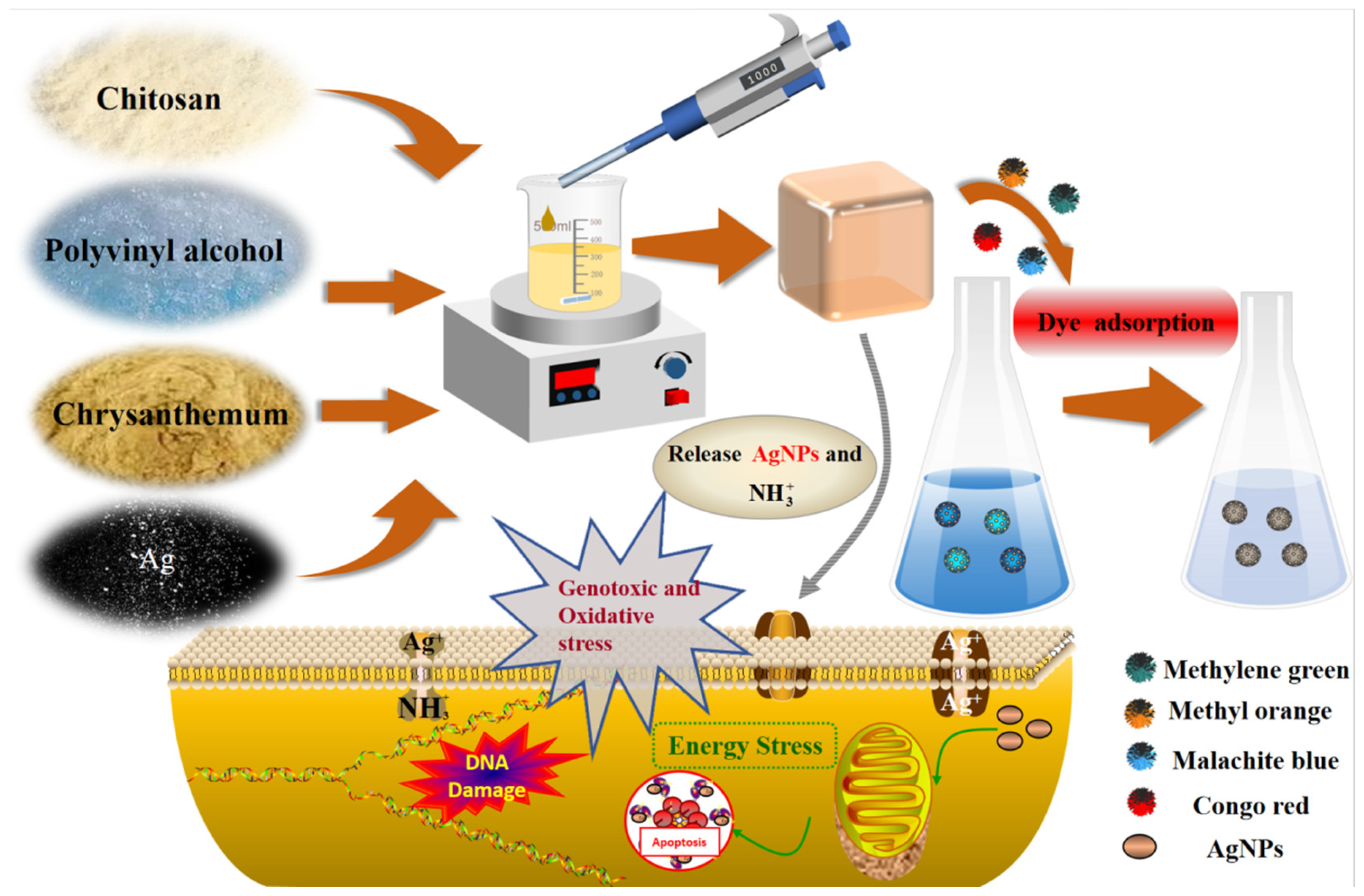
Preparation and Application of Multifunctional Chitosan–Polyvinyl Alcohol–Nanosilver–Chrysanthemum Extract Composite Gel

by
Kejian Shen
and
Yucai He
*
School of Pharmacy, Changzhou University, Changzhou 213164, China
*
Author to whom correspondence should be addressed.
Processes 2025, 13(2), 517; https://doi.org/10.3390/pr13020517
Submission received: 5 January 2025 / Revised: 22 January 2025 / Accepted: 28 January 2025 / Published: 12 February 2025

Abstract

:
In this study, we designed the preparation method and application study of chitosan–polyvinyl alcohol–chrysanthemum extract–nanosilver composite gel (CTS/PVA/Ag/CHR), constructed a composite gel system with chitosan/polyvinyl alcohol as the carrier, and utilized chrysanthemum extract within the gel to convert silver nitrate into nanosilver via green reduction. In the bacterial inhibition experiments, the CTS/PVA/Ag/CHR gel showed excellent antibacterial properties, and the diameter of the inhibition circle for Escherichia coli, Staphylococcus aureus, and Pseudomonas aeruginosa by the agar diffusion method was 32.5 mm, 30.5 mm, and 34.0 mm, respectively. In the aqueous bacterial inhibition experiments, the gel’s inhibition rate against the three kinds of bacteria was 100% after 5 h. The abundant hydroxyl groups contained in the polyvinyl alcohol (PVA) formed hydrogen bonds with the amino groups present in chitosan (CTS), which maintained the stability of the gel structure and enhanced the moisturizing and water storage properties of the gel. The adsorption curves of the CTS/PVA/Ag/CHR gel were fitted using a proposed pseudo-second-order kinetic model. Methylene blue, methyl orange, Congo red, and malachite green were discovered to have strong adsorption capacities, with the most significant adsorption effect for methyl orange at 205.65 mg/g. Moreover, the CTS/PVA/Ag/CHR gel showed good freshness preservation in milk simulation experiments. Due to its superior adsorption capability and antibacterial qualities, the CTS/PVA/Ag/CHR gels have great potential for applications in wastewater purification and food preservation.

1. Introduction

Pathogenic bacteria (bacteria, fungi, molds, etc.) are ubiquitous in the living environment and often invade drinking water, household hygiene products, food, etc. [1]. In recent years, various antibacterial materials have been developed [2]. Traditional antibacterial materials have a low biodegradation rate and poor biocompatibility. Accordingly, there is an urgent need to develop green, safe, and sustainable multifunctional antibacterial gel materials [3]. Silver ions are extensively used in sterilization and disinfection in medicine because of their super-oxidizing properties, as well as their ability to strongly attract and quickly connect to the bacterial body’s protease sulfhydryl group (-SH) to inactivate the protease [4], resulting in bacterial death. Additionally, silver ions can cause viral death by coagulating the protein molecules of viruses and binding the electron donors on their DNA molecules [5]. In the domains of environmental preservation and medical health, the natural antimicrobial properties and environmental friendliness of silver have been rediscovered. Silver ion-based antibacterial reagents are thought to be an effective antibacterial substance. In contrast to antibiotics, they do not make bacteria resistant. In addition, silver ions are naturally occurring elements that do not pollute the environment [6]. In the application of antimicrobial materials, nanosilver has been used in medical device coatings to effectively reduce the risk of post-operative infection due to its broad-spectrum antimicrobial properties [7]. Nanosilver particles of smaller size exhibit enhanced efficacy in eliminating pathogenic bacteria. However, they tend to accumulate, leading to instability. It has been observed that the controlled release of Ag+ from silver nanoalloys can improve antibacterial efficiency while sustaining antibacterial activity [8].
It is known that chrysanthemum (CHR) extract is rich in flavonoids, phenolic acids, polysaccharides, and other highly effective bioactive ingredients. It has antibacterial, antioxidant, anti-tumor, anti-inflammatory, and other pharmacological effects. Due to the presence of natural antibacterial components in chrysanthemum extract, it can be applied in the field of bacteriostatic materials [9]. Chitosan (CTS) is the only nitrogen-containing alkaline amino polysaccharide known in nature. It is non-toxic, biodegradable, and biocompatible [10], and it has antibacterial properties. Chitosan carries more amino groups on the surface and has cationic characteristics [11]. It can inhibit bacteria by binding negative electrons, which is the key to chitosan’s antibacterial properties. Chitosan is widely used because of its excellent properties and can be used in food packaging, medical materials [12,13], and dye adsorption [14]. Polyvinyl alcohol (PVA) is a polyhydroxyl polymer with excellent performance. It has water-soluble and biodegradable properties. It contains a large number of hydroxyl groups, which can crosslink to form a macromolecular mesh structure [15]. Polyvinyl alcohol is a non-toxic and non-irritating water-soluble polymer material with good biocompatibility [16,17,18]. At present, it is widely used in the pharmaceutical [19], food, and medical industries [20,21,22]. Adding polyvinyl alcohol to chitosan solution to prepare polyvinyl alcohol/chitosan polymers can improve swelling and mechanical properties [23].
Metal nanoparticles (e.g., silver nanoparticles (AgNPs)) can improve the antibacterial ability of antibacterial materials [24]. A number of techniques, such as chemical reduction, thermal breakdown, and UV irradiation reduction, can be used to create AgNPs [24,25,26]. However, these approaches have the drawbacks of being expensive and potentially polluting to the environment. Green techniques using plant extracts as stabilizers and reducing agents have gained an increasing amount of attention in the preparation of AgNPs [26]. Numerous functional groups (hydroxyl, carboxyl, phenolic hydroxyl, etc.) found in chrysanthemums can interact with metal ions via complexation, ion exchange, reduction, and electrostatic contact. Therefore, AgNPs can be created by reducing silver ions in chrysanthemum. In this work, CTS/PVA/Ag/CHR gels were prepared by using chrysanthemum extract as a green reducing agent and a chitosan (CTS)–polyvinyl alcohol (PVA) composite substrate as a carrier. Subsequently, the antibacterial activity, water storage properties, hygroscopicity, swelling properties, cytotoxicity, and dye adsorption capacity of the composite gels were studied. The CTS/PVA/Ag/CHR composite gel prepared in this study was environmentally friendly and had good dye adsorption ability. Finally, the CTS/PVA/Ag/CHR composite gel has potential applications in sewage treatment and food preservation.

2. Materials and Methods

2.1. Materials

Every reagent utilized in this investigation was of analytical grade. The Shanghai Institute of Microbiology in China provided E. coli ATCC 25922, P. aeruginosa ATCC 9027, S. aureus ATCC 6538, silver nitrate (AgNO3 ≥ 99.8%), chitosan (CTS) (deacetylation degree ≥ 85%, CAS 9012-76-4), polyvinyl alcohol (PVA ≥ 97.2%) (CAS 9002-89-5, molecular weight 65000), and glutaraldehyde (CAS 111-30-8, molecular weight 100.12). The chrysanthemum powder was purchased from Evergreen Bio (Xian, China).

2.2. Preparation of CTS/PVA/Ag/CHR Gel

A 2 wt% CTS solution was prepared with a magnetic stirrer at room temperature, and 1 g PVA was dissolved in 50 mL ultrapure water (2 wt%) and stirred in a water bath at 90 °C. The CTS-PVA composite membrane solution was created by adding the CTS solution to the PVA solution, mixing and thoroughly stirring. Then, 0.1 g silver nitrate and 1 mL chrysanthemum extract were added to promote the green reduction reaction. A crosslinking reaction was initiated by adding 1 mL of glutaraldehyde solution (the AgNO3 concentration was 0.2 g/L), and the gel was obtained after reaction at 500–600 rpm for 1–2 h. After cleaning with deionized water, CTS/PVA/Ag/CHR gel was obtained. The CTS/PVA/Ag/CHR gel was dried for 2 to 3 h at 60 °C in an oven. After the drying was over, the CTS/PVA/Ag/CHR was kept at room temperature for an investigation of its physical characteristics.

2.3. Characterization of CTS/PVA/Ag/CHR Gel

The CTS/PVA/Ag/CHR gels were analyzed using a Fourier-transform infrared spectrometer (FT-IR) (Nicolet iS50 Thermo Scientific Co, Waltham, MA, USA) in the 500–4000 cm−1 range. A Zeiss Sigma 300 scanning electron microscope (SEM) (Zeiss, Oberkochen, Baden-Wurtberg, Germany) was utilized to examine the composites’ surface morphology and crosslinking structure. X-ray diffraction (XRD) spectra of the gels were measured with Cu-Ka radiation (λ = 0.154 nm) using an X-ray diffractometer (X ’Pert PRO MPD, PANalytical, Amsterdam, Netherlands). The diffraction angle (2θ) was set to a range of 10–80°.

2.4. Bacteriostatic Test

E. coli ATCC 25922, S. aureus 6538, and P. aeruginosa 9027 were used as experimental bacteria to test the antimicrobial performance of the antibacterial materials. All glassware used for the antimicrobial experiments were sterilized by autoclaving (121 °C, 20 min). The plate count method was utilized to determine the antibacterial activity of the materials, and the pore diffusion method was used to evaluate the antibacterial effect of the materials. The bacterial solution was diluted to 106, and the bacterial suspension (0.10 mL) was evenly coated and distributed on LB solid medium. The holes were drilled to 9.0 mm length with a hole punch. Then, 100 mg CTS/PVA/Ag/CHR gel (dry weight) was added to the wells and incubated at 37 °C. After 24 h, the culture medium was removed and the diameter of antibacterial zone at the bottom of the culture medium was measured with a ruler. The antimicrobial ratio of the materials was determined as follows:
Antibacterial   ratio   ( % ) = A 0 A 1 A 0 × 100
where A0 represents the initial colony count, and A1 represents the number of colonies following CTS/PVA/Ag/CHR gel inhibition.

2.5. Effect of Ag+ and CTS Concentration on CTS/PVA/Ag/CHR Gel Preparation and Gel Preparation Optimization

Different gels were prepared, including C-P-J gel (without silver ions in the components), C-P-A gel (without chrysanthemum extract in the components), C-A-J gel (without polyvinyl alcohol in the components), C-J gel (without silver ions or polyvinyl alcohol in the components), and C-P-A-J gel (containing all components). The antibacterial efficacy of the gels was evaluated using the agar diffusion method. The diffusion method was used to observe the antibacterial activity of C-P-A-J gels against microorganisms, and the effects of Ag+ content (0.001–0.4 wt%) and CTS content (0.5–3.0 wt%) on the antibacterial activity were investigated.

2.6. Effect of CTS/PVA/Ag/CHR Gel Dose and Reaction Time on Antimicrobial Properties

P. aeruginosa, S. aureus, and E. coli were used as test bacteria for testing the effects of gel dose and antibacterial duration on the antibacterial activity. The initial concentration of the bacterial suspension was 108 CFU/mL. Here, 50 mL of bacterial suspension was mixed with the bacteriostatic gels, and the samples were incubated on a 37 °C constant-temperature oscillator. The plate counting method was used to determine the bacteriostatic activity [27].

2.7. Reusability and Stability of CTS/PVA/Ag/CHR Gels

One crucial feature of antibacterial gels is reusability. P. aeruginosa, S. aureus, and E. coli were evaluated for the gel’s antimicrobial properties in sterile saline. After 1 h of bacterial inhibition, 50 mL of suspension of bacteria (108 CFU/mL) was blended with 100 mg (dry weight) of CTS/PVA/Ag/CHR gel. The bacterial content of the suspension was then assessed using the plate streaking method. The gel was cleaned with deionized or ultrapure water following each antimicrobial examination, and it was then kept dry until the subsequent test. Following eight antimicrobial test batches, the gels’ inhibitory effects on the three bacteria were noted and examined. Another crucial aspect of bacteriostatic materials is their stability. The manufactured CTS/PVA/Ag/CHR gels were kept inside and their antimicrobial qualities were assessed on various days. Each time, the antibacterial impact was noted and examined to determine the antimicrobial materials’ stability.

2.8. Milk Preservation Test

The milk preservation test was conducted to assess the antimicrobial preservation properties of the gel material. We prepared 10 plastic Petri dishes (sterilized with ultraviolet light for 30 min) and 15 mL of food-grade pure milk (pasteurized and stored in a refrigerator at 4 °C). Then, 100 mg of the prepared C-P-A, C-A-J, C-P-A-J, C-J, and C-P-J gels were placed into each Petri dish. The aforementioned milk samples were kept in an incubator at 37 °C for 14 days, while the remaining 5 served as controls. The milk samples were collected on alternate days, and photographs were taken during the collection process. The surface shape and colony expansion were noted at room temperature.

2.9. Cytotoxicity Test

The effects of CTS/PVA/Ag/CHR gel and control hydrogels on cells were studied via the MTT method, and the cytotoxicity of the hydrogels was evaluated. A PK-15 cell suspension (6.5 × 10⁵ cells/mL) was inoculated on a 96-well plate. After 1 day, the original medium was removed, and the gel materials (C-P-A-J, C-P-A, C-A-J, C-P-J, and C-J) with different concentrations (0.1–10 mg/mL) were added. After 24 h of culture, 10 μL of the MTT solution was added to each hole of the 96-well plate. After 4 h, the supernatant was centrifuged and 150 μL DMSO was added to each well to dissolve formamide crystals. The absorbance was measured at 570 nm using an enzyme-linked immunosorbent assay (ELISA) reader. There was a positive correlation between cell metabolic activity and optical density. The cytotoxicity of the hydrogels was evaluated by using the optical density values.

2.10. Swelling Ratio, Water Loss, Moisture Adsorption, and Moisture Content of CTS/PVA/Ag/CHR Gels

The way in which gel materials expand in water is one of the most crucial ways to describe their physical characteristics [28,29]. CTS/PVA/Ag/CHR gels with different PVA contents (2, 4, 6, 8, 10 and 12 wt%) were weighed at 0.10 g (dry weight) each and prepared for the swelling property experiment. After being soaked in deionized water, the gels were put into the water and dehydrated continuously at hourly intervals, and the water on the surface was absorbed by filter paper and weighed. The swelling ratio (ER) formula is defined as below:
Swelling   ratio   ( % ) = M 2 M 1 M 1 × 100
where M2 denotes the mass of the gel after swelling in water, and M1 indicates the initial dry gel mass.
The water-blocking property of the gel structure is an important aspect for practical applications. After the preparation of gels with varying PVA concentrations (2.0–12.0%), 0.10 g (dry weight) of gel was weighed and placed in an oven set to 60 °C for 1–30 min, and then they were removed and weighed to record the mass loss. The impact of polyvinyl alcohol dosage on the ratio of water loss in the gels was investigated. The experiment was repeated three times. The following is the water loss ratio (WLR) equation:
Water   loss   ratio   ( % ) = W 1 W 2 W 1 × 100
where W2 is the mass of the gel at a given point. W1 is the mass of the starting gel.
One of the key metrics for assessing gel performance is its hygroscopicity during regular storage. CTS/PVA/Ag/CHR gels prepared with different PVA concentrations (2.0–12.0%) were dried. Then, 100 mg of the gels were placed in a desiccator at a temperature of 20 °C and a constant humidity of 34%. The gels were weighed once after 2–72 h to observe the morphological changes of the gels with different PVA concentrations and determine their water content. The data were then processed to analyze the effect of PVA concentration on the water absorption properties of the gels. The moisture absorption ratio (MAR) formula was given as below:
Moisture   adsorption   ratio   ( % ) = M n M 0 M 0 × 100
M0 is the mass of the sample prior to placement, and Mn is the mass of the sample after n hours of placement.
One of the key indicators used to assess a gel’s moisturizing qualities is its water content [30]. The water content of the gels at different PVA concentrations (2.0–12.0 wt%) was determined to evaluate the moisturizing performance of CTS/PVA/Ag/CHR gels. The formula for the water content is given below:
Moisture   content   ( % ) = W n W 0 W 0 × 100
where the mass of the dry gel is W0 and the mass of the wet gel is Wn.

2.11. Degradation Characteristics of CTS/PVA/Ag/CHR Gels

The gel material’s ability to degrade well embodies both energy conservation and environmental protection. The prepared CTS/PVA/Ag/CHR gel was buried in natural soil without the addition of any active enzyme, and the temperature and humidity were maintained at 37 °C and 80%. The gel samples were gathered for examination every day for 5 days, washed with purified water to remove dirt, and the morphological features of the gels were captured on camera. After drying in the oven, the gels were weighed, and the ratio of degradation was calculated as follows:
Degradation   ratio   ( % ) = W 0 W 1 W 0 × 100
where W0 is the mass of the gel before degradation, and W1 is the mass of the gel after degradation.

2.12. Adsorption of Dyes onto CTS/PVA/Ag/CHR Gels

In this experiment, the effects of the gels on the adsorption of dye, including malachite green (15.0 mg/L), Congo red (12.0 mg/L), and methyl orange (5.0 mg/L), were investigated. The C-P-A-J composite gel (0.10 g) was used to individually adsorb these dyes. Specifically, 20.0 mL of each dye solution was put into a screw-cap container, followed by the addition of the prepared gel. The adsorption of dyes was conducted on a thermostatic oscillator (35 °C, 180 rpm), and samples were withdrawn at a fixed point within 24 h of oscillation. The absorbance of the supernatant obtained from the withdrawn samples was measured by a UV-VIS spectrophotometer [31]. The CTS/PVA/Ag/CHR gel’s equilibrium adsorption quantity (mg/g) was determined as follows:
q e = ( C 0 C e ) V M
where the starting dye concentration is C0 (mg/L). The equilibrium dye content is Ce (mg/L). V (L) is the volume of the dye solution. The mass of the gel is denoted by M (g). The capacity of equilibrium adsorption is denoted by qe (mg/g).
As the adsorption time is t (h), the adsorption amount is denoted by qt (mg/g). When the adsorption equilibrium is achieved, the adsorption amount is qe (mg/g). The pseudo-first-order and pseudo-second-order dynamics mode rates are denoted by k1 and k2. The following is an expression of the equations for the suggested first- and second-order dynamics models:
q t = q e ( 1 exp k 1 t )
q t = k 2 q e 2 t 1 + k 2 q e t

2.13. Statistical Analysis

All experiments were independently repeated at least three times. SPSS 25.0 software was used to process and analyze the data. The significance of the difference was as follows: * indicates p < 0.05, ** indicates p < 0.01, *** indicates p < 0.001.

3. Results and Discussion

3.1. Characterization of CTS/PVA/Ag/CHR Gels

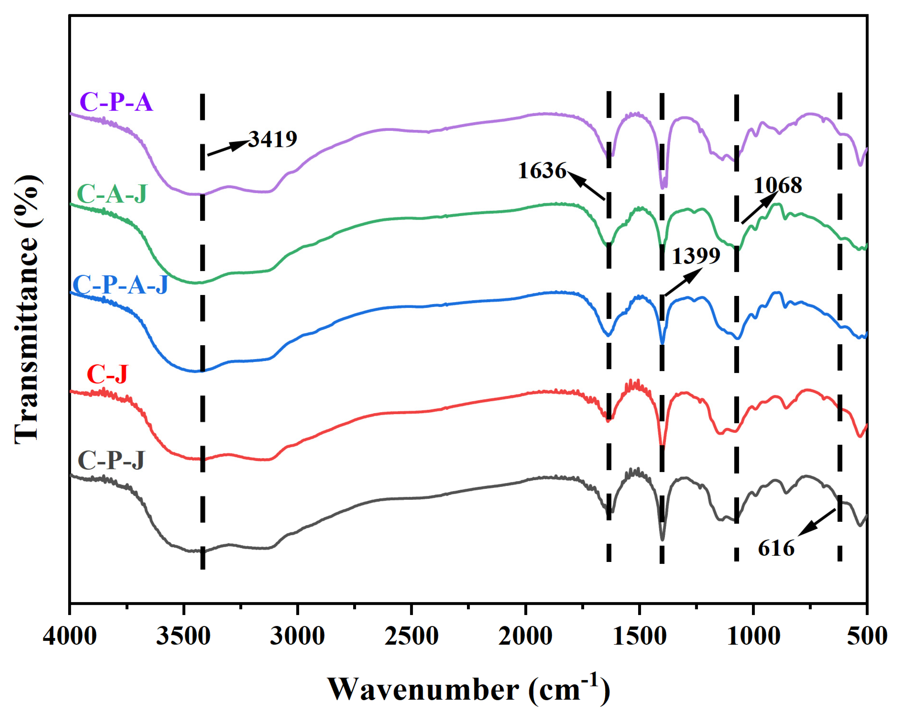
3.1.1. Fourier-Transform Infrared (FT-IR) Spectrometry Analysis
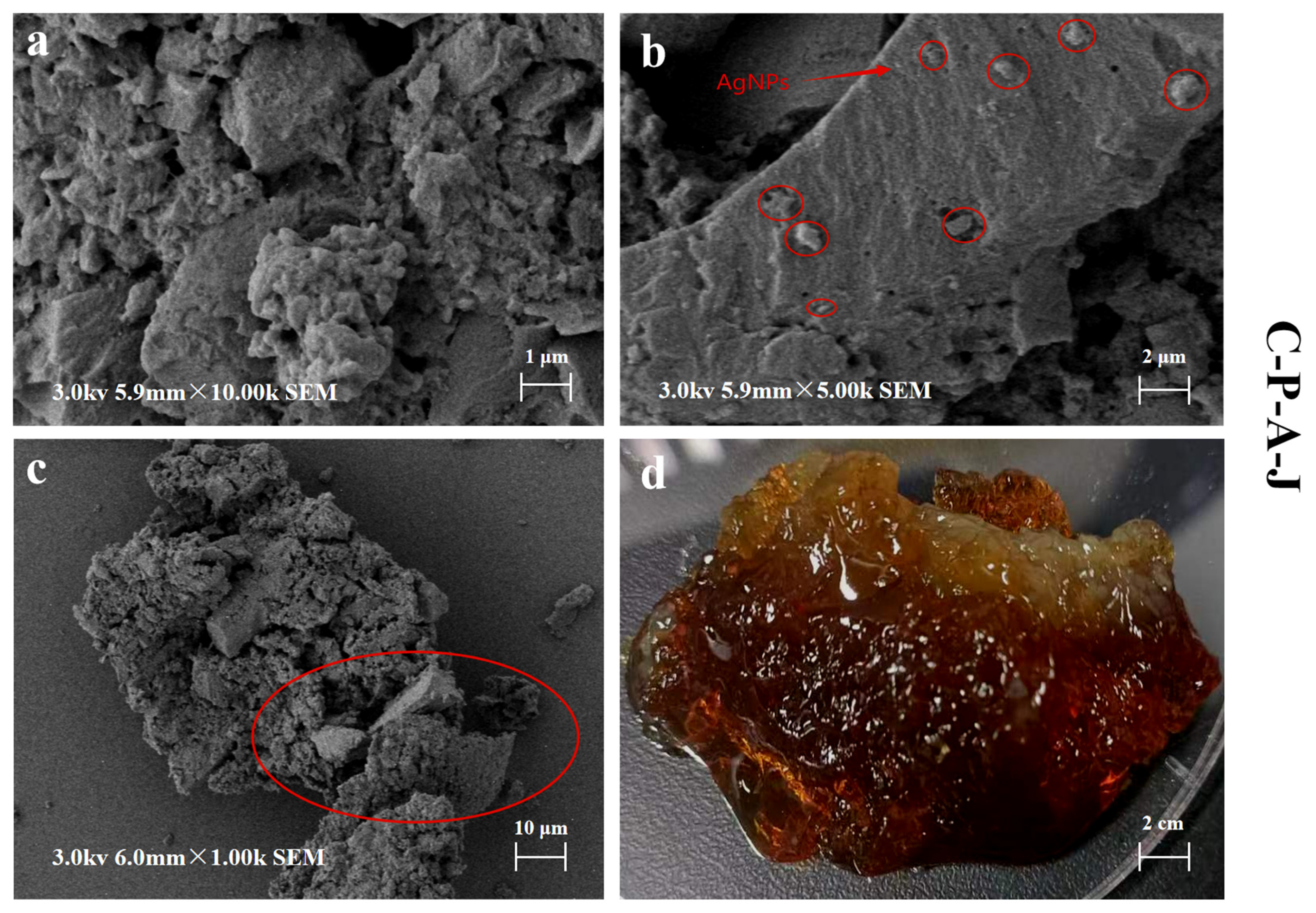
The resolved FT-IR spectra can allow the measurement of the functional group structure and chemical properties of the gel material [30,31,32,33,34], as well as the determination of the molecular-scale interactions of PVA, CTS, and AgNPs. The absorption peak about 3419 cm−1 is associated with the interaction of the -NH2 group in CTS with the -OH group of PVA to form the vibration of O-H stretching [35] (Figure 1). The absorption peak near 1399 cm−1 is attributed to the C-C stretching vibration, and the characteristic peak around 1636 cm−1 is related to the C=O stretching of amide I [36]. At 1068 cm−1, the peak was related to the C-O stretching vibration.

3.1.2. SEM Analysis

The surface structure and morphology of the CTS/PVA/Ag/CHR gel could be determined through the observation of scanning electron microscopy (SEM) (Figure 2). In Figure 2a, it can be observed that the surface roughness of CTS/PVA/Ag/CHR gel and the appearance of some large pores are attributed to AgNPs aggregating to form agglomerates during the gel recrystallization process. These aggregates cause an uneven rough structure to form on the gel surface [35]. Flavonoids, organic acids, and terpenoids in chrysanthemum contain a large number of hydroxyl groups, which can form hydrogen bonds with other hydroxyl groups or amino groups in the polymer chain, thus enhancing the intermolecular interaction and forming a closer crosslinked network. In Figure 2b, the fine granular crystals marked in red circles are silver nanoparticles, which are dispersed and fixed on the gel surface. As shown in Figure 2c, the red circle marks can observe the interaction between the polymers inside the gel and form a tight cross-linked network, making the structure more stable. The SEM image shows that the structure of the composite gel is stable, and the pore size structure of the surface is conducive to the loading of silver ions. The increase in the specific surface area of the gel surface may contribute to the enhancement of the gel adsorption capacity [37].

3.1.3. XRD Analysis

The measurement of gel surfaces made from various materials (C-P-A, C-A-J, C-P-A-J, C-J, and C-P-J) using XRD is displayed in Figure 3. The XRD pattern was examined and analyzed. Characteristic diffraction peaks were observed at 18.01°, 37.00°, 41.31°, and 72.60° (2θ), which are assigned to the related planes of the face-centered cubic structure of silver. The presence of these diffraction peaks provides strong evidence for the incorporation of silver nanoparticles within the gel matrix.

3.2. Antibacterial Characteristics

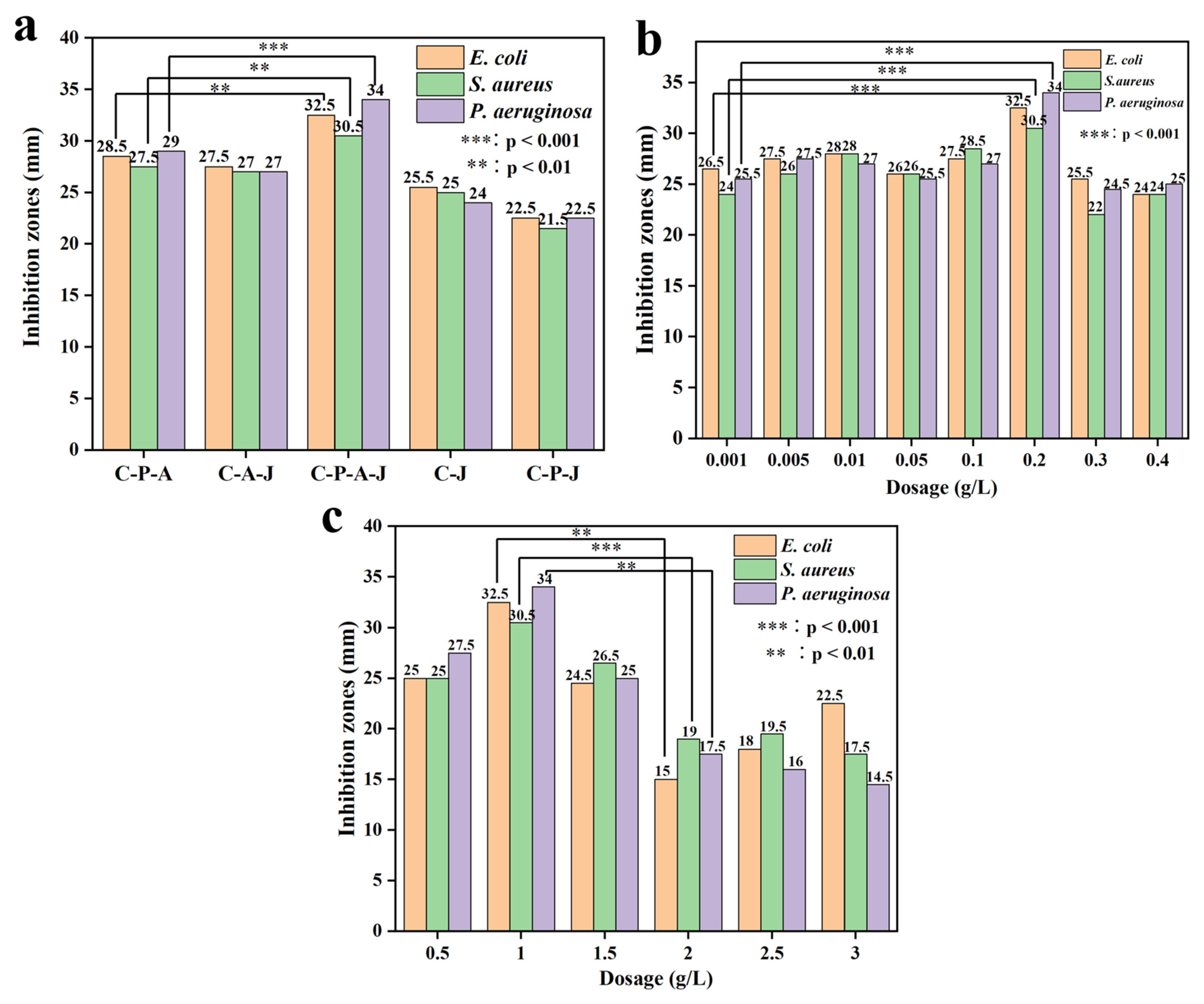
3.2.1. Enhancement of Gel Methods of Preparation and the Impact of CTS and Ag+ Dose on Antibacterial Efficacy in CTS/PVA/Ag/CHR Gels

The amount of antibacterial components and the materials used to assemble the gels affect their antibacterial properties [38,39]. As shown in Figure 4a, three bacteria were weakly inhibited by C-J and C-P-J gel, the antibacterial effect of C-P-A-J gel was higher than that of other composite gels (p < 0.05). The positively charged amino groups on the CTS may react with the negative charges on the bacterial cell membranes, which can interfere with the synthesis of the bacterial cell wall and cause the bacteria to lyse and die. The interaction between CTS and PVA forms a network adhesion structure, which can act as a silver ion release channel, enhancing the antibacterial effect.
It is evident from Figure 4b that the inhibitory effect of Ag+ on bacteria became increasingly stronger as the dose increased within a certain range. The highest level of antibacterial activity was observed at 0.2 g/L of AgNPs (Figure 4b), with significant inhibition (p < 0.05). When the AgNPs concentration reached a certain dose, AgNPs became embedded in the C-P-A-J gel, which can limit the release of AgNPs and reduce the antibacterial ability. When the concentration of added CTS was 1 g/L, the antibacterial activity of the composite gel reached the highest, with significant inhibition (p < 0.05) (Figure 4c). For a high CTS loading, the three-dimensional network structure generated by CTS and PVA through crosslinking can become compact and dense. This drastically reduces the material’s antibacterial activity and influences the release of AgNPs.

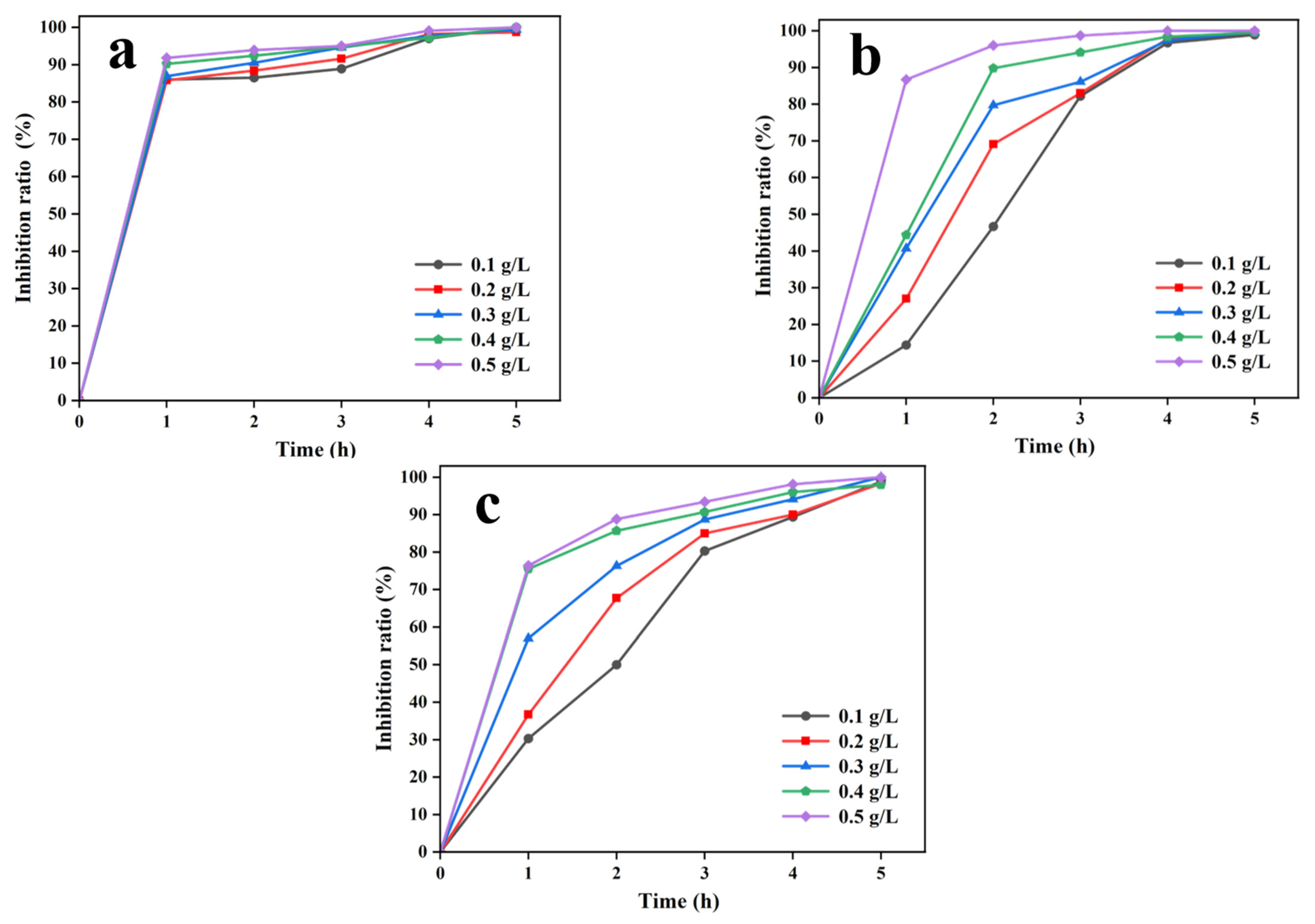
3.2.2. Impact of Incubation Duration and CTS/PVA/Ag/CHR Gel Dosage

Increasing the C-P-A-J gel loading and gradually promoting its liberation duration may enhance its antimicrobial effect. As displayed in Figure 5a, the antimicrobial efficiency changed clearly by increasing C-P-A-J gel loading and its antimicrobial duration. E. coli is a typical Gram-negative, parthenogenetic anaerobic bacterium that is widely found in humans and other mammals [40,41,42], as well as in small amounts in daily drinking water. Increasing the dosage of C-P-A-J gel resulted in a significant enhancement in the antimicrobial effect within 1 h. However, the increase in the dosage of the gels did not greatly enhance the antibacterial effect, and the bacteriostatic ratio reached 100% after 5 h, indicating that the gel dosage had reached the optimal value. According to other composite gel experiments, the antimicrobial ratio of the gel material (1000 mg/L) was less than 100% after 5 h. In this study, 0.1 g/L of C-P-A-J gel resulted in 100% inhibition after 5 h. S. aureus is a common foodborne pathogenic microorganism that is widespread in nature and can live on the surface of human skin for a long period. S. aureus inhibitors have been extensively studied [43]. With the increase in action duration and C-P-A-J gel loading, the inhibition efficiency gradually increased. The antibacterial efficacy of 0.5 g/L C-P-A-J gel reached its maximum inhibition strength within 2 h (Figure 5b), and maintained near-stability after 2 h. The inhibition ratio elevated gradually when the increase in action duration increased (C-P-A-J gel was <0.5 g/L). After 5 h, the inhibition ratio exceeded 99%, indicating that the C-P-A-J gel had significant inhibitory activity against S. aureus. It is known that P. aeruginosa is a major hospital-acquired pathogen that is extremely resistant to antibiotics [44]. The inhibition ratio increased to 76% when the gel dosage was 0.5 g/L and the action time was 1 h (Figure 5c). For 1 h, the inhibition of the gel had the fastest onset of action. When the inhibition time was more than 2 h, the inhibitory effect gradually smoothened. After just 5 h, the inhibition ratio reached 100%.
The above results illustrated that the C-P-A-J gel had a highly effective inhibitory effect on both Gram-positive and Gram-negative bacteria, especially on E. coli, which had a significant inhibitory effect. The gels demonstrated outstanding antibacterial activity for an extended period of inhibition at modest dosages.

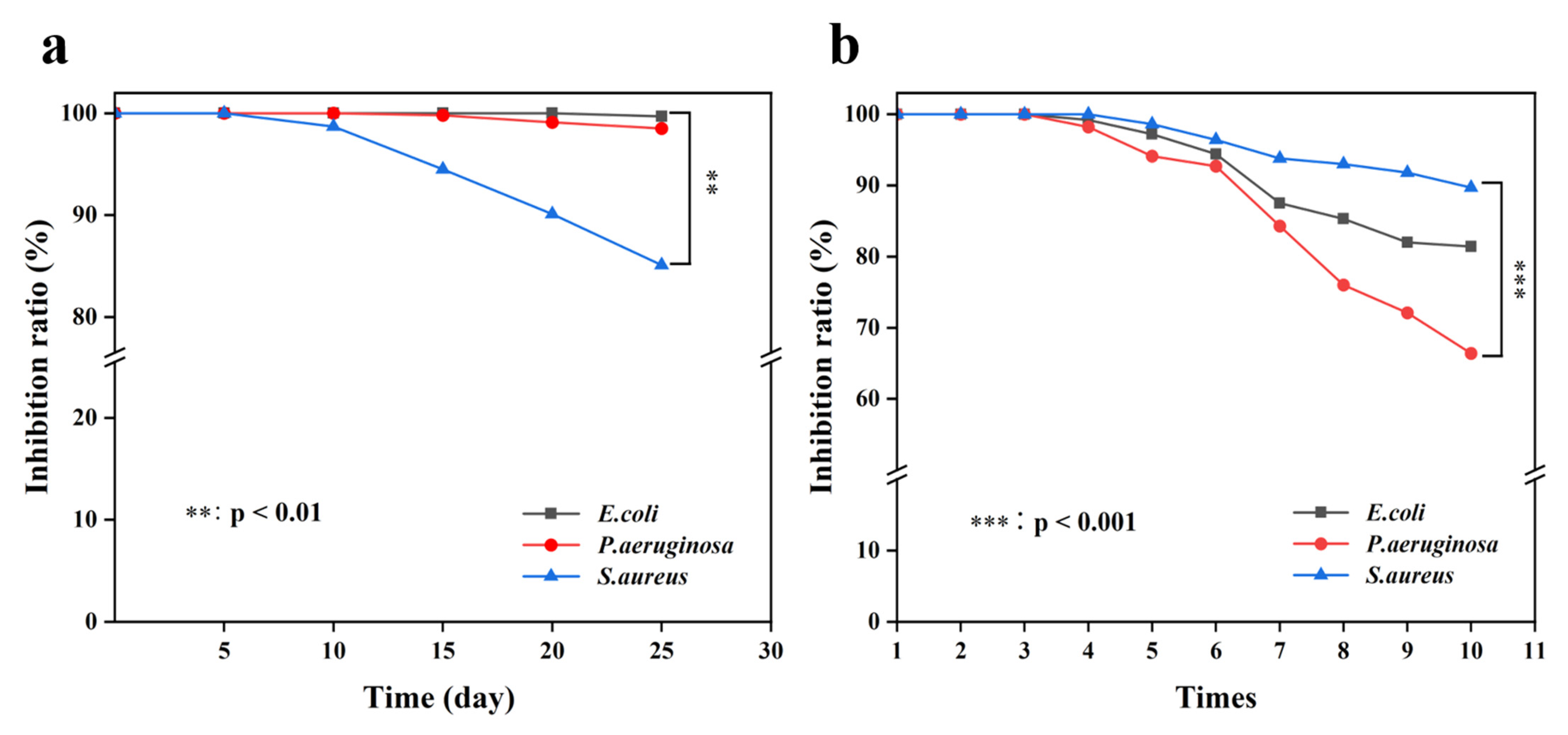
3.2.3. Stability and Reusability of CTS/PVA/Ag/CHR Gels

Antimicrobial materials are used in a wide range of practical application scenarios, and the overall performance steadiness and reproducibility of antimicrobial gels are regarded as vital elements for adaptation to specific environments [45,46]. Antimicrobial gels have high stability, good inhibition strength, and low application costs. As shown in Figure 6a, the C-P-A-J gel had long-term stable antimicrobial performance. For S. aureus, the inhibition ratio of the composite gel after 10 days was significantly lower (p < 0.05), and the inhibition ratio remained above 85% after 25 days of preservation. The gels had better inhibition efficacy for E. coli and P. aeruginosa, and the inhibition ratio of the two bacteria remained above 98% after 25 days. As illustrated in Figure 6b, the antimicrobial effect started to diminish during the antimicrobial test. This phenomenon was attributed to the depletion of antimicrobial compounds resulting from the repeated application of C-P-A-J gel. After 10 cycles of antimicrobial experiments, the inhibition ratio of the gel remained above 80% for E. coli and S. aureus, which was significantly higher than that of P. aeruginosa (p < 0.05). The natural fiber antimicrobial material was less durable, and the antimicrobial impact reduced extensively after five assessments. These results indicated that the C-P-A-J gel had good stability for antibacterial performance.

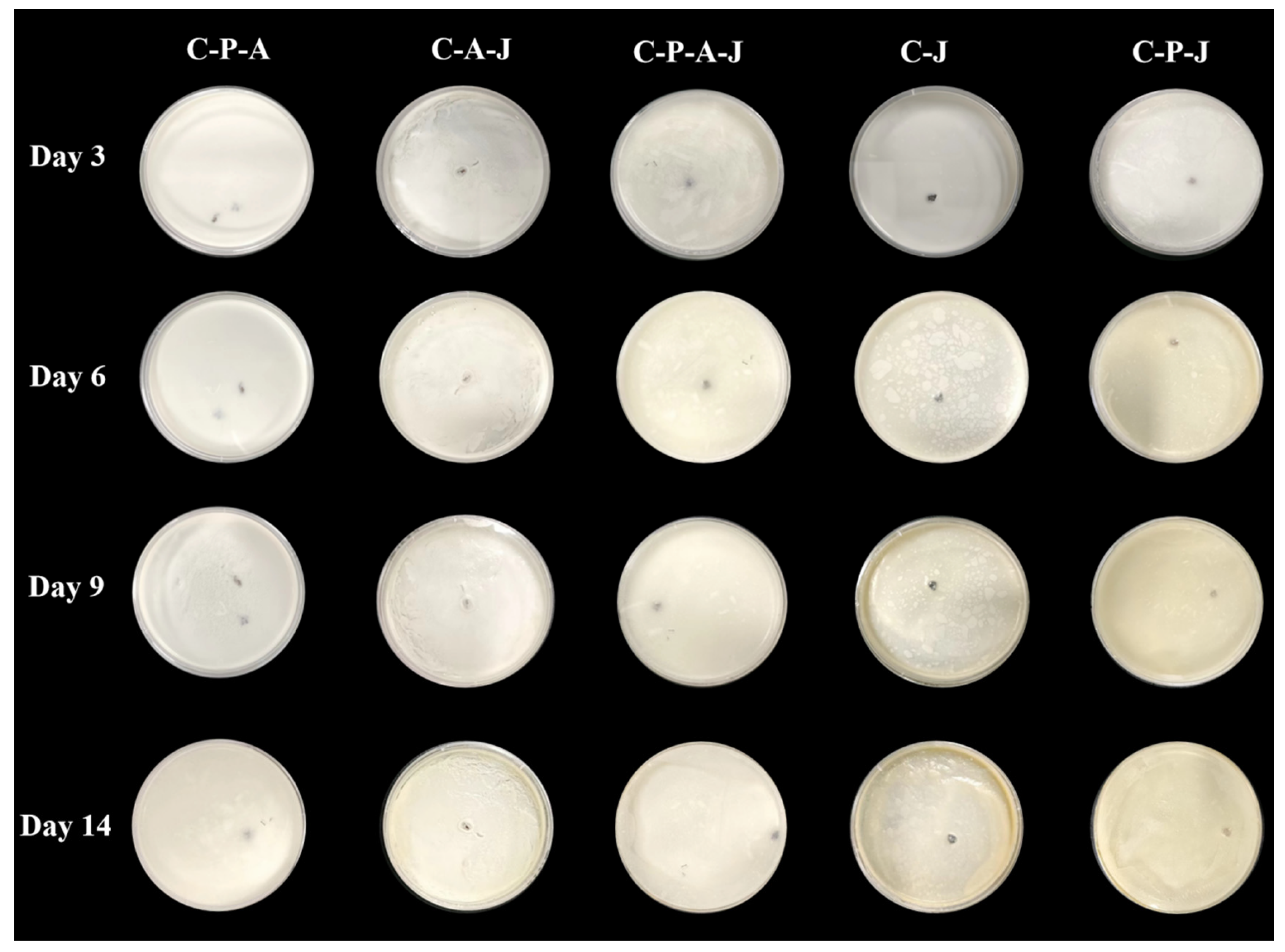
3.3. Preservation Applications of Complex Gels

Milk is a food product rich in nutrients and high in protein, and therefore susceptible to bacterial infestation [47]; thus, fresh milk was chosen to analyze the preservation properties of the gels. As illustrated in Figure 7, the condition of the milk did not change significantly in the first few days (3 days). On the sixth day, yellow colonies and solid milk residues began to appear on the surface of the milk samples added to the C-P-J gel, accompanied by the production of a slight odor. The samples with the addition of C-P-A-J gel, C-P-A gel and C-A-J gel did not undergo any significant changes. This indicated that the gels without AgNPs did not possess antibacterial properties. On the ninth day, the color of the milk samples with the addition of C-P-J and C-J gels continued to deepen and the odor increased. For the C-P-J and C-J gels, the milk samples underwent a significant deterioration on the 14th day. In contrast, the samples containing C-P-A-J gel, C-A-J gel and C-P-A gel exhibited a yellowish surface color and emitted a slight odor, indicating that the composite gels had good bacteriostatic activity. The C-P-A-J gel showed excellent antimicrobial activity because the hydrogel’s antibacterial ingredients prevented a variety of microorganisms from growing. Therefore, it appeared that the C-P-A-J gel could prolong the milk’s shelf life. In the future, edible and biocompatible antibacterial gels can be prepared for potential applications in food preservation.

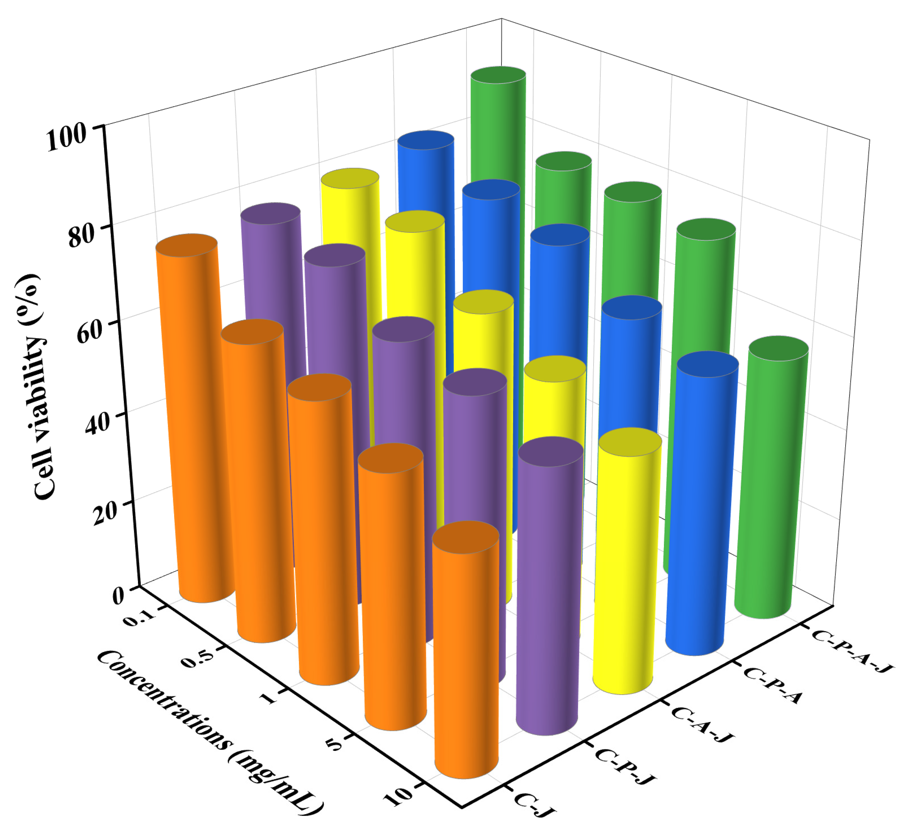
3.4. Cytotoxicity of Complex Gels

To assess the biocompatibility of the C-P-A-J hydrogel, the MTT assay was employed to evaluate its cytotoxicity [48]. As illustrated in Figure 8, cytotoxicity tests were conducted using various concentrations (0.1–10 mg/mL) of C-P-A-J, C-P-A, C-A-J, C-P-J, and C-J hydrogels. The results demonstrated that the CTS/PVA/Ag/CHR gel exhibited minimal cytotoxicity when the concentration was 0.1 mg/mL. The cell viability of the composite hydrogel remained above 75%, which may meet the safety standard of biological materials. These results indicated that the CTS/PVA/Ag/CHR hydrogels had no obvious cytotoxicity.

3.5. Synergistic Antimicrobial Mechanism of CTS/PVA/Ag/CHR Gel

In the CTS/PVA/Ag/CHR gel, CTS and PVA provided support for the attachment of AgNPs by crosslinking to create a thick network structure in three dimensions, and chrysanthemum extract was a green reducing agent for AgNO3. In the gel structure, silver ions have broad-spectrum antimicrobial properties, and CTS is a naturally occurring bacteriostatic agent. The synergistic effect of the two components significantly enhances the antibacterial efficiency (Figure 9). When the gel is immersed in water, its internal stable network crosslinking structure promotes the penetration of water molecules to swell the gel. Water molecules are readily absorbed by PVA’s hydrophilic groups, and the water body receives the amino groups and AgNPs that are liberated from the gels, thus rapidly exerting the function of bacteriostatic inhibition, whereas the gel’s porous shape facilitates the absorption. Ultimately, ion transport channels are destroyed as a result of AgNPs and amino groups interacting with mitochondrial respiratory enzymes [27], which inhibit the metabolic activities inside the cell. The internal adhesion structure after the expansion of the gel can provide a space for the interaction between the bacteria and the nanosilver. A composite network structure can form through the crosslinking of PVA and CTS. AgNPs may load into the community shape to extend the contact location between microorganism and antibacterial materials [25,49]. Therefore, it is essential to fully leverage the synergistic antibacterial effects of CTS and AgNPs. Chrysanthemum extracts are environmentally friendly and can be used as biocompatible and stable antimicrobial materials. These prepared gels have good prospects in terms of antibacterial application.

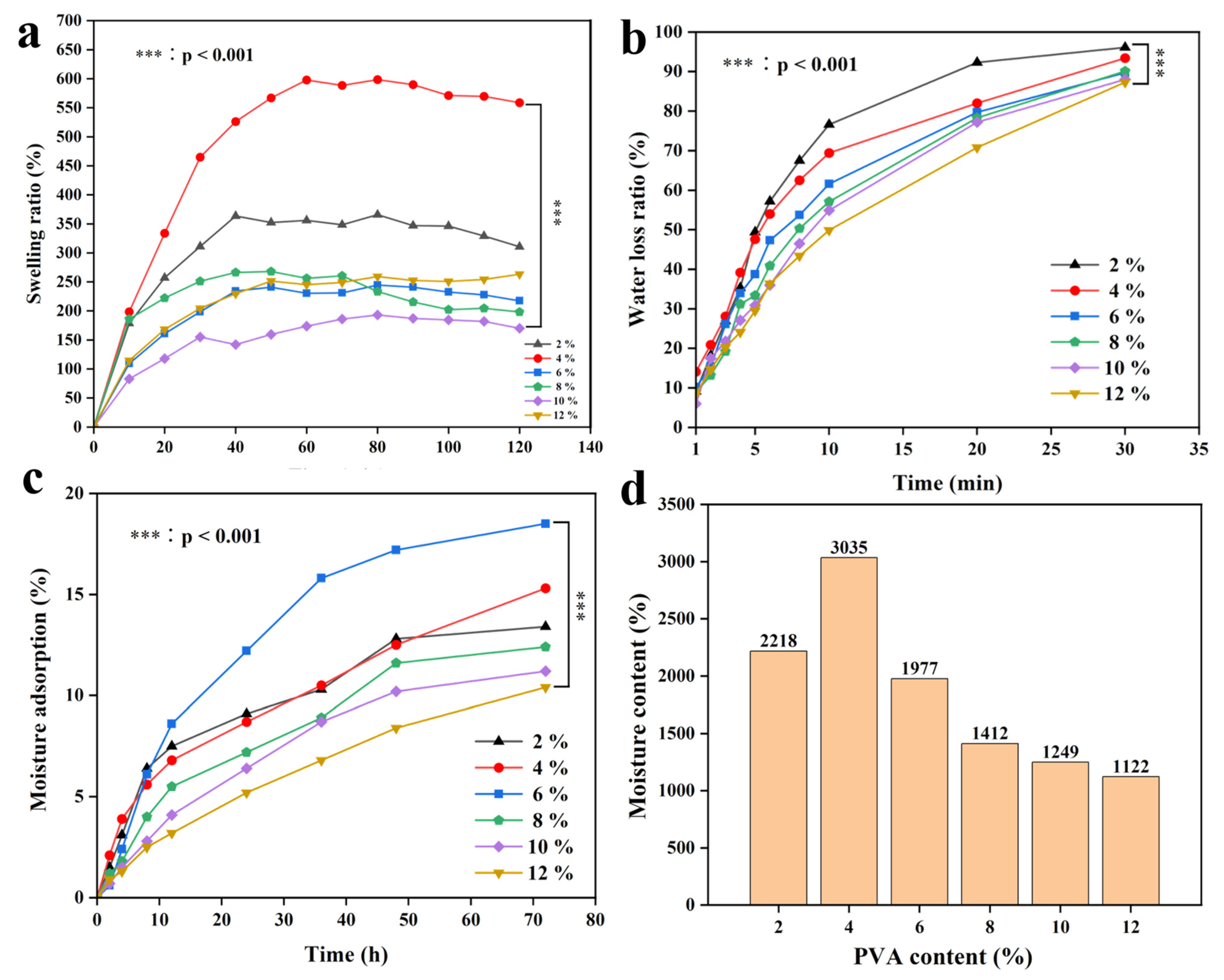
3.6. Swelling Ratio, Moisture Content, and Moisture Adsorption of CTS/PVA/Ag/CHR Gel

One important metric for describing the gel’s physical characteristics is its ratio of swelling [50,51]. According to Figure 10a, the gel exhibited the most significant increase in its swelling ratio during the initial 30 min. When the PVA content reached 6.0 wt%, 8.0 wt%, and 12.0 wt%, the swelling equilibrium could be observed within 50 min. This phenomenon was attributed to the fact that PVA (as a crosslinking agent) might facilitate the formation and expansion of the gel’s pore structure, thereby enhancing gel swelling. The highest swelling ratio (nearly 600%) was achieved when the PVA content was 4.0 wt%, which was significantly higher than other PVA content composite gels (p < 0.05).
In the water loss tests, the water within the C-P-A-J hydrogel started to evaporate. Figure 10b shows that the dosage of PVA may have affected the water loss ratio of the gels. The water loss ratio gradually decreased with the increase in the PVA dosage in a certain range of PVA concentrations. It was observed that the gel’s water loss ratio peaked at a PVA content of 2.0%, which was significantly higher than other PVA content composite gels (p < 0.05). When the PVA concentration was 12.0%, the hydrogel exhibited the minimal ratio of water loss (p < 0.05). When the concentration was 8.0% and 10.0%, the ratio of water loss was less than 90%. These findings suggest that adding an appropriate loading of PVA could significantly enhance the gel’s capacity to retain water and other compounds [52,53].
In the moisture absorption experiments, it was observed that the PVA concentration had an impact on the water content and moisture absorption ratio of the C-P-A-J gels (Figure 10c). PVA material has hygroscopic properties in the moderate concentration range. The hygroscopicity of the gels exhibited a positive correlation with the increasing concentration of PVA. When the concentration of PVA exceeded 6%, the hygroscopicity of the gel diminished as the concentration increased. Additionally, higher concentrations of PVA can form crosslinks with CTS [54], thereby inhibiting the gel’s ability to absorb moisture from the air. When the concentration of PVA was 6%, the hygroscopic performance of the gels was the highest, which was significantly higher than other PVA content composite gels (p < 0.05). This implies that a moderate PVA content enhances the gel’s ability to absorb moisture.
In the water content test, the C-P-A-J gel showed excellent water retention performance. As shown in Figure 10d, as the PVA dosage increased from 2% to 4%, the gel’s water content rose, when the concentration of PVA is 4%, the moisture content of gels is the highest, which is significantly higher than that of other gels with PVA concentration (p < 0.05). By increasing the PVA concentration from 4% to 12%, the gel’s water content declined clearly. This resulted was ascribed to the fact that PVA can affect the gel’s crosslinking strength and structural shape [54,55]. With the increase in the PVA concentration, the network structure of the gel was more stable, resulting in a reduced capacity to retain water. Conversely, at lower PVA concentrations, the network structure was relatively looser, allowing for greater water retention. This was due to the interaction between the PVA and the -OH and -NH2 groups in the CTS molecule. The crosslinking of polymer chains may enhance the hydrophilicity of materials [54]. The C-P-A-J gel had exceptional physical characteristics with a sophisticated porous three-dimensional network structure, which may successfully regulate the flow of water molecules, according to measurements of water loss and water content.

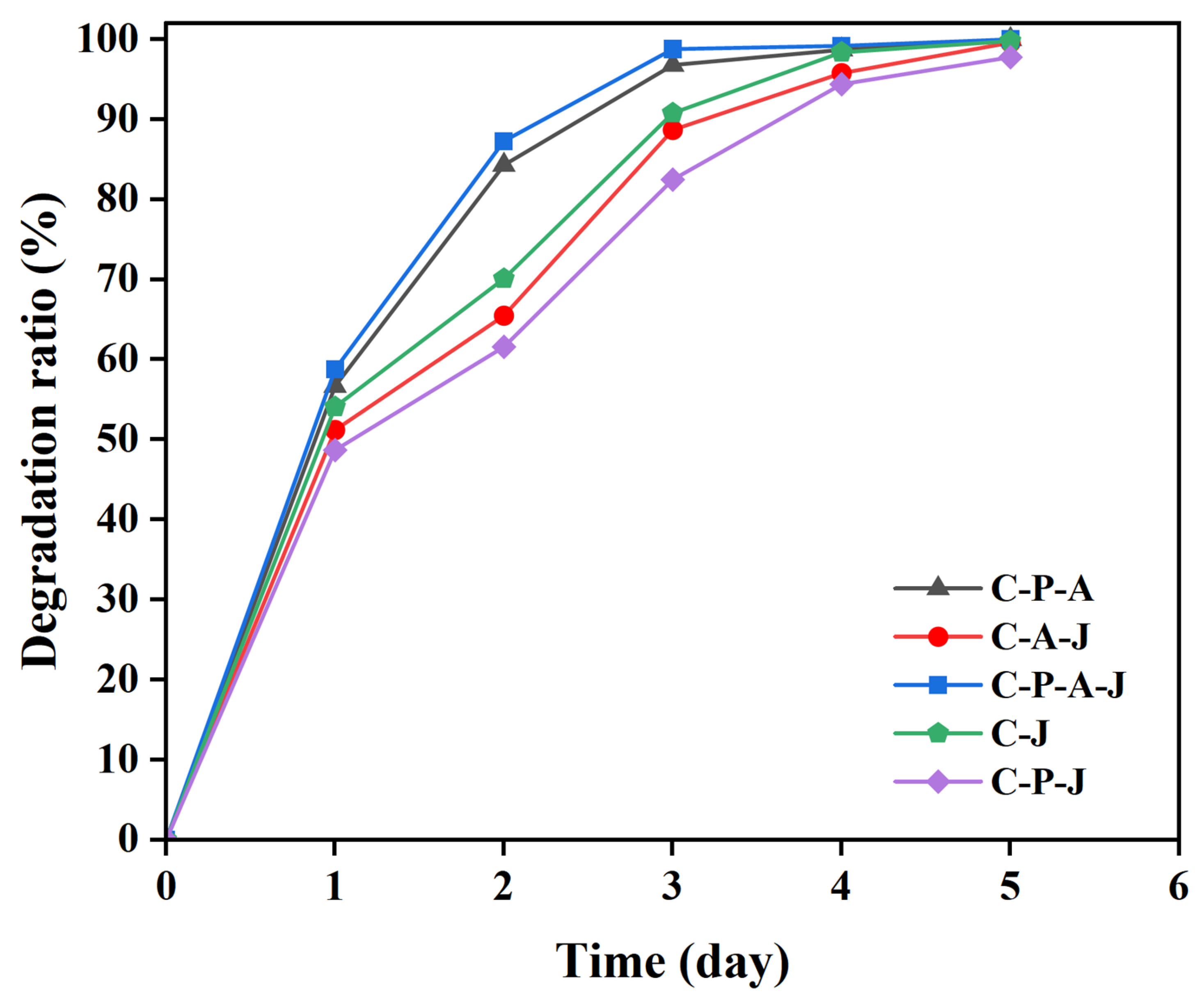
3.7. CTS/PVA/Ag/CHR Gel Degradation

Degradation performance is an important environmental protection index for assessing antimicrobial compounds’ effectiveness, which reflects the ideology of green environmental protection [56,57]. The absorbent nature of PVA might facilitate the absorption of water from the soil and accelerate degradation. The chrysanthemum-extracted components in the gel structure had a certain moisturizing effect; thus, the degradation ratio of the C-P-A-J gel reached the highest. The total degradation ratio reached 100% on the fifth day. In the C-P-A gels, the addition of PVA resulted in larger pores on the gel’s surface, which increased the efficiency of breakdown by allowing oxygen and microbes to enter (Figure 11). The addition of AgNPs to C-J gel further improved the mechanical properties of the gels, and the gel structure was more compact and stable [27], which made it difficult for the water to penetrate and reduced the degradation efficiency of the C-A-J gel. In the C-P-J gels, CTS and PVA formed a dense crosslinked structure, which blocked the entry of some oxygen and moisture. Accordingly, the degradation ratio of the C-P-J gel was slow. C-P-A-J gel is not harmful and has good compatibility with the natural ecological environment. Thus, these prepared gels are an environmentally friendly antimicrobial material with potential application value.

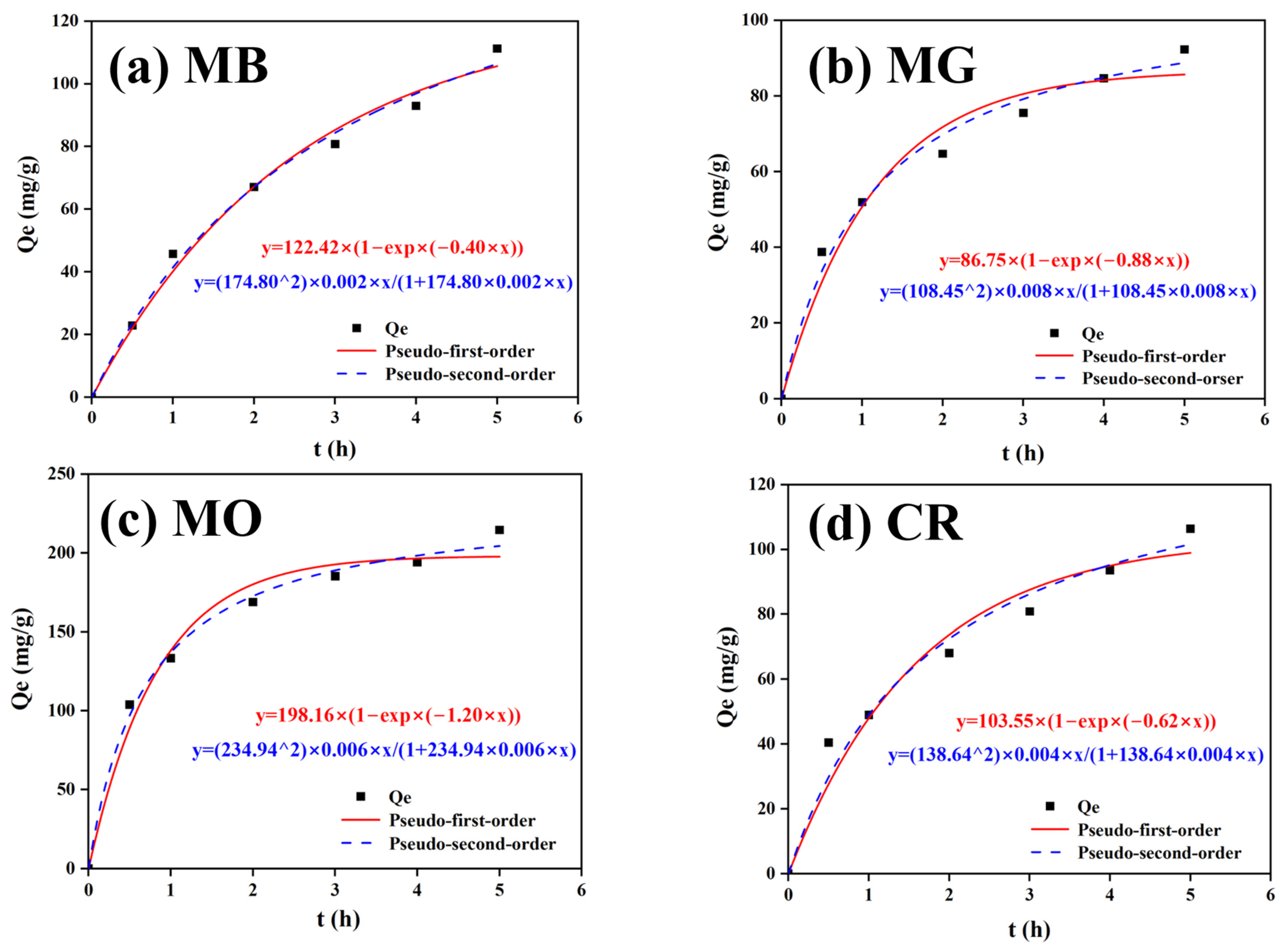
3.8. CTS/PVA/Ag/CHR Gel Dye Adsorption Properties

According to the adsorption results, the adsorption of the gel for MG, MO, and CR reached equilibrium in about 4 h (Figure 12), demonstrating that a significant quantity of dye was absorbed by the gel’s pores during the adsorption process. The gels had a strong affinity for MO with a maximum adsorption of 214.52 mg/g. The C-P-A-J gel’s adsorption kinetics are adequately represented by the fitted second-order equations. As shown in Table 1, the R2 values of the fitted pseudo-second-order kinetic equations of the four groups were all equal to 0.99.
The equilibrium adsorption capacities of the gel for MB, MG, MO, and CR calculated from the fitted pseudo-first-order kinetic model were 122.42, 86.75, 198.16, and 103.55 mg/g, respectively. The equilibrium adsorption capacities of the gel for MB, MG, MO, and CR calculated from the fitted pseudo-second-order kinetic model were 174.80, 108.45, 234.94, and 138.64 mg/g. The experimental maximum adsorption capacities for MB, MG, MO, and CR were 111.28, 92.26, 214.52, and 106.38 mg/g, respectively. It can be seen that the fitted curves were in good agreement with the proposed pseudo-second-order kinetic equation.
The correlation coefficient (R2) of the curve (near 1) indicated the adsorption model’s dependability, and the experimental data demonstrated good dependability and a strong connection. Consequently, the entire adsorption process is well described by the suggested pseudo-second-order kinetic equation. The C-P-A-J gel’s good adsorption capacity for dyes was demonstrated by the adsorption experiments’ findings, underscoring its potential for use in a variety of real-world situations. It is of great interest to prepare the biobased materials with strong adsorption and antibacterial qualities in wastewater treatment [58,59,60,61,62,63,64,65,66].

4. Conclusions

In the present study, C-P-A-J gels with antimicrobial and dye adsorption capacities were prepared using the reduced extraction of pure natural biomass from chrysanthemum. The considerable swelling properties and hydrophobic structure of the gel enhanced its stability, degradability, and antimicrobial effect, and encouraged the release of AgNPs and amino groups to enhance the synergistic antibacterial effect. The water bacterial inhibition experiment and the milk preservation test validated the C-P-A-J gel’s strong antibacterial activity, which delayed the period of milk spoilage. The tests demonstrated that the gel could increase food preservation. In addition, the gels showed good adsorption capacity for dyes, with a maximum adsorption capacity of 214.52 mg/g for methyl orange under optimal conditions. The composite gel exhibited clear benefits in bacteriostasis and dye adsorption, and its extensive research and development opportunities should yield fresh concepts for resolving contemporary issues in food preservation and environmental protection.

Author Contributions

Conceptualization, Methodology, Data Curation, Software, and Writing—Original Draft, K.S.; Supervision and Writing—Review and Editing, Y.H. All authors have read and agreed to the published version of the manuscript.

Funding

This research received no external funding.

Data Availability Statement

The original contributions presented in this study are included in the article. Further inquiries can be directed to the corresponding author.

Acknowledgments

The authors thank the Analysis and Testing Center (Changzhou University) for measuring samples using FT-IR, XRD, SEM, etc.

Conflicts of Interest

The authors declare no conflicts of interest.

References

  1. Jin, F.; Liao, S.; Li, W.; Jiang, C.; Wei, Q.; Xia, X.; Wang, Q. Amphiphilic sodium alginate-polylysine hydrogel with high antibacterial efficiency in a wide pH range. Carbohydr. Polym. 2023, 299, 120195. [Google Scholar] [CrossRef] [PubMed]
  2. Pan, H.; Jiang, B.; Chen, J.; Jin, Z. Blend-modification of soy protein/lauric acid edible films using polysaccharides. Food Chem. 2014, 151, 1–6. [Google Scholar] [CrossRef]
  3. Wang, Y.; Zhang, T.T.; Huang, M.; Zhang, M.; He, Y.C. Preparation of dandelion flower extract-based polyvinyl alcohol-chitosan-dandelion-CuNPs composite gel for efficient bacteriostatic and dye adsorption. In. J. Biol. Macromol. 2024, 281, 136512. [Google Scholar] [CrossRef] [PubMed]
  4. Su, X.R.; Xue, Y.Y.; Li, Q. Advances in the research of antibacterial properties of silver ion and its application in wound treatment. Zhonghua Shao Shang Za Zhi = Zhonghua Shaoshang Zazhi = Chin. J. Burn. 2018, 34, 183–186. [Google Scholar]
  5. Fuhrmann, P.L.; Powell, J.; Rousseau, D. Structure and rheology of oil-continuous capillary suspensions containing water-swellable cellulose beads and fibres. Food Hydrocoll. 2023, 139, 108503. [Google Scholar] [CrossRef]
  6. Kumaraswamy, S.; Patil, S.L.; Mallaiah, S.H. In vitro biocompatibility evaluation of radiolytically synthesized silver/polyvinyl hydrogel nanocomposites for wound dressing applications. J. Bioact. Compat. Polym. 2020, 35, 435–450. [Google Scholar] [CrossRef]
  7. Yang, Z.; Li, K.R.; Zhang, Y.Y.; Hu, J.L.; Li, T.Y.; Weng, Z.X.; Wu, L.X. Antibiotic Silver Particles Coated Graphene Oxide/polyurethane Nanocomposites Foams and Its Mechanical Properties. Chin. J. Struct. Chem. 2022, 41, 2203125–2203131. [Google Scholar]
  8. Qin, Z.; Zheng, Y.; Wang, Y.; Du, T.; Li, C.; Wang, X.; Jiang, H. Versatile roles of silver in Ag-based nanoalloys for antibacterial applications. Coord. Chem. Rev. 2021, 449, 214218. [Google Scholar] [CrossRef]
  9. Sassi, A.B.; Harzallah-Skhiri, F.; Bourgougnon, N.; Aouni, M. Antimicrobial activities of four Tunisian Chrysanthemum species. Indian. J. Med. Res. 2008, 127, 183–192. [Google Scholar] [PubMed]
  10. Ma, G.; Qian, B.; Yang, J.; Hu, C.; Nie, J. Synthesis and properties of photosensitive chitosan derivatives. Int. J. Biol. Macromol. 2010, 46, 558–561. [Google Scholar] [CrossRef]
  11. Yang, S.F.; Wen, Y.; Yi, P.; Xiao, K.; Dong, C.F. Effects of chitosan inhibitor on the electrochemical corrosion behavior of 2205 duplex stainless steel. Int. J. Miner. Metall. Mater. 2017, 24, 1260–1266. [Google Scholar] [CrossRef]
  12. Yang, D.; Gong, L.; Li, Q.; Fan, B.; Ma, C.L.; He, Y.C. Preparation of a biobased polyelectrolyte complex from chitosan and sodium carboxymethyl cellulose and its antibacterial characteristics. Int. J. Biol. Macromol. 2023, 227, 524–534. [Google Scholar] [CrossRef] [PubMed]
  13. Li, Q.; Lu, F.; Zhou, G.; Yu, K.; Lu, B.; Xiao, Y.; Dai, F.; Wu, D.; Lan, G. Silver Inlaid with Gold Nanoparticle/Chitosan Wound Dressing Enhances Antibacterial Activity and Porosity, and Promotes Wound Healing. Biomacromolecules 2017, 18, 3766–3775. [Google Scholar] [CrossRef] [PubMed]
  14. Sivararnan, A.; Ganti, S.S.; Nguyen, H.X.; Birk, G.; Wieber, A.; Lubda, D.; Banga, A.K. Development and evaluation of a polyvinyl alcohol based topical gel. J. Drug Deliv. Sci. Technol. 2017, 39, 210–216. [Google Scholar] [CrossRef]
  15. Ingle, J.; Patel, U.D. Electrochemical reduction of nitrate in the presence of silver-coated polyvinyl alcohol beads as a spatially suspended catalyst. J. Water Process Eng. 2022, 49, 103082. [Google Scholar] [CrossRef]
  16. Zhang, K.; Liu, Y.; Zhao, Z.; Shi, X.; Zhang, R.; He, Y.; Zhang, H.; Wang, W. Magnesium-doped nano-hydroxyapatite/polyvinyl alcohol/chitosan composite hydrogel: Preparation, characterization. Int. J. Nanomed. 2024, 19, 651–671. [Google Scholar] [CrossRef]
  17. Rodriguez-Rodriguez, R.; Espinosa-Andrews, H.; Velasquillo-Martinez, C.; Garcia-Carvajal, Z.Y. Composite hydrogels based on gelatin, chitosan and polyvinyl alcohol to biomedical applications: A review. Int. J. Polym. Mater. Polym. Biomater. 2020, 69, 1–20. [Google Scholar] [CrossRef]
  18. Kuila, S.B.; Ray, S.K. Dehydration of dioxane by pervaporation using filled blend membranes of polyvinyl alcohol and sodium alginate. Carbohydr. Polym. 2014, 101, 1154–1165. [Google Scholar] [CrossRef]
  19. Lewis, K.; Lee, R.E.; Broetz-Oesterhelt, H.; Hiller, S.; Rodnina, M.V.; Schneider, T.; Weingarth, M.; Wohlgemuth, I. Sophisticated natural products as antibiotics. Nature 2024, 632, 39–49. [Google Scholar] [CrossRef]
  20. Tuyen, N.V.; Ryu, J.H.; Yae, J.B.; Kim, H.G.; Hong, S.W.; Ahn, D.H. Nitrogen removal performance of anammox process with PVA–SA gel bead crosslinked with sodium sulfate as a biomass carrier. J. Ind. Eng. Chem. 2018, 67, 326–332. [Google Scholar] [CrossRef]
  21. Wang, S.W.; Dong, X.X.; Liu, M.T.; Ren, D.D.; Wang, T.T.; Chen, C.; Leng, Y.T.; Li, Q. High strength polyvinyl alcohol/polyacrylic acid-based hydrogels with multiple networks. Polym.-Plast. Technol. Mater. 2021, 60, 392–404. [Google Scholar] [CrossRef]
  22. Kim, Y.J.; Min, J. Property modulation of the alginate-based hydrogel via semi-interpenetrating polymer network (semi-IPN) with poly(vinyl alcohol). Int. J. Biol. Macromol. 2021, 193, 1068–1077. [Google Scholar] [CrossRef] [PubMed]
  23. Nagarpita, M.V.; Roy, P.; Shruthi, S.B.; Sailaja, R.R.N. Synthesis and swelling characteristics of chitosan and CMC grafted sodium acrylate-co-acrylamide using modified nanoclay and examining its efficacy for removal of dyes. Int. J. Biol. Macromol. 2017, 102, 1226–1240. [Google Scholar] [CrossRef]
  24. Velidandi, A.; Pabbathi, N.P.P.; Dahariya, S.; Baadhe, R.R. Green synthesis of novel Ag–Cu and Ag–Znbimetallic nanoparticles and their in vitro biological, eco-toxicity and catalytic studies. Nano-Struct. Nano-Objects 2021, 26, 100687. [Google Scholar] [CrossRef]
  25. Chen, Q.; Ma, J.; Hu, Y.; Fei, P. Multifunctional antibacterial films with silver nanoparticles reduced in situ by lemon juice. Food Chem. 2021, 365, 130517. [Google Scholar] [CrossRef] [PubMed]
  26. Jiang, B.; Tian, C.; Song, G.; Pan, Q.; Wang, Z.; Shi, L.; Qiao, Y.; Fu, H. A green route to synthesize novel Ag/C antibacterial agent. Mater. Res. Bull. 2012, 47, 458–463. [Google Scholar] [CrossRef]
  27. Zhang, Z.; He, Y. Synthesis and characteristics of a fish scale-based biochar–nanosilver antibacterial material. Processes 2023, 11, 1992. [Google Scholar] [CrossRef]
  28. Afshari, M.J.; Sabzi, M.; Jiang, L.; Behshad, Y.; Zanjanijam, A.R.; Mahdavinia, G.R.; Ahmadi, M. Incorporation of dynamicboronate links and Ag nanoparticles into PVA hydrogels for pH-Regulated and prolonged release of methotrexate. J. Drug Deliv. Sci. Technol. 2021, 63, 102502. [Google Scholar] [CrossRef]
  29. Bahrami, A.; Mokarram, R.R.; Khiabani, M.S.; Ghanbarzadeh, B.; Salehi, R. Physico-mechanical and antimicrobial properties of tragacanth/hydroxypropyl methylcellulose/beeswax edible films reinforced with silver nanoparticles. Int. J. Biol. Macromol. 2019, 129, 1103–1112. [Google Scholar] [CrossRef] [PubMed]
  30. Salam, A.; Pawlak, J.J.; Venditti, R.A.; El-tahlawy, K. Synthesis and Characterization of Starch Citrate-Chitosan Foam with Superior Water and Saline Absorbance Properties. Biomacromolecules 2010, 11, 1453–1459. [Google Scholar] [CrossRef]
  31. Zhao, T.; Li, X.; Gong, Y.; Guo, Y.; Quan, F.; Shi, Q. Study on polysaccharide polyelectrolyte complex and fabrication of alginate/chitosan derivative composite fibers. Int. J. Biol. Macromol. 2021, 184, 181–187. [Google Scholar] [CrossRef]
  32. Huang, Z.H.; Sun, X.Y.; Li, Y.; Ge, W.; Wang, J.D. Adsorption behaviors of chitosan and the analysis of FTIR spectra. Spectrosc. Spectr. Anal. 2005, 25, 698–700. [Google Scholar]
  33. Ionita, M.; Pandele, M.A.; Iovu, H. Sodium alginate/graphene oxide composite films with enhanced thermal and mechanical properties. Carbohydr. Polym. 2013, 94, 339–344. [Google Scholar] [CrossRef] [PubMed]
  34. Koneru, A.; Dharmalingam, K.; Anandalakshmi, R. Cellulose based nanocomposite hydrogel films consisting of sodium carboxymethylcellulose–grapefruit seed extract nanoparticles for potential wound healing applications. Int. J. Biol. Macromol. 2020, 148, 833–842. [Google Scholar] [CrossRef]
  35. Quadrado, R.F.N.; Gohlke, G.; Oliboni, R.S.; Smaniotto, A.; Fajardo, A.R. Hybrid hydrogels containing one-step biosynthesized silver nanoparticles: Preparation, characterization and catalytic application. J. Ind. Eng. Chem. 2019, 79, 326–337. [Google Scholar] [CrossRef]
  36. Cheng, Y.; Far, B.F.; Jahanbakhshi, M.; Bahrami, S.; Tamimi, P.; Sedaghat, M.; Ghazizadeha, E. Exploring the potential of a polyvinyl alcohol/chitosan-based nanofibrous matrix for erythromycin delivery: Fabrication, in vitro and in vivo evaluation. RSC Adv. 2023, 13, 18450–18460. [Google Scholar] [CrossRef]
  37. Rahaman, M.S.; Hasnine, S.M.M.; Ahmed, T.; Sultana, S.; Bhuiyan, M.A.Q.; Manir, M.S.; Ullah, N.; Sen, S.K.; Hossain, M.N.; Hossain, M.S.; et al. Radiation crosslinked polyvinyl alcohol/polyvinyl pyrrolidone/acrylic acid hydrogels: Swelling, crosslinking and dye adsorption study. Iran. Polym. J. 2021, 30, 1101–1116. [Google Scholar] [CrossRef]
  38. Spagnol, C.; Fragal, E.H.; Pereira, A.G.B.; Nakamura, C.V.; Muniz, E.C.; Follmann, H.D.M.; Silva, R.; Rubira, A.F. Cellulose nanowhiskers decorated with silver nanoparticles as an additive to antibacterial polymers membranes fabricated by electrospinning. J. Colloid. Interface Sci. 2018, 531, 705–715. [Google Scholar] [CrossRef] [PubMed]
  39. Saha, S.; Malik, M.M.; Qureshi, M.S. Study of synergistic effects of antibiotics and triangular shaped silver nanoparticles, synthesized using UV-light irradiation, on S. aureus and P. aeruginosa. Mater. Today Proc. 2019, 18, 920–927. [Google Scholar] [CrossRef]
  40. Yu, X.; Wang, Y.; Zhang, J.; Liu, J.; Wang, A.; Ding, L. Recent development of copper- based nanozymes for biomedical applications. Adv. Healthc. Mater. 2024, 13, 2302023. [Google Scholar] [CrossRef] [PubMed]
  41. Zeng, A.R.; Wang, Y.T.; Li, D.J.; Guo, J.D.; Chen, Q.W. Preparation and antibacterial properties of polycaprolactone/quaternized chitosan blends. Chin. J. Chem. Eng. 2021, 32, 462–471. [Google Scholar] [CrossRef]
  42. Sun, N.; Wang, T.; Yan, X. Self-assembled supermolecular hydrogel based on hydroxyethyl cellulose: Formation, in vitro release and bacteriostasis application. Carbohydr. Polym. 2017, 172, 49–59. [Google Scholar] [CrossRef]
  43. Chen, G.; He, L.; Zhang, P.; Zhang, J.; Mei, X.; Wang, D.; Zhang, Y.; Ren, X.; Chen, Z. Encapsulation of green tea polyphenol nanospheres in PVA/alginate hydrogel for promoting wound healing of diabetic rats by regulating PI3K/AKT pathway. Mater. Sci. Eng. C-Mater. Biol. Appl. 2020, 110, 110686. [Google Scholar] [CrossRef] [PubMed]
  44. Troïa, T.; Siad, J.; Di Giorgio, C.; Brunel, J.M. Design and synthesis of new polyamine quinoline antibiotic enhancers to fight resistant gram-negative P. aeruginosa bacteria. Eur. J. Med. Chem. Rep. 2022, 5, 100054. [Google Scholar] [CrossRef]
  45. Kumar, M.; Kaushik, D.; Kumar, A.; Gupta, P.; Proestos, C.; Oz, E.; Orhan, E.; Kaur, J.; Khan, M.R.; Elobeid, T.; et al. Green synthesis of copper nanoparticles from Nigella sativa seed extract and evaluation of their antibacterial and antiobesity activity. Int. J. Food Sci. Technol. 2023, 58, 2883–2892. [Google Scholar] [CrossRef]
  46. Gasti, T.; Hiremani, V.D.; Kesti, S.S.; Vanjeri, V.N.; Goudar, N.; Masti, S.P.; Thimmappa, S.C.; Chougale, R.B. Physicochemical and antibacterial evaluation of poly (vinyl alcohol)/guar gum/silver nanocomposite films for food packaging applications. J. Polym. Environ. 2021, 29, 3347–3363. [Google Scholar] [CrossRef]
  47. Bugatti, V.; Zuppardi, F.; Viscusi, G.; Gorrasi, G. Active Packaging Based on Coupled Nylon/PE Pouches Filled with Active Nano-Hybrid: Effect on the Shelf Life of Fresh Milk. Nanomaterials 2021, 11, 1881. [Google Scholar] [CrossRef] [PubMed]
  48. Wang, Z.; Koirala, B.; Hernandez, Y.; Zimmerman, M.; Park, S.; Perlin, D.S.; Brady, S.F. A naturally inspired antibiotic to target multidrug-resistant pathogens. Nature 2022, 601, 606. [Google Scholar] [CrossRef] [PubMed]
  49. Meng, Z.; Fan, B.; Yan, P.; Liu, C. Research on the antibacterial mechanism of medical nanosilver latex composite materials. Front. Mater. 2024, 11, 1349870. [Google Scholar] [CrossRef]
  50. Vityazev, F.V.; Khramova, D.S.; Saveliev, N.Y.; Ipatova, E.A.; Burkov, A.A.; Beloserov, V.S.; Belyi, V.A.; Kononov, L.O.; Martinson, E.A.; Litvinets, S.G.; et al. Pectin–glycerol gel beads: Preparation, characterization and swelling behaviour. Carbohydr. Polym. 2020, 238, 116166. [Google Scholar] [CrossRef] [PubMed]
  51. Lin, D.; Kelly, A.L.; Miao, S. The impact of pH on mechanical properties, storage stability and digestion of alginate-based and soy protein isolate-stabilized emulsion gel beads with encapsulated lycopene. Food Chem. 2022, 372, 131262. [Google Scholar] [CrossRef] [PubMed]
  52. Wang, J.; Liang, J.; Sun, L.; Li, G.; Temmink, H.; Rijnaarts, H.H.M. Granule-based immobilization and activity enhancement of anammox biomass via PVA/CS and PVA/CS/Fe gel beads. Bioresour. Technol. 2020, 309, 123448. [Google Scholar] [CrossRef]
  53. Hu, S.; Hsieh, Y.-L. Silver nanoparticle synthesis using lignin as reducing and capping agents: A kinetic and mechanistic study. Int. J. Biol. Macromol. 2016, 82, 856–862. [Google Scholar] [CrossRef] [PubMed]
  54. Chen, Q.; Wang, W.; Chen, G.; Li, X.; Wang, Y.; Xiong, J.; Wei, L. Preparation and characterization of polyvinyl alcohol-chitosan/cerium hydrogel with significant antibacterial activity. Starch–Stärke 2021, 73, 2000253. [Google Scholar] [CrossRef]
  55. Nan, N.; DeVallance, D.B.; Xie, X.F.; Wang, J.X. The effect of bio-carbon addition on the electrical, mechanical, and thermal properties of polyvinyl alcohol/biochar composites. J. Compos. Mater. 2016, 50, 1161–1168. [Google Scholar] [CrossRef]
  56. Li, K.; Zhang, B.; Yang, Z.; Jiang, X.; Li, X. Degradation behaviors of silicone gel encapsulation material with moisture intrusion. Polym. Degrad. Stab. 2022, 206, 110197. [Google Scholar] [CrossRef]
  57. An, N.; Li, K.; Wang, Y.; Shen, W.; Huang, X.; Xu, S.; Wu, L.; Huang, H. Biodegradable bio-film based on Cordyceps militaris and metal-organic frameworks for fruit preservation. Int. J. Biol. Macromol. 2024, 262, 130095. [Google Scholar] [CrossRef]
  58. Liu, B.T.; Li, X.; Wu, Y.H.; Shi, Y. Removal of organic pollutants and antibacterial properties of modified chitosan and their composites. Desalination Water Treat. 2022, 276, 70–82. [Google Scholar] [CrossRef]
  59. Gonçalves, J.O.; da Silva, K.A.; Rios, E.C.; Crispim, M.M.; Dotto, G.L.; Pinto, L.A.D. Single and Binary Adsorption of Food Dyes on Chitosan/Activated Carbon Hydrogels. Chem. Eng. Technol. 2019, 42, 454–464. [Google Scholar] [CrossRef]
  60. Ren, L.; Xu, J.; Zhang, Y.; Zhou, J.; Chen, D.; Chang, Z. Preparation and characterization of porous chitosan microspheres and adsorption performance for hexavalent chromium. Int. J. Biol. Macromol. 2019, 135, 898–906. [Google Scholar] [CrossRef]
  61. Chen, T.; Wen, X.; Li, X.; He, J.; Yan, B.; Fang, Z.; Zhao, L.; Liu, Z.; Han, L. Single/co-adsorption and mechanism of methylene blue and lead by beta-cyclodextrin modified magnetic alginate/biochar. Bioresour. Technol. 2023, 381, 129130. [Google Scholar] [CrossRef] [PubMed]
  62. Chen, S.; Zhu, H.; Luo, Y. Chitosan-based oral colon-specific delivery systems for polyphenols: Recent advances and emerging trends. J. Mater. Chem. B 2022, 10, 7328–7348. [Google Scholar] [CrossRef] [PubMed]
  63. Ayutthaya, N.M.N.; Ummartyotin, S.; Inprasit, T.; Pisitsak, P. Genipin-crosslinked water hyacinth/chitosan sponges as green adsorbents for reactive dye removal. Fibers Polym. 2023, 25, 13–25. [Google Scholar] [CrossRef]
  64. Kunwar, S.; Roy, A.; Bhusal, U.; Gacem, A.; Abdullah, M.M.S.; Sharma, P.; Yadav, K.K.; Rustagi, S.; Chatterjee, N.; Deshwal, V.K.; et al. Bio-fabrication of Cu/Ag/Zn nanoparticles and their antioxidant and dye degradation activities. Catalysts 2023, 13, 891. [Google Scholar] [CrossRef]
  65. Chen, Z.; Ni, Y.Z.; Zhang, W.W.; Wang, Z.Y.; Ma, R.J.; Jia, Z.H.; Chen, K.L. Fabrication of multiple protective fabric based on ZnO/chitosan composite microcapsules via thiol-ene click modification. Cellulose 2023, 30, 8023–8036. [Google Scholar] [CrossRef]
  66. He, Q.; Zhang, H.; Ma, M.; He, Y.; Jia, J.; Hu, Q.; Gong, Y. Critical assessment of protozoa contamination and control measures in mass culture of the diatom Phaeodactylum tricornutum. Bioresour. Technol. 2022, 359, 127460. [Google Scholar] [CrossRef] [PubMed]
Figure 1. FT-IR spectra of C-P-A, C-A-J, C-P-A-J, C-J, and C-P-J gels.
Processes 13 00517 g001
Figure 2. CTS/PVA/Ag/CHR (C-P-A-J) gel images from the SEM at 10,000 times magnification (a), 5000 times magnification (b), and 1000 times magnification (c); picture of CTS/PVA/Ag/CHR gel (d).
Processes 13 00517 g002
Figure 3. XRD images of C-P-A, C-A-J, C-P-A-J, C-J, and C-P-J gels.
Processes 13 00517 g003
Figure 4. Antibacterial effect of gels made from a variety of materials (C-P-A, C-A-J, C-P-A-J, C-J, and C-P-J) (a), dosage of Ag+ (0.001, 0.005, 0.01, 0.05, 0.1, 0.2, 0.3, and 0.4 g/L) (b), and dosage of chitosan (0.5, 1.0, 1.5, 2.0, 2.5, and 3.0 g/L) (c).
Processes 13 00517 g004
Figure 5. The antimicrobial activity of CTS/PVA/Ag/CHR gels was measured against E. coli (a), S. aureus (b), and P. aeruginosa (c) at various doses (0.1–0.5 g/L) and for different durations (1.0–5.0 h).
Processes 13 00517 g005
Figure 6. Antibacterial ability of CTS/PVA/Ag/CHR gels for 5–25 days (a); antibacterial ability of CTS/PVA/Ag/CHR gels for repetitive antibacterial performance (1–10 times) (b).
Processes 13 00517 g006
Figure 7. Status of milk containing C-P-A, C-A-J, C-P-A-J, C-J, and C-P-J gels after 3–14 days.
Processes 13 00517 g007
Figure 8. Effect of different gels (C-P-A-J, C-P-A, C-A-J, C-P-J, and C-J) at different concentrations (0.1, 0.5, 1.0, 5.0, 10.0 mg/mL) on PK-15 cell activity.
Processes 13 00517 g008
Figure 9. Synergistic antimicrobial mechanism of CTS/PVA/Ag/CHR gels.
Processes 13 00517 g009
Figure 10. Images of swelling ratio (a), water loss ratio (b), moisture adsorption (c), and moisture content (d) for various PVA dosages (2.0–12.0 wt%).
Processes 13 00517 g010
Figure 11. Degradation ratio of C-P-A, C-A-J, C-P-A-J, C-J, and C-P-J gels within 5 days.
Processes 13 00517 g011
Figure 12. Kinetic fitting results for adsorption of methylene blue (a), malachite green (b), methyl orange (c), and Congo red (d) onto CTS/PVA/Ag/CHR gels.
Processes 13 00517 g012
Table 1. Kinetic variables for dye adsorption on CTS/PVA/Ag/CHR gel.
Model of Pseudo-First OrderModel of Pseudo-Second Order
Qe exp (mg/g)Qe cal (mg/g)k1 (min−1)R2Qe cal (mg/g)k2 (g/mg·min)R2
MB111.28122.420.400.99174.800.0020.99
MG92.2686.750.880.99108.450.0080.99
MO214.52198.161.200.98234.940.0060.99
CR106.38103.550.620.96138.640.0040.99
Disclaimer/Publisher’s Note: The statements, opinions and data contained in all publications are solely those of the individual author(s) and contributor(s) and not of MDPI and/or the editor(s). MDPI and/or the editor(s) disclaim responsibility for any injury to people or property resulting from any ideas, methods, instructions or products referred to in the content.

