Article

Production of Natural Pigment from Bacillus subtilis KU710517 Using Agro-Industrial Wastes and Application in Dyeing of Wool Fabrics

1
Department of Chemistry, College of Science, Qassim University, Buraidah 51452, Saudi Arabia
2
National Research Centre, Textile Research and Technology Institute, Dyeing, Printing, and Auxiliaries Department, Dokki, Giza P.O. Box 12622, Egypt
3
Chemistry of Natural and Microbial Products Department, National Research Centre, Dokki, Giza P.O. Box 12622, Egypt
*
Author to whom correspondence should be addressed.
Processes 2025, 13(11), 3453; https://doi.org/10.3390/pr13113453
Submission received: 19 September 2025 / Revised: 18 October 2025 / Accepted: 22 October 2025 / Published: 27 October 2025
(This article belongs to the Section Manufacturing Processes and Systems)

Abstract

A comparative study was performed between some waste materials to assess their ability to produce natural pigment from Bacillus subtilis KU710517 isolated from the marine sponge Pseudoceratina arabica. Bacillus subtilis KU710517 was able to produce a yellowish-brown pigment with wheat bran and molokhia stems in both water and synthetic media. Some factors affecting the pigment production by Bacillus subtilis KU710517 were studied. The pigments produced had been assessed for their use in dyeing wool fabrics (at a liquor ratio of 50:1 across various pH levels), and the color strength values of samples were examined. The highest color strength value of dyed wool fabrics was obtained when using water containing 6% molokhia stems (K/S 6.98) for 2 days at pH 9. Also, good fastness properties were obtained with molokhia stems. Therefore, the yellowish-brown pigment produced from Bacillus subtilis KU710517 is highly appropriate for dyeing and printing wool textiles and serves as a safe alternative to synthetic dyes that create environmental issues. Moreover, using waste materials and water in the production of dye is an economical and ecofriendly method. HPLC analysis of the pigment produced from molokhia stems in a water medium indicated the presence of rutin and syringic acid, which are responsible for the yellowish-brown color. The antimicrobial properties of the produced pigment were examined with the cup agar diffusion technique. Nutrient agar plates were inoculated with 0.1 mL of 105–106 cells/mL of yeast and bacteria. Czapek-Dox agar plates were heavily inoculated with 0.1 mL (106 cells/mL) of fungal culture. 100 microliters of the dye sample were added to each cup. The pigment showed considerable antimicrobial activity against bacteria, yeast, and fungi and displayed the strongest antimicrobial activity against E. coli (28 mm zone of inhibition). Therefore, the produced pigment can be used in the pharmaceutical field, especially in the dyeing of surgical dressings and clothing.

1. Introduction

According to the Food and Agriculture Organization (FAO), the processing of various agricultural products generates approximately 250 million tons of agro-industrial waste annually worldwide. This waste includes straw, stems, stalks, leaves, bran, husks, peels, legumes, and bagasse [1]. Utilizing these byproducts not only helps address waste disposal challenges but also reduces environmental pollution and its harmful effects on human and animal health [2,3,4].
Synthetic dyes are increasingly facing market resistance due to the hazardous chemicals involved in their production, which are associated with allergies, toxicity, and carcinogenicity [5]. Furthermore, these dyes typically rely on petroleum-derived precursors, making them unsustainable [6]. These concerns have driven interest in exploring eco-friendly, natural alternatives to conventional dyes.
Natural pigments derived from microbial sources, particularly bacteria, are considered safe when they are non-toxic, non-allergenic, non-carcinogenic, and biodegradable. These pigments have wide-ranging applications in the food, pharmaceutical, textile, and dairy industries [3,5]. Bacteria, in particular, offer distinct advantages due to their rapid growth, resistance to seasonal changes, ability to produce a wide variety of pigment colors, and high yields.
Recent research on bacterial pigments is focused on identifying low-cost and sustainable growth media to make the industrial-scale production economically viable [7]. Among pigment-producing bacteria, Bacillus species are notable for their ability to form colorful endospores [8,9,10,11]. For example, Bacillus subtilis commonly produces riboflavin, a yellow pigment used in food products, vitamin-enriched milk, and energy drinks [3,12]. Strain B. subtilis 168 produces a distinct brownish pigment, particularly in the presence of copper ions, which has been suggested to have photoprotective properties similar to melanin [13].
In recent years, agro-industrial residues have been increasingly used as low-cost carbohydrate sources for microbial metabolite production. Synthetic media supplemented with such residues can significantly enhance pigment synthesis through either solid-state or submerged fermentation [3].
The present study presents a method for producing safe natural bacterial pigments using selected agro-industrial wastes as substrates. This approach is both environmentally friendly and economically efficient, providing a sustainable solution to waste management and replacement of synthetic dyes, which are associated with allergies, toxicity, and carcinogenicity. The isolate Bacillus subtilis KU710517 used in this study was previously isolated from the marine sponge Pseudoceratina arabica, as it is well known that the natural products of this sponge had been used as antimicrobial agents [14]. To our knowledge, this work is the first study that has produced natural pigment from the new isolate Bacillus subtilis KU710517 and from molokhia stems also. Furthermore, the produced pigment exerted antimicrobial activity against bacteria, yeast, and fungi, which encourages their potential application in the pharmaceutical field, especially in the dyeing of surgical dressings and clothing.

2. Materials and Methods

2.1. Microorganism

Bacillus subtilis KU710517 used in this study was isolated from the marine sponge Pseudoceratina arabica. It was identified, and the nucleotide sequence was submitted to NCBI Gene Bank with the accession number KU710517 [15].

2.2. The Solid Waste Used for Pigment Production

Potato peels, molokhia stems, and wheat bran were used as solid waste substrates in the production medium. Potato peels and molokhia stems obtained from kitchen waste were washed, sliced into small pieces, and dried in an oven overnight at 80 °C; then they were ground in a mixer grinder and stored in a dry place at room temperature. Wheat bran was purchased from a bakery shop, stored in the refrigerator, and used as it is. The dry substrates were used in the media for pigment production.

2.3. Fabric Used

100% woven wool fabric was used in the study. The fabric had a plain weave structure, an areal density of 260 g/m2, and a bulk density of approximately 0.30 g/cm3. and it was purchased from a fabric store in Al-Balah Agency, Cairo, Egypt.

2.4. Production of Pigment

The synthetic medium constituents used for production of pigment were as follows (g/L): solid substrate, 50; yeast extract, 4.0; MgSO4·7H2O, 0.5; KH2PO4, 2.5; and glucose, 5.0.
The water medium contained 2.5 g of each solid substrate in a 250 mL Erlenmeyer flask with 50 mL distilled water. The same for the synthetic medium: 2.5 g of each solid substrate was introduced into a 250 mL Erlenmeyer flask in 50 mL of the above-mentioned medium. The medium was autoclaved at 121 °C for 20 min, and then it was left to cool at room temperature.
A slant of 2-days-old Bacillus subtilis KU710517 culture was scratched with 10 mL of distilled water, and 2.5 mL of cell suspension were used to inoculate 50 mL of the sterile medium in a 250 mL Erlenmeyer flask. The inoculated flasks were incubated in a rotatory shaker for 48 h at 150 rpm and 37 °C. After the incubation period, the cells were centrifuged at 3000 rpm for 10 min to separate the pigment from other medium components. The supernatant containing the pigment was collected, dried using the freeze-drying technique, and then dissolved in 5 mL of distilled water and used as the crude pigment extract [16].

2.5. pH Adjustment

The pH of the dyeing solutions and culture media was measured using a calibrated digital pH meter (Jenway Model 3510, Cole-Parmer Ltd., Staffordshire, UK) equipped with a glass electrode. Before each series of measurements, the pH meter was standardized using buffer solutions of pH 4.0, 7.0, and 9.0 at room temperature (25 ± 2 °C). The electrode was rinsed thoroughly with distilled water between samples to avoid cross-contamination. For each dye bath, the pH was adjusted using glacial acetic acid or 0.1 M Na2CO3, and the final pH value was recorded immediately after adjustment.

2.6. Dyeing of Wool Fabrics

Dyeing tests were conducted on wool textiles using the natural dyes that were produced. The dye bath contained the extracted dye at a liquor ratio of 50:1 across various pH levels. Dyeing was initiated at 50 °C for 10 min, after which the temperature was increased to boiling for 30 min, and dyeing proceeded for an additional 45 min. Following the dyeing process, the temperature was decreased to 60 °C. The dyed wool samples were washed in a 2 g/L non-ionic detergent (Hospital CV) solution at 60 °C with a liquor ratio of 50:1 for 30 min, then rinsed and air dried [17].

2.7. Color Strength and Color Parameters Measurements

A Data Color 1200–1079 SF600 Plus-CT Spectra flash Spectrophotometer 100–240 V (Data color International, Lawrenceville, NJ 08648, USA) was used to measure the reflectance values of the dyed wool samples. The relative color strength (K/S value) was determined, and the color difference (ΔE) values were calculated using the CIE ΔE*94 formula [18,19,20,21].

2.8. Fastness Testing

The fastness properties of the dyed fabrics were evaluated according to ISO standard procedures. Color fastness to washing was assessed following ISO 105-C06, and both color change and staining were rated using the grey scale from 1 (very poor) to 5 (excellent). Color fastness to perspiration was evaluated according to ISO 105-E04 under acidic and alkaline conditions, with results similarly rated on the grey scale (1 = very poor, 5 = excellent). Color fastness to light was determined following ISO 105-B02 using a xenon arc lamp, and the degree of fading was assessed using the blue wool scale from 1 (very poor) to 8 (excellent). Finally, color fastness to rubbing was tested according to ISO 105-X12 [22], under both dry and wet conditions, with staining evaluated on the grey scale from 1 (severe) to 5 (excellent) [23,24,25,26].

2.9. Antimicrobial Test

The antimicrobial properties of the produced yellowish-brown pigment were examined with the cup agar diffusion technique. Staphylococcus aureus ATCC 6538-P served as the Gram-positive bacteria, Escherichia coli ATCC 25933 represented the Gram-negative bacteria, Candida albicans ATCC 10231 was used as yeast, and the filamentous fungus Aspergillus niger NRRL-A326 was also used. Nutrient agar plates were inoculated thoroughly with 0.1 mL of 105–106 cells/mL for yeast and bacteria. Czapek-Dox agar plates were heavily inoculated with 0.1 mL (106 cells/mL) of fungal culture to evaluate antifungal effectiveness. A hole was made in every inoculated plate, and 100 microliters of the sample were added to each cup. The plates were stored in a refrigerator at 4 °C for 2 h to enhance diffusion, and then they were placed upright at the optimal incubation temperature to enhance microbial growth. The antimicrobial activity was assessed by measuring the diameter of inhibition zones expressed in millimeters [27].

2.10. Partial Characterization of Bacillus subtilis KU710517 Pigment

2.10.1. FTIR Spectroscopy (Fourier Transform Infrared Spectroscopy)

The functional groups present in the bacterial pigment extract were identified using Fourier Transform Infrared (FTIR) spectroscopy (PerkinElmer Spectrum, Shelton, CT 06484-4794, USA). Samples were analyzed in the range of 4000–400 cm−1 at a resolution of 4 cm−1. A powdered form of the pigment extract (obtained by freeze-drying) sample (approximately 2 mg) was finely ground with 200 mg of spectroscopic-grade potassium bromide (KBr) and pressed into a transparent pellet using a hydraulic press. The background spectrum of pure KBr was recorded under identical conditions and automatically subtracted.

2.10.2. HPLC Analysis

HPLC analysis was carried out using an Agilent 1260 series. The separation was carried out using a Zorbax Eclipse Plus C8 column (4.6 mm × 250 mm i.d., 5 μm). The mobile phase consisted of water (A) and 0.05% trifluoroacetic acid in acetonitrile (B) at a flow rate of 0.9 mL/min. The mobile phase was programmed consecutively in a linear gradient as follows: 0 min (82% A); 0–1 min (82% A); 1–11 min (75% A); 11–18 min (60% A); min (82% A); 22–24 min (82% A). The multi-wavelength detector was monitored at 280 nm. The injection volume was 5 μL. The column temperature was maintained at 40 °C.

2.11. Statistical Analysis

All experiments were performed in triplicate, and the results were expressed as means.

3. Results

3.1. Screening for Pigment Produced by Bacillus subtilis KU710517 on Liquid Media with Different Waste Materials

As mentioned in the literature, B. subtilis produces either Riboflavin yellow pigment or a brownish pigmentation (melanin-like compound) [3,12,13]. A comparative study was performed between some waste materials including potato peels, molokhia stems and wheat bran to assess their ability with Bacillus subtilis KU710517 to produce natural pigment in both water and synthetic media. As summarized in Table 1, Bacillus subtilis KU710517 could not produce any pigment on Potato peels. A yellowish-brown pigment formation was observed with Wheat bran and Molokhia stems in both water and synthetic medium. Therefore, Bacillus subtilis KU710517 was used for further studies using wheat bran and molokhia stems in water and synthetic liquid media.

3.2. Effect of Wheat Bran and Molokhia Stems Concentration in the Synthetic Liquid Medium on the Color Strength (K/S) of Dyed Wool Fabrics

Figure 1 and Table 2 illustrate the effect of varying wheat bran concentrations (ranging from 1% to 6%) in a synthetic medium on the color strength (K/S) and colorimetric values (L*, a*, b*, and E) of dyed wool fabrics, respectively. The results clearly show that wheat bran concentration plays a significant role in dye uptake and the resulting color characteristics of the fabric. The K/S value, which represents the color strength or dye fixation on the fabric, increases steadily from 0.5 at 1% wheat bran to the highest value of 4.36 at 5% concentration. This suggests that wheat bran acts as an effective agent in enhancing dye adsorption. A decline in K/S was observed at 6% concentration, indicating that higher concentrations may inhibit further dye absorption, possibly due to saturation effects or interference with dye-fiber interactions. Correspondingly, the L* values, which indicate lightness, decrease with increasing K/S values, confirming the visual darkening of the fabric as dye uptake increases. The lowest L* value of 60.9 is observed at 1% wheat bran, and the highest value of 73.5 is noted at 5%, reinforcing that the fabric becomes darker and more saturated with color as more dye is absorbed. The a* and b* values, which indicate red-green and yellow-blue color dimensions, respectively, also increase with wheat bran concentration, suggesting a shift toward warmer tones (redder and yellower hues). Particularly, the b* value rises from 13.23 at 1% to a significant 20.7 at 5%, which may reflect the nature of the dye used and its interaction with the medium. The overall color difference (ΔE) values also support these findings, with the greatest color change (ΔE = 75.6) observed at 5% wheat bran, indicating the most intense coloration and deviation from the un-dyed or initial state.
Figure 1 and Table 3 present the influence of varying molokhia stalk concentrations (1% to 5%) on the color strength (K/S) and associated colorimetric values (L*, a*, b*, and ΔE) of wool fabric dyed with microbial pigment from Bacillus subtilis KU710517. The results indicate a clear relationship between molokhia concentration and dyeing performance. The K/S value, an indicator of dye fixation and color depth, shows a consistent increase from 3.9 at 1% concentration to a maximum of 6.39 at 4%. This trend suggests that molokhia stalks serve as a beneficial component in the dyeing medium, possibly acting as a natural nutrient or cofactor that promotes pigment production or enhances dye–fiber binding. However, at 5% concentration, a decrease in K/S to 5.24 is observed. This reduction may be due to oversaturation or interference caused by excessive organic matter, which could hinder dye uptake or affect pigment stability. The L* values also support this interpretation, increasing steadily with concentration, indicating lighter and more vivid coloration. The highest L* value (80.1) at 4% molokhia concentration correlates with the peak K/S, suggesting an optimal balance between brightness and color depth. The slight drop in L* to 78.4 at 5% reflects the observed decline in K/S. a* and b* values, which represent red-green and yellow-blue color coordinates, respectively, also rose with increasing molokhia concentration, indicating a shift towards warmer and more saturated hues. Notably, the b* value (yellowness) jumps significantly from 17.6 at 1% to 30.6 at 4%, then slightly decreases at 5%, reinforcing the idea that 4% is the optimal concentration for maximum chromatic intensity. Similarly, the total color difference (ΔE) reaches its peak at 4% (85.9), confirming that this concentration results in the most significant overall color transformation compared to the initial fabric state. The decline to 82.3 at 5% further supports the finding that dye performance plateaus or slightly deteriorates beyond the 4% threshold.

3.3. Effect of Wheat Bran and Molokhia Stem Concentration in Water on the Color Strength (K/S) and Color Parameters of Dyed Wool Fabric

Figure 2 and Table 4 illustrate the impact of different concentrations of wheat bran in water on the color strength (K/S) and associated colorimetric values (L*, a*, b*, and ΔE) of wool fabric dyed using pigment derived from Bacillus subtilis KU710517 at pH 6.0. The data reflect a clear correlation between wheat bran concentration and dye performance up to a certain point, after which the effect plateaus or declines. The K/S value increases steadily from 1.1 at 1% wheat bran to a peak of 3.6 at 5% concentration. This pattern indicates that wheat bran supports microbial pigment production or enhances the dyeing interaction between the pigment and wool fibers. It likely serves as a nutritional source for the bacterium, improving pigment biosynthesis. However, at 6% concentration, the K/S value decreases to 2.8, suggesting that excessive wheat bran may lead to suboptimal fermentation conditions, pigment degradation, or a saturation effect on the fiber surface. The lightness value (L*) increases from 61.9 at 1% to 72.5 at 5%, indicating a progression toward brighter shades due to more effective dye uptake. A slight drop to 71.3 at 6% wheat bran aligns with the decline in K/S, confirming reduced dye absorption. Color coordinates a* (redness) and b* (yellowness) also increase as wheat bran concentration rises, with b* showing a notable increase from 11.23 at 1% to 19.7 at 5%, which points to the development of warmer, more saturated colors. These values also slightly decline at 6%, again suggesting an optimum at 5%. The overall color difference (ΔE) mirrors the trends seen in K/S and chromatic values. ΔE rises from 50.1 at 1% to a maximum of 74.6 at 5%, before falling to 70.9 at 6%. This observation confirms that the most significant visual transformation in fabric color occurs at 5% wheat bran concentration, with any further increase offering diminishing returns.
Table 5 illustrates that the highest value of color strength was observed at 6% molokhia stem concentration in water. From the previous results it was observed that both wheat bran and molokhia stems exhibited higher values of color strength with the synthetic medium at pH 9. However, lower values of color strength were observed with water (pH 6). This might be due to the pH difference between the synthetic medium and water, which might affect the bacterial growth and pigment production. It is evident that pH 9 is more suitable than pH 6 for pigment production by Bacillus subtilis KU710517.
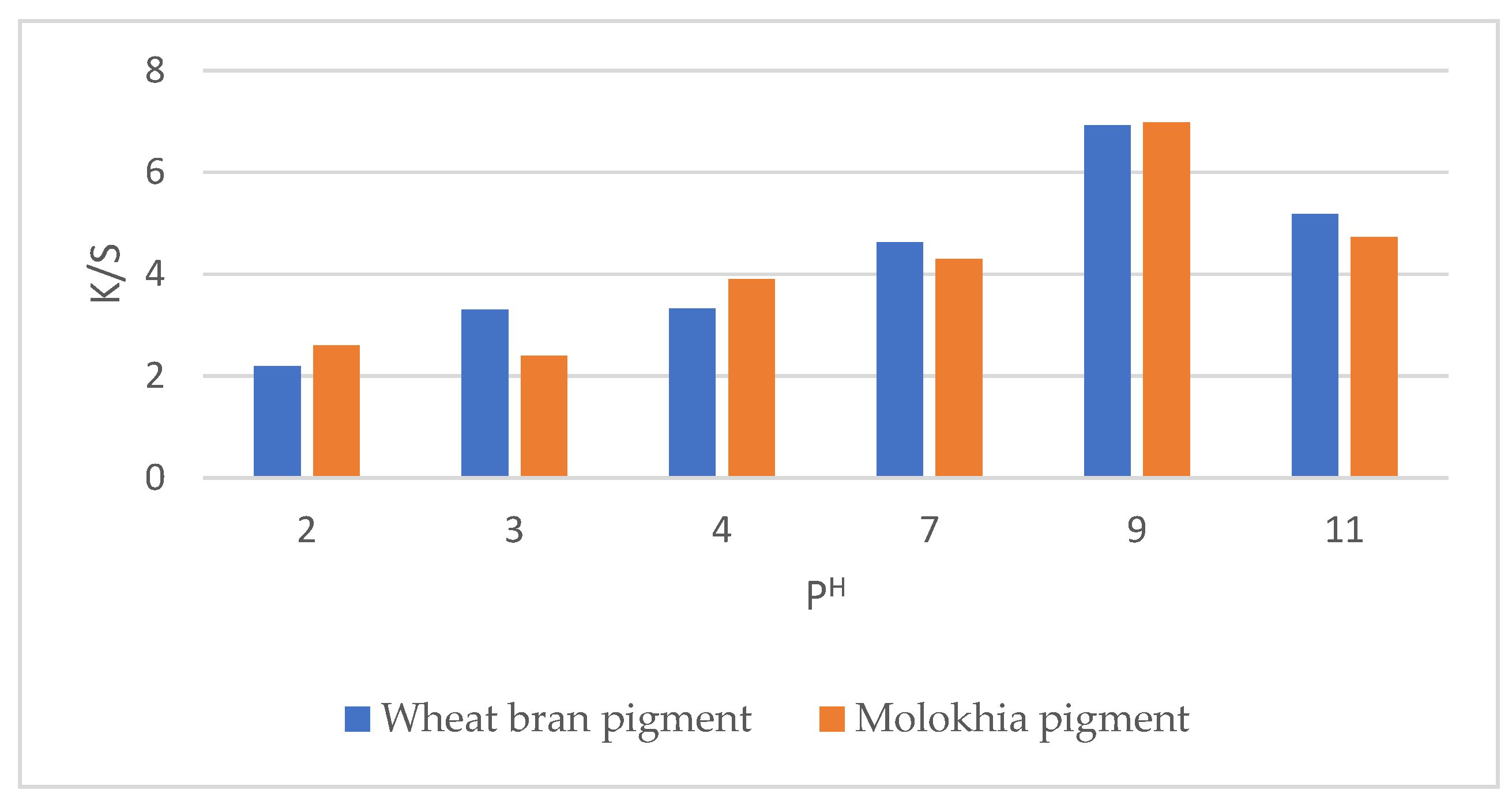
It is well known that using water instead of synthetic medium for pigment production is a cost-effective and economic approach. Therefore, further studies were performed by using the pigment produced from Bacillus subtilis KU710517 and waste substrates in water for dyeing wool samples at different pH (Table 6 and Table 7) and Figure 3. The color characteristics of dyed wool fabrics varied notably with changes in dye pH for both pigment sources, molokhia stalk (Table 6) and wheat bran (Table 7).
For molokhia stalk, lightness (L*) values ranged between 74.01 and 80.18, with the highest lightness observed at pH 4. Increasing pH above or below this level led to a decrease in L* values, indicating darker shades. The redness (a*) and yellowness (b*) parameters increased with rising pH, reaching maximum values at pH 9 (a* = 6.29; b* = 28.21), producing more reddish-yellow tones. Correspondingly, chroma (c) also increased, with the highest saturation (28.9) at pH 9, while hue angle (h) slightly decreased with alkalinity. The total color difference (ΔE) values were highest under acidic (pH 2–3) and alkaline (pH 9) conditions.
For wheat bran, a similar trend was noted, though with more pronounced variations. The lightness (L*) increased from 68.29 at pH 2 to 78.05 at pH 11. Both a* and b* values increased with pH, peaking at pH 9 (a* = 3.85; b* = 29.4), suggesting enhanced color intensity. The chroma (c) reached its maximum (28.35) at pH 9, while hue (h) showed slight variations. The ΔE values also indicated noticeable color changes at both acidic and alkaline extremes.
The pigment produced by using water containing 5% wheat bran and water with 6% molokhia stems was represented by Figure 4. However, Figure 5 illustrates the natural wool fabric after dyeing using pigment produced from Bacillus subtilis KU710517 with both molokhia stalk and wheat bran in water.

3.4. Fastness Properties of Dyed Wool Fabric at Optimum Conditions

Table 8 represents the fastness properties of dyed wool fabrics using the yellowish-brown dye produced by Bacillus subtilis KU710517 in water medium. The best fastness properties and the highest color strength value (6.98) were obtained when using molokhia stems in water. Therefore, the pigment produced from molokhia stems will be used for further investigations.

3.5. Antimicrobial Activity of the Bacillus subtilis KU710517 Pigment

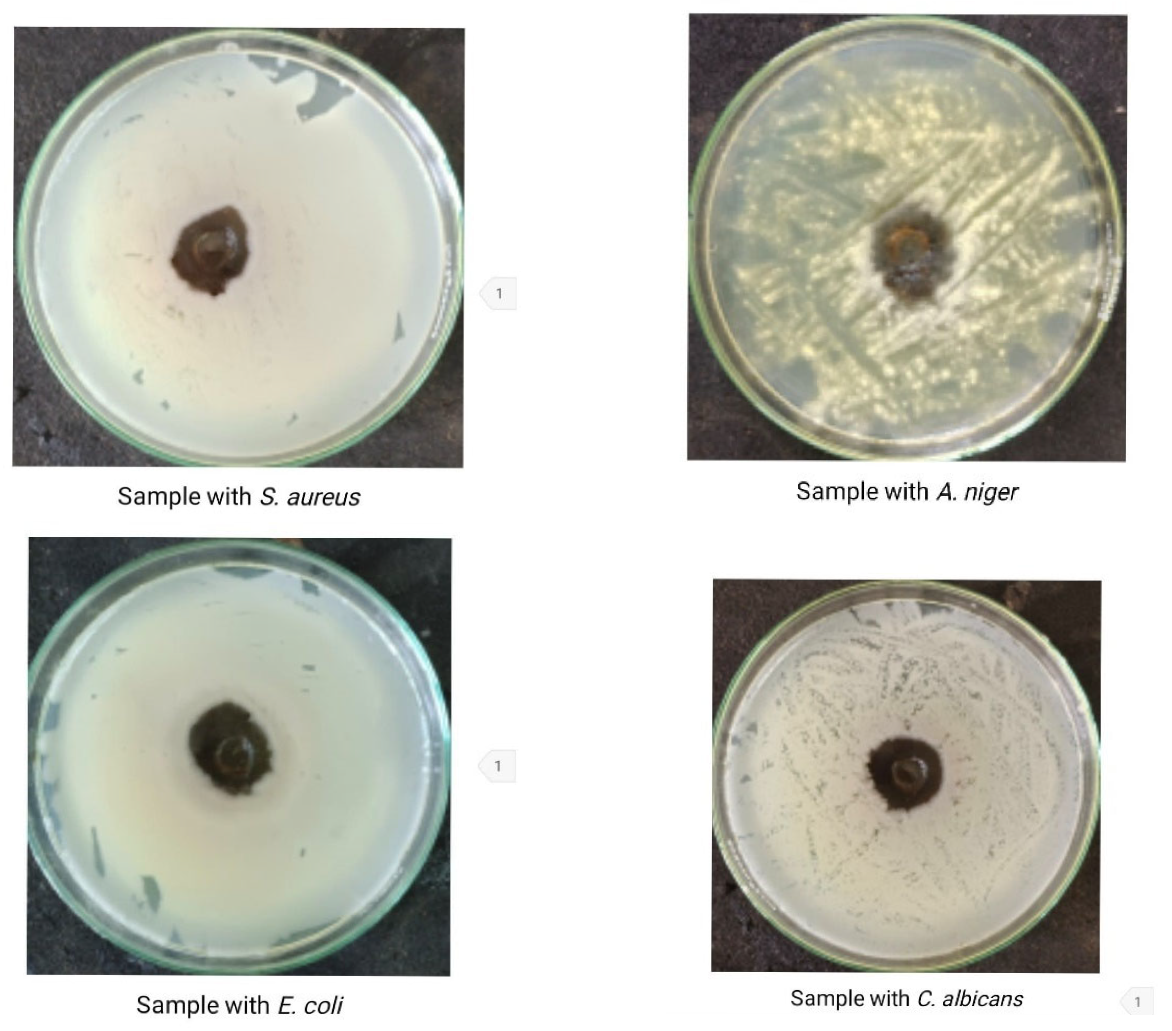
The yellowish-brown pigment produced by Bacillus subtilis KU710517 grown on molokhia stems-water medium showed considerable activity with Gram-negative and Gram-positive bacteria (Escherichia coli ATCC 25933 and Staphylococcus aureus ATCC 6538-P, respectively) as well as the fungus Aspergillus niger NRRL-A326 and the yeast Candida albicans ATCC 10231 (Table 9, Figure 6). It showed the strongest antimicrobial activity against E. coli (28 mm zone of inhibition).

3.6. Partial Characterization of Bacillus subtilis KU710517 Pigment

3.6.1. FTIR Spectroscopy

The FTIR spectrum of the bacterial pigment extract exhibited a broad absorption band around 3300–3500 cm−1 corresponding to O–H stretching vibrations of hydroxyl groups, typical of phenolic compounds. The peaks observed near 1600–1650 cm−1 are attributed to aromatic C=C stretching and conjugated C=O groups, while those between 1000–1300 cm−1 correspond to C–O stretching of alcohols, phenols, or glycosidic bonds. Additional weak peaks in the region 2850–2950 cm−1 are due to aliphatic C–H stretching vibrations.
These spectral characteristics confirm the presence of hydroxyl, carbonyl, and aromatic functionalities that are generally associated with polyphenolic and flavonoid structures. However, the FTIR spectrum of a crude extract represents an overlapping signal of many constituents and therefore cannot be used to identify a single specific compound. The observed bands are consistent with functional groups found in phenolic acids (such as gallic and chlorogenic acids) and flavonoid glycosides (such as rutin), indicating the polyphenolic nature of the bacterial pigment (Figure 7).

3.6.2. HPLC Analysis

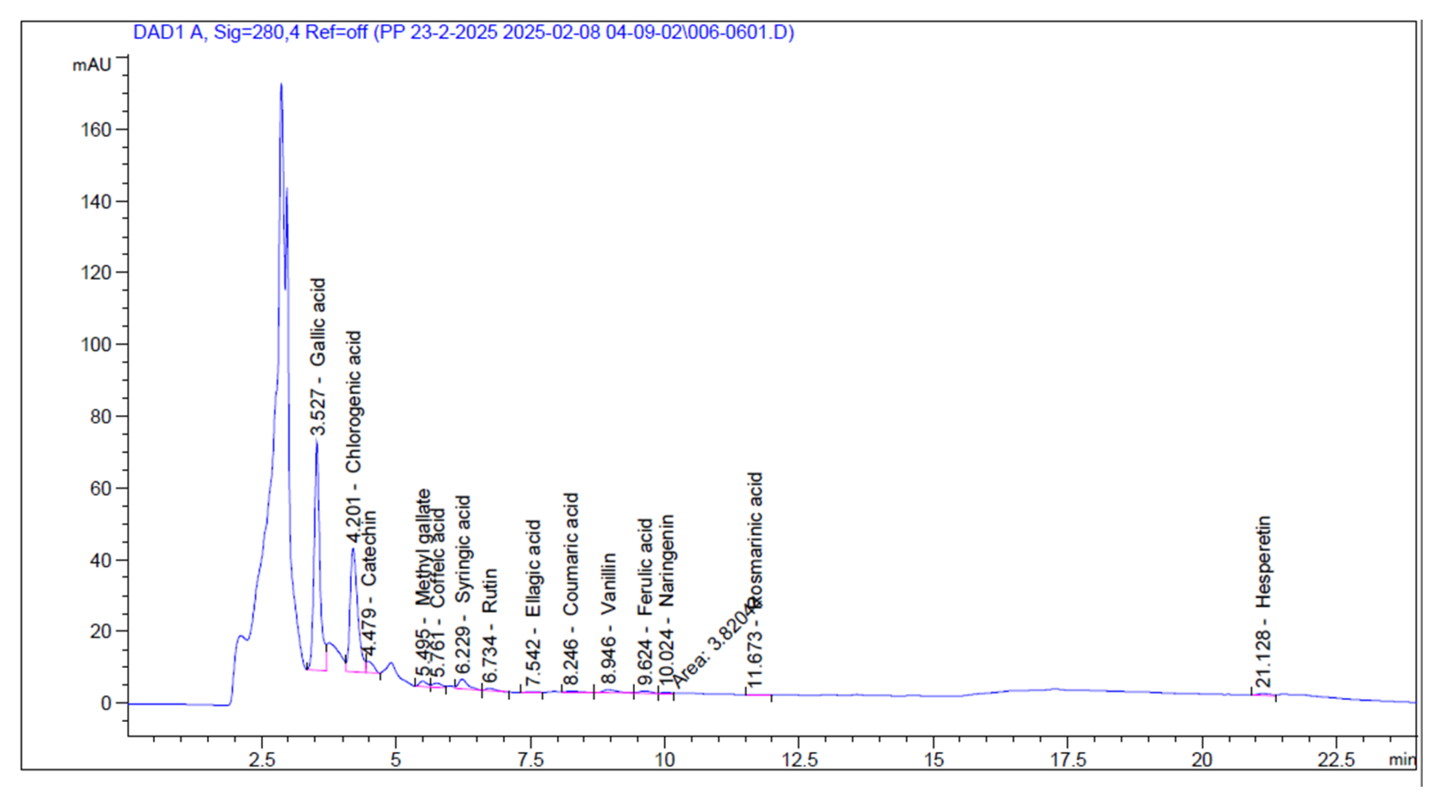
On the basis of chemical structure, the produced dye is from the flavonoids class. According to the HPLC analysis, most of the pigment constituents are polyphenols. Chlorogenic acid (CA) represented the highest concentration (475.65 µg/mL) followed by Gallic acid (345.20 µg/mL). Syringic acid, which is a Gallic acid derivative, is also found (19.31 µg/mL). The flavonoid Catechin is found in high concentration (62.40 µg/mL). Rutin is found in a concentration of (12.44 µg/mL). Other flavonoids such as Hesperetin and Naringenin were found in concentrations less than 10 µg/mL (Table 10, Figure 8). Pigmentation might be due to the presence of different polyphenolic compounds such as rutin (yellow color) and syringic acid (light brown color).
Several studies have reported that rutin, catechin, chlorogenic acid (CA) and Gallic acid (GA) are known to have antimicrobial effect [28,29,30,31,32,33,34,35]. Syringic acid possesses anti-oxidant, antimicrobial, anti-inflammatory activities [36]. This could explain the antimicrobial effect of the produced pigment, which is related to the presence of rutin, catechin, chlorogenic acid, gallic acid and syringic acid together in the pigment.

4. Discussion

As mentioned in the literature, B. subtilis produces either Riboflavin yellow pigment or a brownish pigmentation (melanin-like compound) [3,12,13]. This study highlights the potential of agro-waste materials—specifically wheat bran and molokhia stalks—for microbial pigment production by Bacillus subtilis KU710517 and their application in wool dyeing. Among the tested wastes, only wheat bran and molokhia stalks supported pigment production, whereas potato peels showed no effect.
Potato peels contain 10.6% carbohydrate, 1.8% protein, 1.3% ash content, 84.2% moisture content, 0.3% fat, 2.5% fiber and 7.8% starch [37]. Molokhia stems contain 44% carbohydrate, 22% protein, 16% ash content, 5% moisture content, 2% fat, 11% fiber and vitamins, 35% lignin, 50% cellulose, and 15% hemicelluloses. Therefore, it can be considered a rich substrate for microbial bioactive compounds production [38,39]. In wheat bran the dietary fiber content varied from 40 to 53% of the dry matter, and the starch content from 9 to 25%. Around 55% arabinoxylan, cellulose (9–12%), lignin (3–5%), fructan (3–4%), and mixed-linked β-glucan (2.2–2.6%). The ash content of wheat bran was 5.5–6.5%. Wheat bran also contains about 4–6% di- and tri-saccharides such as sucrose and raffinose [40]. The difference in pigment production with different wastes might be due to differences in available sugars and nutrients or due to inhibitory compounds in potato peels that limit pigment biosynthesis.
The concentration of both wheat bran and molokhia stalks in the synthetic medium had a significant impact on dye uptake and color strength (K/S) of wool fabrics. For wheat bran, pigment production and dye intensity increased with concentration up to 5%, after which a slight decrease was noted. Similarly, molokhia stems showed an optimal concentration at 4%, with K/S peaking at 6.39 before declining at 5%. These declines beyond optimal levels may be due to oversaturation or interference with pigment production or dye binding, likely caused by excessive organic material.
Colorimetric values (L*, a*, b*, and ΔE) supported these trends. In both cases, higher concentrations led to increased brightness and yellowness (higher L* and b* values), indicating warmer and more saturated colors. For molokhia stalks in particular, b* rose sharply from 17.6 at 1% to 30.6 at 6%, showing strong color enhancement. The ΔE values, which represent total color difference, peaked at the same optimal concentrations for both materials, confirming the most intense and effective dyeing occurred at 5% wheat bran and 4% molokhia stalks.
The concentration of both wheat bran and molokhia stalks in water also had a significant impact on color strength (K/S) of wool fabrics. For wheat bran, pigment production and dye intensity increased with concentration up to 5%, after which a slight decrease was noticed. Molokhia stalks showed an optimal concentration at 6%, with K/S peaking at 4.25 before declining at 7%.
From the previous results, it was observed that both wheat bran and molokhia stems exhibited higher values of color strength with the synthetic medium (pH 9). However, lower values of color strength were observed with water (pH 6). This might be due to the pH difference between the synthetic medium and water, which might affect the bacterial growth and pigment production. It is evident that pH 9 is more suitable than pH 6 for pigment production by Bacillus subtilis KU710517. Other researchers also observed low pigment formation with acidic pH, while moderate pigment formation was obtained with alkaline pH [17].
It is well known that using water instead of synthetic medium for pigment production is a cost-effective and economic approach. Therefore, further studies were performed by using the pigment produced from Bacillus subtilis KU710517 and waste substrates in water for dyeing wool samples at different pH. The results in Table 6 and Table 7 indicate that both molokhia stems and wheat bran in water exhibited the highest values of color strength at pH 9 (K/S of 6.98 and 6.93, respectively). These results proved that pH 9 is the most suitable for the dyeing process and also for pigment production by Bacillus subtilis KU710517.
The results demonstrate that pH has a significant effect on the color characteristics of wool fabrics dyed with pigments derived from Bacillus subtilis KU710517, regardless of the plant substrate used. In both systems, alkaline conditions (pH 9) produced the most vivid and saturated colors, characterized by higher a* and b* values and higher chroma (c). This can be attributed to the enhanced ionization of pigment molecules and improved dye-fiber interactions in alkaline media, which favor the fixation of pigment on wool fibers.
Conversely, acidic conditions (pH 2–3) resulted in duller shades with lower chroma and lightness values. This may be due to limited pigment binding at low pH, where the wool surface carries a positive charge that repels cationic pigment species or inhibits hydrogen bonding. When comparing the two pigment sources, the wheat bran pigment produced slightly deeper and more intense colors (lower L*, higher ΔE) than the molokhia stalk pigment, indicating that pigment composition or concentration may differ between substrates. Overall, the optimum dyeing performance was observed at pH 9, where both color intensity and uniformity were highest, suggesting that mildly alkaline conditions are most favorable for achieving bright, stable coloration of wool using microbial pigments. The results indicated that both wheat bran and molokhia stems in water exhibited the highest values of color strength at pH 9. These results proved that pH 9 is the most suitable for the dying process and also for pigment production by Bacillus subtilis KU710517.
Therefore, the optimum conditions for production of yellowish-brown dye from Bacillus subtilis KU710517 which gave the highest color strength value of dyed wool fabrics are: using water containing 5% wheat bran (K/S 6.93) or 6% molokhia stems (K/S 6.98), for 2 days, at pH 9. These values of color strength were much higher than the color strength value obtained when cotton fabrics were dyed by the natural quercetin dye extracted from Hibiscus sabdarifa (K/S 0.94) [26].
The yellowish-brown pigment produced by Bacillus subtilis KU710517 grown on molokhia stems-water medium showed considerable activity with Gram-negative, Gram-positive bacteria, yeast, and fungi. It displayed the strongest antimicrobial activity against E. coli (28 mm zone of inhibition). Ali et al. [17] also had observed considerable antimicrobial activity against E. coli and S. aureus by using the brown pigment produced from Streptomyces virginiae. On the contrary, other studies represented producing a red pigment with weak antimicrobial activity against E. coli [41]. The present investigation presented good results (28 mm zone of inhibition with E. coli) when compared with Kazi et al. [42], where their brown pigment revealed 4 mm zone of inhibition against E. coli.
The FTIR and HPLC analysis indicated that the pigmentation is due to the presence of polyphenolic compounds: chlorogenic acid, gallic acid, catechin, and rutin, which produce a yellow to brown color. Many previous scientific studies had confirmed the antimicrobial activity of polyphenolic compounds against both Gram-positive and Gram-negative bacterial strains [28,29,30,43]. Catechin has a bactericidal effect, as it can damage the bacterial cell membrane [31]. Chlorogenic acid (CA) is also one of the most essential compounds that possess antibacterial activity [32]. Chlorogenic acid attacks bacterial strains by inducing irreversible alterations in cell membrane permeability, leading to the cells’ inability to sustain membrane potential and cytoplasmic macromolecules [33]. Gallic acid (GA) is one of the most important natural polyphenols. Several studies were carried out on the function of GA and its derivatives [44]. It was found that it had broad-spectrum biological activities such as anti-inflammatory, antimicrobial, and anticancer activities [34]. GA produces pores in the bacterial membrane and causes irreversible changes in bacteria by changing membrane permeability, hydrophobicity, and physicochemical properties of bacteria [35]. Syringic acid possesses antioxidant, antimicrobial, and anti-inflammatory activities [36]. The aforementioned data could explain the antimicrobial effect of the produced pigment, which is related to the presence of rutin, catechin, chlorogenic acid, gallic acid, and syringic acid together in the pigment. Although washing or disinfection resistance was not evaluated, the dyed wool antimicrobial properties make the material potentially useful for hygienic or protective textiles including healthcare support items, functional clothing, or antimicrobial home fabrics. Further work is required to focus on improving the dye fastness and stability under washing and disinfection processes according to standard method.

5. Conclusions

This study successfully demonstrated the use of agro-waste materials, specifically wheat bran and molokhia stems, as effective, low-cost substrates for the production of yellowish-brown pigments by Bacillus subtilis KU710517. Molokhia stem wastes are easily obtained since Molokhia or Jew’s Mallow, also known as Corchorus olitorius plant, is cheap and abundant in Egypt. Among the tested conditions, molokhia stems and wheat bran in water at pH 9 yielded the highest color strength (K/S = 6.98 and K/S = 6.93, respectively) and exhibited excellent fastness properties on wool fabrics. The results suggest that both wheat bran and molokhia stems can act not only as low-cost substrates for pigment production but also as enhancers of dyeing performance. Optimizing their concentration and pH is essential for obtaining better dyeing performance. The produced pigment also showed strong antimicrobial activity against a broad spectrum of pathogens, particularly E. coli. Chemical characterization confirmed that the pigment is rich in bioactive flavonoids and polyphenols such as chlorogenic acid, gallic acid, catechin, rutin, and syringic acid, which contribute to its color and biological activity. These results highlight the pigment’s potential for use in eco-friendly and functional textile applications.

Author Contributions

Conceptualization, K.A.A. and H.R.W.; Methodology, H.M.E.-H. and H.R.W.; Formal analysis, H.M.E.-H.; Investigation, K.A.A. and H.R.W.; Resources, H.R.W.; Data curation, H.M.E.-H.; Writing—original draft, K.A.A.; Writing—review & editing, H.R.W. All authors have read and agreed to the published version of the manuscript.

Funding

This research received no external funding.

Data Availability Statement

The original contributions presented in this study are included in the article. Further inquiries can be directed to the corresponding author.

Acknowledgments

The Researchers would like to thank the Deanship of Graduate Studies and Scientific Research at Qassim University for financial support (QU-APC-2025).

Conflicts of Interest

The authors declare no conflicts of interest.

References

  1. Heredia-Guerrero, J.A.; Heredia, A.; Domínguez, E. Cutin from agro-waste as a raw material for the production of bioplastics. J. Exp. Bot. 2017, 68, 5401–5410. [Google Scholar] [CrossRef]
  2. Nayak, A.; Bhushan, B. An overview of the recent trends on the waste valorization techniques for food wastes. J. Environ. Manag. 2019, 233, 352–370. [Google Scholar] [CrossRef]
  3. Panesar, R.; Kaur, S.; Panesar, P.S. Production of microbial pigments utilizing agro-industrial waste: A review. Curr. Opin. Food Sci. 2015, 1, 70–76. [Google Scholar] [CrossRef]
  4. Zihare, L.; Spalvins, K.; Blumberga, D. Multi criteria analysis for products derived from agro-industrial by-products. Energy Procedia 2018, 147, 452–457. [Google Scholar] [CrossRef]
  5. Sen, T.; Barrow, C.J.; Deshmukh, S.K. Microbial pigments in the food industry—Challenges and the way forward. Front. Nutr. 2019, 6, 7. [Google Scholar] [CrossRef] [PubMed]
  6. Kumar, A.; Vishwakarma, H.S.; Singh, J.; Dwivedi, S.; Kumar, M. Microbial pigments: Production and their applications in various industries. Int. J. Pharm. Chem. Biol. Sci. 2015, 5, 203–212. Available online: https://www.ijpcbs.com/articles/microbial-pigments-production-and-their-applications-invarious-industries.pdf (accessed on 19 October 2025).
  7. Ahmad, W.A.; Ahmad, W.Y.W.; Zakaria, Z.A.; Yusof, N.Z. Application of Bacterial Pigments as Colorant. In Application of Bacterial Pigments as Colorant: The Malaysian Perspective; Springer: Berlin/Heidelberg, Germany, 2011; pp. 57–74. [Google Scholar] [CrossRef]
  8. Barnett, T.A.; Valenzuela, D.; Riner, S.; Hageman, J.H. Production by Bacillis subtilis of brown sporulation-associated pigments. Can. J. Microbiol. 1983, 29, 96–101. [Google Scholar] [CrossRef]
  9. Hoti, S.; Balaraman, K. Formation of melanin pigment by a mutant of Bacillus thuringiensis H-14. Microbiology 1993, 139, 2365–2369. [Google Scholar] [CrossRef]
  10. Liu, Y.T.; Sui, M.J.; Ji, D.D.; Wu, I.H.; Chou, C.C.; Chen, C.C. Protection from ultraviolet irradiation by melanin of mosquitocidal activity of Bacillus thuringiensis var. israelensis. J. Invertebr. Pathol. 1993, 62, 131–136. [Google Scholar] [CrossRef]
  11. Mitchell, C.; Iyer, S.; Skomurski, J.F.; Vary, J.C. Red pigment in Bacillus megaterium spores. Appl. Environ. Microbiol. 1986, 52, 64–67. [Google Scholar] [CrossRef]
  12. Venil, C.K.; Zakaria, Z.A.; Ahmad, W.A. Bacterial pigments and their applications. Process Biochem. 2013, 48, 1065–1079. [Google Scholar] [CrossRef]
  13. Moeller, R.; Horneck, G.; Facius, R.; Stackebrandt, E. Role of pigmentation in protecting Bacillus sp. endospores against environmental UV radiation. FEMS Microbiol. Ecol. 2005, 51, 231–236. [Google Scholar] [CrossRef]
  14. Jang, J.H.; van Soest, R.W.; Fusetani, N.; Matsunaga, S. Pseudoceratins A (Ia) and B (Ib), Antifungal Bicyclic Bromotyrosine-Derived Metabolites from the Marine Sponge Pseudoceratina purpurea. J. Org. Chem. 2007, 72, 1211–1217. [Google Scholar] [CrossRef]
  15. Wehaidy, H.R.; Abdel-Naby, M.; Shousha, W.G.; El Mallah, M.I.; Shawky, M.M. Optimization of the production and characterization of milk clotting enzyme from Bacillus subtilis isolated from marine sponge. Egypt. Pharm. J. 2016, 15, 158–166. [Google Scholar] [CrossRef]
  16. Parmar, R.S.; Singh, C. A comprehensive study of ecofriendly natural pigment and its applications. Biochem. Biophys. Rep. 2018, 13, 22–26. [Google Scholar] [CrossRef]
  17. Ali, A.M.; Keera, A.A.; Helmy, S.M.; Abd El-Nasser, N.H.; Ahmed, K.A.; El-Hennawi, H.M. Selection of Pigment (Melanin) production in Streptomyces and their application in Printing and Dyeing of Wool Fabrics. Res. J. Chem. Sci. 2011, 1, 22–28. Available online: https://www.isca.me/rjcs/Archives/v1/i5/ISCA_RJCS_04_2011_90.pdf (accessed on 21 October 2025).
  18. Chrysler, L. Methods of Test for Colour Fastness of Textiles and Leather, 7th ed.; British Standards Institution: London, UK, 1990; Available online: https://knowledge.bsigroup.com/products/methods-of-test-for-colour-fastness-of-textiles-and-leather (accessed on 19 October 2025).
  19. Abdou, E.S.; El-Hennawi, H.M.; Ahmed, K.A. Edible Coloured Printing Paste for Food Packaging Materials Using Carboxymethyl Cellulose/Chitosan nanoparticles Composite as Thickeners. Egypt. J. Chem. 2024, 67, 637–645. [Google Scholar] [CrossRef]
  20. Ahmed, K.; Shahin, A.; Ragheb, A.; El-Hennawi, H. A Facile Synthesis with One Step of Disperse Azo Dyes to be Applied as Nano-Inks in Textile Printing. Biointerface Res. Appl. Chem. 2021, 11, 11713–11723. [Google Scholar] [CrossRef]
  21. Ahmed, E.A.; Elgemeie, G.; Ahmed, K.A.-E. Microwave-assisted synthesis of disazothiazolone dyes as antibacterial agents and their application in polyester printing. Pigment Resin Technol. 2022, 51, 1–5. [Google Scholar] [CrossRef]
  22. ISO 105-X19:2020; Textiles—Tests for Colour Fastness—Part X19: Colour Fastness to Rubbing. ISO: Geneva, Switzerland, 2013. Available online: https://www.iso.org/standard/77287.html?utm_ (accessed on 21 October 2025).
  23. ISO 105-C06:2010; Textiles—Tests for Colour Fastness—Part C06: Colour Fastness to Domestic and Commercial Laundering. ISO: Geneva, Switzerland, 2010. Available online: https://www.iso.org/standard/51276.html?utm_ (accessed on 19 October 2025).
  24. ISO 105-E04:2013; Textiles—Tests for Colour Fastness—Part E04: Colour Fastness to Perspiration. ISO: Geneva, Switzerland, 2013. Available online: https://www.iso.org/standard/57973.html?utm_ (accessed on 19 October 2025).
  25. ISO 105-B02:2014; Textiles—Tests for Colour Fastness—Part B02: Colour Fastness to Artificial Light: Xenon Arc Fading Lamp Test. ISO: Geneva, Switzerland, 2014. Available online: https://www.iso.org/standard/65209.html?utm_ (accessed on 19 October 2025).
  26. Elsaba, Y.M.; El-Hennawi, H.M.; Ibrahim, M.M.; Wehaidy, H.R. Production of a novel laccase from Ceratorhiza hydrophila and assessing its potential in natural dye fixation and cytotoxicity against tumor cells. J. Genet. Eng. Biotechnol. 2023, 21, 14. [Google Scholar] [CrossRef] [PubMed]
  27. Tamam, E.; Fadda, A.A.; El-Sawy, E.R.; Abdel-Aziz, M.S.; Tawfik, E.H. Design, synthesis, antimicrobial and docking studies of benzothiazoles bearing pyrazole and pyrimidine moieties. ChemistrySelect 2023, 8, e202302468. [Google Scholar] [CrossRef]
  28. Ganeshpurkar, A.; Saluja, A.K. The pharmacological potential of rutin. Saudi Pharm. J. 2017, 25, 149–164. [Google Scholar] [CrossRef]
  29. Rodríguez-Valdovinos, K.Y.; Salgado-Garciglia, R.; Vázquez-Sánchez, M.; Álvarez-Bernal, D.; Oregel-Zamudio, E.; Ceja-Torres, L.F.; Medina-Medrano, J.R. Quantitative analysis of rutin by HPTLC and in vitro antioxidant and antibacterial activities of phenolic-rich extracts from Verbesina sphaerocephala. Plants 2021, 10, 475. [Google Scholar] [CrossRef]
  30. Jhanji, R.; Singh, A.; Kumar, A. Antibacterial potential of selected phytomolecules: An experimental study. Microbiol. Immunol. 2021, 65, 325–332. [Google Scholar] [CrossRef] [PubMed]
  31. Gopal, J.; Muthu, M.; Paul, D.; Kim, D.H.; Chun, S. Bactericidal activity of green tea extracts: The importance of catechin containing nano particles. Sci. Rep. 2016, 6, 19710. [Google Scholar] [CrossRef]
  32. Miao, M.; Xiang, L. Pharmacological action and potential targets of chlorogenic acid. Adv. Pharmacol. 2020, 87, 71–88. [Google Scholar] [CrossRef]
  33. Lou, Z.; Wang, H.; Zhu, S.; Ma, C.; Wang, Z. Antibacterial activity and mechanism of action of chlorogenic acid. J. Food Sci. 2011, 76, M398–M403. [Google Scholar] [CrossRef] [PubMed]
  34. Fernandes, F.H.A.; Salgado, H.R.N. Gallic acid: Review of the methods of determination and quantification. Crit. Rev. Anal. Chem. 2016, 46, 257–265. [Google Scholar] [CrossRef] [PubMed]
  35. Borges, A.; Ferreira, C.; Saavedra, M.J.; Simões, M. Antibacterial activity and mode of action of ferulic and gallic acids against pathogenic bacteria. Microb. Drug Resist. 2013, 19, 256–265. [Google Scholar] [CrossRef]
  36. Cheemanapalli Srinivasulu, C.S.; Mopuri Ramgopal, M.R.; Golla Ramanjaneyulu, G.R.; Anuradha, C.M.; Kumar, C.S. Syringic acid (SA)—A review of its occurrence, biosynthesis, pharmacological and industrial importance. Pharma Innov. J. 2018, 12, 2217–2225. [Google Scholar] [CrossRef]
  37. Kinoka, M. Potato and potato peels: A comprehensive review on potato peels nutritional composition, bioactive compounds and its application in the food industry. Pharma Innov. J. 2023, 12, 2217–2225. Available online: https://www.thepharmajournal.com/archives/2023/vol12issue7/PartZ/12-6-182-370.pdf (accessed on 19 October 2025).
  38. Mostafa, F.A.; Wehaidy, H.R.; Sharaf, S.; El-Hennawi, H.M.; Mahmoud, S.A.; Saleh, S.A. Aspergillus awamori MK788209 cellulase: Production, statistical optimization, pea peels saccharification and textile applications. Microb. Cell Factories 2024, 23, 11. [Google Scholar] [CrossRef]
  39. Ahmed, S.A.; Saleh, S.A.A.; Mostafa, F.A.; Abd El Aty, A.A.; Ammar, H.A.M. Characterization and valuable applications of xylanase from endophytic fungus Aspergillus terreus KP900973 isolated from Corchorus olitorius. Biocatal. Agric. Biotechnol. 2016, 7, 134–144. [Google Scholar] [CrossRef]
  40. Esposito, F.; Arlotti, G.; Bonifati, A.M.; Napolitano, A.; Vitale, D.; Fogliano, V. Antioxidant activity and dietary fibre in durum wheat bran by-products. Food Res. Int. 2005, 38, 1167–1173. [Google Scholar] [CrossRef]
  41. Teiba, I.I.; Mamdouh, I.; Yousef, M.I.; Hussein, A.; El-Bilawy, E.H. Antimicrobial activity of Monascus purpureus-derived red pigments against Salmonella typhimurium, Escherichia coli, and Enterococcus faecalis. AMB Express 2025, 15, 6. [Google Scholar] [CrossRef]
  42. Kazi, Z.; Hungund, B.S.; Yaradoddi, J.S.; Banapurmath, N.R.; Yusuf, A.A.; Kishore, K.L.; Soudagar, M.E.M.; Khan, T.M.Y.; Elfasakhany, A.; Buyondo, K.A. Production, characterization, and antimicrobial activity of pigment from Streptomyces species. J. Nanomater. 2022, 2022, 3962301. [Google Scholar] [CrossRef]
  43. Wang, Z.; Ding, Z.; Li, Z.; Ding, Y.; Jiang, F.; Liu, J. Antioxidant and antibacterial study of 10 flavonoids revealed rutin as a potential antibiofilm agent in Klebsiella pneumoniae strains isolated from hospitalized patients. Microb. Pathog. 2021, 159, 105121. [Google Scholar] [CrossRef] [PubMed]
  44. Yang, K.; Zhang, L.; Liao, P.; Xiao, Z.; Zhang, F.; Sindaye, D.; Xin, Z.; Tan, C.; Deng, J.; Yin, Y.; et al. Impact of gallic acid on gut health: Focus on the gut microbiome, immune response, and mechanisms of action. Front. Immunol. 2020, 11, 580208. [Google Scholar] [CrossRef] [PubMed]
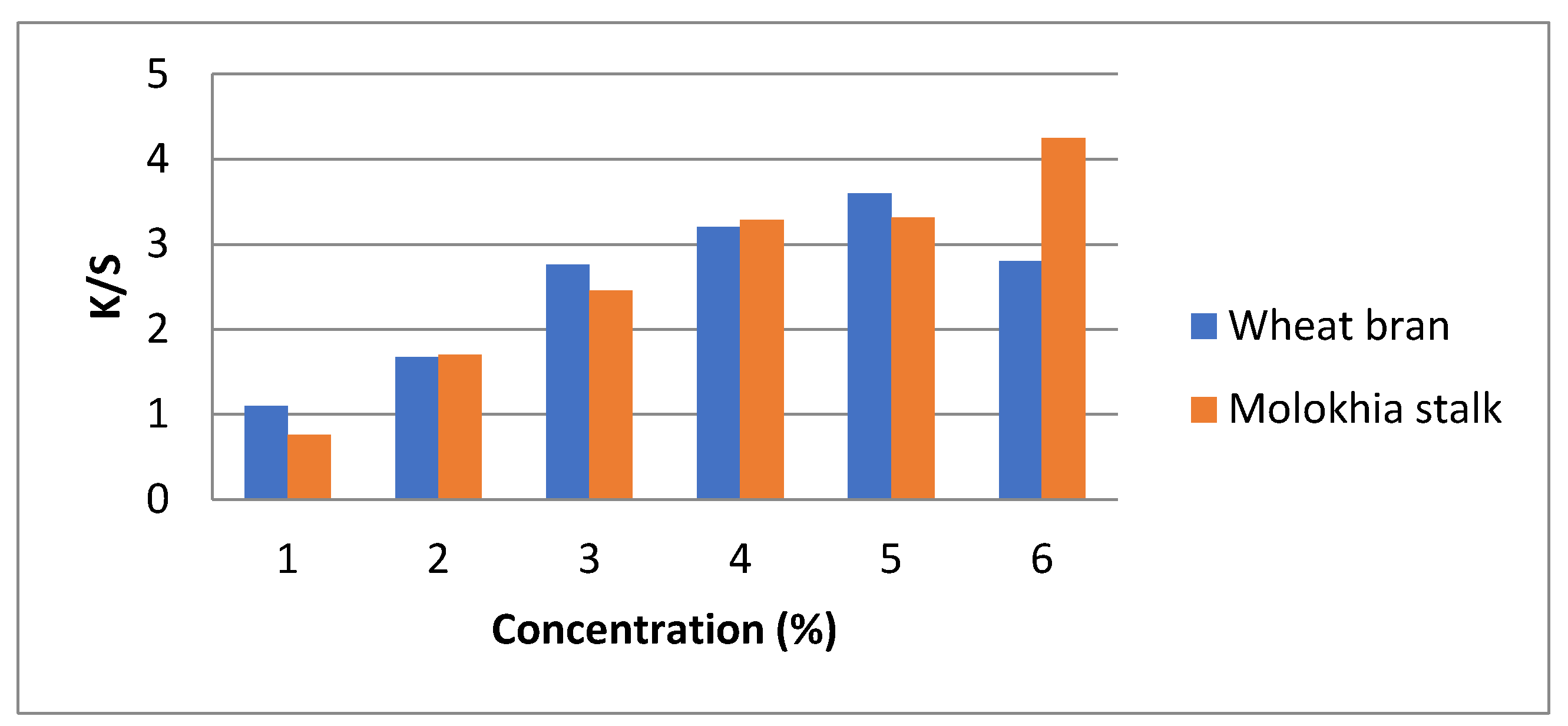
Figure 1. Effect of molokhia stalk and Wheat bran concentration in the synthetic medium on the color strength (K/S) of dyed wool fabric (pH 9.0).
Processes 13 03453 g001
Figure 2. Effect of molokhia stalk and Wheat bran concentration in water on the color strength (K/S) of dyed wool fabric (pH 9.0).
Processes 13 03453 g002
Figure 3. Effect of pH on color strength of dyed wool fabrics using pigment produced from Bacillus subtilis KU710517 using wheat bran and molokhia stem in water.
Processes 13 03453 g003
Figure 4. Represents the pigment produced by using water containing 5% Wheat bran and water with 6% Molokhia stems.
Processes 13 03453 g004
Figure 5. Natural wool fabric (a), dyed wool fabrics using pigment produced from Bacillus subtilis KU710517 with molokhia stalk (b), and wheat bran (c) in water.
Processes 13 03453 g005
Figure 6. The inhibition zone of Bacillus subtilis KU710517 pigment produced in water-molokhia stems medium.
Processes 13 03453 g006
Figure 7. FTIR spectrum of pigment produced by Bacillus subtilis KU710517 in water-molokhia stems medium.
Processes 13 03453 g007
Figure 8. Represent the HPLC analysis of Bacillus subtilis KU710517 pigment.
Processes 13 03453 g008
Table 1. Screening for pigment produced by Bacillus subtilis KU710517 on liquid media with different waste materials.
Liquid Medium *Pigment Production
Water with potato peels−ve
Synthetic medium with potato peels−ve
Water with wheat bran +ve
Synthetic medium with wheat bran+ve
Water with molokhia stalks+ve
Synthetic medium with molokhia stalks+ve
* Cultures were incubated on a rotary shaker (150 rpm) for two days at 37 °C and pH 9.0.
Table 2. Effect of wheat bran concentration in the synthetic liquid medium on the color parameters of dyed wool fabric.
Wheat Bran
Concentration (%)
L*a*b*ΔE
160.92.413.2350.1
264.42.7814.770.55
369.93.115.3572.38
470.23.516.673.8
573.54.220.775.6
671.4417.174.9
Table 3. Effect of molokhia stem concentration in the synthetic liquid medium on the color parameters of dyed wool fabric (pH 9.0).
Molokhia Stem
Concentration (%)
L*a*b*ΔE
172.23.017.676.8
274.54.221.879.6
377.24.922.880.6
480.16.130.685.9
578.45–829.382.3
Table 4. Effect of wheat bran concentration in water on the color parameters (K/S) of dyed wool fabric (pH 6.0).
Wheat Bran
Concentration (%)
L*a*b*ΔE
161.92.411.2350.1
2632.613.754.55
365.93.115.462.38
469.23.216.171.8
572.54.219.774.6
671.33.918.170.9
Table 5. Effect of molokhia stalk concentration in water on the color parameters of dyed wool fabric.
Molokhia Stalk
Concentration
(%)
L*a*b*ΔE
159.71.49.2350.1
2621.610.754.55
364.92.112.462.38
471.24.214.171.8
575.54.7814.774.6
680.34.916.170.9
778.92.411.2370.1
Table 6. Effect of pH on color parameters of dyed wool fabrics using pigment produced from Bacillus subtilis KU710517 and molokhia stalk in water.
pHL*a*b*chΔE
274.942.4916.7116.981.5376.82
374.622.2116.5616.782.3976.46
480.181.7511.7011.8381.5381.05
774.013.8523.323.6280.6277.69
974.066.2928.2128.977.4379.5
1174.734.9325.7626.2379.1677.2
Table 7. Effect of pH on color parameters of dyed wool fabrics using pigment produced from Bacillus subtilis KU710517 and wheat bran in water.
pHL*a*b*chΔE
268.291.7517.9518.0484.4380.34
369.393.0723.4232.6282.5282.83
469.993.1018.2918.5580.3872.38
775.16.321.924.778.573.8
973.103.8529.428.3529.6282.15
1178.054.5127.3927.7680.6679.84
Table 8. Fastness properties of dyed wool fabrics at optimum conditions.
SampleK/SRubbing FastnessWashing FastnessPerspiration FastnessLight Fastness
WetDryAltStAcidAlkaline
AltStAltSt
Wool Fabric (100%)
5% wheat bran 6.393–444444–544–55–6
6% molokhia stems 6.983–444444–544–55–6
Table 9. The antimicrobial activity of Bacillus subtilis KU710517 pigment produced in water-Molokhia stems medium.
Test MicrobeClear Zone
(mm)
Staphylococcus aureus24
Escherichia coli28
Candida albicans26
Aspergillus niger16
Table 10. HPLC analysis of Bacillus subtilis KU710517 pigment.
ComponentsAreaConc. (mg/mL)
Gallic acid471.47345.20
Chlorogenic acid341 35475.65
Catechin29.0462.40
Methyl gallate15.238 52
Coffeic acid14.787.58
Syringic acid32.8319.31
Rutin8.3112.44
Ellagic acid1.511.53
Coumaric acid6.182.22
Vanillin16.355.93
Ferulic acid10.165.90
Naringenin3.823.53
Rosmarinic acid1.641.59
Daidzein0.000.00
Querectin0.000.00
Cinnamic acid0.000.00
Kaempferol0.000.00
Hesperetin9.064.24
Disclaimer/Publisher’s Note: The statements, opinions and data contained in all publications are solely those of the individual author(s) and contributor(s) and not of MDPI and/or the editor(s). MDPI and/or the editor(s) disclaim responsibility for any injury to people or property resulting from any ideas, methods, instructions or products referred to in the content.