
Novel Hydrazide Hydrazone Derivatives as Antimicrobial Agents: Design, Synthesis, and Molecular Dynamics


Abstract
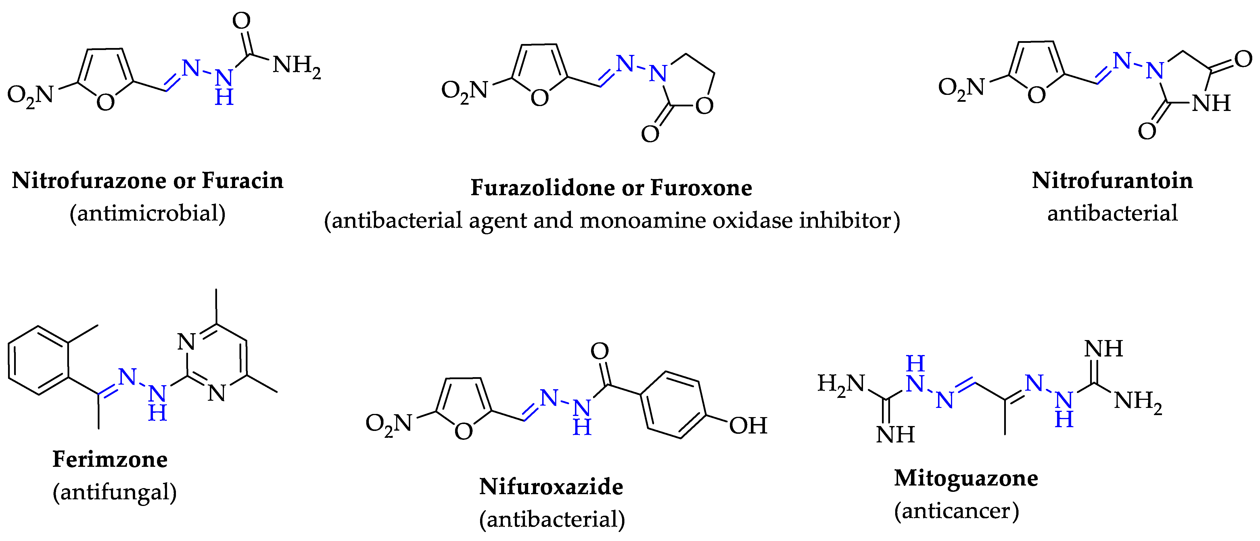
1. Introduction
2. Results and Discussion
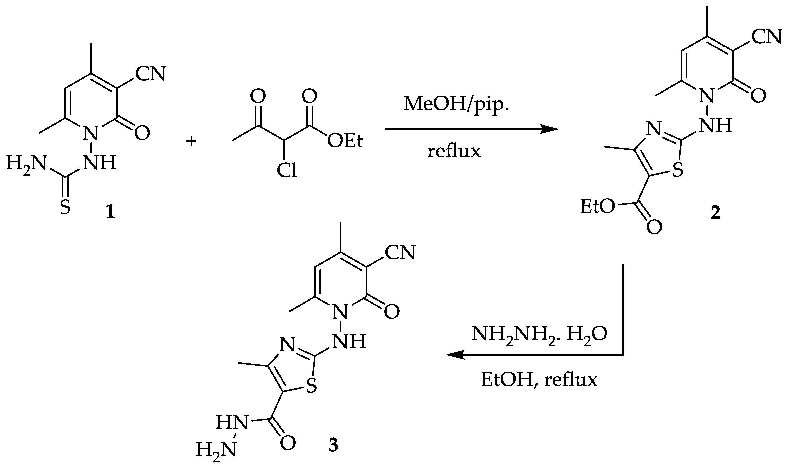
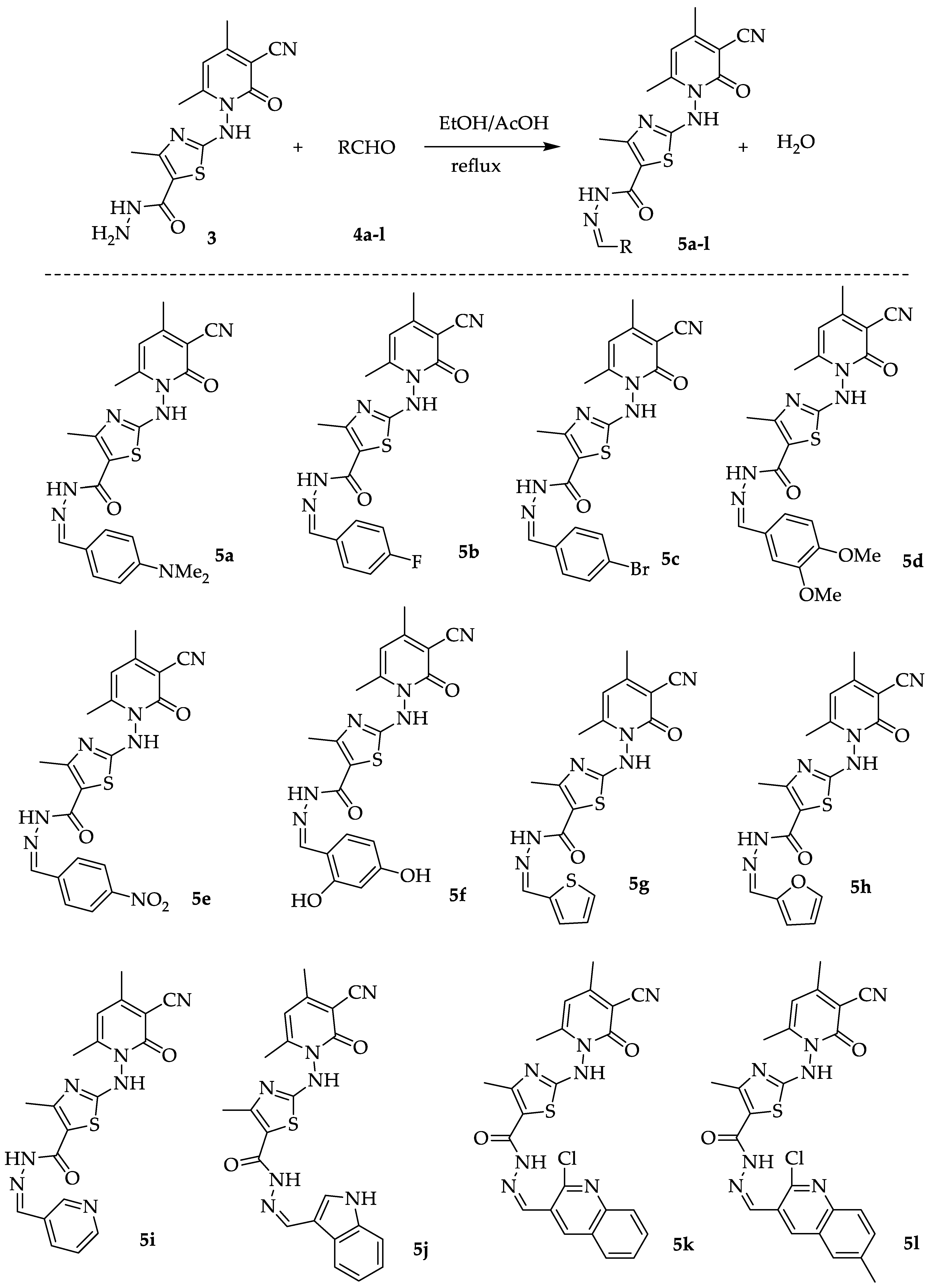
2.1. Chemistry
2.2. Antimicrobial Activity
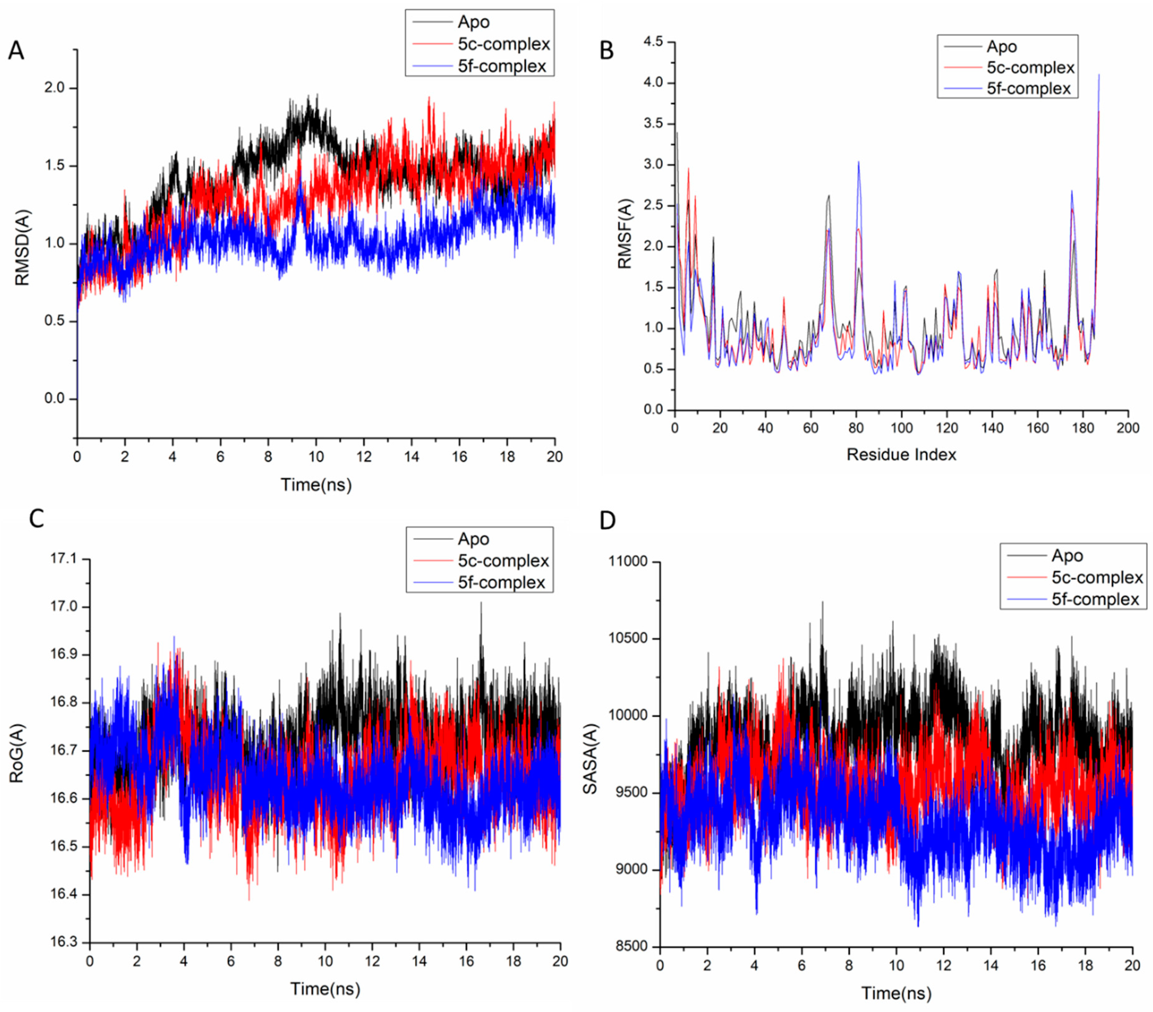
2.3. Molecular Dynamic and System Stability
2.3.1. The Binding Interaction Mechanism
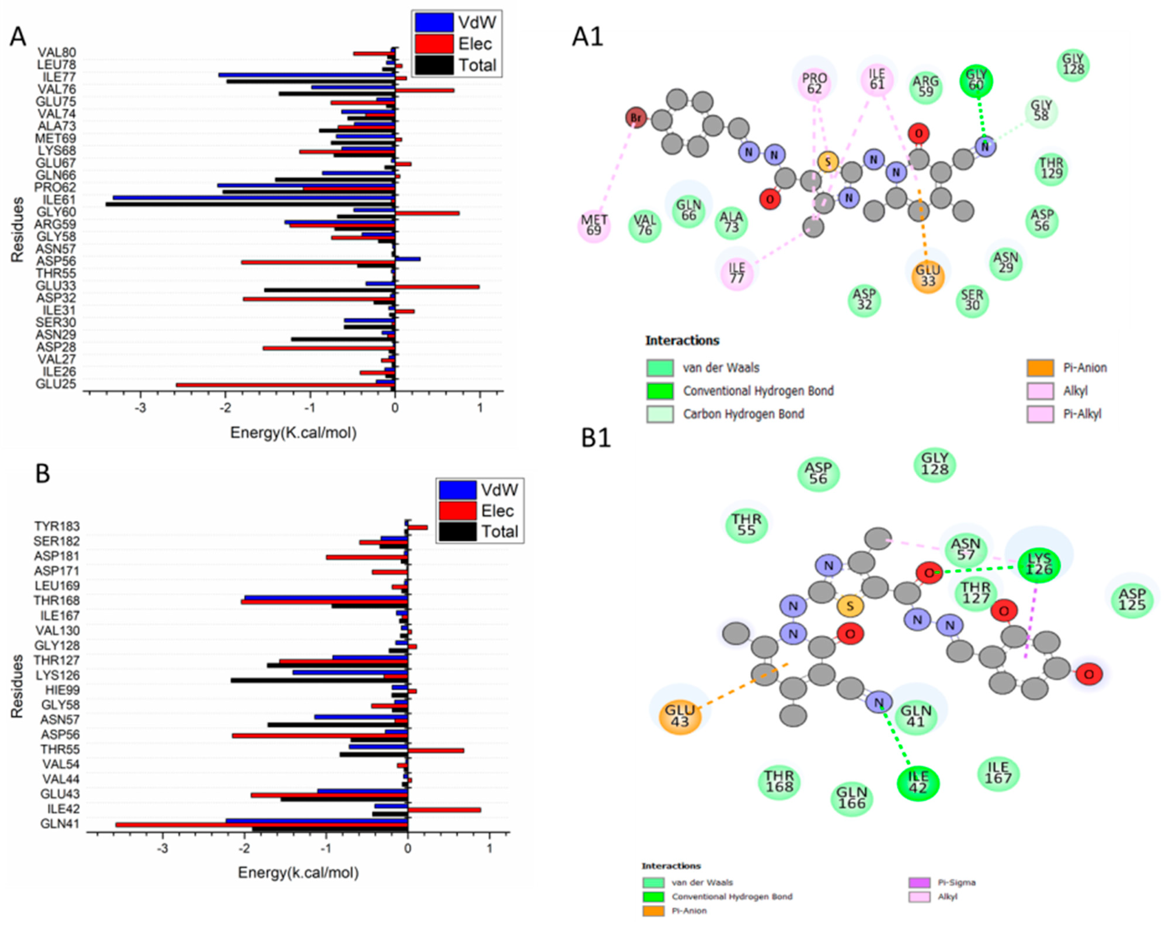
2.3.2. Finding the Essential Residues in Charge of Ligand Binding
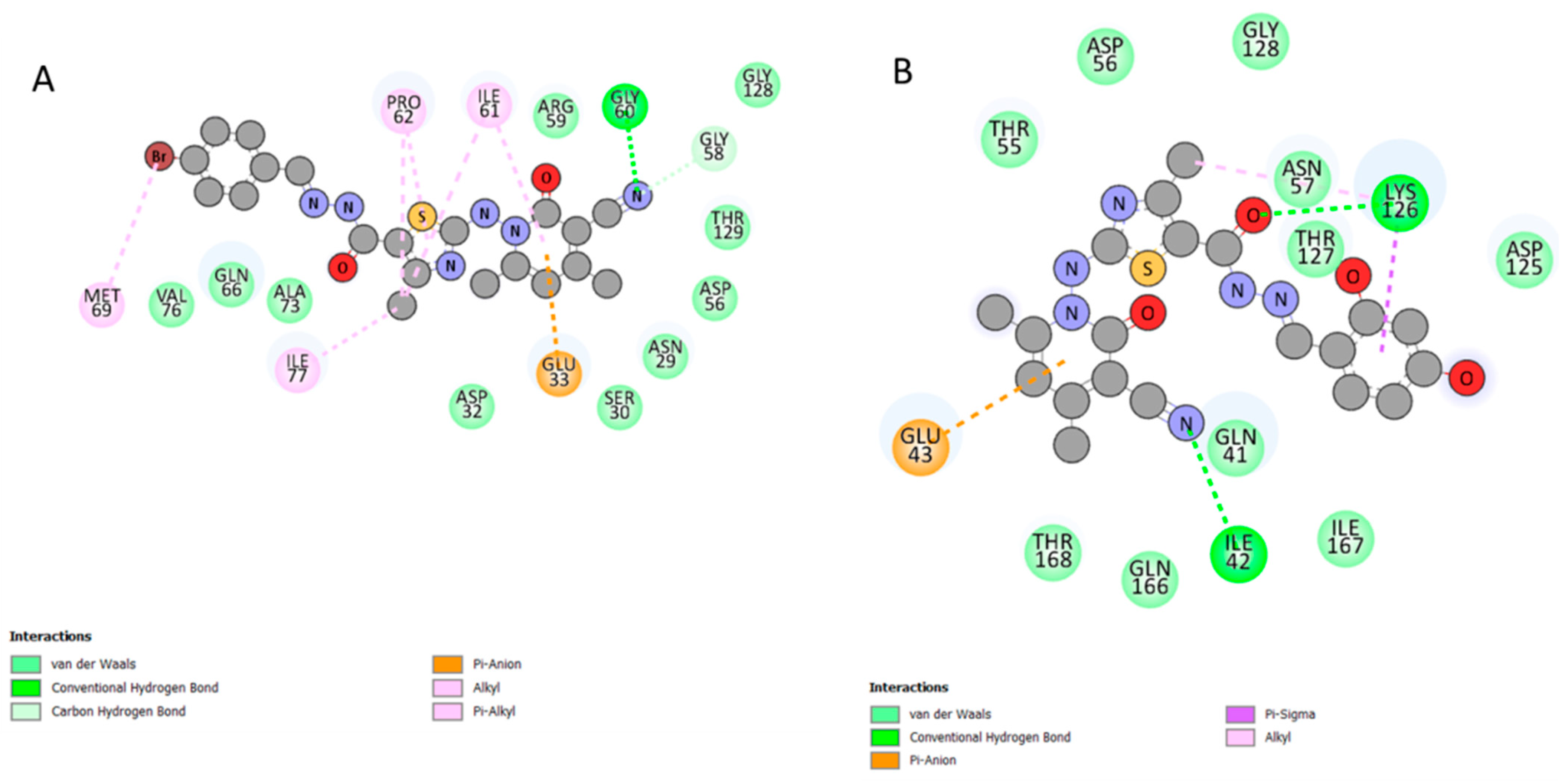
2.3.3. Patterns of Ligand–Residue Interactions in Networks
3. Experimental Section
3.1. Chemistry
3.2. Antimicrobial Activity
Minimum Inhibitory Concentration (MIC)
3.3. Molecular Dynamic and System Stability
3.3.1. System Preparation
3.3.2. Molecular Dynamics (MD) Simulations
3.3.3. Thermodynamic Calculation
4. Conclusions
Funding
Data Availability Statement
Acknowledgments
Conflicts of Interest
References
- Tang, K.W.K.; Millar, B.C.; Moore, J.E. Antimicrobial Resistance (AMR). Br. J. Biomed. Sci. 2023, 80, 11387. [Google Scholar] [CrossRef]
- Levin-Reisman, I.; Ronin, I.; Gefen, O.; Braniss, I.; Shoresh, N.; Balaban, N.Q. Antibiotic tolerance facilitates the evolution of resistance. Science 2017, 355, 826–830. [Google Scholar] [CrossRef]
- Doherty, T.M.; Hausdorff, W.P.; Kristinsson, K.G. Effect of vaccination on the use of antimicrobial agents: A systematic literature review. Ann. Med. 2020, 52, 283–299. [Google Scholar] [CrossRef]
- Rollas, S.; Küçükgüzel, S.G. Biological Activities of Hydrazone Derivatives. Molecules 2007, 12, 1910–1939. [Google Scholar] [CrossRef]
- Angelova, V.; Karabeliov, V.; Andreeva-Gateva, P.A.; Tchekalarova, J. Recent Developments of Hydrazide/Hydrazone Derivatives and Their Analogs as Anticonvulsant Agents in Animal Models. Drug Dev. Res. 2016, 77, 379–392. [Google Scholar] [CrossRef]
- Popiołek, Ł. Hydrazide–hydrazones as potential antimicrobial agents: Overview of the literature since 2010. Med. Chem. Res. 2017, 26, 287–301. [Google Scholar] [CrossRef]
- Sharma, P.C.; Sharma, D.; Sharma, A.; Saini, N.; Goyal, R.; Ola, M.; Chawla, R.; Thakur, V.K. Hydrazone comprising compounds as promising anti-infective agents: Chemistry and structure-property relationship. Mater. Today Chem. 2020, 18, 100349. [Google Scholar] [CrossRef]
- Guilherme, F.D.; Simonetti, J.E.; Folquitto, L.R.S.; Reis, A.C.C.; Oliver, J.C.; Dias, A.L.T.; Dias, D.F.; Carvalho, D.T.; Brandao, G.C.; de Souza, T.B. Synthesis, chemical characterization and antimicrobial activity of new acylhydrazones derived from carbohydrates. J. Mol. Struct. 2019, 1184, 349–356. [Google Scholar] [CrossRef]
- Popiołek, Ł. Updated information on antimicrobial activity of hydrazide–hydrazones. Int. J. Mol. Sci. 2021, 22, 9389. [Google Scholar] [CrossRef]
- Rakesh, K.P.; Vivek, H.K.; Manukumar, H.M.; Shantharam, C.S.; Bukhari, S.N.A.; Qin, H.-L.; Sridhara, M.B. Promising bactericidal approach of dihydrazone analogues against bio-film forming Gram-negative bacteria and molecular mechanistic studies. RSC Adv. 2018, 8, 5473–5483. [Google Scholar] [CrossRef]
- Li, C.; Sridhara, M.B.; Rakesh, K.P.; Vivek, H.K.; Manukumar, H.M.; Shantharam, C.S.; Qin, H.-L. Multi-targeted dihydrazones as potent biotherapeutics. Bioorg. Chem. 2018, 81, 389–395. [Google Scholar] [CrossRef]
- Ullas, B.J.; Rakesh, K.P.; Shivakumar, J.; Gowda, D.C.; Chandrashekara, P.G. Multitargeted quinazolinone-Schiff’s bases as potent bio-therapeutics. Results Chem. 2020, 2, 100067. [Google Scholar] [CrossRef]
- Popiołek, Ł.; Tuszynska, K.; Biernasiuk, A. Searching for novel antimicrobial agents among hydrazide-hydrazones of 4-iodosalicylic acid. Biomed. Pharmacother. 2022, 153, 113302. [Google Scholar] [CrossRef]
- Popiołek, Ł.; Piątkowska-Chmiel, I.; Gawrońska-Grzywacz, M.; Biernasiuk, A.; Izdebska, M.; Herbet, M.; Sysa, M.; Malm, A.; Dudka, J.; Wujec, M. New hydrazide-hydrazones and 1,3-thiazolidin-4-ones with 3-hydroxy-2-naphthoic moiety: Synthesis, in vitro and in vivo studies. Biomed. Pharmacother. 2018, 103, 1337–1347. [Google Scholar] [CrossRef]
- Popiołek, Ł.; Biernasiuk, A. Hydrazide-hydrazones of 3-methoxybenzoic acid and 4-tert-butylbenzoic acid with promising antibacterial activity against Bacillus spp. J. Enzyme Inhib. Med. Chem. 2016, 31, 62–69. [Google Scholar] [CrossRef]
- Popiołek, Ł.; Biernasiuk, A. Synthesis and investigation of antimicrobial activities of nitrofurazone analogues containing hydrazide-hydrazone moiety. Saudi Pharm. J. 2017, 25, 1097–1102. [Google Scholar] [CrossRef]
- Popiołek, Ł.; Biernasiuk, A.; Berecka, A.; Gumieniczek, A.; Malm, A.; Wujec, M. New hydrazide-hydrazones of isonicotinic acid: Synthesis, lipophilicity and in vitro antimicrobial screening. Chem. Biol. Drug Des. 2018, 91, 915–923. [Google Scholar] [CrossRef]
- Popiołek, Ł.; Rysz, B.; Biernasiuk, A.; Wujec, M. Synthesis of promising antimicrobial agents: Hydrazide-hydrazones of 5-nitrofuran-2-carboxylic acid. Chem. Biol. Drug Des. 2020, 90, 260–269. [Google Scholar] [CrossRef]
- Paruch, K.; Popiołek, Ł.; Biernasiuk, A.; Berecka-Rycerz, A.; Malm, A.; Gumieniczek, A.; Wujec, M. Novel derivatives of 4-methyl-1,2,3-thiadiazole-5-carboxylic acid hydrazide: Synthesis, lipophilicity, and in vitro antimicrobial activity screening. Appl. Sci. 2021, 11, 1180. [Google Scholar] [CrossRef]
- Popiołek, Ł.; Biernasiuk, A.; Malm, A. Design, Synthesis, and in vitro Antimicrobial Activity of New Furan/Thiophene-1,3-Benzothiazin-4-one Hybrids. J. Heterocycl. Chem. 2016, 53, 479–486. [Google Scholar] [CrossRef]
- Majumdar, P.; Pati, A.; Patra, M.; Behera, R.K.; Behera, A.K. Acid Hydrazides, Potent Reagents for Synthesis of Oxygen-, Nitrogen-, and/or Sulfur-Containing Heterocyclic Rings. Chem. Rev. 2014, 114, 2942–2977. [Google Scholar] [CrossRef]
- He, L.-Y.; Qiu, X.-Y.; Cheng, J.-Y.; Liu, S.-J.; Wu, S.-M. Synthesis, characterization, and crystal structures of vanadium(V) complexes derived from halido-substituted tridentate hydrazone compounds with antimicrobial activity. Polyhedron 2018, 156, 105–110. [Google Scholar] [CrossRef]
- Rocha, C.S.; Bomfim Filho, L.F.O.; de Souza, A.E.; Diniz, R.; Denadai, Â.M.L.; Beraldo, H.; Teixeira, L.R. Structural studies and investigation on the antifungal activity of silver(I) complexes with 5-nitrofuran-derived hydrazones. Polyhedron 2019, 170, 723–730. [Google Scholar] [CrossRef]
- Kendel, A.; Miljanic, S.; Kontrec, D.; Soldin, Z.; Galic, N. Copper(II) complexes of aroylhydrazones: Preparation and structural characterization. J. Mol. Struct. 2020, 1207, 127783. [Google Scholar] [CrossRef]
- Neethu, K.S.; Sivaselvam, S.; Theetharappan, M.; Ranjitha, J.; Bhuvanesh, N.S.P.; Ponpandian, N.; Neelakantan, M.A.; Kaveri, M.V. In vitro evaluations of biomolecular interactions, antioxidant and anticancer activities of Nickel(II) and Copper(II) complexes with 1:2 coordination of anthracenyl hydrazone ligands. Inorg. Chim. Acta 2021, 524, 120419. [Google Scholar] [CrossRef]
- Borik, R.M. Novel chalcone derivatives containing pyridone and thiazole moieties: Design, synthesis, molecular docking, antibacterial, and antioxidant activities. Curr. Org. Chem. 2023, 27, 1960–1977. [Google Scholar] [CrossRef]
- Shehta, W.; Agili, F.; Farag, B.; Said, S.A.; Youssif, S.; Abdraboh, M.E.; El-Kalyoubi, S. Design, synthesis and antitumor activity of novel pyran-functionalized uracil derivatives: Docking studies. Future Med. Chem. 2023, 15, 421–436. [Google Scholar] [CrossRef]
- El-Kalyoubi, S.; Gomaa, H.A.M.; Abdelhafez, E.M.N.; Ramadan, M.; Agili, F.; Youssif, B.G.M. Design, Synthesis, and Anti-Proliferative Action of Purine/Pteridine-Based Derivatives as Dual Inhibitors of EGFR and BRAFV600E. Pharmaceuticals 2023, 16, 716. [Google Scholar] [CrossRef]
- Elbatrawy, O.R.; El Deeb, M.A.; Hagras, M.; Agili, F.; Hegazy, M.; El-Husseiny, A.A.; Elkady, M.A.; Eissa, I.H.; El-Kalyoubi, S. New thiouracil derivatives as histone deacetylase inhibitors and apoptosis inducers: Design, synthesis and anticancer evaluation. Future Med. Chem. 2023, 15, 1019–1035. [Google Scholar] [CrossRef]
- Elbatrawy, O.R.; Hagras, M.; El Deeb, M.A.; Agili, F.; Hegazy, M.; El-Husseiny, A.A.; Mokhtar, M.M.; Elkhawaga, S.Y.; Eissa, I.H.; El-Kalyoubi, S. Discovery of New Uracil and Thiouracil Derivatives as Potential HDAC Inhibitors. Pharmaceuticals 2023, 16, 966. [Google Scholar] [CrossRef]
- El-Kalyoubi, S.; El-Sebaey, S.A.; El-Sayed, A.A.; Abdelhamid, M.S.; Agili, F.; Elfeky, S.M. Novel pyrimidine Schiff bases and their selenium-containing nanoparticles as dual inhibitors of CDK1 and tubulin polymerase: Design, synthesis, anti-proliferative evaluation, and molecular modelling. J. Enzym. Inhib. Med. Chem. 2023, 38, 2232125. [Google Scholar] [CrossRef] [PubMed]
- Agili, F. Novel Thiazole Derivatives Containing Imidazole and Furan Scaffold: Design, Synthesis, Molecular Docking, Antibacterial, and Antioxidant Evaluation. Molecules 2024, 29, 1491. [Google Scholar] [CrossRef] [PubMed]
- Khidre, R.E.; Radini, I.A.M. Design, synthesis and docking studies of novel thiazole derivatives incorporating pyridine moiety and assessment as antimicrobial agents. Sci. Rep. 2021, 11, 7846. [Google Scholar] [CrossRef] [PubMed]
- Casanova, B.B.; Muniz, M.N.; De Oliveira, T.; De Oliveira, L.F.; Machado, M.M.; Fuentefria, A.M.; Gosmann, G.; Gnoatto, S.C.B. Synthesis and Biological Evaluation of Hydrazone Derivatives as Antifungal Agents. Molecules 2015, 20, 9229–9241. [Google Scholar] [CrossRef] [PubMed]
- Machaba, K.E.; Mhlongo, N.N.; Soliman, M.E.S. Induced Mutation Proves a Potential Target for TB Therapy: A Molecular Dynamics Study on LprG. Cell Biochem. Biophys. 2018, 76, 345–356. [Google Scholar] [CrossRef] [PubMed]
- Richmond, T.J. Solvent accessible surface area and excluded volume in proteins: Analytical equations for overlapping spheres and implications for the hydrophobic effect. J. Mol. Biol. 1984, 178, 63–89. [Google Scholar] [CrossRef]
- Cournia, Z.; Allen, B.; Sherman, W. Relative Binding Free Energy Calculations in Drug Discovery: Recent Advances and Practical Considerations. J. Chem. Inf. Model. 2017, 57, 2911–2937. [Google Scholar] [CrossRef] [PubMed]
- Maxwell, A.; Lawson, D. The ATP-binding site of type II topoisomerases as a target for antibacterial drugs. Curr. Top. Med. Chem. 2003, 3, 283–303. [Google Scholar] [CrossRef]
- Hossain, M.A.; Shah, M.D.; Sang, S.V.; Sakari, M. Chemical composition and antibacterial properties of the essential oils and crude extracts of Merremia borneensis. J. King Saud Univ. Sci. 2012, 24, 243–249. [Google Scholar] [CrossRef]
- Bauer, A.W.; Kirby, W.M.; Sherris, J.C.; Turck, M. Antibiotic susceptibility testing by a standardized single disk method. Am. J. Clin. Pathol. 1966, 45, 493–496. [Google Scholar] [CrossRef]
- Gunes, H.; Gulen, D.; Mutlu, R.; Gumus, A.; Tas, T.; Topkaya, A.E. Antibacterial effects of curcumin: An in vitro minimum inhibitory concentration study. Toxicol. Ind. Health. 2016, 32, 246–250. [Google Scholar] [CrossRef] [PubMed]
- Pettersen, E.F.; Goddard, T.D.; Huang, C.C.; Couch, G.S.; Greenblatt, D.M.; Meng, E.C.; Ferrin, T.E. UCSF Chimera-a visualization system for exploratory research and analysis. J. Comput. Chem. 2004, 25, 1605–1612. [Google Scholar] [CrossRef] [PubMed]
- Li, H.; Robertson, A.D.; Jensen, J.H. Very fast empirical prediction and rationalization of protein pKa values. Proteins 2005, 61, 704–721. [Google Scholar] [CrossRef] [PubMed]
- Bethany, H. Reflections On ChemDraw. Chem. Eng. News Arch. 2014, 92, 26–27. [Google Scholar] [CrossRef]
- Hospital, A.; Goñi, J.R.; Orozco, M.; Gelpí, J.L. Molecular dynamics simulations: Advances and applications. Adv. Appl. Bioinform. Chem. 2015, 8, 37–47. [Google Scholar] [CrossRef] [PubMed]
- Wang, J.; Wang, W.; Kollman, P.A.; Case, D.A. Automatic atom type and bond type perception in molecular mechanical calculations. J. Mol. Graph. Model. 2006, 25, 247–260. [Google Scholar] [CrossRef] [PubMed]
- Hasanin, M.; Hashem, A.H.; El-Rashedy, A.A.; Kamel, S. Synthesis of novel heterocyclic compounds based on dialdehyde cellulose: Characterization, antimicrobial, antitumor activity, molecular dynamics simulation and target identification. Cellulose 2021, 28, 8355–8374. [Google Scholar] [CrossRef]
- Ylilauri, M.; Pentikäinen, O.T. MMGBSA as a tool to understand the binding affinities of filamin-peptide interactions. J. Chem. Inf. Model. 2013, 53, 2626–2633. [Google Scholar] [CrossRef]
- Hayes, M.J.; Archontis, G. MM-GB(PB)SA Calculations of Protein-Ligand Binding Free Energies. In Molecular Dynamics—Studies of Synthetic and Biological Macromolecules; InTech: Singapore, 2012. [Google Scholar]
- Hou, T.; Wang, J.; Li, Y.; Wang, W. Assessing the performance of the MM/PBSA and MM/GBSA methods. 1. The accuracy of binding free energy calculations based on molecular dynamics simulations. J. Chem. Inf. Model. 2011, 51, 69–82. [Google Scholar] [CrossRef]

| Entry | Cpd No. | Antimicrobial Activity (mm) | ||||
|---|---|---|---|---|---|---|
| E. coli ATCC 25922 | S. aureus ATCC 25923 | B. subtilis ATCC 6051 | K. pneumoniae ATCC 13883 | C. albicans ATCC 10231 | ||
| 1 | 2 | (+) 15.5 ± 0.29 | (+) 9.53 ± 0.29 | (+) 12.7 ± 0.45 | (+) 10.4 ± 0.26 | (−) |
| 2 | 3 | (−) | (−) | (−) | (−) | (−) |
| 3 | 5a | (+)8.5 ± 0.2 | (+) 9.93 ± 0.12 | (+) 6.1 ± 0.2 | (+) 9.43 ± 0.15 | (−) |
| 4 | 5b | (−) | (−) | (−) | (−) | (−) |
| 5 | 5c | (+) 13.4 ± 0.29 | (+) 18.5 ± 0.29 | (+) 19.8 ± 0.25 | (+) 18.1 ± 0.6 | (−) |
| 6 | 5d | (+) 13.2 ± 0.78 | (+) 11.2 ± 0.21 | (+) 20.0 ± 0.1 | (+) 18.3 ± 0.4 | (−) |
| 7 | 5e | (−) | (+) 5.1 ± 0.17 | (−) | (−) | (−) |
| 8 | 5f | (+) 16.9 ± 0.29 | (+) 16.0 ± 0.31 | (+) 20.4 ± 0.25 | (+) 19.9 ± 0.71 | (−) |
| 9 | 5g | (−) | (−) | (−) | (−) | (−) |
| 10 | 5h | (−) | (−) | (−) | (−) | (−) |
| 11 | 5i | (−) | (−) | (−) | (−) | (−) |
| 12 | 5j | (−) | (−) | (−) | (−) | (−) |
| 13 | 5k | (−) | (−) | (−) | (−) | (−) |
| 14 | 5l | (−) | (−) | (−) | (−) | (−) |
| 15 | Gentamycin (10 µg/mL) | (+) 12.6 ± 0.06 | (+) 14.3 ± 0.26 | (+) 23.6 ± 0.06 | (+) 18.4 ± 0.41 | (−) |
| Entry | Cpd No. | Concentration (mg/mL) | Antimicrobial Activity (mm) | |||
|---|---|---|---|---|---|---|
| E. coli ATCC 25922 | S. aureus ATCC 25923 | B. subtilis ATCC 6051 | K. pneumoniae ATCC 13883 | |||
| 1 | 2 | 10 | (+) 15.5 ± 0.29 | (+) 9.53 ± 0.29 | (+)12.7 ± 0.45 | (+) 10.4 ± 0.26 |
| 5 | (+) 6.2 ± 0.12 | (+) 3.1 ± 0.17 | (+) 5.0 ± 0.10 | (+) 3.4 ± 0.21 | ||
| 2.5 | (−) | (−) | (−) | (−) | ||
| 2 | 5a | 10 | (+) 8.5 ± 0.2 | (+) 9.93 ± 0.12 | (+) 6.1 ± 0.2 | (+) 9.43 ± 0.15 |
| 5 | (+) 4.8 ± 0.12 | (+) 5.7 ± 0.2 | (-) | (+) 4.3 ± 0.1 | ||
| 2.5 | (−) | (−) | (−) | (−) | ||
| 3 | 5c | 10 | (+) 13.4 ± 0.29 | (+) 18.5 ± 0.29 | (+) 19.8 ± 0.25 | (+) 18.1 ± 0.6 |
| 5 | (+) 5.4 ± 0.12 | (+) 9.3 ± 0.55 | (+) 9.0 ± 0.26 | (+) 6.7 ± 0.26 | ||
| 2.5 | (−) | (−) | (+) 3.0 ± 0.25 | (−) | ||
| 4 | 5d | 10 | (+) 13.2 ± 0.78 | (+) 11.2 ± 0.21 | (+) 20.0 ± 0.1 | (+) 18.3 ± 0.4 |
| 5 | (+) 6.4 ± 0.15 | (+) 4.3 ± 0.23 | (+) 5.2 ± 0.42 | (+) 7.2 ± 0.38 | ||
| 2.5 | (−) | (−) | (−) | (−) | ||
| 5 | 5f | 10 | (+) 16.9 ± 0.29 | (+) 16.0 ± 0.31 | (+) 20.4 ± 0.25 | (+) 19.9 ± 0.71 |
| 5 | (+) 8.1 ± 0.42 | (+) 5.7 ± 0.36 | (+) 8.3 ± 0.3 | (+)10.1 ± 0.25 | ||
| 2.5 | (+) 3.4 ± 0.31 | (−) | (−) | (+) 4.0 ± 0.36 | ||
| Energy Components (kcal/mol) | |||||
|---|---|---|---|---|---|
| Complex | ΔEvdW | ΔEelec | ΔGgas | ΔGsolv | ΔGbind |
| 5c | −44.42 ± 0.48 | −20.80 ± 0.37 | −65.22 ± 0.30 | 35.35 ± 1.31 | −29.87 ± 0.48 |
| 5f | −43.92 ± 0.55 | −5.704 ± 0.13 | −49.62 ± 0.20 | 16.62 ± 0.84 | −32.99 ± 0.65 |
Disclaimer/Publisher’s Note: The statements, opinions and data contained in all publications are solely those of the individual author(s) and contributor(s) and not of MDPI and/or the editor(s). MDPI and/or the editor(s) disclaim responsibility for any injury to people or property resulting from any ideas, methods, instructions or products referred to in the content. |
© 2024 by the author. Licensee MDPI, Basel, Switzerland. This article is an open access article distributed under the terms and conditions of the Creative Commons Attribution (CC BY) license (https://creativecommons.org/licenses/by/4.0/).
Share and Cite
Agili, F. Novel Hydrazide Hydrazone Derivatives as Antimicrobial Agents: Design, Synthesis, and Molecular Dynamics. Processes 2024, 12, 1055. https://doi.org/10.3390/pr12061055
Agili F. Novel Hydrazide Hydrazone Derivatives as Antimicrobial Agents: Design, Synthesis, and Molecular Dynamics. Processes. 2024; 12(6):1055. https://doi.org/10.3390/pr12061055
Chicago/Turabian StyleAgili, Fatimah. 2024. "Novel Hydrazide Hydrazone Derivatives as Antimicrobial Agents: Design, Synthesis, and Molecular Dynamics" Processes 12, no. 6: 1055. https://doi.org/10.3390/pr12061055
APA StyleAgili, F. (2024). Novel Hydrazide Hydrazone Derivatives as Antimicrobial Agents: Design, Synthesis, and Molecular Dynamics. Processes, 12(6), 1055. https://doi.org/10.3390/pr12061055