Article

Synthesis of Polymeric Nanoparticles Using Fungal Biosurfactant as Stabilizer

by
Angélica Ribeiro Soares
1,2,
Juliano Camurça de Andrade
2,3,
Caroline Dutra Lacerda
2,
Sidney Gomes Azevedo
4,
Maria Tereza Martins Pérez
4,5,
Lizandro Manzato
4,5,
Sergio Duvoisin Junior
2 and
Patrícia Melchionna Albuquerque
1,2,3,5,*
1
Programa de Pós-Graduação em Biotecnologia e Recursos Naturais da Amazônia, Universidade do Estado do Amazonas, Manaus 69050-020, Brazil
2
Grupo de Pesquisa Química Aplicada à Tecnologia, Escola Superior de Tecnologia, Universidade do Estado do Amazonas, Manaus 69050-020, Brazil
3
Programa Multicêntrico de Pós-Graduação em Bioquímica e Biologia Molecular, Universidade do Estado do Amazonas, Manaus 69050-020, Brazil
4
Laboratório de Síntese e Caracterização de Nanomateriais, Instituto Federal do Amazonas, Manaus 69075-351, Brazil
5
Programa de Pós-Graduação em Biodiversidade e Biotecnologia da Rede Bionorte, Universidade do Estado do Amazonas, Manaus 69050-020, Brazil
*
Author to whom correspondence should be addressed.
Processes 2024, 12(12), 2739; https://doi.org/10.3390/pr12122739
Submission received: 21 October 2024 / Revised: 29 November 2024 / Accepted: 1 December 2024 / Published: 3 December 2024
(This article belongs to the Special Issue 2nd Edition of Innovation in Chemical Plant Design)

Abstract

:
Polymeric nanoparticles (PNPs) are highly valuable across various industries due to their advantageous properties, including biocompatibility and enhanced release control, which are particularly important for pharmaceutical and cosmetic applications. Fungi, through secondary metabolism, are capable of producing biosurfactants (BSs)—amphiphilic molecules that reduce surface tension and can therefore substitute synthetic surfactants in PNP stabilization. In this study, we investigated the production of biosurfactants by the endophytic fungus Aspergillus welwitschiae CG2-16, isolated from the Amazon region, as well as its use as a PNP stabilizer. The fungus exhibited a 36% reduction in the surface tension of the culture medium during growth, indicative of BS production. The partially purified biosurfactant demonstrated an emulsification of 24%, a critical micelle concentration (CMC) of 280 mg/L, and an FTIR spectrum suggesting a lipopeptide composition. The biosurfactant was employed in the synthesis of poly-ε-caprolactone (PCL) nanoparticles via nanoprecipitation and emulsion/diffusion methods. Nanoprecipitation yielded spherical nanoparticles with a low polydispersity index (0.14 ± 0.04) and a high zeta potential (−29.10 ± 8.70 mV), indicating suspension stability. These findings highlight the significant role of biosurfactants in polymeric nanoparticle formation and stabilization, emphasizing their potential for diverse applications in pharmaceutical, cosmetic, and other industrial sectors.

1. Introduction

The synthesis of polymeric nanoparticles (PNPs) has attracted great interest in research involving nanomaterials due to their versatility in applications across areas such as medicine, agriculture, and engineering, among others [1,2]. Biodegradable polymers are preferred for obtaining nanoformulations as they offer advantages such as control over size, shape, and surface charge, in addition to being cost-effective, having low toxicity, and possessing good biocompatibility and biodegradability properties [3,4]. These characteristics enable their use in health and well-being applications, such as drug delivery, cosmetic formulations, and food products [5,6]. Poly-ε-caprolactone (PCL) is a semi-crystalline polymer that stands out in this context because, in addition to being approved by the Food and Drug Administration (FDA) for use as a biomaterial, it is easy to process, has a prolonged degradation rate, and exhibits high biocompatibility [7,8,9].
The stability and functionalization of polymeric nanoparticles are critical aspects of their performance in specific applications [2,10]. The addition of surfactants—amphiphilic molecules—during the manufacture of nanoparticles has been considered of paramount importance since it contributes to the stabilization of the system [11]. Many synthetic surfactants may have low biodegradability and be toxic. Moreover, in most cases, their production does not meet the requirements of ecological synthesis [12,13]. As a more sustainable alternative, it is increasingly necessary to replace synthetic surfactants with biosurfactants, which are surfactants produced by microorganisms such as bacteria and fungi [14,15].
Fungi are diverse organisms known to exist in a wide range of morphologies, lifestyles, and development patterns and in various habitats, such as soil, water, air, animals, and plants, including environments with extreme conditions [16]. Such adaptive flexibility benefits them in terms of metabolite production, such as in the case of endophytic fungi—those associated with plant hosts and known for producing biologically active substances [17].
The production of fungal biosurfactants has been described in the literature, and many studies demonstrate the potential of using these microorganisms in the production of surfactant molecules [18,19]. Fungi produce a remarkable diversity of biosurfactants, including several unique to them, such as sophorolipids, mannosylerythritol lipids, polyol lipids, cellobiose lipids, and hydrophobins [20,21]. The distinct properties and versatility of these biosurfactants facilitate their application across various industries, including personal care, food production, agriculture, and pharmaceuticals [22,23,24,25]. However, studies on the production of biosurfactants from endophytic fungi remain scarce, highlighting the need to deepen the understanding of the relationship between these microorganisms and their host plants [26].
Some studies have investigated the efficiency of biosurfactants as substitutes in the production of metallic nanoparticles, demonstrating that they can be superior stabilizers compared to synthetic ones [27]. However, no studies have yet analyzed the behavior of biosurfactants during the synthesis of polymeric nanoparticles.
Therefore, this study aimed to investigate the synthesis of PNPs using the biosurfactant produced by an Amazonian endophytic fungus as a stabilizer. We determined the physicochemical properties of the fungal biosurfactant and performed a comparative analysis of the nanoprecipitation and emulsion/diffusion methods to produce PCL nanoparticles. The produced PNPs were characterized by dynamic light scattering (DLS), scanning electron microscopy (SEM) and transmission electron microscopy (TEM). The integration of fungal biosurfactants into the nanomaterials manufacturing process could represent a significant advance in materials science and enable the development of more sustainable formulations.

2. Materials and Methods

2.1. Production of Biosurfactants

For the production of the biosurfactant, the endophytic fungus Aspergillus welwitschiae CG2-16, deposited in the Central Microbiological Collection of the Amazonas State University (CCM/UEA), was used. This fungus was isolated in 2019 from the Amazonian species Fridericia chica [28]. The fungal identification details can be accessed in the GenBank database under the code PP336937.
To standardize the fungal inoculum, the culture, which had been preserved using the Castellani method [29], was reactivated on potato dextrose agar (PDA) to produce a suspension of asexual spores (conidia). Conidia were collected from fully sporulating colonies on a PDA plate, added to sterile distilled water, and standardized using a Neubauer chamber to achieve a concentration of 105 cells/mL. Experiments were conducted using conidia suspensions no older than one week [30].
The conidia suspension (400 μL) was transferred to 1 L Erlenmeyer flasks containing 400 mL of culture medium composed of 0.5 g/L of MgSO4, 3.0 g/L of Na2HPO4, 1.0 g/L of KH2PO4, and 2.0 g/L of yeast extract. After autoclaving, soybean oil was added at 2.0 g/L, having been previously filtered through a membrane with a pore size of 0.22 µm. The flasks were incubated in a shaker under constant agitation at 170 rpm and 36 °C for 96 h [31].
After the cultivation period, the surfactant compounds were isolated from the cell-free culture medium using ethanol precipitation as the extraction method. The cell-free broth was mixed with cold ethanol (1:4 v/v) and kept at 4 °C for 48 h. The precipitate was collected via centrifugation at 5000 rpm for 20 min, treated with chloroform-methanol (2:1 v/v) to remove residual oils, and then dried at 60 °C until the weight remained constant [32].

2.2. Surface Tension Measurement

The Du Nouy ring method was used to determine the surface tension (ST) of the fungal culture medium using a semi-automatic force tensiometer (K20, Krűss GmbH, Hamburg, Germany). For each measurement, 25 mL of the cell-free supernatant was used. The equipment performed ten readings at a speed of 25%. Ultrapure water was used to calibrate the equipment [33].

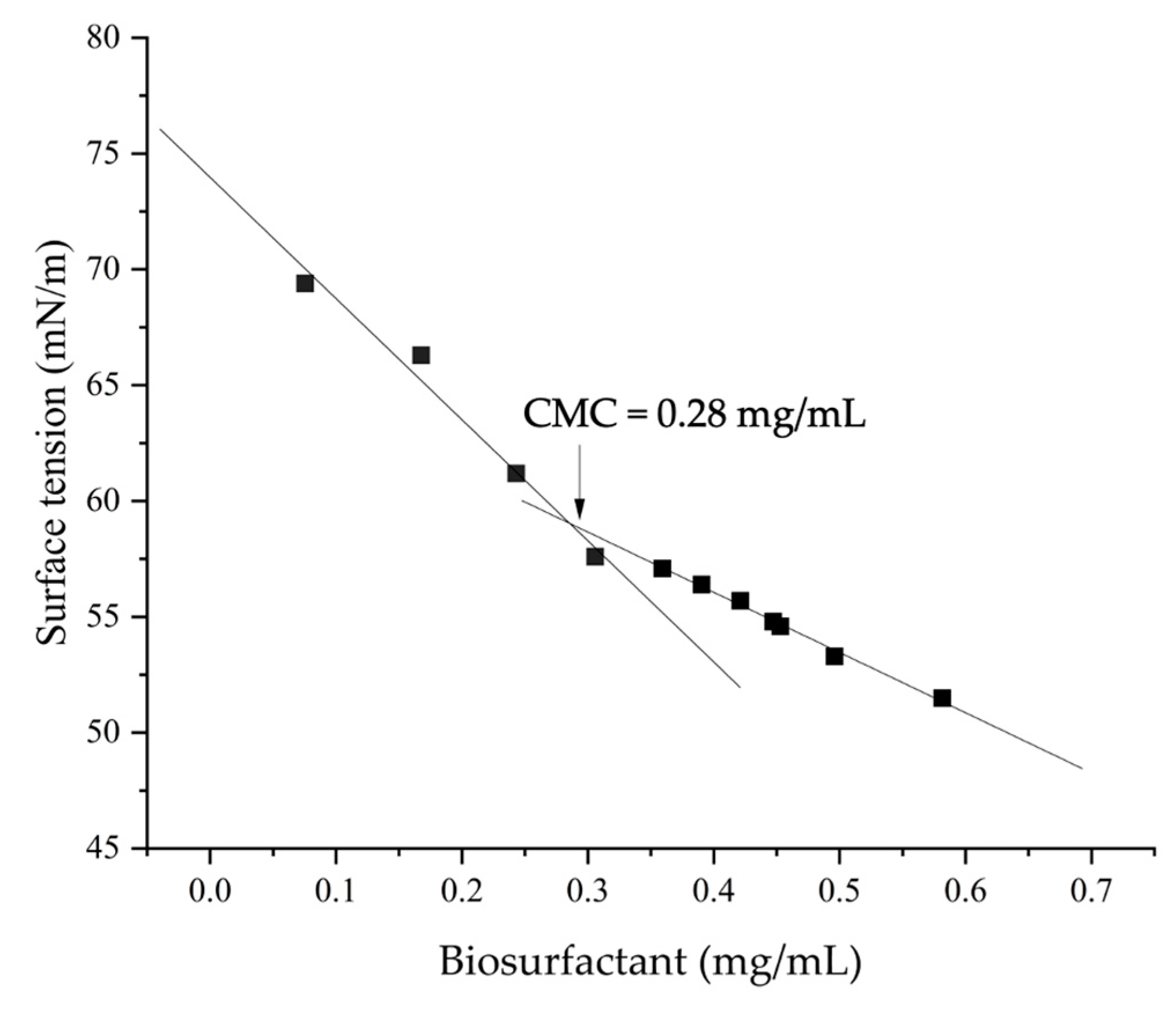
2.3. Determination of CMC

To determine the critical micellar concentration (CMC) of the biosurfactant, a stock solution of 0.9 mg/mL was prepared. This solution was successively diluted with ultrapure water and analyzed using the tensiometer to determine the surface tension. The CMC was obtained from the plot of biosurfactant concentration versus surface tension, where the inflection point of the curve represents the CMC [34].

2.4. Emulsification Index

The analysis of emulsifying capacity was performed by calculating the emulsification index. For this, a mixture of 6 mL of kerosene and 4 mL of a solution containing 10 mg of the biosurfactant was mixed using a vortex for 2 min and then allowed to stand at 28 °C for 24 h [35]. The assays were performed in triplicate, and the synthetic surfactant sodium dodecyl sulfate (SDS) at 10% was used as a positive control. After the waiting period, the height of the emulsion layer and the total height were measured with the aid of a metal universal caliper (150 M, MTX, Suzano, Brazil), and the values were used to calculate E24 (Equation (1)).
Emulsification   index   E 24 = Emulsion   layer   height Total   height     ×   100

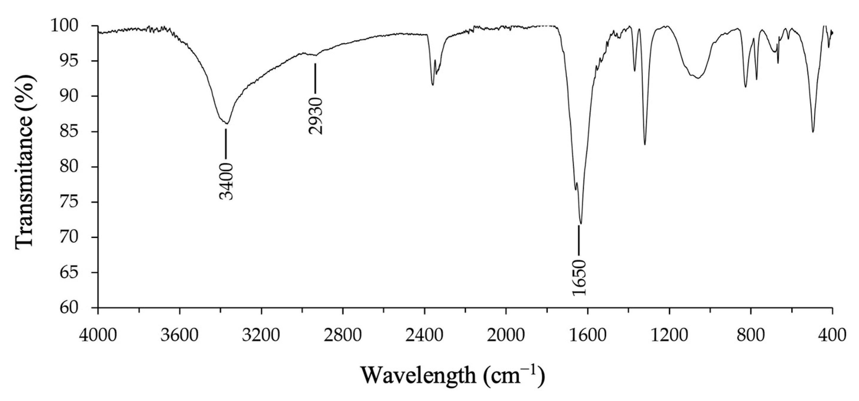
2.5. Fourier-Transform Infrared Spectroscopy (FTIR)

The extracted biosurfactant was analyzed using a spectrophotometer (IRAffinity-1s, Shimadzu, Kyoto, Japan) to identify the main functional groups present in the fungal metabolite. The sample was analyzed with the aid of the attenuated total reflection (ATR) accessory, and the spectrum in the infrared region was obtained in the wavelength range of 400 to 4000 cm−1, with a resolution of 1 cm−1 and an average of 70 scans.

2.6. Synthesis of Polymeric Nanoparticles

Two classical methods were used to obtain the PNPs: nanoprecipitation [7,36,37] and emulsion/diffusion [3,37,38]. The nanoparticle system was developed from two solutions: the organic phase and the aqueous phase. In both methods, analytical-grade reagents were used, which did not undergo additional purification. Poly-ε-caprolactone (PCL) was used as the wall material, Tween 80 was used as a non-ionic surfactant, and acetone was used for solubilization. At the end of the preparation of each formulation, the nanoparticle suspension was left overnight under constant agitation at room temperature until the complete removal of the organic solvents. The samples were coded as follows: NP—nanoparticles without biosurfactants; NPPB—nanoparticles containing the fungal biosurfactant, prepared using the nanoprecipitation method; and NPEB—nanoparticles prepared using the emulsion/diffusion method with the biosurfactant. Figure 1 summarizes the synthesis of nanoformulations I and II using nanoprecipitation or emulsion/diffusion.

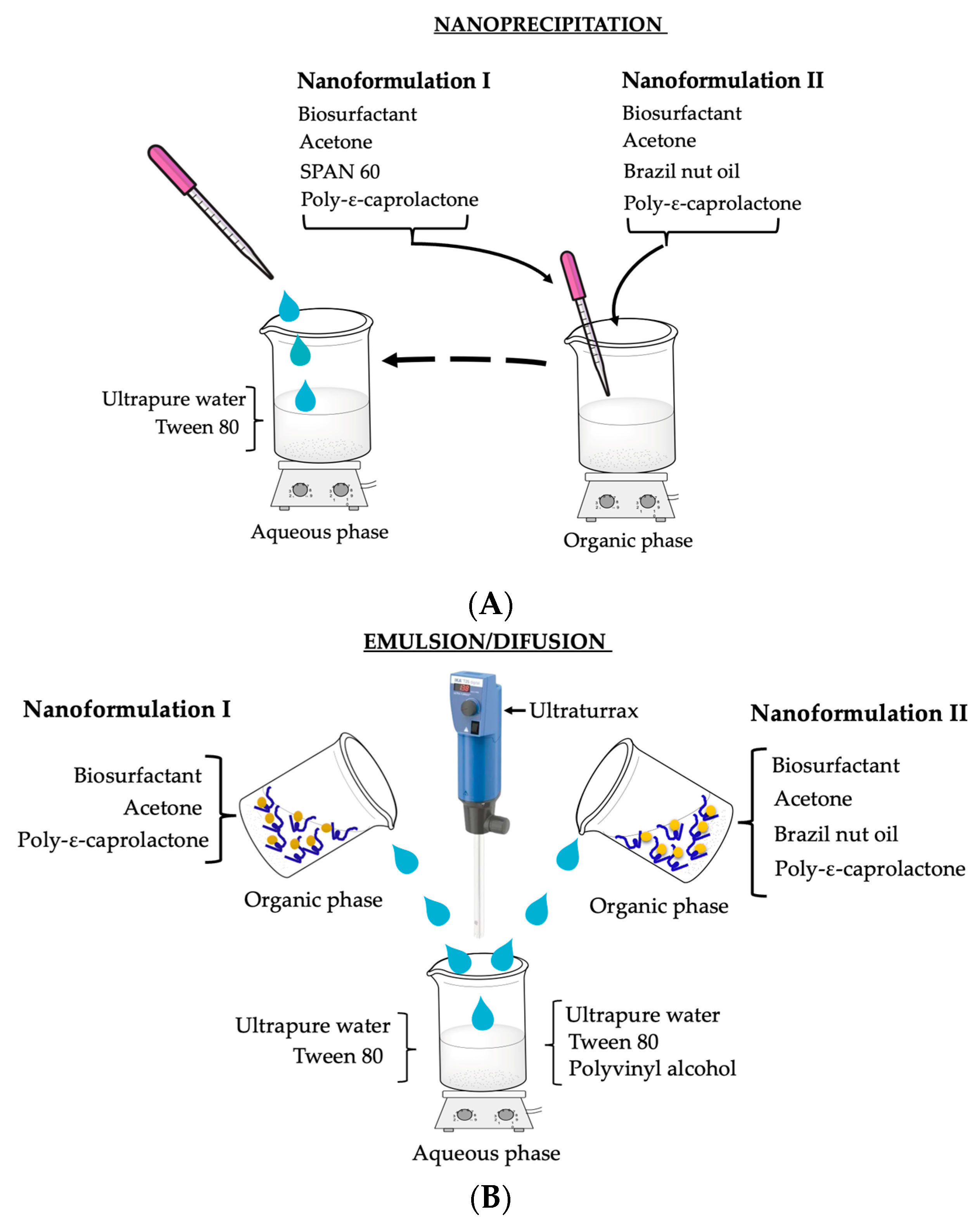
2.6.1. Nanoprecipitation Method—Nanoformulation I (NPPBI) and II (NPPBII)

For the preparation of the aqueous phase of nanoformulation I, 20 mg of Tween 80 was solubilized in 150 mL of ultrapure water. This phase was maintained under constant magnetic stirring at room temperature (28 ± 2 °C). The organic phase was prepared in three stages, using a magnetic stirrer (C-MAG HS 7, IKA, Campinas, Brazil) at 40 °C. Initially, 6 mg of the biosurfactant (BS) was solubilized in 700 µL of ultrapure water. Then, 10 mL of acetone was added, and the material was kept under stirring (solution I). In parallel, 60 mg of PCL was dissolved in 20 mL of acetone (solution II), and 40 mg of Span 60 was added to 10 mL of acetone (solution III). The preparation of the organic phase was completed by combining the three solutions. For nanoparticle formation, the organic phase was added to the aqueous phase dropwise with the aid of a Pasteur pipette.
For the preparation of nanoformulation II, the aqueous phase consisted of 20 mg of Tween 80 solubilized in 150 mL of ultrapure water. For the organic phase, 30 mg of PCL was added to 10 mL of acetone, and after solubilization, 25 mg of Brazil nut oil (BNO) was added (solution I). Solution II was prepared by dissolving 5 mg of the fungal biosurfactant in 10 mL of acetone. After solubilization, solution II was gently poured into solution I. Once the organic phase was completely solubilized, it was added to the aqueous phase dropwise with the aid of a Pasteur pipette.

2.6.2. Emulsion/Diffusion—Nanoformulation I (NPEBI) and II (NPEBII)

For the aqueous phase of nanoformulation I, 20 mg of Tween 80 was solubilized in 100 mL of ultrapure water. The mixture was kept under constant magnetic stirring until complete solubilization. For the preparation of the organic phase, all the solutions were kept under constant stirring at 40 °C until complete homogenization. Initially, 30 mg of PCL was added to 20 mL of acetone. After complete solubilization, 25 mg of BNO was added (solution I). Then, 5 mg of the BS was solubilized in 1 mL of soybean oil (solution II). After solubilization, solution II was gently added to solution I with the aid of a homogenizer (Ultra-Turrax, IKA, Campinas, Brazil), at 10,000 rpm for 30 s.
The preparation of the aqueous phase of nanoformulation II used two solutions. Initially, 20 mg of Tween 80 was solubilized in 50 mL of deionized water at 40 °C under constant stirring (solution I). Then, 100 mL of distilled water was added to 500 mg of polyvinyl alcohol (PVA) at 90 °C under slow stirring. After partial solubilization, the temperature was reduced to 40 °C, and the solution was placed in an ice bath so that the crystals could be macerated with the aid of a glass stick. Subsequently, the remaining material was stirred at 40 °C until complete solubilization (solution II). For the preparation of the organic phase, the solutions were kept under constant stirring at 40 °C until complete homogenization. The BS was solubilized in 10 mL of acetone (solution III), and 0.02 g of PCL was solubilized in 20 mL of acetone (solution IV). After the solubilization of solution IV, 25 mg of BNO was added to the phase with PCL, and solution III was poured into solution IV. The organic phase was then poured into the aqueous phase with the aid of a homogenizer (Ultra-Turrax, IKA, Campinas, Brazil), at 10,000 rpm for 30 s.

2.7. Characterization of Polymer Nanoparticles

2.7.1. Particle Size Measurement by Dynamic Light Scattering (DLS) and Zeta Potential (ζ)

The average particle diameter and polydispersity index (PDI) were measured using the dynamic light scattering (DLS) technique on a nanoparticle analyzer (SZ-100-Z, Horiba Scientific, Tokyo, Japan). The analysis was performed at 25 °C at an angle of 90° to the laser. In the same equipment, the zeta potential was measured via electrophoretic mobility using the Zeta-Meter system at a temperature of 25 ± 2 °C.
To measure the mean particle diameter and PDI of the samples, 1 mL of each sample was diluted in 3 mL of deionized water. For the analysis of nanoparticles containing the BS, 200 µL of the formulation was diluted in 5 mL of deionized water. For the analysis of nanoparticles without the BS, 200 µL of the formulation was diluted in 4 mL of deionized water. The final solution of each sample was filtered through a membrane with a pore size of 0.45 µm. Subsequently, the solution was inserted into a cuvette, and five readings were taken on the DLS equipment.
To characterize the surface charges of the nanoparticles using the ζ-potential, double capillary cuvettes were used. The samples were inserted individually using a 1 mL syringe and analyzed at a voltage of 34 mV, positioned at an angle of 90° with respect to the laser beam. The intensity-weighted mean value was measured, and the average was calculated from three distinct measurements.

2.7.2. Scanning Electron Microscopy (SEM)

The nanoformulation was prepared by placing a drop of the NPPBII suspension onto a watch glass and spreading it evenly. The suspension was then dried in an oven at 40 °C until fully dry, forming a thin film. This nanoparticle film was attached to a metal stub using double-sided carbon tape, coated with a 10 nm layer of platinum using a vacuum sputter coater (EM ACE600, Leica Microsystems, Wetzlar, Germany), and subsequently examined with a scanning electron microscope (JSM-IT500H, Jeol Ltd., Tokyo, Japan) in secondary electron mode.

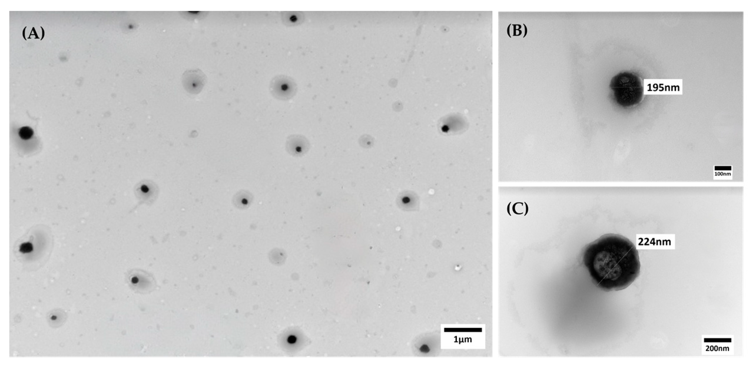
2.7.3. Transmission Electron Microscopy (TEM)

The sample was subjected to sonication for 5 min, and subsequently, 5 µL was deposited onto copper grids (300 mesh) coated with Formvar/Carbon. The grids were left to dry at room temperature for 2 h. The images were acquired using a transmission electron microscope (JEM 1400Flash, Jeol Ltd., Tokyo, Japan) operating at 120 kV.

2.8. Statistical Analysis

Statistical analysis was performed to compare the results of particle size, PDI, and ζ-potential of the various formulations. The data were presented as mean and standard deviation. All readings were performed in triplicate using independently prepared samples. The analyzed data were compared using a one-way ANOVA, followed, in cases of differences, by the Tukey test (p < 0.05), with the aid of SigmaPlot 15.0 (Systat Software, San Jose, CA, USA).

3. Results

3.1. Production and Characterization of the Fungal Biosurfactant

After 96 h of growth of the fungus Aspergillus welwitschiae CG2-16, the maximum reduction in the surface tension of the culture medium was observed, which was 36%, resulting in 36.0 mN/m. After the extraction and partial purification of the fungal biosurfactant, a yield of 0.87 mg/mL was obtained. The biosurfactant extracted from the culture medium showed an emulsification index of 24% and a CMC of 280 mg/L (Figure 2).
The FTIR spectrum of the fungal biosurfactant (Figure 3) revealed important structural characteristics of this metabolite. A low-intensity band is observed between 2960 and 2850 cm−1, which corresponds to the aliphatic C-H bond; a broad and intense band is observed at 3400 cm−1 that indicates the asymmetric elongation of N-H bonds in secondary amides; and a characteristic peak is observed at 1650 cm−1 for the amide C=O bond [39].

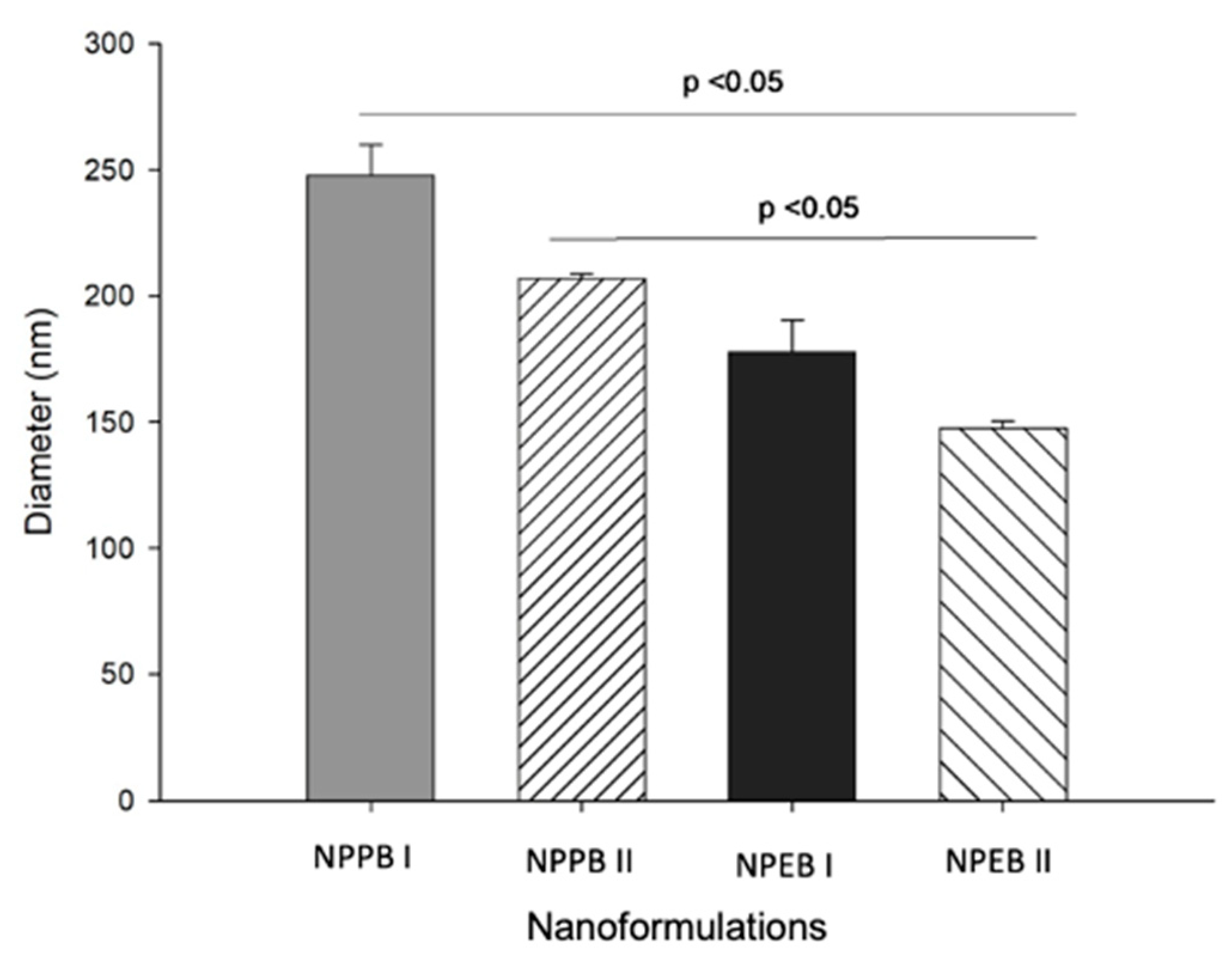
3.2. Production and Characterization of the Nanoformulations

The average particle diameter, PDI and zeta potential values of the nanoformulations produced via nanoprecipitation and emulsion/diffusion methods are shown in Table 1.
The synthesis process of the nanoformulations without the biosurfactant, composed only of Tween 80 (NPPII, NPEI, and NPEII), prevented the formation of nanoparticles, as the analysis via DLS indicated that the average size of the particles present was greater than the measurement limit of the equipment (10 µm). NPPI had a mean diameter of 237.6 ± 9.3 and was the only nanoformulation without the biosurfactant that resulted in the formation of nanoparticles. However, in this formulation, the surfactant Span 60 was added, which acted to stabilize the nanosystem.
The mean diameters of the nanoparticles with biosurfactants (NPPBI and NPPBII) were compared with the value obtained for NPPI (the formulation without the BS). It is observed that the nanoformulations prepared with the same methodology did not show a significant difference between them (p > 0.05) regarding the average diameter of the particles. In addition, it can be noted that the method selected for the synthesis influences the size of the nanoparticles (Figure 4). This idea is supported by the significant difference between the average particle size in the NPEBII nanoformulation and the particle sizes in the NPPBI and NPPBII nanoformulations. The NPEBII nanoformulation resulted in the smallest nanoparticle size among the nanoformulations.
Figure 5 shows the PDI of the nanoformulations. The NPPBI and NPPBII samples presented a PDI of 0.11 ± 0.08 and 0.14 ± 0.04, respectively, indicating a homogeneous distribution of the particles. On the other hand, the formulations prepared using the emulsion/diffusion method, with and without the biosurfactant, showed a higher PDI, with NPEBI and NPEBII ranging from 0.27 ± 0.06 to 0.32 ± 0.09.
For the zeta potential, it is observed that the nanoformulations prepared with the same methodology presented significant differences among themselves (Figure 6), ranging from −29.10 to −45.00 mV for the NP nanoformulations and from −21.90 to −43.39 mV for those prepared using ED. In addition, significant differences were observed between NPPBI and NPEBII and between NPPBII and NPEBI (p < 0.05). The NPPBI and NPPBII formulations presented zeta potentials of −45.00 ± 1.10 mV and −29.10 ± 8.67 mV, respectively. In comparison, NPPI showed a zeta potential of −48.50 ± 3.42 mV.
The nanoparticle prepared using the nanoprecipitation method containing the fungal biosurfactant (NPPBII) was investigated by SEM and TEM. The micrographs are shown in Figure 7 and Figure 8, respectively. The SEM image reveals a spherical shape, which is a known characteristic of PCL nanoparticles. The diameter size of the spheres ranged between 179 and 181 nm.
TEM analysis confirmed the size and morphology of the nanoparticles produced using the nanoprecipitation method, incorporating the fungal biosurfactant (NPPBII). The representative TEM image (Figure 8) reveals distinct spherical nanoparticles, uniformly distributed without signs of aggregation or adhesion—characteristic features of PCL nanoparticles. The particle diameters ranged from 156 to 228 nm, consistent with the SEM analysis (Figure 7).

4. Discussion

The surface tension reduction and emulsification index are screening tests frequently used to determine the presence of surfactants in culture media [40]. The 36% reduction in surface tension demonstrates the potential of the biosurfactant produced by the fungus A. welwitschiae CG2-16. This result not only confirms the effectiveness of the fungus in producing biosurfactants but also indicates its potential for applications in processes that require the reduction of ST, such as in the cleaning, cosmetics, and pharmaceutical industries [34]. In addition, the production of BSs in only 96 h of cultivation represents an important result, especially for a fungal metabolite, which generally produces these compounds in longer cultivation times. The endophytic fungus Aspergillus niger, isolated from the Piper hispidum plant, for example, which also led to a 36% reduction in the surface tension of the culture medium, obtained this result only after 8 days [26].
The biosurfactant produced by A. welwitschiae CG2-16 can be considered a promising surfactant when considering the CMC value (280 mg/L)—a crucial parameter that indicates the efficiency of the biosurfactant in practical applications, as it defines the minimum concentration required for the formation of micelles. Other biosurfactants have CMC values in the same range, and some are even lower than synthetic surfactants [41,42], which is one of the advantages of using biosurfactants. In addition, the BS obtained from A. welwitschiae CG2-16 presented an emulsification index of 24%, which demonstrates its ability to stabilize emulsions. However, considering the low E24 value, it can be suggested that its main application is as a surfactant and not as a bioemulsifier [43,44].
The results obtained from the FTIR analysis allow us to suggest that the biosurfactant extracted from A. welwitschiae CG2-16 belongs to the class of lipopeptides [39,45]. This class of biosurfactants is commonly found in fungi [18] and consists of a fatty acid chain linked to a peptide moiety. Lipopeptides are among the most popular biosurfactants, with high structural diversity and various functional activities [42]. Due to the intensity of the bands observed, the BS obtained from A. welwitschiae CG2-16 is possibly composed of a major peptide chain and a small lipid portion. The low intensity of the C-H bond bands indicates the presence of a small hydrophobic portion, while the characteristic bands of peptide bonds have a high intensity. According to Mnif et al. [42], the variation in the composition of the fatty acid component, as well as the type, quantity, and arrangement of amino acids within the peptide chain, contributes significantly to the structural diversity of lipopeptides. Moreover, each lipopeptide family, defined by a consistent peptide core, can be further subdivided into different isoforms based on variations in the length of the fatty acid chain.
The fungal biosurfactant proved to be essential for the formation of polymeric nanoparticles from the nanoformulations tested in the present study, which were prepared using two different methods. The emulsion/diffusion method produced nanoparticles with a smaller diameter (147.6 and 177.8 nm) when compared to the nanoprecipitation method (206.9 and 247.8 nm). This size difference between the nanoparticles prepared using different methodologies has also been observed in other studies. Abriata et al. [46] developed PNPs with PCL via the emulsion/diffusion method for loading paclitaxel and obtained a mean particle diameter of 140 nm and a PDI of 0.1. On the other hand, PCL nanoparticles containing the essential oil of palmarosa (Cymbopogon martinii) and the monoterpene geraniol, prepared using the nanoprecipitation method, presented an average size of 282.1 and 289.3 ± 2 nm, respectively, with a PDI below 0.14 [7]. These results are in accordance with those found in the present study.
The results for the PDI are important for determining the degree of homogeneity of the particle diameters present in the suspension: the closer the value is to zero, the more homogeneous the system. Values below 0.1 represent a monodisperse system [47]. The type and concentration of the oil used in the formulation may interfere with the PDI [48]. In the present work, two types of oils were used: soybean oil for the solubilization of the biosurfactant in NPEII and Brazil nut oil for the greater stability of the organic phase in NPPII, NPEI, and NPEII. NPPI was the only nanoformulation in which no oil was used, and it presented the lowest PDI value, corroborating the fact that the PDI can be affected by the use of oils.
The PDI values of the nanoformulations obtained show that the nanoprecipitation method, especially when combined with the biosurfactant, produces nanoparticles with a more uniform size distribution, which is corroborated by the SEM findings. This conclusion is also supported by data from the literature, which demonstrate that nanoprecipitation is an effective method for producing homogeneous nanoparticles [49].
The zeta potential is related to the surface charge of the nanoparticles—an important parameter for evaluating the stability of the colloidal suspension and the interaction between the nanoparticles. Dispersions with zeta potential values greater than 30 mV (in modulus) are considered highly stable [50]. The nanoformulations obtained in our study have high negative ζ-potential values, indicating that they are stable systems. The NPPBI formulation stood out for having a zeta potential of −45.00 ± 1.13 mV and is thus a promising system for future applications. Negative values of ζ-potential indicate the presence of negative charges on the nanoparticle surface and can lead to an increase in solubility, as observed by Bouallegue et al. [51]. A less negative value for the ζ-potential was observed for the NPPBII formulation, which suggests that the introduction of the biosurfactant can alter the surface charge of the nanoparticles, potentially influencing their interaction with the surrounding environment. Studies have shown that biosurfactants can alter the surface of nanoparticles, modulating their zeta potential and, consequently, their stability and biological interaction [27,52,53].
With the exception of the NPPI formulation, it was not possible to define the particle size, PDI, and zeta potential of the other formulations without the biosurfactant using the DLS technique. The particles present in these formulations must have a diameter greater than 10 µm, which is the maximum limit that the equipment can evaluate. In contrast, the NPPBI formulation presented a size of 247.8 nm, a PDI of 0.11, and a zeta potential of −45.00, thus demonstrating the importance of the biosurfactant in stabilizing the nanoformulation.
The SEM image of NPPBII revealed that the PCL nanoparticles containing the fungal biosurfactant were homogeneously distributed and had a spherical shape, with diameters ranging around 180 nm. Comparing these results with those obtained from DLS, an inevitable difference in size is observed, which can be explained by the different approaches and conditions of each technique. SEM provides a direct analysis of particle size, without the influence of the solvent, observing the actual particle size. On the other hand, DLS measures the hydrodynamic size of the particles, considering the solvent layer or the interaction of the nanoparticles with the molecules of the dispersing medium, which increases the apparent particle size [54], as observed in our study.
TEM (transmission electron microscopy) generates images by transmitting an electron beam through a sample. This technique is especially valuable for characterizing polymeric nanoparticles, as it offers sub-nanometer resolution, providing detailed insights into nanoparticle morphology [38]. TEM micrographs complement the findings of SEM and DLS analyses, revealing spherical nanoparticles that are uniformly distributed, free from aggregation, and approximately 200 nm in diameter.
The biosurfactant produced by the fungus A. welwitschiae CG2-16 assisted in the stabilization and uniformity of the synthesized nanoparticles, probably by decreasing the surface tension, thereby facilitating the formation of smaller and more uniform spherical nanoparticles [55,56,57], as corroborated by SEM and TEM images. Surfactants have been widely used for this purpose and are commonly employed as stabilizers to prevent the agglomeration of colloidal suspensions and allow for the acquisition of well-structured nanosystems. In addition, these surface-active molecules have the advantage of allowing the reduction of ST and increasing the affinity of lipid structures [58]. Some of these compounds have even demonstrated the ability to reduce the diameter of nanoparticles [59].
According to Vecino et al. [53], biosurfactants reduce the formation of aggregates and facilitate a uniform morphology of the nanoparticles. SEM and TEM images of NPPBII show a uniform spherical shape of the PCL nanoparticle formed with the use of the A. welwitschiae CG2-16 biosurfactant, without agglomeration. On the other hand, the FTIR analysis suggests that the A. welwitschiae CG2-16 BS is a lipopeptide. Therefore, it can be inferred that the lipid structure of the BS interacts with the apolar moiety of the PCL, stabilizes the surface by reducing charges, and leads to a uniform spherical nanoparticle.
The use of biosurfactants as stabilizers has already been evaluated in the synthesis of metal nanoparticles as a way of developing more sustainable nanosystems with lower risks to health. The use of a rhamnolipid extracted from Pseudomonas aeruginosa for coating silver nanoparticles applied in enhanced oil recovery has been reported. The silver nanoparticles based on the biosurfactant were non-toxic and obtained greater stability than chemically synthesized nanoparticles [52]. In another study, in addition to the greater stability, silver and iron nanoparticles coated with biosurfactants showed reduced cell adhesion by modifying the hydrophobicity of the surface and, because of this, demonstrated excellent antibiofilm activity [60].
The presence of the fungal biosurfactant was crucial for the composition of the NPPBII and NPEBII nanoformulations. These did not have any other surfactant in their composition besides the nonionic surfactant Tween 80, which was considered inefficient for the stability of these nanoformulations without the presence of the biosurfactant. NPPI showed the formation of stable nanoparticles because it was composed of Span 60 and Tween 80. Thus, it is suggested that the interaction between Tween 80 and the biosurfactant was able to keep the other nanoformulations stable. The results obtained in this research revealed a promising perspective on the use of fungal biosurfactants in combination with poly-ε-caprolactone (PCL) for the synthesis of polymeric nanoparticles.
PCL is recognized for its easy accessibility, biodegradability, and high biocompatibility and has become a material of interest in several areas of study [61]. The efficacy of PCL nanoparticles was demonstrated for loading Nigella sativa oil and applying it in the treatment of leishmaniasis, which evidences the ability of nanoparticles to provide a continuous and enhanced release of oil molecules and result in a significant inhibition of the growth of Leishmania infantum [62]. In addition, according to Morsy [63], stabilizers are key in the preparation of polymeric nanoparticles, which shows the importance of the choice and proper concentration of components in the formulation.
Due to the physicochemical characteristics of polymer nanoparticles stabilized with biosurfactants, they could be used for various applications. In the area of drug delivery, nanoparticles with reduced sizes and improved colloidal stability are ideal for the controlled release of drugs since they allow for constant and safe administration and improve therapeutic efficacy. This system can be particularly useful in the treatment of chronic and infectious diseases, where continuous and controlled administration is crucial [64,65]. In nanocosmetics, the stabilizing properties of biosurfactants can be used in the formulation of products such as photoprotective and anti-aging creams and lotions, which require prolonged stability and the uniform distribution of active ingredients.
The biodegradability of biosurfactants also meets the growing demand for products that generate fewer environmental impacts [15,48]. Nanopesticides have been studied as an option to enable the use of biopesticides and thus reduce the environmental impacts caused by synthetic agrochemicals [66]. Essential oils are considered an advantageous alternative to pesticides, and the nanoencapsulation of these could increase availability and enhance their efficiency [67,68,69]. In this sense, biosurfactants can be used to stabilize nanoparticles containing essential oils and increase the efficiency of the use of these natural resources [70].
The ability of biosurfactants to reduce surface tension can also be explored in the remediation of contaminated soils and waters, facilitating the degradation and removal of pollutants [71,72]. Stabilized nanoparticles can be employed for the targeted delivery of remediation agents to improve the efficiency of environmental cleanup processes [73]. Therefore, polymeric nanoparticles stabilized with biosurfactants present themselves as an interesting solution for diverse applications in different areas, such as pharmaceuticals (controlled drug release), cosmetics (active compound delivery), agriculture (biopesticide dispersion), environmental (the remediation of contaminated sites), and food (the encapsulation of volatile compounds), among many others.

5. Conclusions

The biosurfactant produced by the Amazonian endophytic fungus A. welwitschiae CG2-16 can be used in the synthesis of polymer nanoparticles with PCL, ensuring the stability of the nanoformulations. It was observed that the methodology used in the preparation of the nanoformulations influences the size of the PNPs, with the smallest particle diameters obtained using the emulsion/diffusion method, while the nanoformulation produced using the nanoprecipitation method presented a higher zeta potential. The application of biosurfactants not only improves the properties of the nanoparticles but also contributes to the expansion of the use of this class of biomolecules.
The vast application potential of these formulations opens the way for future research projects and technological development to further explore the benefits of fungal biosurfactants as an alternative material that causes fewer environmental impacts than synthetic surfactants. However, further studies are needed to fully characterize these polymeric nanoparticles and explore their potential applications. Limitations include the need to optimize the synthesis processes and further investigate the long-term stability and environmental impact of these PNPs. Nonetheless, these biosurfactants offer a sustainable route for developing advanced materials with reduced ecological footprints.

Author Contributions

Conceptualization, P.M.A. and A.R.S.; methodology, A.R.S., M.T.M.P. and S.G.A.; validation, A.R.S., J.C.d.A. and S.G.A.; formal analysis, A.R.S. and S.G.A.; investigation, A.R.S., S.G.A., M.T.M.P., C.D.L. and J.C.d.A.; resources, P.M.A., S.D.J. and L.M.; data curation, A.R.S. and C.D.L.; writing—original draft preparation, A.R.S., S.G.A. and C.D.L.; writing—review and editing, P.M.A. and J.C.d.A.; supervision, P.M.A. and L.M.; project administration, P.M.A.; funding acquisition, P.M.A., S.D.J. and L.M. All authors have read and agreed to the published version of the manuscript.

Funding

This research was funded by Fundação de Amparo à Pesquisa do Estado do Amazonas (FAPEAM) (grant numbers 01.02.016301.00568/2021-05 and 01.02.016301.00101/2024-08) and by Coordenação de Aperfeiçoamento de Pessoal de Nível Superior (CAPES) (grant nunber 88881.510151/2020-01—PDPG Amazônia Legal and finance code 001).

Data Availability Statement

The original contributions presented in the study are included in the article, further inquiries can be directed to the corresponding author.

Acknowledgments

The authors gratefully acknowledge Universidade do Estado do Amazonas—UEA, Instituto Federal do Amazonas—IFAM, Sistema Nacional de Laboratórios em Nanotecnologias—SisNANO, Centro Multiusuário para Análise de Fenômenos Biomédicos—CMABIO, FAPEAM and CAPES for supporting this research.

Conflicts of Interest

The authors declare no conflict of interest.

References

  1. Altammar, K.A.A. Review on nanoparticles: Characteristics, synthesis, applications, and challenges. Front. Microbiol. 2023, 14, 1155622. [Google Scholar] [CrossRef] [PubMed]
  2. Zielinska, A.; Carreiró, F.; Oliveira, A.M.; Neves, A.; Pires, B.; Nagasamy Venkatesh, D.; Durazzo, A.; Lucarini, M.; Eder, P.; Silva, A.M.; et al. Polymeric nanoparticles: Production, characterization, toxicology and ecotoxicology. Molecules 2020, 25, 3731. [Google Scholar] [CrossRef] [PubMed]
  3. Azevedo, S.G.; Rocha, A.L.F.; de Aguiar Nunes, R.Z.; da Costa Pinto, C.; Ţălu, Ş.; da Fonseca Filho, H.D.; de Araújo Bezerra, J.; Lima, A.R.; Guimarães, F.E.G.; Campelo, P.H.; et al. Pulsatile controlled release and stability evaluation of polymeric particles containing Piper nigrum essential oil and preservatives. Materials 2022, 15, 5415. [Google Scholar] [CrossRef]
  4. Begines, B.; Ortiz, T.; Pérez-Aranda, M.; Martínez, G.; Merinero, M.; Argüelles-Arias, F.; Alcudia, A. Polymeric nanoparticles for drug delivery: Recent developments and future prospects. Nanomaterials 2020, 10, 1403. [Google Scholar] [CrossRef]
  5. Krishnaswamy, K.; Orsat, V. Sustainable delivery systems through green nanotechnology. In Nano- and Microscale Drug Delivery Systems: Design and Fabrication; Grumezescu, A.M., Ed.; Elsevier: Amsterdam, The Netherlands, 2017; pp. 17–32. [Google Scholar] [CrossRef]
  6. Parupudi, A.; Mulagapati, S.H.R.; Subramony, J.A. Nanoparticle technologies: Recent state of the art and emerging opportunities. In Nanoparticle Therapeutics: Production Technologies, Types of Nanoparticles, and Regulatory Aspects; Kesharwani, P., Singh, K.K., Eds.; Elsevier: Amsterdam, The Netherlands, 2022; pp. 3–46. [Google Scholar] [CrossRef]
  7. Jummes, B.; Sganzerla, W.G.; da Rosa, C.G.; Noronha, C.M.; Nunes, M.R.; Bertoldi, F.C.; Barreto, P.L.M. Antioxidant and antimicrobial poly-ε-caprolactone nanoparticles loaded with Cymbopogon martinii essential oil. Biocatal. Agric. Biotechnol. 2020, 23, 101499. [Google Scholar] [CrossRef]
  8. MacCraith, E.; O’Brien, F.J.; Davis, N.F. Biodegradable materials for surgical management of stress urinary incontinence: A narrative review. Eur. J. Obstet. Gynecol. Reprod. Biol. 2021, 259, 153–160. [Google Scholar] [CrossRef]
  9. Mulinti, P.; Brooks, J.E.; Lervick, B.; Pullan, J.E.; Brooks, A.E. Strategies to improve the hemocompatibility of biodegradable biomaterials. In Hemocompatibility of Biomaterials for Clinical Applications: Blood-Biomaterials Interactions; Siedlecki, C.A., Ed.; Elsevier: Amsterdam, The Netherlands, 2017; pp. 253–278. [Google Scholar] [CrossRef]
  10. Pulingam, T.; Foroozandeh, P.; Chuah, J.A.; Sudesh, K. Exploring Various techniques for the chemical and biological synthesis of polymeric nanoparticles. Nanomaterials 2022, 12, 576. [Google Scholar] [CrossRef]
  11. Cortés, H.; Hernández-Parra, H.; Bernal-Chávez, S.A.; Del Prado-Audelo, M.L.; Caballero-Florán, I.H.; Borbolla-Jiménez, F.V.; González-Torres, M.; Magaña, J.J.; Leyva-Gómez, G. Non-ionic surfactants for stabilization of polymeric nanoparticles for biomedical uses. Materials 2021, 14, 3197. [Google Scholar] [CrossRef] [PubMed]
  12. Badmus, S.O.; Amusa, H.K.; Oyehan, T.A.; Saleh, T.A. Environmental risks and toxicity of surfactants: Overview of analysis, assessment, and remediation techniques. Environ. Sci. Pollut. R. 2021, 28, 62085–62104. [Google Scholar] [CrossRef]
  13. Johnson, P.; Trybala, A.; Starov, V.; Pinfield, V.J. Effect of synthetic surfactants on the environment and the potential for substitution by biosurfactants. Adv. Colloid Interface Sci. 2021, 288, 102340. [Google Scholar] [CrossRef]
  14. Drakontis, C.E.; Amin, S. Biosurfactants: Formulations, properties, and applications. Curr. Opin. Colloid Interface Sci. 2020, 48, 77–90. [Google Scholar] [CrossRef]
  15. Santos, A.C.; Morais, F.; Simões, A.; Pereira, I.; Sequeira, J.A.D.; Pereira-Silva, M.; Veiga, F.; Ribeiro, A. Nanotechnology for the development of new cosmetic formulations. Expert Opin. Drug Deliv. 2019, 16, 313–330. [Google Scholar] [CrossRef] [PubMed]
  16. Gautam, A.K.; Verma, R.K.; Avasthi, S.; Sushma, B.Y.; Devadatha, B.; Niranjan, M.; Suwannarach, N. Current insight into traditional and modern methods in fungal diversity estimates. J. Fungi 2022, 8, 226. [Google Scholar] [CrossRef]
  17. Gurgel, R.S.; de Melo Pereira, D.Í.; Garcia, A.V.F.; Fernandes de Souza, A.T.; Mendes da Silva, T.; de Andrade, C.P.; Lima da Silva, W.; Nunez, C.V.; Fantin, C.; de Lima Procópio, R.E.; et al. Antimicrobial and antioxidant activities of endophytic fungi associated with Arrabidaea chica (Bignoniaceae). J. Fungi 2023, 9, 864. [Google Scholar] [CrossRef]
  18. Sanches, M.A.; Luzeiro, I.G.; Alves Cortez, A.C.; Simplício de Souza, É.; Albuquerque, P.M.; Chopra, H.K.; Braga de Souza, J.V. Production of biosurfactants by Ascomycetes. Int. J. Microbiol. 2021, 2021, 6669263. [Google Scholar] [CrossRef]
  19. Silva, A.C.S.; Santos, P.N.; Silva, T.A.L.; Andrade, R.F.S.; Campos-Takaki, G.M. Biosurfactant production by fungi as a sustainable alternative. Arq. Inst. Biol. 2018, 85, e0502017. [Google Scholar] [CrossRef]
  20. Abdel-Mawgoud, A.M.; Stephanopoulos, G. Simple glycolipids of microbes: Chemistry, biological activity and metabolic engineering. Synth. Syst. Biotechnol. 2018, 3, 3–19. [Google Scholar] [CrossRef] [PubMed]
  21. Garay, L.A.; Sitepu, I.R.; Cajka, T.; Xu, J.; Teh, H.E.; German, J.B.; Pan, Z.; Dungan, S.R.; Block, D.E.; Boundy-Mills, K.L. Extracellular fungal polyol lipids: A new class of potential high value lipids. Biotechnol. Adv. 2018, 36, 397–414. [Google Scholar] [CrossRef]
  22. Bae, I.; Lee, E.S.; Yoo, J.W.; Lee, S.H.; Ko, J.Y.; Kim, Y.J.; Lee, T.R.; Kim, D.; Lee, C.S. Mannosylerythritol lipids inhibit melanogenesis via suppressing ERK-CREB-MiTF-tyrosinase signalling in normal human melanocytes and a three-dimensional human skin equivalent. Exp. Dermatol. 2019, 28, 738–741. [Google Scholar] [CrossRef]
  23. Maniglia, B.C.; Laroque, D.A.; Andrade, L.M.; Carciofi, B.A.M.; Tenório, J.A.S.; Andrade, C.J. Production of active cassava starch films; effect of adding a biosurfactant or synthetic surfactant. React. Funct. Polym. 2019, 144, 104368. [Google Scholar] [CrossRef]
  24. Shah, V.; Daverey, A. Effects of sophorolipids augmentation on the plant growth and phytoremediation of heavy metal contaminated soil. J. Clean. Prod. 2021, 280, 124406. [Google Scholar] [CrossRef]
  25. Chuo, S.C.; Ahmad, A.; Mohd-Setapar, S.H.; Mohamed, S.F.; Rafatullah, M. Utilization of green sophoro-lipids biosurfactant in reverse micelle extraction of antibiotics: Kinetic and mass transfer studies. J. Mol. Liq. 2019, 276, 225–232. [Google Scholar] [CrossRef]
  26. Silva, M.E.T.; Duvoisin, S., Jr.; Oliveira, R.L.; Banhos, E.F.; Souza, A.Q.L.; Albuquerque, P.M. Biosurfactant production of Piper hispidum endophytic fungi. J. Appl. Microbiol. 2021, 130, 561–569. [Google Scholar] [CrossRef]
  27. Nitschke, M.; Marangon, C.A. Microbial surfactants in nanotechnology: Recent trends and applications. Crit. Rev. Biotechnol. 2022, 42, 294–310. [Google Scholar] [CrossRef]
  28. De Melo Pereira, D.Í.; Gurgel, R.S.; de Souza, A.T.F.; Matias, R.R.; de Souza Falcão, L.; Chaves, F.C.M.; da Silva, G.F.; Martínez, J.G.; de Lima Procópio, R.E.; Fantin, C.; et al. Isolation and identification of pigment-producing endophytic fungi from the Amazonian Species Fridericia chica. J. Fungi 2024, 10, 77. [Google Scholar] [CrossRef]
  29. Castellani, A. The viability of some pathogenic fungi in sterile distilled water. J. Trop. Med. Hyg. 1939, 42, 225–226. [Google Scholar]
  30. Wen, G.; Cao, R.; Wan, Q.; Tan, L.; Xu, X.; Wang, J.; Huang, T. Development of fungal spore staining methods for flow cytometric quantification and their application in chlorine-based disinfection. Chemosphere 2020, 243, 125453. [Google Scholar] [CrossRef]
  31. Soares, A.R. Produção de Biossurfactantes Fúngicos e Aplicação No Desenvolvimento de Nanopartículas Poliméricas. Master Thesis, Amazonas State University, Manaus, Brazil, 24 June 2024. [Google Scholar]
  32. Monteiro, A.S.; Bonfim, M.R.Q.; Domingues, V.S.; Corrêa, A.; Siqueira, E.P.; Zani, C.L.; Santos, V.L. Identification and characterization of bioemulsifier-producing yeasts isolated from effluents of a dairy industry. Bioresour. Technol. 2010, 101, 5186–5193. [Google Scholar] [CrossRef]
  33. Pereira, D.D.F.; Duvoisin Junior, S.; Albuquerque, P.M. Study of biosurfactants production by Amazon fungi. J. Eng. Exact Sci. 2017, 3, 688–695. [Google Scholar]
  34. Makkar, R.S.; Cameotra, S.S. Biosurfactant production by a thermophilic Bacillus subtilis strain. J. Ind. Microbiol. Biotechnol. 1997, 18, 37–42. [Google Scholar] [CrossRef]
  35. Pornsunthorntawee, O.; Wongpanit, P.; Chavadej, S.; Abe, M.; Rujiravanit, R. Structural and physicochemical characterization of crude biosurfactant produced by Pseudomonas aeruginosa SP4 isolated from petroleum contaminated soil. Bioresource Technol. 2008, 99, 1589–1595. [Google Scholar] [CrossRef]
  36. Noronha, C.M.; Granada, A.F.; de Carvalho, S.M.; Lino, R.C.; Matheus, M.V.; Barreto, P.L.M. Optimization of α-tocopherol loaded nanocapsules by the nanoprecipitation method. Ind. Crops Prod. 2013, 50, 896–903. [Google Scholar] [CrossRef]
  37. Costa, Í.C.; Azevedo, S.G.; Sanches, E.A.; da Fonseca Filho, H.D. Characterization of polymeric nanoparticles filled with Piper nigrum essential oil by atomic force microscopy. Matéria 2021, 26, e12981. [Google Scholar] [CrossRef]
  38. Crucho, C.I.C.; Barros, M.T. Polymeric nanoparticles: A study on the preparation variables and characterization methods. Mater. Sci. Eng. C. 2017, 80, 771–784. [Google Scholar] [CrossRef]
  39. Chittepu, O.R. Isolation and characterization of biosurfactant producing bacteria from groundnut oil cake dumping site for the control of foodborne pathogens. Grain Oil Sci. Technol. 2019, 2, 15–20. [Google Scholar] [CrossRef]
  40. Nayarisseri, A.; Singh, P.; Singh, S.K. Screening, isolation and characterization of biosurfactant producing Bacillus subtilis strain ANSKLAB03. Bioinformation 2018, 14, 304–314. [Google Scholar] [CrossRef]
  41. Jahan, R.; Bodratti, A.M.; Tsianou, M.; Alexandridis, P. Biosurfactants, natural alternatives to synthetic surfactants: Physicochemical properties and applications. Adv. Colloid Interface Sci. 2020, 275, 102061. [Google Scholar] [CrossRef]
  42. Mnif, I.; Segovia, R.; Bouallegue, A.; Ghribi, D.; Rabanal, F. Identifcation of diferent lipopeptides isoforms produced by Bacillus mojavensis BI2 and evaluation of their surface activities for potential environmental application. J. Polym. Environ. 2023, 31, 2668–2685. [Google Scholar] [CrossRef]
  43. Luft, L.; Confortin, T.C.; Todero, I.; Zabot, G.L.; Mazutti, M.A. An overview of fungal biopolymers: Bioemulsifiers and biosurfactants compounds production. Crit. Rev. Biotechnol. 2020, 40, 1059–1080. [Google Scholar] [CrossRef]
  44. Uzoigwe, C.; Burgess, J.G.; Ennis, C.J.; Rahman, P.K.S.M. Bioemulsifiers are not biosurfactants and require different screening approaches. Front. Microbiol. 2015, 6, 245. [Google Scholar] [CrossRef]
  45. Al-Sahlany, S.T.G.; Al-Kaabi, W.J.; Al-Manhel, A.J.A.; Niamah, A.K.; Altemimi, A.B.; Al-Wafi, H.; Cacciola, F. Effects of β-glucan extracted from Saccharomyces cerevisiae on the quality of bio-yoghurts: In vitro and in vivo evaluation. J. Food Meas. Charact. 2022, 16, 3607–3617. [Google Scholar] [CrossRef]
  46. Abriata, J.P.; Turatti, R.C.; Luiz, M.T.; Raspantini, G.L.; Tofani, L.B.; do Amaral, R.L.F.; Swiech, K.; Marcato, P.D.; Marchetti, J.M. Development, characterization and biological in vitro assays of paclitaxel-loaded PCL polymeric nanoparticles. Mater. Sci. Eng. C 2019, 96, 347–355. [Google Scholar] [CrossRef]
  47. Filippov, S.K.; Khusnutdinov, R.; Murmiliuk, A.; Inam, W.; Zakharova, L.Y.; Zhang, H.; Khutoryanskiy, V.V. Dynamic light scattering and transmission electron microscopy in drug delivery: A roadmap for correct characterization of nanoparticles and interpretation of results. Mater Horiz 2023, 10, 5354–5370. [Google Scholar] [CrossRef]
  48. Khayata, N.; Abdelwahed, W.; Chehna, M.F.; Charcosset, C.; Fessi, H. Preparation of vitamin e loaded nanocapsules by the nanoprecipitation method: From laboratory scale to large scale using a membrane contactor. Int. J. Pharm. 2012, 423, 419–427. [Google Scholar] [CrossRef] [PubMed]
  49. Alshamsan, A. Nanoprecipitation is more efficient than emulsion solvent evaporation method to encapsulate cucurbitacin I in PLGA nanoparticles. Saudi Pharm. J. 2014, 22, 219–222. [Google Scholar] [CrossRef]
  50. Bhattacharjee, S. DLS and zeta potential—What they are and what they are not? J. Control. Release 2016, 235, 337–351. [Google Scholar] [CrossRef]
  51. Bouallegue, A.; Sabbah, M.; Di Pierro, P.; Salamatullah, A.M.; Bourhia, M.; Ellouz-Chaabouni, S. Properties of active levan-bitter vetch protein films for potential use in food packaging applications. ACS Omega 2023, 8, 42787–42796. [Google Scholar] [CrossRef]
  52. Elakkiya, V.T.; SureshKumar, P.; Alharbi, N.S.; Kadaikunnan, S.; Khaled, J.M.; Govindarajan, M. Swift production of rhamnolipid biosurfactant, biopolymer and synthesis of biosurfactant-wrapped silver nanoparticles and its enhanced oil recovery. Saudi J. Biol. Sci. 2020, 27, 1892–1899. [Google Scholar] [CrossRef]
  53. Vecino, X.; Rodríguez-López, L.; Rincón-Fontán, M.; Cruz, J.M.; Moldes, A.B. Chapter Six—Nanomaterials synthesized by biosurfactants. In Comprehensive Analytical Chemistry; Verma, S.K., Das, A.K., Eds.; Elsevier: Amsterdam, The Netherlands, 2021; pp. 267–301. [Google Scholar] [CrossRef]
  54. Bootz, A.; Vogel, V.; Schubert, D.; Kreuter, J. Comparison of scanning electron microscopy, dynamic light scattering and analytical ultracentrifugation for the sizing of poly(butyl cyanoacrylate) nanoparticles. Eur. J. Pharm. Biopharm. 2004, 57, 369–375. [Google Scholar] [CrossRef]
  55. Bezza, F.A.; Tichapondwa, S.M.; Chirwa, E.M.N. Synthesis of biosurfactant stabilized silver nanoparticles, characterization and their potential application for bactericidal purposes. J. Hazard. Mater. 2020, 393, 122319. [Google Scholar] [CrossRef]
  56. Femina, C.C.; Kamalesh, T. Advances in stabilization of metallic nanoparticle with biosurfactants- a review on current trends. Heliyon 2024, 10, e29773. [Google Scholar] [CrossRef]
  57. Gómez-Graña, S.; Perez-Ameneiro, M.; Vecino, X.; Pastoriza-Santos, I.; Perez-Juste, J.; Cruz, J.M.; Moldes, A.B. Biogenic synthesis of metal nanoparticles using a biosurfactant extracted from corn and their antimicrobial properties. Nanomaterials 2017, 7, 139. [Google Scholar] [CrossRef]
  58. Belletti, D.; Grabrucker, A.M.; Pederzoli, F.; Menrath, I.; Cappello, V.; Vandelli, M.A.; Forni, F.; Tosi, G.; Ruozi, B. Exploiting the versatility of cholesterol in nanoparticles formulation. Int. J. Pharm. 2016, 511, 331–340. [Google Scholar] [CrossRef]
  59. Bonaccorso, A.; Musumeci, T.; Carbone, C.; Vicari, L.; Lauro, M.R.; Puglisi, G. Revisiting the role of sucrose in PLGA-PEG nanocarrier for potential intranasal delivery. Pharm. Dev. Technol. 2018, 23, 265–274. [Google Scholar] [CrossRef]
  60. Khalid, H.F.; Tehseen, B.; Sarwar, Y.; Hussain, S.Z.; Khan, W.S.; Raza, Z.A.; Bajwa, S.Z.; Kanaras, A.G.; Hussain, I.; Rehman, A. biosurfactant coated silver and iron oxide nanoparticles with enhanced anti-biofilm and anti-adhesive properties. J. Hazard. Mater. 2019, 364, 441–448. [Google Scholar] [CrossRef]
  61. Woodruff, M.A.; Hutmacher, D.W. The return of a forgotten polymer—Polycaprolactone in the 21st Century. Prog. Polym. Sci. 2010, 35, 1217–1256. [Google Scholar] [CrossRef]
  62. Abamor, E.S.; Tosyali, O.A.; Bagirova, M.; Allahverdiyev, A. Nigella sativa oil entrapped polycaprolactone nanoparticles for leishmaniasis treatment. IET Nanobiotechnol. 2018, 12, 1018–1026. [Google Scholar] [CrossRef]
  63. Morsy, S.M.I. Role of surfactants in nanotechnology and their applications. Int. J. Curr. Microbiol. App. Sci. 2014, 3, 237–260. [Google Scholar]
  64. Banik, B.L.; Fattahi, P.; Brown, J.L. Polymeric nanoparticles: The future of nanomedicine. Wiley Interdiscip. Rev. Nanomed. Nanobiotechnol. 2016, 8, 271–299. [Google Scholar] [CrossRef]
  65. El-Say, K.M.; El-Sawy, H.S. Polymeric nanoparticles: Promising platform for drug delivery. Int. J. Pharm. 2017, 528, 675–691. [Google Scholar] [CrossRef]
  66. Usman, M.; Farooq, M.; Wakeel, A.; Nawaz, A.; Cheema, S.A.; Rehman, H.U.; Ashraf, I.; Sanaullah, M. Nanotechnology in agriculture: Current status, challenges and future opportunities. Sci. Total Environ. 2020, 721, 137778. [Google Scholar] [CrossRef] [PubMed]
  67. Albuquerque, P.M.; Azevedo, S.G.; de Andrade, C.P.; D’Ambros, N.C.d.S.; Pérez, M.T.M.; Manzato, L. Biotechnological applications of nanoencapsulated essential oils: A review. Polymers 2022, 14, 5495. [Google Scholar] [CrossRef]
  68. Fierascu, R.C.; Fierascu, I.C.; Dinu-Pirvu, C.E.; Fierascu, I.; Paunescu, A. The application of essential oils as a next-generation of pesticides: Recent developments and future perspectives. Z. Naturforsch. C J. Biosci. 2020, 75, 183–204. [Google Scholar] [CrossRef] [PubMed]
  69. Maes, C.; Bouquillon, S.; Fauconnier, M.L. Encapsulation of essential oils for the development of biosourced pesticides with controlled release: A review. Molecules 2019, 24, 2539. [Google Scholar] [CrossRef] [PubMed]
  70. Karam, T.K.; Ortega, S.; Ueda Nakamura, T.; Auzély-Velty, R.; Nakamura, C.V. Development of chitosan nanocapsules containing essential oil of Matricaria chamomilla L. for the treatment of cutaneous leishmaniasis. Int. J. Biol. Macromol. 2020, 162, 199–208. [Google Scholar] [CrossRef]
  71. Bezza, F.A.; Nkhalambayausi Chirwa, E.M. Biosurfactant-enhanced bioremediation of aged polycyclic aromatic hydrocarbons (PAHs) in creosote contaminated soil. Chemosphere 2016, 144, 635–644. [Google Scholar] [CrossRef] [PubMed]
  72. Gayathiri, E.; Prakash, P.; Karmegam, N.; Varjani, S.; Awasthi, M.K.; Ravindran, B. Biosurfactants: Potential and eco-friendly material for sustainable agriculture and environmental safety—A review. Agronomy 2022, 12, 662. [Google Scholar] [CrossRef]
  73. Vázquez-Núñez, E.; Molina-Guerrero, C.E.; Peña-Castro, J.M.; Fernández-Luqueño, F.; de la Rosa-Álvarez, M.G. Use of nanotechnology for the bioremediation of contaminants: A review. Processes 2020, 8, 826. [Google Scholar] [CrossRef]
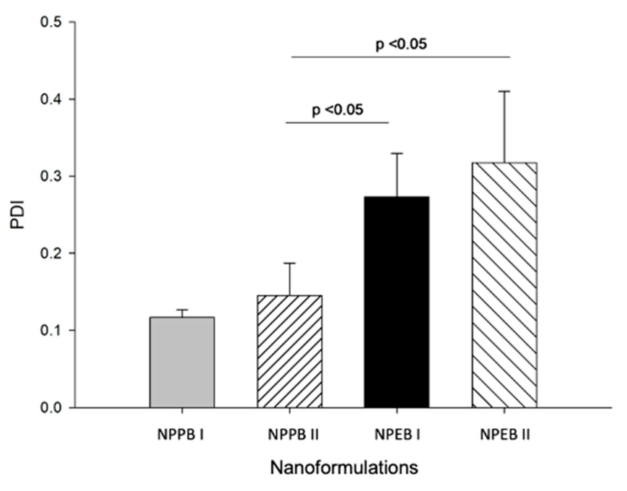
Figure 1. Illustration of the synthesis process of the nanoformulations prepared using the nanoprecipitation method (A) and the emulsion/diffusion method (B).
Processes 12 02739 g001
Figure 2. Variation of surface tension as a function of the concentration of the biosurfactant produced by the endophytic fungus Aspergillus welwitschiae CG2-16. The inflection point indicates the critical micellar concentration.
Processes 12 02739 g002
Figure 3. FTIR spectrum of the biosurfactant produced by the endophytic fungus Aspergillus welwitschiae CG2-16.
Processes 12 02739 g003
Figure 4. Average particle diameter in the nanoformulations prepared using the nanoprecipitation (NP) and emulsion/diffusion (ED) methods, with two different compositions (I and II), containing the biosurfactant produced by the fungus Aspergillus welwitschiae CG2-16. Data are expressed as mean and standard deviation. A significant difference (p < 0.05) in indicated, according to the Tukey test.
Processes 12 02739 g004
Figure 5. Polydispersity index (PDI) of the nanoformulations prepared using the nanoprecipitation (NP) and emulsion/diffusion (ED) method, with two different compositions (I and II), containing the biosurfactant produced by the fungus Aspergillus welwitschiae CG2-16. Data are expressed as mean and standard deviation. A significant difference (p < 0.05) in indicated, according to the Tukey test.
Processes 12 02739 g005
Figure 6. Zeta potential (mV) of the nanoformulations prepared using the method of nanoprecipitation (NP) and emulsion/diffusion (ED), with two different compositions (I and II), containing the biosurfactant produced by the fungus Aspergillus welwitschiae CG2-16. Data are expressed as mean and standard deviation. A significant difference (p < 0.05) in indicated, according to the Tukey test.
Processes 12 02739 g006
Figure 7. Morphology of the NPPBII nanoparticles, determined by SEM (A). Isolated particles at higher magnification (B,C). Bar: 500 nm (A), 100 nm (B,C).
Processes 12 02739 g007
Figure 8. Morphology of the NPPBII nanoparticles, determined by TEM (A). Isolated particles at higher magnification (B,C). Bar: 1 μm (A), 100 nm (B) and 200 nm (C).
Processes 12 02739 g008
Table 1. Average particle diameter, polydispersity index (PDI) and zeta potential of nanoformulations produced by nanoprecipitation and emulsion/diffusion methods, with two different compositions (I and II), containing the biosurfactant produced by the fungus Aspergillus welwitschiae CG2-16. Data are expressed as mean and standard deviation.
NanoformulationAverage Particle Diameter
(nm)
PDIZeta Potential
(mV)
NPPI237.6 ± 9.30.12 ± 0.07−48.50 ± 3.42
NPPII---
NPEI---
NPEII---
NPPBI247.8 ± 12.2 a0.11 ± 0.08 a,b−45.00 ± 1.13 a
NPPBII206.9 ± 1.9 a0.14 ± 0.04 a−29.10 ± 8.67 b
NPEBI177.8 ± 12.8 a,b0.27 ± 0.06 b−43.39 ± 7.40 a
NPEBII147.6 ± 2.7 b0.32 ± 0.09 b−21.90 ± 2.50 b
NPP = Nanoparticles without biosurfactant, formulated using the nanoprecipitation method. NPE = Nanoparticles without biosurfactant, formulated using the emulsion/diffusion method. B = with biosurfactant. Different letters indicate a significant difference (p < 0.05) between variables in the same column, according to the Tukey test.
Disclaimer/Publisher’s Note: The statements, opinions and data contained in all publications are solely those of the individual author(s) and contributor(s) and not of MDPI and/or the editor(s). MDPI and/or the editor(s) disclaim responsibility for any injury to people or property resulting from any ideas, methods, instructions or products referred to in the content.

