Article

Evaluation of Polyphenolic Profile and Antioxidant Activity of Sea Buckthorn (Elaeagnus rhamnoides (L.) A. Nelson) Leaf and Berry Extracts Obtained via Optimized Microwave-Assisted and Accelerated Solvent Extraction

by
Patricija Čulina
1,
Maja Repajić
2,
Ivona Elez Garofulić
2,
Verica Dragović-Uzelac
2 and
Sandra Pedisić
1,*
1
Faculty of Food Technology and Biotechnology, University of Zagreb, P. Kasandrića 3, 23000 Zadar, Croatia
2
Faculty of Food Technology and Biotechnology, University of Zagreb, Pierottijeva 6, 10000 Zagreb, Croatia
*
Author to whom correspondence should be addressed.
Processes 2024, 12(1), 126; https://doi.org/10.3390/pr12010126
Submission received: 8 December 2023 / Revised: 28 December 2023 / Accepted: 30 December 2023 / Published: 3 January 2024

Abstract

:
The aim of this study was to optimize parameters of microwave-assisted extraction (MAE) and accelerated solvent extraction (ASE) in terms of extraction temperature and time, microwave power and cycle numbers on the phenolic content of sea buckthorn leaves and berries, using 70% ethanol (v/v) as an extraction solvent. The characterization of phenolic composition in leaf and berry extracts obtained at optimal MAE and ASE conditions was performed with UPLC/ESI-MS2, while antioxidant activity was determined using the ORAC method. The optimal extraction conditions for MAE were 60 °C, 500 W and 15 min for leaves and 60 °C, 300 W, and 10 min for berries. The optimal extraction conditions for ASE from both leaves and berries were 120 °C, 15 min, and 3 cycles. Total phenolic content (TPC) in MAE and ASE extracts from leaves was similar to the TPC determined in extracts obtained by conventional extraction (60 °C/30 min); however, ASE contributed to the higher TPC of the berry extracts. The flavonols kaempferol-3-rutinoside in the leaves and kaempferol in the berries were the most abundant phenols of sea buckthorn. A higher antioxidant activity was found in the leaf extracts obtained by ASE and it correlated with the phenolic content. In general, ASE favored the extraction of all polyphenols from leaves, while MAE was more suitable for the extraction of flavonols from berries, suggesting that the choice of the optimal extraction method is crucial with regard to the target molecules and future applications.

1. Introduction

Sea buckthorn (Elaeagnus rhamnoides (L.) A. Nelson, genus Hippophae, family Eleagnaceae) (SB) is a shrubby species of native Eurasian plant communities capable of growing on poor soils and temperatures from −40 to +40 °C [1]. Considered to be a valuable source of various bioactive molecules (BAMs), SB’s parts include a variety of polyphenols, carotenoids, tocopherols, sterols, fatty acids, minerals, vitamins, and other compounds [2,3,4,5]. These BAMs are responsible for many of the plant’s bioactive qualities, including those that are antioxidant, anti-inflammatory, antitumor, immunomodulatory, antimicrobial and others [6,7,8]. However, a number of factors, such as the variety of species, the plant’s part, the location of growth, the soil’s composition, the application of fertilizers, and the degree of ripeness, substantially influence the chemical composition of different SB parts [5]. The leaves are thought to be the most valuable source of phenols, followed by the pulp, pericarp, and seeds [9], even though the entire plant contains a variety of polyphenols, including flavonoids, hydrolyzable tannins, and phenolic acids [10,11]. The most prevalent subclass of flavonoids are flavonols, primarily present in glycosylated forms of the aglycones of kaempferol, isorhamnetin, quercetin, and myricetin [11,12,13,14]. The most abundant phenolic acids in SB are gallic acid, syringic acid, protocatechuic acid, salicylic acid, vanillic acid, gentisic acid, caffeic acid, ferulic acid, and chlorogenic acid. Tannins have also been found in SB, including strictinin, hyporhamnin, isostrictinin, pedunculagin, hippophaenin A, and hippophaenin B [15]. The interest and demand for natural substances, particularly plant polyphenols, which could be used as food ingredients or pharmaceuticals, have increased in recent years [16,17] and SB has a great potential to be included in a variety of food and drug formulations. Extraction is the initial step for isolating the target BAMs and due to the different phenolic groups in SB and their physical and chemical properties, it is crucial to find the most efficient extraction methods. The most commonly used methods are conventional (CE) such as maceration, heat reflux, and Soxhlet extraction, but as their disadvantages are high energy and solvent consumption and the decomposition or degradation of the thermolabile BAMs (phenolics and carotenoids), they have led to the increasing use of advanced extraction methods such as ultrasound-assisted extraction, microwave-assisted extraction (MAE), infrared irradiation, pulsed electric fields, enzyme-assisted extraction, and accelerated solvent extraction (ASE). MAE and ASE are widely used advanced extraction methods to obtain the highest extraction yields and to ensure the stability of BAMs [18,19,20,21,22,23]. The advantage of using MAE lies in the microwave irradiation’s heating mechanism, which homogeneously heats the sample and effectively disrupts the cell due to the internal overheating caused by the ion conduction and dipole rotation [24]. ASE is a rapid extraction method that uses solvents at high pressures and temperatures to keep the solvent as a liquid throughout the extraction process. The main advantages of ASE are that increasing the temperature of extraction positively affects the solubility of analytes, improves the rate of mass transfer, and lowers the viscosity and surface tension of the used solvent, thereby increasing the extraction rates, although caution should be taken with thermally labile compounds [25]. However, many factors such as conditions of extraction, solvent type and polarity, extraction temperature and time, size of particles, solvent to sample volume ratio, influence the extraction yield and extract composition [26]. Thus, in order to obtain the highest yield of target compounds, it is crucial to optimize the extraction conditions. Extraction studies have shown that the use of hydroalcoholic solvents improves the recovery of phenolic compounds compared to pure solvents, and the use of an ethanol–water mixture is recommended for MAE and ASE [27]. For example, microwave power, irradiation time, and temperature are important factors affecting extraction yield when using MAE, as the microwave power increasing generally improves extraction yield and leads to a shorter extraction time, but can also lead to an increase in temperature and the degradation of thermolabile compounds. It has also been shown that when extracting thermolabile compounds, it is best to use solvents with lower dielectric constants [28]. In the study of Fan et al. [29], an improved extraction yield of SB flavonoids was determined under optimal MAE conditions: 50% ethanol as extraction solvent, material to liquid ratio of 1:40 (g/mL), microwave power of 550 W, and microwave time of 5 min. The highest total phenolic content (TPC) of SB leaves was achieved under optimal MAE conditions: 50% ethanol as extraction solvent, solvent to plant ratio of 20:1 and temperature of 90 °C [30]. ASE also improves the extraction efficiency through controlled conditions of temperature, static extraction time and number of extraction cycles, which when optimized can increase the content of target BAMs extracted from the plant [31]. It has been observed that the highest yield of phenolic acids was obtained at higher temperatures while lower temperatures were more effective in extracting high yields of flavonoids [31]. ASE and MAE have been used for the extraction of polyphenols from many plants and fruits [32], but to our knowledge, data of the polyphenol content and profile of SB produced at optimal conditions are limited.
Therefore, the aim of this study was to determine the optimal conditions for the extraction of polyphenols from SB leaves and berries using MAE ((temperature (40, 60, and 80 °C), microwave power (300, 500, and 700 W), and irradiation time (5, 10 and 15 min)) and ASE ((temperature (80, 100, and 120 °C), static extraction time (5, 10, and 15 min) and number of cycles (1, 2, and 3)) and to compare the extraction efficiency with conventional extraction (60 °C, 30 min) in terms of polyphenol content. The solvent used for extraction was 70% ethanol. In extracts produced under optimal extraction conditions, the individual phenolic compounds were determined using UPLC/ESI-MS2 and the antioxidant activity using the ORAC method.

2. Materials and Methods

2.1. Plant Material

The samples of leaves and berries of SB (Elaeagnus rhamnoides (L.) A. Nelson) were harvested in Croatia, Hrvatsko Zagorje region in July 2020. The samples were freeze-dried and stored in the dark at room temperature. Prior to extraction, the samples were ground to powder using an electric mill (AR 1105; Moulinex, Saint-Lo, France) and sieved through a sieve with a size of 0.5 to 1 mm.

2.2. Chemicals and Reagents

Ethanol, acetonitrile, and formic acid used for extraction were HPLC grade and were procured from BDH Prolabo (Lutterworth, UK). The sodium phosphate (96%) and anhydrous sodium carbonate (99.5%) were purchased from Kemika (Zagreb, Croatia), Folin–Ciocalteu reagent was obtained from Merck (Darmstadt, Germany), Trolox (6-hydroxy-2,5,7,8-tetramethylchroman-2-carboxylic acid) from Acros Organics (Thermo Fisher Scientific, Geel, Belgium), 2,20-Azobis (2-amidinopropane) hydrochloride was purchased from Sigma-Aldrich (Steinheim, Germany), and fluorescein sodium salt from Honeywell Riedel-de-Haën (Bucharest, Romania). Authentic phenolic standards of caffeic, gallic, p-coumaric and vanillic acid, quercetin-3-glucoside, and kaempferol-3-rutinoside were purchased from Sigma-Aldrich (Steinheim, Germany); epicatechin and catechin were obtained from Extrasynthese (Genay, France); and from Acros Organics (Thermo Fisher Scientific, Geel, Belgium), quercetin-3-rutinoside. Distilled Milli-Q water (Millipore Corp., Bedford, NY, USA) was used.

2.3. Microwave-Assisted Extraction (MAE)

The MAE was performed with a Milestone microwave reactor (Start S Microwave Labstation for Synthesis; Sorisole, Italy) with flexible microwave power using 70% ethanol (v/v) as extraction solvent. The procedure was conducted according to the method previously described by Elez Garofulić et al. [18]. The ground samples of leaves and berries (1 ± 0.001 g) were mixed with 40 mL extraction solvent in the extraction vessel with the addition of a magnetic stir bar and placed in a microwave reactor with a cooling system. The stirring was constant and set to 50%. To determine the optimal extraction conditions in terms of the highest yield, the following parameters were varied: temperature (40, 60, and 80 °C), microwave power (300, 500, and 700 W), and irradiation time (5, 10, and 15 min) according to the experimental design. After extraction, the cooled and filtered extract was transferred and made up with the extraction solvent in a 50 mL flask. All extractions were carried out in duplicate (n = 2) and stored in nitrogen gas atmosphere at −18 °C until analyzed.

2.4. Accelerated Solvent Extraction (ASE)

The ASE was performed with the Dionex™ ASE™ 350 extractor (Thermo Fisher Scientific Inc., Sunnyvale, CA, USA) using 70% ethanol as an extraction solvent (v/v). The procedure was carried out in accordance with the methodology described in our previous study [33]. The mixture of sample (1 ± 0.001 g) and diatomaceous earth (0.5 g) was added to 34 mL stainless steel cells fitted with 2 cellulose filters at the bottom of the cell. To determine the optimal extraction conditions with the highest yield, the following parameters were varied: temperature (80, 100, and 120 °C), static extraction time (5, 10, and 15 min) and number of cycles (1, 2, and 3) according to the experimental design. The pressure was set to a fixed value of 10.34 MPa. The extracts obtained were filled with the extraction solvent in a 50 mL flask. Extractions were carried out in a duplicate (n = 2) and stored in nitrogen gas atmosphere at −18 °C until analyzed.

2.5. Conventional Extraction

The efficiency of the MAE and ASE methods was compared with heat-reflux extraction (CE): 1 ± 0.001 g of SB leaves and berries were placed into the flask containing 40 mL of 70% ethanol solution (v/v), extracted for 30 min, filtered through filter paper, and made up to 50 mL with extraction solvent. Extractions were carried out in duplicate (n = 2) and stored in a nitrogen gas atmosphere at −18 °C until analyzed.

2.6. Analysis of Polyphenols

2.6.1. Total Phenolic Content (TPC) Determination

The TPC of the SB leaves and berries was obtained using the spectrophotometric method described by Shortle et al. [34]. Briefly, 100 μL of the extract, 200 μL of the Folin–Ciocalteu reagent, and 2 mL of distilled water were mixed with 1 mL of 20% sodium carbonate. The mixture was incubated at 50 °C for 25 min and absorbance was measured at 765 nm (UV-VIS UviLine 9400; Secomam, Ales, France). Calculation of TPC was performed using the gallic acid standard calibration (50–500 mg/L) and TPC was expressed in mg gallic acid equivalents (GAE) per 100 g of dry mass (mg/100 g dm) (n = 2).

2.6.2. Polyphenol Characterization by UPLC/ESI-MS2 Analysis

The characterization of polyphenols was conducted using an Agilent 1290 RRLC instrument (Agilent, Santa Clara, CA, USA) and triple quadrupole mass spectrometer (6430 QqQ) with ESI ion source, according to our previous study [33]. The phenolic compounds such as quercetin-3-rutinoside (rutin), kaempferol-3-rutinoside, quercetin-3-glucoside, catechin, epicatechin, caffeic acid, chlorogenic acid, gallic acid, p-coumaric acid, and vanillic acid were identified by comparing the mass spectra and fragmentation patterns of the authentic standards, while the identification of other compounds was based on the previously reported data [35,36,37,38,39]. The concentrations obtained were expressed in mg per 100 g dry mass (mg/100 g dm) (n = 2).

2.6.3. Oxygen Radical Absorbance Capacity (ORAC) Assay

The ORAC assay was performed using a Clariostar fluorescence microplate reader (BMG LABTECH, Offenburg, Germany) according to the method described in our previous study [33]. The results were expressed as µmol Trolox equivalent per g dry mass (µmol TE/g dm) (n = 2).

2.7. Statistical Analysis

Statistica ver. 12.0 software (StatSoft Inc., Tulsa, OK, USA) was used for the statistical analysis. The influence of MAE (microwave power, temperature, and irradiation time) and ASE (static extraction time, temperature, and number of cycles) parameters on the TPC of SB leaves and berries was evaluated using a three-level full factorial design with 54 experimental trials (Table 1 and Table 2). To determine the data’s normality and homoscedasticity, the Shapiro–Wilk and Levene tests were used. Then, data were analyzed using ANOVA (parametric data) or the Kruskal–Wallis test (non-parametric data), and mean differences between groups were compared using Tukey’s HSD test or the Kruskal–Wallis test. At a significance level of p ≤ 0.05, all tests were carried out and the statistical analysis results are shown as least squares (LS) mean and standard error (SE). To compare the efficiency of the MAE and ASE in terms of TPC, mean values were compared using one-way ANOVA and Tukey’s HSD test (post-hoc).

3. Results and Discussion

In this study, the influence of the advanced extraction methods MAE and ASE on the phenolic content of SB leaf and berry extracts was investigated. The results of TPC in SB leaf and berry extracts obtained under different conditions of MAE (temperature of 40, 60, and 80 °C; microwave power of 300, 500, and 700 W; and extraction time of 5, 10, and 15 min) and ASE (temperature of 80, 100, and 120 °C; static extraction time of 5, 10, and 15 min; and cycle numbers of 1, 2, and 3) are shown in Table 1 and Table 2. The results of the statistical analysis used to determine optimal extraction conditions are shown in Table 3 and Table 4 and the TPC of leaf and berry extracts obtained at optimal MAE and ASE conditions was compared with extracts obtained by CE (Figure 1). Phenolic characterization in leaf and berry extracts obtained at optimal MAE and ASE conditions by UPLC-MS2 are shown in Table 5, while the phenolic characterization of leaf and berry extracts obtained by CE is shown in Table S1 (Supplementary Materials). The antioxidant capacity in leaf and berry extracts obtained at optimal MAE and ASE conditions is shown in Figure 2.
The TPC in SB leaf extracts obtained with MAE ranged from 6514 to 10779 mg GAE/100 g dm (Table 1) and from 6905 to 10,918 mg GAE/100 g dm in extracts obtained with ASE (Table 2). The TPC in the MAE berry extracts was significantly lower compared to the leaves, ranging from 246 to 406 mg GAE/100 g dm and from 220 to 688 mg GAE/100 g dm when ASE was used. Both extraction methods resulted in significant yields of phenolic compounds in SB leaf extracts, which is in line with the earlier research of Galan et al. [40] and Michal et al. [41], who reported TPC in MAE leaf extracts ranging from 87.37 to 157.63 mg GAE/g and from 53.0 to 92.0 mg GAE/g in ASE leaf extracts, respectively. Compared with our results, the study of Périno-Issartier et al. [23] determined higher TPC in the SB berry extracts obtained with solvent-free MAE (1147 mg GAE/100 g dm) and using conventional extraction with methanol as solvent (741.9 mg GAE/100 g dm). Although the mechanism of MAE and ASE is similar since the solvent in both systems is heated and pressurized [42], ASE contributed to higher TPC compared to MAE, especially in berry extracts. The same trend was observed in the study of Rodríguez-Pérez et al. [43] for the extraction of BAMs from Moringa oleifera leaves. The good performance of ASE is probably the result of a combination of high extraction temperatures above the boiling point of the solvent and multiple extraction cycles leading to complete extraction of the target compounds [44]. In MAE, the microwave energy absorbed through the plant cell walls leads to internal overheating, which allows faster diffusion of analytes in the solvent [45]. The rapid rise in temperature during microwave heating avoids the thermal gradient, which increases the risk of degradation of thermolabile BAMs [46]. In addition, plant matrix characteristics also affect MAE performance [45].

3.1. Effect of MAE on the TPC of Sea Buckthorn Leaves and Berries

The optimal MAE conditions for the extraction of SB leaf and berry polyphenols were determined via varying the temperature (40, 60, and 80 °C), microwave power (300, 500, and 700 W), and time (5, 10, and 15 min). As shown in Table 1, the lowest TPC was obtained in the MAE extracts of leaves and berries at an extraction temperature of 40 °C, a microwave power of 700 W, and an extraction time of 5 min. On the other hand, the highest TPC for leaves was obtained under the following MAE conditions: temperature of 60 °C, microwave power of 500 W, and extraction time of 15 min and for berry extracts at a temperature of 80 °C, microwave power of 500 W, and extraction time of 10 min.
Temperature is considered a crucial parameter in MAE, and for the extraction of phenolic compounds with MAE the most frequently used temperature range is between 30 and 180 °C, although a decrease in polyphenol content has been observed at temperatures above 100 °C. Lower extraction temperatures or shorter extraction times are more often used to avoid the co-extraction of interfering chemicals, even though better polyphenol recoveries can be achieved at higher temperatures and times [47]. Therefore, a moderate temperature range from 40 to 80 °C was used in this study. According to the results in Table 3, temperature had a statistically significant effect (p ≤ 0.05) on the TPC of both leaves and berry extracts. The increase in phenolic content in the obtained MAE leaf and berry extracts was noted when the temperature increased from 40 to 60 °C.
In the study of Galan et al. [40], the same trend was observed when the extraction temperature was increased from 40 to 90 °C. Higher temperature allows better solubility of phenolic compounds and the mass transfer coefficient between the sample matrix and the extraction medium increases [48]. Asofei et al. [30] reported an optimal MAE temperature of 90 °C for the extraction of polyphenols from SB leaves and a solvent/plant ratio of 20:1 and 50% ethanol as the extraction solvent. The two most fluctuating parameters in MAE are the microwave power and the extraction time which change depending on the plant species, BAMs, and various parts of the same plant [49]. Microwave power has considerable impact on the extraction of BAMs since microwave irradiation can speed the rupture of plant cells by rapidly raising the temperature and pressure [50]. However, excessive exposure to microwave irradiation leads to overheating and the degradation of phenolic compounds. To avoid oxidation and degradation of thermolabile compounds due to overheating of the system, the duration of extraction must be adjusted to the properties of the sample. In the present study, microwave power and extraction time had no statistically significant influence on the TPC of the extracts (Table 3). However, increasing the microwave power from 300 to 500 W had a positive effect on the TPC in the leaf extracts. In general, increasing the microwave power increases the efficiency of extraction by maximizing the molecular interaction between the sample and the electromagnetic field [51]. A further increase in microwave power to 700 W led to a decrease in TPC in both leaf and berry extracts (Table 3). Similar results were reported by Dahmoune et al. [52] for the MAE of phenolic compounds from SB leaves and by Alara et al. [53] for Vernonia amygdalina leaves. Regarding the extraction time, the results of the statistical analysis showed no significant effect, but increasing the extraction time from 5 to 10 min increased the TPC in the berry fruit extracts and in the leaf extracts after 15 min (Table 3). A similar trend was observed in the study of Rafiea et al. [54], where the highest TPC was obtained in three different varieties of olive leaves in aqueous MAE extracts after 15 min of extraction. The MAE extraction yield could be enhanced by increasing the extraction temperature, time, and microwave power up to a certain limit, after which the risk of considerable loss of thermolabile BAMs increases. The stability of the extracted compounds is a crucial factor in choosing the optimal extraction conditions. Considering the results of the statistical analysis, the optimal MAE conditions for total phenolics from SB leaves were 60 °C, 500 W, and 15 min and from berries 60 °C, 300 W, and 10 min, respectively.

3.2. Effect of ASE on the TPC of Sea Buckthorn Leaves and Fruits

The optimal ASE conditions for the extraction of SB leaf and berry polyphenols were determined by varying the temperature (80, 100, and 120 °C), static extraction time (5, 10, and 15 min) and number of cycles (1, 2, and 3). As shown in Table 2, the lowest TPC was obtained in ASE leaf and berry extracts at an extraction temperature of 100 °C, static time of 5 min, and number of cycles, 1. On the other hand, the highest TPC in ASE leaf and berry extracts was obtained at 120 °C, a static time of 15 min, and number of cycles, 3. The use of high temperatures enhanced extraction efficiency via disrupting analyte–sample matrix interactions, which is one of the key elements impacting efficiency and selectivity of ASE [44]. Solvents remain liquid under the high pressure of ASE even at temperatures above their boiling point. In this study, according to the results of statistical analysis, temperature had a statistically significant effect (p < 0.001) on the TPC in both leaf and berry extracts (Table 4).
In leaf extracts, TPC increased proportionally with the increase in extraction temperature; meanwhile, in berry extracts, a significant increase in TPC was noted when the temperature was increased from 100 to 120 °C. The obtained results are consistent with the study of Repajić et al. [19], who reported that nettle leaves had a significantly higher content of analyzed phenolic compounds when the temperature was raised from 20 to 110 °C. Raising the temperature during ASE improves compound solubility, diffusion rate, and mass transfer and allows the solvent to enter the matrix more easily [55]. Despite the high temperature and pressure used, the rapid penetration of the solvent protects the phenolic compounds from degradation [56]. However, in order to improve ASE efficiency, the effect of temperature should be studied with static time and cycles [57]. In the present study, the static extraction time had a statistically significant effect (p < 0.001) on the TPC of the berry extracts (Table 4). In general, increasing the static time increased the yield of phenolic compounds in both leaves and berries, and the highest TPC was obtained after 15 min. Prolongation of the static extraction time at elevated temperatures promotes diffusion of analytes into the extraction solvent [57]. A similar trend was observed in the study of ASE of phenolic compounds from fennel seeds and grape skins, where the application of a longer static time resulted in a higher TPC [19,58]. The static cycles also had a statistically significant effect (p < 0.001) on the TPC in leaf and berry extracts and an increase in the number of cycles resulted in a higher yield of phenolic compounds (Table 4). The use of static cycles helps to maintain a favorable extraction equilibrium without diluting the sample by adding fresh solvent during the extraction process [57]. According to Repajić et al. [19], the phenolic yield from nettle leaves was significantly influenced by static time, and the highest yields were obtained during the third cycle, which is in accordance with the results of our study. The addition of a fresh solvent positively affects the extraction of phenolic compounds from Passiflora species in the study of Gomez et al. [59], and optimal extraction conditions were achieved with five extraction cycles. The results of the statistical analysis showed that the optimal ASE conditions for total phenolics from SB leaves and berries were 120 °C, 15 min, and 3 cycles.

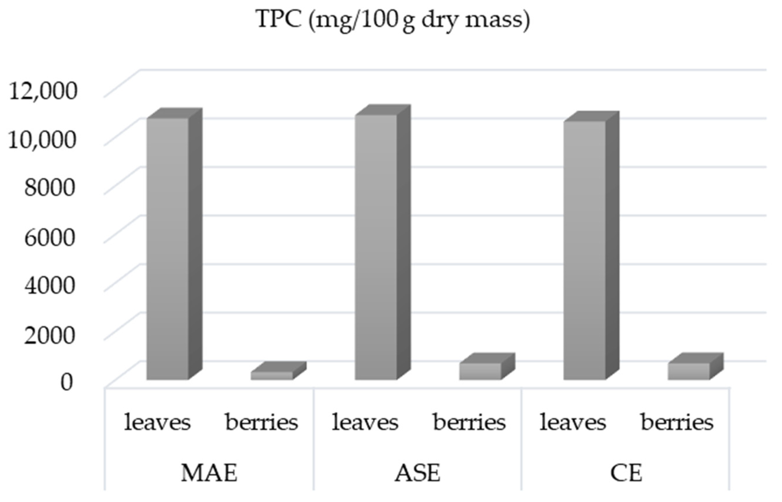
3.3. Comparison between CE, MAE and ASE

The TPC of the leaf extracts was higher than that of the berries, and ASE contributed more to the higher TPC than MAE, especially in the berry extracts. It is likely that the higher TPC yields in ASE are due to accelerated diffusion favored by the breaking of intermolecular forces, ensuring a continuous flow of solvent through the solid matrix, as well as the effects of the higher extraction temperature [60]. The TPC in the berry extracts obtained with CE (691.5 mg/100 g dm) was similar to the TPC of berry extracts obtained with ASE, while the berry extracts obtained with MAE showed a significantly lower TPC value (Figure 1). Chaves et al. [60] reported that MAE and ASE are similar technologies with little difference in yield, as the solvent is pressurized and heated in both extractions, but in addition to the operating conditions, the characteristics of the plant matrix affect the performance of MAE. On the other hand, the TPC content in leaf extracts obtained by MAE and ASE was almost identical to those obtained after 30 min of CE (10,652.7 mg/100 g), demonstrating the advantages of MAE and ASE, namely the decrease in extraction time and solvent consumption. A similar trend was observed by Ince et al. [61] for aerial parts of dry nettle extracts, by Elez Garofulić et al. [18] for Pistacia lentiscus L. fruit and leaves extracts, by Georgiopoulou et al. [62] for Chlorella vulgaris extracts, and by Alhallaf et al. [63] for nonotus obliquus (chaga) sclerotia extracts.

3.4. UPLC/ESI-MS2

UPLC/ESI-MS2 was used to compare the polyphenolic profile and extraction efficiency of SB leaf and berry extracts obtained under optimal MAE and ASE conditions. A total of 29 compounds were identified, including 19 flavonols, 2 flavan-3-ols, and 8 phenolic acids (Table 5). The polyphenolic profile of leaf and berry extracts obtained by CE was also determined (Table S1).
Compound (Cp) 1, characterized as isorhamnetin, exhibited specific ESI/MS2 fragmentation, which is common for flavonols. As previously reported in the literature, the MS fingerprint of this compound was dominated by the loss of small neutral fragments (−28 Da (CO), −18 Da (H2O), −44 Da (CO2), or their combination) [64]. Cp 2–8 were characterized as isorhamnetin glycosides with a fragment ion at m/z 317 corresponding to the aglycone isorhamnetin. Cp 2 was tentatively proposed as isorhamnetin-3-sinapoyglucose-glucoside-7-rhamnoside according to its fragmentation pattern, which produced fragment ions at m/z 463 after the loss of the sinapoylglucose (−368 amu) and one glucose (−146 amu) moiety from the C-3 position, and at m/z 317 after the loss of the sinapoylglucose (−368 amu), glucose (−146 amu), and rhamnose (−146 amu) moiety. Cp 3 and 4 identified as ishorhamnetin-3-sophoroside-7-rhamnoside and isorhamnetin-3-rutinoside-7-glucoside were characterized by losses corresponding to the sophorose (324 amu) and rhamnose moiety (−146 amu) and the rutinose (−308 amu) and hexose (−146) moiety, respectively [37]. Cp 5–8, identified as isorhamnetin-3-hexoside, isorhamnetin-3-rhamnoside, isorhamnetin-3,7-dihexoside, and isorhamnetin-3-rutinoside, were characterized by losses corresponding to hexoside (−146 amu), rhamnoside (−146 amu) and rutinoside (−308 amu) moieties [36,37]. Isorhamnetin-3-hexoside was the most abundant isorhamnetin glycoside in berry extracts obtained by ASE (23.6 mg/100 g dm) and in leaf and berry extracts obtained by MAE (12.8 and 45.4 mg/100 g dm, respectively), while isorhamnetin-3,7-dihexoside was the most abundant in leaf extracts obtained by ASE (40.4 mg/100 g dm). In the extracts obtained by CE, the most abundant isorhamnetin glycosides were isorhamnetin-3-hexoside (49 mg/100 g dm) in the leaf extract and isorhamnetin-3,7-dihexoside (41.1 mg/100 g dm) in the berry extract. Pop et al. [37] also identified isorhamnetin-3-hexoside and isorhamnetin-3,7-dihexoside in SB leaves and berries, while Wang et al. [65] reported isorhamnetin-3-hexoside as the main isorhamnetin glycoside in sea buckthorn leaves and berries. Cp 10–13 were characterized as kaempferol glycosides based on a fragment ion at m/z 287 corresponding to aglycone kaempferol (Cp 9) with a specific fragment ion at m/z 145 obtained by losses of 2 CO (−56 amu) and 2 C2H2O (−84 amu) [39]. Cp 12 was identified as kaempferol-3-rutinoside via comparison with authentic standards. Cp 10, 11, and 13 were characterized by losses corresponding to the sophoroside (−178 amu) and rhamnoside (−146 amu) moieties as kaempferol-3-O-sophorose-7-O-rhamnoside, by losses corresponding to the hexoside (−162 amu) and rhamnoside (−146 amu) moieties as kaemferol-3-O-glucoside-7-O-rhamnoside, and by losses corresponding to the rutinoside moiety (−308 amu) as kaempferol rhamnoside [35,36,37]. Among the kaempferol glycosides, kaempferol-3-rutinoside was found in the highest concentration in the leaves (300 mg/100 g dm in ASE extracts and 110.8 mg/100 g dm in MAE extracts), while kaempferol-3-O-glucoside-7-O-rhamnoside was the most abundant in the berries (10.3 mg/100 g dm in ASE extracts and 11.0 mg/100 g dm in MAE extracts). The same trend was observed in the extracts of leaves and berries obtained by CE, with kaempferol-3-rutinoside being the most abundant in the leaves (303.1 mg/100 g dm), while kaempferol-3-O-glucoside-7-O-rhamnoside was the most abundant in the berries (5.8 mg/100 g dm) (Table S1). Rosch et al. [36] reported the presence of kaempferol-3-rutinoside and kaemferol-3-O-glucoside-7-O-rhamnoside in sea buckthorn pomace. Pop et al. [37] also identified kaempferol-3-rutinoside in leaves at concentrations ranging from 23.9 to 89.4 mg/100 g dm. Kaempferol was determined in high concentrations in all berry extracts as follows: 51.3 mg/100 g dm in extracts obtained by ASE; 81.2 mg/100 g dm in extracts obtained by MAE and 30.9 mg/100 g dm in extracts obtained by CE. The presence of kaempferol was reported in our previous study, under reference Čulina et al. [33]. Cp 16 and 17 were identified as quercetin-3-glucoside and quercetin-3-rutinoside (rutin) via comparison with authentic standards. Cp 14, 15, 18, and 19 were characterized as quercetin glycosides based on fragment ions at m/z 303. They were characterized by losses corresponding to the sophoroside (−178 amu) and rhamnoside (−146 amu) moieties as quercetin-3-sophoroside-7-rhamnoside, by losses corresponding to the rhamnoside (−146 amu) and hexoside (−162 amu) moieties as quercetin-3-rhamglucoside-7-rhamnoside, by the loss corresponding to rhamnoside (−146 amu) as quercetin-3-rhamnoside and by the loss corresponding to pentoside (−132 amu) as quercetin-3-pentoside [36,37]. Among quercetin glycosides, quercetin-3-rutinoside was the most abundant in leaf and berry extracts obtained by ASE (185.69 and 15.61 mg/100 g dm, respectively), MAE (76.55 and 12.55 mg/100 g dm, respectively), and CE (14.29 and 19.89 mg/100 g dm). Perk et al. [66] and Li et al. [67] confirmed the presence of rutin in SB leaves and berries in high concentration. The cp 20 and 21 were identified as the flavanols catechin and epicatechin by comparison with authentic standards. Epicatechin was more abundant than catechin in the leaves (10.26 mg/100 g dm in ASE extracts, 2.12 mg/100 g dm in MAE extracts and 10.69 mg/100 g dm in CE extracts), whereas neither catechin nor epicatechin were detected in the berry extracts. Wang et al. [35] also identified catechin and epicatechin in SB leaves. Among phenolic acids, Cp 22, 23, 25, 27, and 29 were identified by comparison with authentic standards as caffeic acid, chlorogenic, gallic acid, p-coumaric acid, and vanillic acid. Related to the fragmentation pattern, the ESI-MS signals at m/z 301, 153, and 137 were tentatively assigned as ellagic, protocatechuic, and 4-hydroxybenzoic acids, with fragment ions at m/z 257, 109, and 93 corresponding to the loss of CO2 from their precursor ions [38]. The most abundant phenolic acid in SB leaf extracts obtained by ASE was gallic acid (87.11 mg/100 g dm), while in berry extracts obtained by ASE it was vanillic acid (52.81 mg/100 g dm). According to Arimboor et al. [68], gallic acid was the most abundant phenolic acid in SB berry parts and leaves. Caffeic acid, p-hydroxybenzoic acid, and vanillic acid were determined in the leaf extracts obtained by MAE, with vanillic acid being the most abundant (20.8 mg/100 g dm). Compared to the leaves, lower concentrations of caffeic acid, p-coumaric acid, and vanillic acid were found in the MAE extracts of the berries, and vanillic acid was also the most abundant (22.1 mg/100 g dm). The most abundant phenolic acid in the extracts obtained by CE was p-hydroxybenzoic acid in the leaf extract (39.4 mg/100 g dm) and gallic acid in the berry extract (91.8 mg/100 g dm) (Table S1). Chen et al. [69] also reported the presence of caffeic, gallic, p-coumaric, protocatechuic, and vanillic acids in SB leaves and berries in a lower concentration than in our study. The presence of ellagic acid was confirmed in the study of Wang et al. [35].
According to results, the predominant phenolic group in both the ASE and MAE leaf and berry extracts were flavonols, with kaempferol-3-rutinoside being the most abundant in the leaves and kaempferol in the berries. The ASE leaf extracts had higher flavonol contents than MAE and CE leaf extracts. Among the phenolic acids, gallic acid dominated in the ASE and CE leaf extracts and vanillic acid in the MAE leaf extracts and vanillic acid in the berry extracts. Flavan-3-ols were only determined in the leaf extracts and epicatechin was present in higher contents. Contradictory results are reported in the literature concerning the extraction efficiency of phenolic compounds using ASE and MAE and some authors have concluded that the extraction method should be selected depending on the target molecules, as not all phenolics follow the same trend [70,71]. Biesaga [72] reported that hydroxyl groups promoted the degradation of flavonoids during MAE, while sugar and methoxyl groups protected them from degradation. According to Liazit et al. [73], ASE was more effective than MAE in the extraction of thermolabile phenolic compounds and those with a higher number of hydroxyl-type substituents. Some studies have emphasized that the extraction method should be selected based on the target molecules to be isolated, but no general conclusion can be drawn due to the influence of the sample matrix [60].

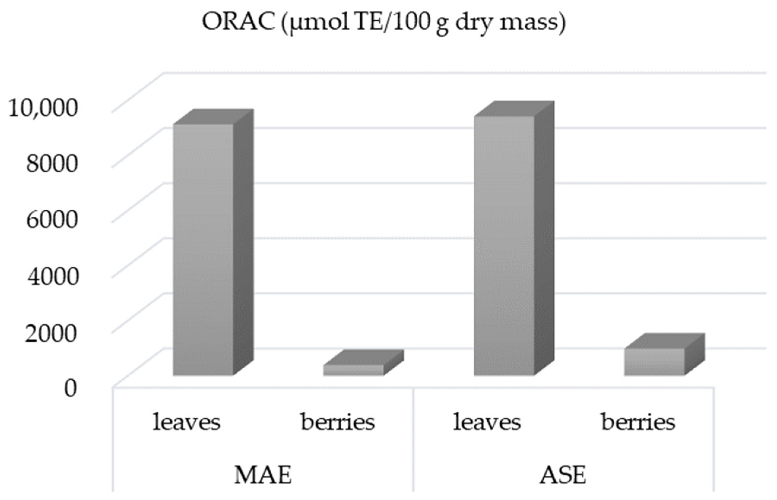
3.5. Antioxidant Activity

An ORAC assay was performed to determine the antioxidant activity of the extracts obtained under optimal MAE and ASE conditions. The ORAC assay is the most biologically relevant and can measure lipophilic and hydrophilic antioxidants [74]. The ORAC value of the SB leaf extracts was 9129 μmol TE/100 g dm for MAE and 9417 μmol TE/100 g dm for ASE, while it was lower for the berries, namely 388 μmol TE/100 g dm for MAE and 985 μmol TE/100 g dm for ASE (Figure 2). The ORAC values correlate with the phenolic content determined, which was higher in the leaves than in the berries and higher in the extracts obtained by ASE than in the MAE extracts. Tkacz et al. [6] reported higher ORAC values for SB berry cultivars grown in Poland that ranged from 15 mmol to 35 mmol TE/100 g dm. Differences in antioxidant activity could be attributed to genetic differences, environmental conditions, the type of plant part, the pre-treatment, the extraction method, and the solvent as well as the amount of BAMs contained in the extract.

4. Conclusions

MAE and ASE extraction were optimized to obtain the highest phenolic yield from SB leaves and berries. The differences in extraction conditions in MAE between leaves and berries were related to microwave power and irradiation time, which were more evident for leaves, while ASE conditions were the same for both leaves and berries. The TPC in ASE leaf and berry extracts and in MAE leaf extracts obtained at optimal conditions was similar to extracts obtained with CE, confirming the advantages of ASE and MAE extraction methods in terms of extraction time and reduction in energy consumption. The polyphenolic profile of SB leaves and berries included 29 compounds from the classes of flavonols, flavan-3-ols, and phenolic acids. The predominant phenolic group in both the ASE and MAE leaf and berry extracts were flavonols, with kaempferol-3-rutinoside being the most abundant in the leaves and kaempferol aglycone in the berries. Among the phenolic acids, gallic acid dominated in the ASE leaf extracts, and vanillic acid in the MAE leaf and berry extracts. Unlike berry extracts, the leaf extracts contained flavan-3-ols, with epicatechin in the highest concentration. ASE contributed to a higher TPC than MAE, but the extraction method should be selected depending on the target molecules, as not all phenolics followed the same trend; in particular, in the berry extracts. The ORAC antioxidant activity was higher in the leaf than in the berry extracts as well as in the extracts obtained by ASE. The results of our study indicate that ASE and MAE can be considered as a good alternative to conventional methods due to the short extraction time, lower energy and cost consumption, and higher efficiency. The use of SB leaf and berry extracts, which are rich in phenols with high antioxidant activity, is likely to be the focus of future research due to the increased demand for value-added products and functional foods.

Supplementary Materials

The following supporting information can be downloaded at: https://www.mdpi.com/article/10.3390/pr12010126/s1, Table S1: UPLC/ESI-MS2 characterization of polyphenolic compounds in sea buckthorn leaves and berries extracts obtained by convectional extraction (CE).

Author Contributions

Conceptualization, P.Č. and S.P.; methodology, P.Č. and S.P.; formal analysis, P.Č., I.E.G. and M.R.; investigation, I.E.G. and M.R.; data curation, I.E.G. and M.R.; writing—original draft preparation, P.Č. and S.P.; writing—review and editing, M.R., I.E.G. and V.D.-U. All authors have read and agreed to the published version of the manuscript.

Funding

This work was supported by the project “Bioactive molecules of medical plant as natural antioxidants, microbicides and preservatives” (KK.01.1.1.04.0093), co-financed by the Croatian Government and the European Union through the European Regional Development Fund-Operational Programme Competitiveness and Cohesion (KK.01.1.1.04).

Data Availability Statement

Data are contained within the article.

Conflicts of Interest

The authors declare no potential financial or other interests that could be perceived to influence the outcomes of this research. No conflict, informed consent, human or animal rights applicable.

References

  1. Tiitinen, K.M.; Hakala, M.A.; Kallio, H.P. Quality components of sea buckthorn (Hippophae rhamnoides L.) varieties. J. Agric. Food Chem. 2005, 53, 1692–1699. [Google Scholar] [CrossRef] [PubMed]
  2. Jain, M.; Ganju, L.; Katiyal, A.; Padwad, Y.; Mishra, K.P.; Chanda, S.; Karan, D.; Yogendra, K.M.S.; Sawhney, R.C. Effect of leaf extract against Dengue virus infection in human blood-derived macrophages. Phytomedicine 2008, 15, 793–799. [Google Scholar] [CrossRef] [PubMed]
  3. Kumar, M.S.Y.; Dutta, R.; Prasad, D.; Misra, K. Subcritical water extraction of antioxidant compounds from Seabuckthorn (Hippophae rhamnoides L.) leaves for the comparative evaluation of antioxidant activity. Food Chem. 2011, 127, 1309–1316. [Google Scholar] [CrossRef] [PubMed]
  4. Saggu, S.; Divekar, H.M.; Gupta, V.; Sawhney, R.C.; Banerjee, P.K.; Kumar, R. Adaptogenic and safety evaluation of seabuckthorn (Hippophae rhamnoides L.) leaf extract: A dose dependent study. Food Chem. Toxicol. 2007, 45, 609–617. [Google Scholar] [CrossRef] [PubMed]
  5. Ciesarová, Z.; Murkovic, M.; Cejpek, K.; Kreps, F.; Tobolková, B.; Koplík, R.; Belajová, E.; Kukurová, K.; Daško, K.; Panovská, Z.; et al. Why is sea buckthorn (Hippophae rhamnoides L.) so exceptional? A review. Food Res. Int. 2020, 133, 109170. [Google Scholar] [CrossRef] [PubMed]
  6. Tkacz, K.; Wojdylo, A.; Turkiewicz, I.P.; Bobak, L.; Nowicka, P. Anti-Oxidant and Anti-Enzymatic Activities of Sea Buckthorn (Hippophae rhamnoides L.) Fruits Modulated by Chemical Components. Antioxidants 2019, 8, 618. [Google Scholar] [CrossRef]
  7. Cao, J.G.; Zheng, Y.X.; Xia, X.; Wang, Q.X.; Xiao, J.B. Total flavonoid contents, antioxidant potential and acetylcholinesterase inhibition activity of the extracts from 15 ferns in China. Ind. Crop Prod. 2015, 75, 135–140. [Google Scholar] [CrossRef]
  8. Chen, C.; Xu, X.M.; Chen, Y.; Yu, M.Y.; Wen, F.Y.; Zhang, H. Identification, quantification and antioxidant activity of acylated flavonol glycosides from sea buckthorn (Hippophae rhamnoides ssp.). Food Chem. 2013, 141, 1573–1579. [Google Scholar] [CrossRef]
  9. Kumar, M.S.Y.; Tirpude, R.J.; Maheshwari, D.T.; Bansal, A.; Misra, K. Antioxidant and antimicrobial properties of phenolic rich fraction of Seabuckthorn (Hippophae rhamnoides L.) leaves. Food Chem. 2013, 141, 3443–3450. [Google Scholar] [CrossRef]
  10. Arimboor, R.; Arumughan, C. HPLC-DAD-MS/MS profiling of antioxidant flavonoid glycosides in sea buckthorn (Hippophae rhamnoides L.) seeds. Int. J. Food Sci. Nutr. 2012, 63, 730–738. [Google Scholar] [CrossRef]
  11. Fatima, T.; Kesari, V.; Watt, I.; Wishart, D.; Todd, J.F.; Schroeder, W.R.; Paliyath, G.; Krishna, P. Metabolite profiling and expression analysis of flavonoid, vitamin C and tocopherol biosynthesis genes in the antioxidant-rich sea buckthorn (Hippophae rhamnoides L.). Phytochemistry 2015, 118, 181–191. [Google Scholar] [CrossRef] [PubMed]
  12. Cosmulescu, S.; Trandafir, I.; Nour, V. Phenolic acids and flavonoids profiles of extracts from edible wild fruits and their antioxidant properties. Int. J. Food Prop. 2017, 20, 3124–3134. [Google Scholar] [CrossRef]
  13. Olas, B. Sea buckthorn as a source of important bioactive compounds in cardiovascular diseases. Food Chem. Toxicol. 2016, 97, 199–204. [Google Scholar] [CrossRef] [PubMed]
  14. Teleszko, M.; Wojdylo, A.; Rudzinska, M.; Oszmianski, J.; Golis, T. Analysis of Lipophilic and Hydrophilic Bioactive Compounds Content in Sea Buckthorn (Hippophae rhamnoides L.) Berries. J. Agr. Food Chem. 2015, 63, 4120–4129. [Google Scholar] [CrossRef]
  15. Ji, M.Y.; Gong, X.; Li, X.; Wang, C.C.; Li, M.H. Advanced Research on the Antioxidant Activity and Mechanism of Polyphenols from Species—A Review. Molecules 2020, 25, 917. [Google Scholar] [CrossRef]
  16. Uttara, B.; Singh, A.V.; Zamboni, P.; Mahajan, R.T. Oxidative Stress and Neurodegenerative Diseases: A Review of Upstream and Downstream Antioxidant Therapeutic Options. Curr. Neuropharmacol. 2009, 7, 65–74. [Google Scholar] [CrossRef]
  17. Wannes, W.A.; Mhamdi, B.; Sriti, J.; Ben Jemia, M.; Ouchikh, O.; Hamdaoui, G.; Kchouk, M.E.; Marzouk, B. Antioxidant activities of the essential oils and methanol extracts from myrtle (Myrtus communis var. L.) leaf, stem and flower. Food Chem. Toxicol. 2010, 48, 1362–1370. [Google Scholar] [CrossRef]
  18. Garofulic, I.E.; Kruk, V.; Martic, A.; Martic, I.; Zoric, Z.; Pedisic, S.; Dragovic, S.; Dragovic-Uzelac, V. Evaluation of Polyphenolic Profile and Antioxidant Activity of Pistacia lentiscus L. Leaves and Fruit Extract Obtained by Optimized Microwave-Assisted Extraction. Foods 2020, 9, 1556. [Google Scholar] [CrossRef]
  19. Repajic, M.; Cegledi, E.; Kruk, V.; Pedisic, S.; Çinar, F.; Kovacevic, D.B.; Zutic, I.; Dragovic-Uzelac, V. Accelerated Solvent Extraction as a Green Tool for the Recovery of Polyphenols and Pigments from Wild Nettle Leaves. Processes 2020, 8, 803. [Google Scholar] [CrossRef]
  20. Chen, Y.L.; Duan, G.L.; Xie, M.F.; Chen, B.; Li, Y. Infrared-assisted extraction coupled with high-performance liquid chromatography for simultaneous determination of eight active compounds in. J. Sep. Sci. 2010, 33, 2888–2897. [Google Scholar] [CrossRef]
  21. Barba, F.J.; Zhu, Z.Z.; Koubaa, M.; Sant’Ana, A.S.; Orlien, V. Green alternative methods for the extraction of antioxidant bioactive compounds from winery wastes and by-products: A review. Trends Food Sci. Technol. 2016, 49, 96–109. [Google Scholar] [CrossRef]
  22. Krishnaswamy, K.; Orsat, V.; Gariépy, Y.; Thangavel, K. Optimization of Microwave-Assisted Extraction of Phenolic Antioxidants from Grape Seeds. Food Bioprocess. Technol. 2013, 6, 441–455. [Google Scholar] [CrossRef]
  23. Périno-Issartier, S.; Zill-e-Huma; Abert-Vian, M.; Chemat, F. Solvent Free Microwave-Assisted Extraction of Antioxidants from Sea Buckthorn (Hippophae rhamnoides) Food By-Products. Food Bioprocess. Technol. 2011, 4, 1020–1028. [Google Scholar] [CrossRef]
  24. Dragovic-Uzelac, V.; Garofulic, I.E.; Jukic, M.; Penic, M.; Dent, M. The Influence of Microwave-Assisted Extraction on the Isolation of Sage (Salvia officinalis L.) Polyphenols. Food Technol. BioTechnol. 2012, 50, 377–383. [Google Scholar]
  25. Herrero, M.; Cifuentes, A.; Ibáñez, E. Extraction techniques for the determination of carotenoids and vitamins in food. In Comprehensive Sampling and Sample Preparation; Pawliszyn, J., Ed.; Elsevier: Amsterdam, The Netherlands, 2012; Volume 4, pp. 181–201. [Google Scholar]
  26. Olas, B. The beneficial health aspects of sea buckthorn (Eleagnus Rhamnoides (L.) A.Nelson) oil. J. Ethnopharmacol. 2018, 213, 183–190. [Google Scholar] [CrossRef] [PubMed]
  27. Osorio-Tobón, J.F. Recent advances and comparisons of conventional and alternative extraction techniques of phenolic compounds. J. Food Sci. Technol. 2020, 57, 4299–4315. [Google Scholar] [CrossRef] [PubMed]
  28. Nour, A.H.; Oluwaseun, A.R.; Nour, A.H.; Omer, M.S.; Ahmed, N. Microwave-Assisted Extraction of Bioactive Compounds. In Microwave Heating—Electromagnetic Fields Causing Thermal and Non-Thermal Effects; Churyumov, G.I., Ed.; IntechOpen: Rijeka, Croatia, 2021. [Google Scholar]
  29. Fan, J.F.; Huang, Y.F.; Zhang, H.R. Extraction of flavonoids from fresh sea buckthorn leaves by microwave-assisted extraction. China Brew. 2009, 8, 94–96. [Google Scholar]
  30. Asofiei, I.; Calinescu, I.; Trifan, A.; David, I.G.; Gavrila, A.I. Microwave-Assisted Batch Extraction of Polyphenols from Sea Buckthorn Leaves. Chem. Eng. Commun. 2016, 203, 1547–1553. [Google Scholar] [CrossRef]
  31. Bitwell, C.; Sen Indra, S.; Luke, C.; Kakoma, M.K. A review of modern and conventional extraction techniques and their applications for extracting phytochemicals from plants. Sci. Afr. 2023, 19, e01585. [Google Scholar] [CrossRef]
  32. Ameer, K.; Shahbaz, H.M.; Kwon, J.H. Green Extraction Methods for Polyphenols from Plant Matrices and Their Byproducts: A Review. Compr. Rev. Food Sci. Food Saf. 2017, 16, 295–315. [Google Scholar] [CrossRef]
  33. Culina, P.; Cvitkovic, D.; Pfeifer, D.; Zoric, Z.; Repajic, M.; Garofulic, I.E.; Balbino, S.; Pedisic, S. Phenolic Profile and Antioxidant Capacity of Selected Medicinal and Aromatic Plants: Diversity upon Plant Species and Extraction Technique. Processes 2021, 9, 2207. [Google Scholar] [CrossRef]
  34. Shortle, E.; O’Grady, M.N.; Gilroy, D.; Furey, A.; Quinn, N.; Kerry, J.P. Influence of extraction technique on the anti-oxidative potential of hawthorn extracts in bovine muscle homogenates. Meat Sci. 2014, 98, 828–834. [Google Scholar] [CrossRef] [PubMed]
  35. Wang, N.N.; Wen, X.F.; Gao, Y.; Lu, S.G.; Li, Y.M.; Shi, Y.B.; Yang, Z.G. Identification and Characterization of the Bioactive Polyphenols and Volatile Compounds in Sea Buckthorn Leaves Tea Together With Antioxidant and α-Glucosidase Inhibitory Activities. Front. Nutr. 2022, 9, 890486. [Google Scholar] [CrossRef] [PubMed]
  36. Rösch, D.; Krumbein, A.; Mügge, C.; Kroh, L.W. Structural investigations of flavonol glycosides from sea buckthorn (Hippophae rhamnoides L.) pomace by NMR Spectroscopy and HPLC-ESI-MSn. J. Agric. Food Chem. 2004, 52, 4039–4046. [Google Scholar] [CrossRef] [PubMed]
  37. Pop, R.M.; Socaciu, C.; Pintea, A.; Buzoianu, A.D.; Sanders, M.G.; Gruppen, H.; Vincken, J.P. UHPLC/PDA-ESI/MS Analysis of the Main Berry and Leaf Flavonol Glycosides from Different Carpathian Hippophae rhamnoides L. Varieties. Phytochem. Analysis 2013, 24, 484–492. [Google Scholar] [CrossRef] [PubMed]
  38. Hossain, M.B.; Rai, D.K.; Brunton, N.P.; Martin-Diana, A.B.; Barry-Ryan, C. Characterization of Phenolic Composition in Lamiaceae Spices by LC-ESI-MS/MS. J. Agric. Food Chem. 2010, 58, 10576–10581. [Google Scholar] [CrossRef] [PubMed]
  39. March, R.E.; Miao, X.S. A fragmentation study of kaempferol using electrospray quadrupole time-of-flight mass spectrometry at high mass resolution. Int. J. Mass. Spectrom. 2004, 231, 157–167. [Google Scholar] [CrossRef]
  40. Galan, A.M.; Calinescu, J.; Trifan, A.; Winkworth-Smith, C.; Calvo-Carrascal, M.; Dodds, C.; Binner, E. New insights into the role of selective and volumetric heating during microwave extraction: Investigation of the extraction of polyphenolic compounds from sea buckthorn leaves using microwave-assisted extraction and conventional solvent extraction. Chem. Eng. Process 2017, 116, 29–39. [Google Scholar] [CrossRef]
  41. Michel, T.; Destandau, E.; Le Floch, G.; Lucchesi, M.E.; Elfakir, C. Antimicrobial, antioxidant and phytochemical investigations of sea buckthorn (Hippophae rhamnoides L.) leaf, stem, root and seed. Food Chem. 2012, 131, 754–760. [Google Scholar] [CrossRef]
  42. Camel, V. Recent extraction techniques for solid matrices-supercritical fluid extraction, pressurized fluid extraction and microwave-assisted extraction: Their potential and pitfalls. Analyst 2001, 126, 1182–1193. [Google Scholar] [CrossRef]
  43. Rodríguez-Pérez, C.; Gilbert-López, B.; Mendiola, J.A.; Quirantes-Piné, R.; Segura-Carretero, A.; Ibáñez, E. Optimization of microwave-assisted extraction and pressurized liquid extraction of phenolic compounds from Moringa oleifera leaves by multiresponse surface methodology. Electrophoresis 2016, 37, 1938–1946. [Google Scholar] [CrossRef] [PubMed]
  44. Mustafa, A.; Turner, C. Pressurized liquid extraction as a green approach in food and herbal plants extraction: A review. Anal. Chim. Acta 2011, 703, 8–18. [Google Scholar] [CrossRef] [PubMed]
  45. Bouras, M.; Chadni, M.; Barba, F.J.; Grimi, N.; Bals, O.; Vorobiev, E. Optimization of microwave-assisted extraction of polyphenols from bark. Ind. Crop Prod. 2015, 77, 590–601. [Google Scholar] [CrossRef]
  46. Wu, J.Y.; Lin, L.D.; Chau, F.T. Ultrasound-assisted extraction of ginseng saponins from ginseng roots and cultured ginseng cells. Ultrason. Sonochem. 2001, 8, 347–352. [Google Scholar] [CrossRef] [PubMed]
  47. Moret, S.; Conchione, C.; Srbinovska, A.; Lucci, P. Microwave-Based Technique for Fast and Reliable Extraction of Organic Contaminants from Food, with a Special Focus on Hydrocarbon Contaminants. Foods 2019, 8, 503. [Google Scholar] [CrossRef] [PubMed]
  48. Dai, J.; Mumper, R.J. Plant Phenolics: Extraction, Analysis and Their Antioxidant and Anticancer Properties. Molecules 2010, 15, 7313–7352. [Google Scholar] [CrossRef] [PubMed]
  49. Arnold, M.; Gramza-Michalowska, A. Recent Development on the Chemical Composition and Phenolic Extraction Methods of Apple (Malus domestica)—A Review. Food Bioprocess. Technol. 2023. [Google Scholar] [CrossRef]
  50. Gahruie, H.H.; Parastouei, K.; Mokhtarian, M.; Rostami, H.; Niakousari, M.; Mohsenpour, Z. Application of innovative processing methods for the extraction of bioactive compounds from saffron petals. J. Appl. Res. Med. Aroma 2020, 19, 100264. [Google Scholar] [CrossRef]
  51. Flórez, N.; Conde, E.; Domínguez, H. Microwave assisted water extraction of plant compounds. J. Chem. Technol. Biot. 2015, 90, 590–607. [Google Scholar] [CrossRef]
  52. Dahmoune, F.; Nayak, B.; Moussi, K.; Remini, H.; Madani, K. Optimization of microwave-assisted extraction of polyphenols from Hippophae rhamnoides L. leaves. Food Chem. 2015, 166, 585–595. [Google Scholar] [CrossRef]
  53. Alara, O.R.; Abdurahman, N.H.; Olalere, O.A. Optimization of microwave-assisted extraction of flavonoids and antioxidants from leaf using response surface methodology. Food Bioprod. Process. 2018, 107, 36–48. [Google Scholar] [CrossRef]
  54. Rafiee, Z.; Jafari, S.M.; Alami, M.; Khomeiri, M. Microwave-Assisted Extraction of Phenolic Compounds from Olive Leaves; a Comparison with Maceration. J. Anim. Plant Sci. 2011, 21, 738–745. [Google Scholar]
  55. Shi, L.H.; Zhao, W.R.; Yang, Z.H.; Subbiah, V.; Suleria, H.A.R. Extraction and characterization of phenolic compounds and their potential antioxidant activities. Environ. Sci. Pollut. Res. 2022, 29, 81112–81129. [Google Scholar] [CrossRef] [PubMed]
  56. Khoddami, A.; Wilkes, M.A.; Roberts, T.H. Techniques for Analysis of Plant Phenolic Compounds. Molecules 2013, 18, 2328–2375. [Google Scholar] [CrossRef] [PubMed]
  57. Mottaleb, M.A.; Sarker, S.D. Accelerated Solvent Extraction for Natural Products Isolation. In Natural Products Isolation, Methods in Molecular Biology, 3rd ed.; Sarker, S.D., Nahar, L., Eds.; Springer: New York, NY, USA, 2012; pp. 75–88. [Google Scholar]
  58. Li, J.; Zhang, S.T.; Zhang, M.N.; Sun, B.S. Novel approach for extraction of grape skin antioxidants by accelerated solvent extraction: Box-Behnken design optimization. J. Food Sci. Technol. 2019, 56, 4879–4890. [Google Scholar] [CrossRef] [PubMed]
  59. Gomes, S.V.F.; Portugal, L.A.; dos Anjos, J.P.; de Jesus, O.N.; de Oliveira, E.J.; David, J.P.; David, J.M. Accelerated solvent extraction of phenolic compounds exploiting a Box-Behnken design and quantification of five flavonoids by HPLC-DAD in Passiflora species. Microchem. J. 2017, 132, 28–35. [Google Scholar] [CrossRef]
  60. Chaves, J.O.; de Souza, M.C.; da Silva, L.C.; Lachos-Perez, D.; Torres-Mayanga, P.C.; Machado, A.P.D.; Forster-Carneiro, T.; Vázquez-Espinosa, M.; González-de-Peredo, A.V.; Barbero, G.F.; et al. Extraction of Flavonoids From Natural Sources Using Modern Techniques. Front. Chem. 2020, 8, 507887. [Google Scholar] [CrossRef]
  61. Ince, A.E.; Sahin, S.; Sumnu, G. Comparison of microwave and ultrasound-assisted extraction techniques for leaching of phenolic compounds from nettle. J. Food Sci. Technol. 2014, 51, 2776–2782. [Google Scholar] [CrossRef]
  62. Georgiopoulou, I.; Tzima, S.; Louli, V.; Magoulas, K. Process Optimization of Microwave-Assisted Extraction of Chlorophyll, Carotenoid and Phenolic Compounds from Chlorella vulgaris and Comparison with Conventional and Supercritical Fluid Extraction. Appl. Sci. 2023, 13, 2740. [Google Scholar] [CrossRef]
  63. Alhallaf, W.; Bishop, K.; Perkins, L.B. Optimization of Accelerated Solvent Extraction of Phenolic Compounds from Chaga Using Response Surface Methodology. Food Anal. Methods 2022, 15, 2777–2790. [Google Scholar] [CrossRef]
  64. Mcnab, H.; Ferreira, E.S.B.; Hulme, A.N.; Quye, A. Negative ion ESI-MS analysis of natural yellow dye flavonoids—An isotopic labelling study. Int. J. Mass. Spectrom. 2009, 284, 57–65. [Google Scholar] [CrossRef]
  65. Wang, H.; Chen, L.J.; Yang, B.R.; Du, J.; Chen, L.; Li, Y.M.; Guo, F.J. Structures, Sources, Identification/Quantification Methods, Health Benefits, Bioaccessibility, and Products of Isorhamnetin Glycosides as Phytonutrients. Nutrients 2023, 15, 1947. [Google Scholar] [CrossRef] [PubMed]
  66. Perk, A.A.; Ceylan, F.D.; Yanar, O.; Boztas, K.; Capanoglu, E. Investigating the antioxidant properties and rutin content of Sea buckthorn (Hippophae rhamnoides L.) leaves and branches. Afr. J. Biotechnol. 2016, 15, 118–124. [Google Scholar]
  67. Li, Y.; Li, P.; Yang, K.L.; He, Q.; Wang, Y.; Sun, Y.H.; He, C.N.; Xiao, P.G. Impact of Drying Methods on Phenolic Components and Antioxidant Activity of Sea Buckthorn (Hippophae rhamnoides L.) Berries from Different Varieties in China. Molecules 2021, 26, 7189. [Google Scholar] [CrossRef]
  68. Arimboor, R.; Kumar, K.S.; Arumughan, C. Simultaneous estimation of phenolic acids in sea buckthorn (Hippophae rhamnoides L.) using RP-HPLC with DAD. J. Pharmaceut. Biomed. 2008, 47, 31–38. [Google Scholar] [CrossRef] [PubMed]
  69. Chen, Y.; Cai, Y.F.; Wang, K.; Wang, Y.S. Bioactive Compounds in Sea Buckthorn and their Efficacy in Preventing and Treating Metabolic Syndrome. Foods 2023, 12, 1985. [Google Scholar] [CrossRef]
  70. Irakli, M.; Skendi, A.; Bouloumpasi, E.; Christaki, S.; Biliaderis, C.G.; Chatzopoulou, P. Sustainable Recovery of Phenolic Compounds from Distilled Rosemary By-Product Using Green Extraction Methods: Optimization, Comparison, and Antioxidant Activity. Molecules 2023, 28, 6669. [Google Scholar] [CrossRef]
  71. Alara, O.R.; Abdurahman, N.H.; Ukaegbu, C.I. Extraction of phenolic compounds: A review. Curr. Res. Food Sci. 2021, 4, 200–214. [Google Scholar] [CrossRef]
  72. Biesaga, M. Influence of extraction methods on stability of flavonoids. J. Chromatogr. A 2011, 1218, 2505–2512. [Google Scholar] [CrossRef]
  73. Liazid, A.; Palma, M.; Brigui, J.; Barroso, C.G. Investigation on phenolic compounds stability during microwave-assisted extraction. J. Chromatogr. A 2007, 1140, 29–34. [Google Scholar] [CrossRef]
  74. Prior, R.L. Oxygen radical absorbance capacity (ORAC): New horizons in relating dietary antioxidants/bioactives and health benefits. J. Funct. Foods 2015, 18, 797–810. [Google Scholar] [CrossRef]
Figure 1. Total phenolic content (TPC) in leaf and berry extracts obtained at optimal MAE and ASE conditions and using conventional extraction (CE).
Processes 12 00126 g001
Figure 2. Antioxidant activity of sea buckthorn MAE and ASE leaf and berry extracts determined using ORAC assay.
Processes 12 00126 g002
Table 1. Total phenolic content in sea buckthorn leaves and berry extracted under different MAE conditions.
Temperature (°C)Power (W)Time (min)Total Phenolic Content
(mg GAE/100 g dm)
LeavesBerries
4030057208 ± 32278 ± 5
106650 ± 74271 ± 9
157255 ± 302306 ± 2
50058197 ± 684309 ± 1
107888 ± 656332 ± 22
159084 ± 506270 ± 13
70056514 ± 225246 ± 1
109671 ± 812328 ± 9
159391 ± 177318 ± 16
6030059477 ± 657373 ± 0
107957 ± 155333 ± 4
157756 ± 115328 ± 13
50057719 ± 417320 ± 16
108488 ± 263329 ± 9
1510,779 ± 557355 ± 21
700510,545 ± 660319 ± 1
107869 ± 31344 ± 17
157935 ± 330253 ± 7
8030058666 ± 431358 ± 12
109192 ± 42377 ± 13
159364 ± 206360 ± 8
50059707 ± 500311 ± 4
108647 ± 625406 ± 20
159955 ± 258372 ± 7
70058198 ± 652333 ± 5
107796 ± 189307 ± 15
158492 ± 531283 ± 7
Results are expressed as mean ± standard deviation; dm—dry mass.
Table 2. Total phenolic content in sea buckthorn leaves and berries extracted under different ASE conditions.
Temperature (°C)Static Time (min)Cycle NumberTotal Phenolic Content
(mg GAE/100 g dm)
LeavesBerries
80517958 ± 169354 ± 11
28151 ± 253390 ± 6
38898 ± 230406 ± 1
1018678 ± 52352 ± 10
28928 ± 171539 ± 22
39207 ± 137573 ± 4
1518605 ± 147401 ± 5
29036 ± 327442 ± 21
39478 ± 253494 ± 2
100516905 ± 0220 ± 17
28587 ± 451428 ± 7
310,163 ± 232436 ± 13
1019035 ± 63427 ± 21
29614 ± 504475 ± 8
39833 ± 52434 ± 3
1519092 ± 85478 ± 13
29626 ± 179539 ± 6
31028 ± 11532 ± 6
120519840 ± 242365 ± 12
29879 ± 211452 ± 3
310,235 ± 169459 ± 21
1019664 ± 84422 ± 13
29712 ± 190525 ± 7
310,238 ± 535642 ± 21
1519295 ± 315470 ± 21
210,497 ± 497580 ± 18
310,918 ± 378688 ± 9
Results are expressed as mean ± standard deviation; dm—dry mass.
Table 3. Influence of MAE parameters on total phenolic content in sea buckthorn leaves and berries.
Source of VariationTotal Phenolic Content
(mg GAE/100 g dm)
LeavesBerries
Temperature (°C)p = 0.043 *p < 0.001 *
407984 ± 282 a295 ± 7 a
608725 ± 290 ab328 ± 8 b
808891 ± 179 b345 ± 9 b
Power (W)p = 0.139p = 0.059
300810 ± 244 a331 ± 9 a
5008915 ± 242 a331 ± 11 a
7008516 ± 298 a306 ± 7 a
Time (min)p = 0.243p = 0.088
58496 ± 315 a319 ± 8 a
108240 ± 215 a336 ± 9 a
158864 ± 257 a313 ± 11 a
* Statistically significant variable at p ≤ 0.05. Results are expressed as mean ± standard error. Means with different letters within column are statistically different at p ≤ 0.05. dm—dry mass.
Table 4. Influence of ASE parameters on total phenolic content in sea buckthorn leaves and berries.
Source of VariationTotal Phenolic Content
(mg GAE/100 g dm)
LeavesBerries
Temperature (°C)p < 0.001 *p < 0.001 *
808771 ± 117 a439 ± 18 a
1009238 ± 240 b441 ± 21 a
12010031 ± 125 c511 ± 25 b
Static time (min)p = 0.271p < 0.001 *
58957 ± 268 a390 ± 17 a
109434 ± 123 a488 ± 21 b
159648 ± 181 a514 ± 20 b
Cycle numberp < 0.001 *p < 0.001 *
18786 ± 209 a388 ± 18 a
29337 ± 175 a486 ± 14 b
39917 ± 151 b518 ± 23 b
* Statistically significant variable at p ≤ 0.05. Results are expressed as mean ± standard error. Means with different letters within column are statistically different at p ≤ 0.05. dm—dry mass.
Table 5. UPLC/ESI-MS2 characterization of polyphenolic compounds in sea buckthorn leaves and berries extracts obtained via optimized MAE and ASE.
Phenolic CompoundsPrecursor Ion (m/z)Fragment Ions (m/z)Ionization ModeMass Concentration (mg/100 g dm)
ASEMAE
LeavesBerriesLeavesBerries
FLAVONOLS
1Isorhamnetin317201positivend2.3 ± 0.10.9 ± 0.0nd
2Isorhamnetin-3-sinapoyglucose-glucoside-7-rhamnoside993463, 317positive1.4 ± 0.11.1 ± 0.20.9 ± 0.11.2 ± 0.1
3Ishorhamnetin-3-sophoroside-7-rhamnoside787463, 317positive5.3 ± 0.11.9 ± 0.11.2 ± 0.17.5 ± 0.2
4Isorhamnetin-3-rutinoside-7-glucoside787479, 317positive6.1 ± 0.21.9 ± 0.10.8 ± 0.14.6 ± 0.5
5Isorhamnetin-3-hexoside479317positive32.7 ± 1.523.6 ± 1.112.8 ± 1.5445.4 ± 1.5
6Isorhamnetin-3-rhamnoside463317positive31.8 ± 1.810.3 ± 0.93.1 ± 0.218.8 ± 0.9
7Isorhamnetin-3,7-dihexoside641479, 317positive40.4 ± 2.438.6 ± 1.04.3 ± 0.112.4 ± 1.5
8Isorhamnetin-3-rutinoside625479, 317positive13.7 ± 0.819.1 ± 1.210.9 ± 1.14.3 ± 0.1
9Kaempferol287145positive29.0 ± 2.451.3 ± 2.44.8 ± 0.181.2 ± 2.8
10Kaempferol-3-O-sophorose-7-O-rhamnoside757287positive23.9 ± 2.88.2 ± 0.01.2 ± 0.111.5 ± 1.7
11Kaemferol-3-O-glucoside-7-O-rhamnoside595433, 287positive11.9 ± 0.810.3 ± 1.14.4 ± 0.111.0 ± 1.5
12Kaempferol-3-rutinoside *595287positive300.0 ± 12.4nd110.0 ± 20.1nd
13Kaempferol-rhamnoside433287positive50.4 ± 2.5ndndnd
14Quercetin-3-sophoroside-7-rhamnoside773611, 303positive6.2 ± 0.12.5 ± 0.50.7 ± 0.14.1 ± 0.1
15Quercetin-3-rhamnosylglucoside-7-rhamnoside757303positive3.9 ± 0.02.7 ± 0.11.3 ± 0.001.9 ± 0.0
16Quercetin-3-glucoside *465303positive12.5 ± 1.18.8 ± 0.55.6 ± 0.119.7 ± 1.1
17Quercetin-3-rutinoside (rutin)611303positive185.7 ± 1.415.6 ± 2.476.6 ± 1.412.6 ± 0.2
18Quercetin-3-rhamnoside (quercitrin)449303positive18.6 ± 2.41.6 ± 0.20.8 ± 0.115.5 ± 1.6
19Quercetin-3-pentoside435303positive8.1 ± 0.5ndnd1.5 ± 0.1
FLAVAN-3-OLS
20Catechin *291139positive4.2 ± 0.1nd0.8 ± 0.0nd
21Epicatechin *291165positive10.3 ± 1.7nd2.1 ± 0.1nd
PHENOLIC ACIDS
22Caffeic acid *179135negative14.1 ± 4.222.5 ± 1.49.4 ± 0.92.5 ± 0.0
23Chlorogenic acid *353191negativend0.30 ± 0.00ndnd
24Ellagic acid301257negative29.9 ± 7.1ndndnd
25Gallic acid *169125negative87.1 ± 5.6ndndnd
26p-hydroxybenzoic acid13793negative22.7 ± 2.430.6 ± 3.8188.9 ± 0.28.1 ± 0.1
27p-coumaric acid *163119negative21.4 ± 1.110.1 ± 1.55.8 ± 0.22.7 ± 0.0
28Protocatechuic acid153109negativend35.1 ± 2.9ndnd
29Vanillic acid *169125positive37.5 ± 2.852.8 ± 10.420.8 ± 5.422.1 ± 1.1
* Identification confirmed using authentic standards. nd—not detected; dm—dry mass. Bold fragment ions—major fragment ions.
Disclaimer/Publisher’s Note: The statements, opinions and data contained in all publications are solely those of the individual author(s) and contributor(s) and not of MDPI and/or the editor(s). MDPI and/or the editor(s) disclaim responsibility for any injury to people or property resulting from any ideas, methods, instructions or products referred to in the content.

