Article

STITCH, Physicochemical, ADMET, and In Silico Analysis of Selected Mikania Constituents as Anti-Inflammatory Agents

by
Narayanaswamy Radhakrishnan
1,*,
Vasantha-Srinivasan Prabhakaran
2,
Mohammad Ahmad Wadaan
3,
Almohannad Baabbad
3,
Ramachandran Vinayagam
4,* and
Sang Gu Kang
4,*
1
Department of Biochemistry, Saveetha Medical College and Hospital, Saveetha Institute of Medical and Technical Sciences (Deemed to be University), Chennai 602105, India
2
Department of Bio-Informatics, Saveetha School of Engineering, Saveetha Institute of Medical and Technical Sciences (Deemed to be University), Chennai 602105, India
3
Department of Zoology, College of Science, King Saud University, P.O. Box 2455, Riyadh 11451, Saudi Arabia
4
Department of Biotechnology, Institute of Biotechnology, College of Life and Applied Sciences, Yeungnam University, 280 Daehak-Ro, Gyeongsan 38541, Gyeongbuk, Republic of Korea
*
Authors to whom correspondence should be addressed.
Processes 2023, 11(6), 1722; https://doi.org/10.3390/pr11061722
Submission received: 5 May 2023 / Revised: 21 May 2023 / Accepted: 23 May 2023 / Published: 5 June 2023
(This article belongs to the Special Issue Natural Compounds Applications in Drug Discovery and Development)

Abstract

:
The Mikania genus has been known to possess numerous pharmacological activities. In the present study, we aimed to evaluate the interaction of 26 selected constituents of Mikania species with (i) cyclooxygenase 2 (COX 2), (ii) human neutrophil elastase (HNE), (iii) lipoxygenase (LOX), matrix metalloproteinase ((iv) MMP 2 and (v) MMP 9), and (vi) microsomal prostaglandin E synthase 2 (mPGES 2) inhibitors using an in silico approach. The 26 selected constituents of Mikania species, namely mikamicranolide, kaurenoic acid, stigmasterol, grandifloric acid, kaurenol, spathulenol, caryophyllene oxide, syringaldehyde, dihydrocoumarin, o-coumaric acid, taraxerol, melilotoside, patuletin, methyl-3,5-di-O-caffeoyl quinate, 3,3′,5-trihydroxy-4′,6,7-trimethoxyflavone, psoralen, curcumene, herniarin, 2,6-dimethoxy quinone, bicyclogermacrene, α-bisabolol, γ-elemene, provincialin, dehydrocostus lactone, mikanin-3-O-sulfate, and nepetin, were assessed based on the docking action with COX 2, HNE, LOX, MMP 2, MMP 9, and mPGES 2 using Discovery Studio (in the case of LOX, the Autodock method was utilized). Moreover, STITCH (Search Tool for Interacting Chemicals), physicochemical, drug-likeness, and ADMET (Absorption, Distribution, Metabolism, Excretion, and Toxicity) analyses were conducted utilizing the STITCH web server, the Mol-inspiration web server, and Discovery Studio, respectively. In the present study, STITCH analysis revealed only six ligands (dihydrocoumarin, patuletin, kaurenol, psoralen, curcumene, and nepetin) that showed interactions with human proteins. Physicochemical analysis showed that seventeen ligands complied well with Lipinski’s rule. ADMET analysis showed eleven ligands to possess hepatotoxic effects. Significantly, the binding free energy estimation displayed that the ligand methyl-3, 5-di-O-caffeoyl quinate revealed the highest binding energy for all the target enzymes, excluding LOX, suggesting that this may have efficacy as a non-steroidal anti-inflammatory drug (NSAID). The current study presents a better understanding of how Mikania is used as a traditional medicinal plant. Specifically, the 26 ligands of the Mikania plant are potential inhibitor against COX 2, HNE, LOX, MMP 2, MMP 9, and mPGES 2 for treatments for acute and/or chronic inflammatory diseases.

1. Introduction

The Mikania genus belongs to the Asteraceae (Daisy) family and it is reported to have around 450 subspecies in the Central America and Asia–Pacific regions [1]. Traditionally, the decoction of M. micrantha leaves has been used indigenously to treat tumors by the ethnic people of Assam, India [2,3]. Moreover, the Mizoram tribes in India have traditionally used M. micrantha juice to treat cuts and open wounds [4]. M. cordata has been used indigenously in Bangladesh to treat various ailments, such as bronchitis, cough, diabetes, fever, influenza, jaundice, muscle spasms, septic sores, and snake bites [5]. Da Silva et al. [6] have reviewed the pharmacological properties of the Mikania genus and reported that it possesses antibacterial, antidiarrheal, antifungal, anti-inflammatory, antinociceptive, antiophidian, antiparasitic, antiprotozoal, antispasmodic, antiulcerogenic, antiviral, bronchodilating, cytotoxic, mutagenic, and vasodilating properties. Recently, Radhakrishnan et al. [7] have reported the mosquitocidal activity of M. scandens.
Our research team identified 26 ligands of the phytoconstituents of Mikania species during the development of mosquito repellents [7]. The present study focuses on Mikania species to demonstrate the relationships among their pharmacological actions and the phytochemicals. Recently, species of Mikania have attracted the interest of researchers due to their numerous pharmacological actions [6]. In this work, therefore, we conducted a docking study with the phytoconstituents of Mikania species, viz., mikamicranolide (sesquiterpene dilactone), kaurenoic acid (diterpenoid), stigmasterol (phytosterol), grandifloric acid (diterpenoid), kaurenol (diterpenoid), spathulenol (sesquiterpenoid), caryophyllene oxide (sesquiterpenoid oxide), syringaldehyde (hydroxybenzaldehyde), dihydrocoumarin (benzopyrone), o-coumaric acid (hydroxycinnamic acid), taraxerol (triterpenoid), melilotoside (phenylpropanoid), patuletin (flavonol), methyl-3,5-di-O-caffeoyl quinate (cyclitol derivative), 3,3′,5-trihydroxy-4′,6,7-trimethoxyflavone (flavonol), psoralen (furanocoumarin), curcumene (sesquiterpenoid), herniarin (coumarin), 2,6-dimethoxyquinone (quinone derivative), bicyclogermacrene (sesquiterpenoid), α-bisabolol (monocyclic sesquiterpene), γ-elemene (triterpenoid), provincialin (sesquiterpene lactone), dehydrocostus lactone (sesquiterpene lactone), mikanin-3-O-sulfate (flavonoid sulfate), and nepetin (flavonoid). The above-mentioned phytoconstituents of Mikania species were investigated for docking with (i) cyclooxygenase 2 (COX 2), (ii) human neutrophil elastase (HNE), (iii) lipoxygenase (LOX), matrix metalloproteinase ((iv) MMP 2 and (v) MMP 9), and (vi) microsomal prostaglandin E synthase 2 (mPGES 2), with an examination of the enzymes’ apparent binding sites using Discovery Studio (in the case of LOX, the Autodock method was applied). Furthermore, STITCH (Search Tool for Interacting Chemicals), physicochemical, drug-likeness, and ADMET analyses were conducted utilizing the STITCH web server, the Mol-inspiration web server, and Discovery Studio, respectively.

2. Results and Discussion

Computational approaches have been emerging as a new tool for evaluating the therapeutic potential of medicinal plants. In particular, molecular docking is used to select protein (enzymes/biomarkers) targets of interest and to identify the docking behavior of particular phytoconstituents on these targets [8]. Computational approaches have great potential for drug repositioning, target identification, ligand profiling, and receptor de-orphanization [9].
Da Silva et al. [6] have demonstrated the anti-inflammatory activity of the Mikania genus and they further reported that Mikania scandens (leaf extract) possesses stronger anti-inflammatory activity than M. scandens (stem extract). Suyenaga et al. [10] have shown the anti-inflammatory activity of Mikania laevigata (leaf decoction) under an in vivo (animal model) approach. Perez-Amador et al. [11] have described the anti-inflammatory activity of Mikania micrantha ethyl acetate (EA) extract in a TPA (12-O-tetradecanoylphorbol-13-acetate)-induced animal model in an in vivo experiment. Della Pasqua et al. [12] have demonstrated that M. laevigata (leaf aqueous extract) possesses superior anti-inflammatory activity compared to M. glomerata (leaf aqueous extract). Thus, the above-summarized anti-inflammatory studies were evaluated to perform the present study.
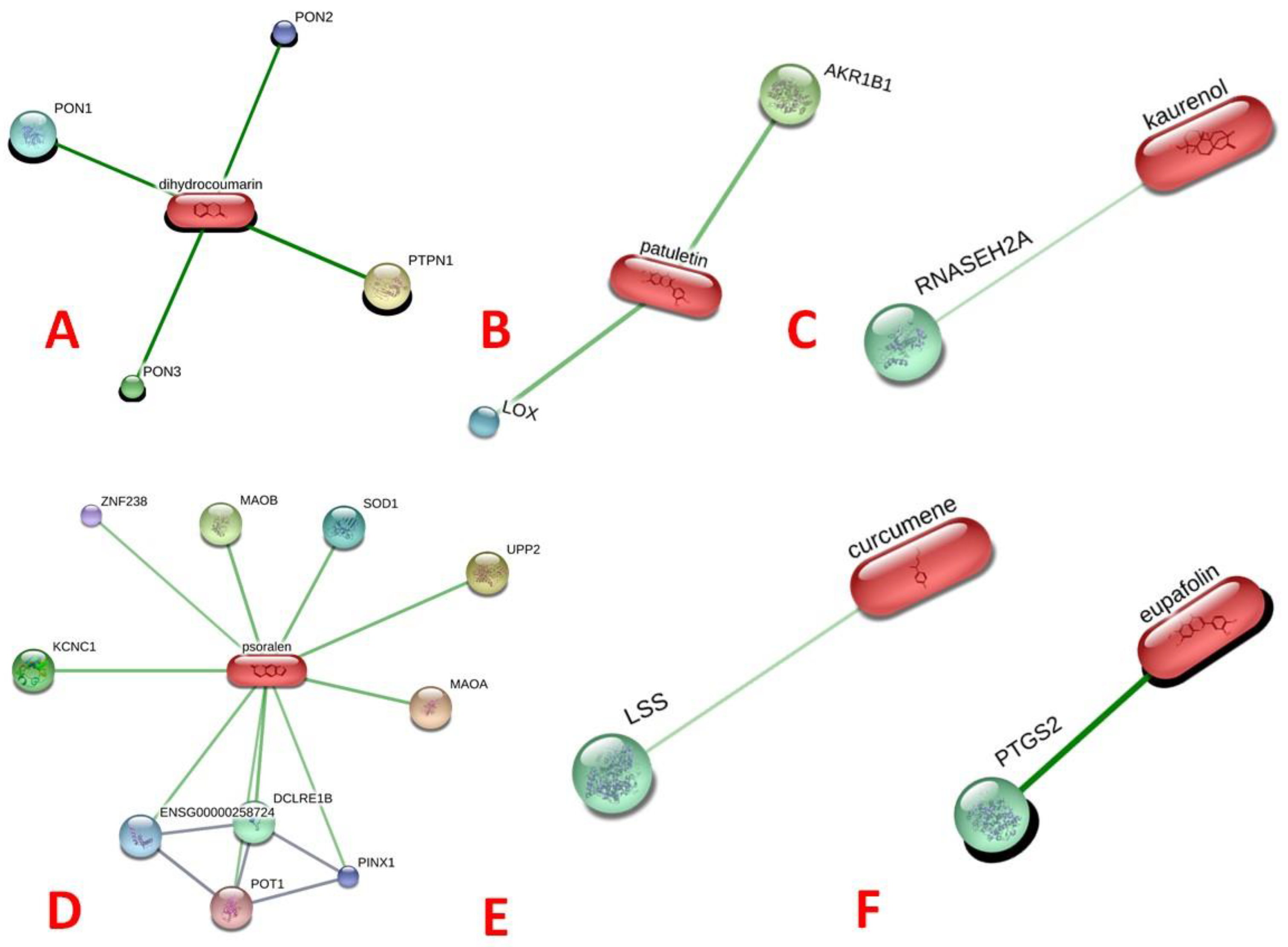
The search tool for interacting chemicals (STITCH) free web server provides comprehensive particulars regarding: (i) metabolic pathways of interactions, (ii) crystal structure information, (iii) binding investigations, and (iv) target–drug correlations [13]. In the present study, the STITCH analysis revealed that only six ligands, namely (a) dihydrocoumarin, (b) patuletin, (c) kaurenol, (d) psoralen, (e) curcumene, and (f) nepetin (eupafolin), showed interactions with human proteins (Figure 1). Interestingly, patuletin interacted with the human lipooxygenase (LOX, inflammatory) protein, as presented in Figure 1b.
Prior to the docking experiments, it is vital to understand the (i) physicochemical, (ii) drug- likeness/bioactivity score, (iii) ADME, and finally, (iv) the toxicity of the 26 chosen phytoconstituents of the Mikania species. These analyses have been shown to help in the computer-aided drug development (CADD) process [14]. Regarding the physicochemical properties, six ligands (stigmasterol, taraxerol, curcumene, bicyclogermacrene, γ-elemene, and provincialin) showed one violation, while only one ligand (3,5-methyl-di-O-caffeoyl quinate) displayed three violations for the rule of five (Table 1). Similarly, with reference to supporting the drug-likeness or the score of the bioactivity analysis, only one ligand (mikamicranolide) revealed a bioactivity score of >0 towards the six descriptors; on the other hand, the other ligands showed a bioactivity score range of active to moderate. Moreover, the other 26 selected ligands showed an inactive score (<−5.0) (Table 2).
Before docking, it is vital to know a compound’s/ligand’s properties, such as (i) physicochemical, (ii) drug-likeness or score of bioactivity, and (iii) ADMET, along with its (iv) toxicity. Moreover, standardized rule (Lipinski’s rule of five) and ADMET are available for determining such properties [15]. Concerning ADMET analysis, eleven ligands (mikamicranolide, spathulenol, caryophyllene oxide, patuletin, 3,3′,5-trihydroxy-4′,6,7-trimethoxyflavone, psoralen, herniarin, 2,6-dimethoxyquinone, dehydrocostus lactone, mikanin-3-O-sulfate, and nepetin) have hepatotoxic properties, as displayed in Table 3.
Regarding the toxicological screening of 26 ligands, as illustrated in Table 4, 5 ligands (dihydrocoumarin, patuletin, 3,3′,5-trihydroxy-4′,6, 7-trimethoxyflavone, 3-O-mikanin-sulfate along with nepetin) are non-degradable in terms of aerobic biodegradability nature. Two ligands (patuletin and 3, 3′, 5-trihydroxy-4′, 6, 7-trimethoxyflavone) are predicated as mutagens.
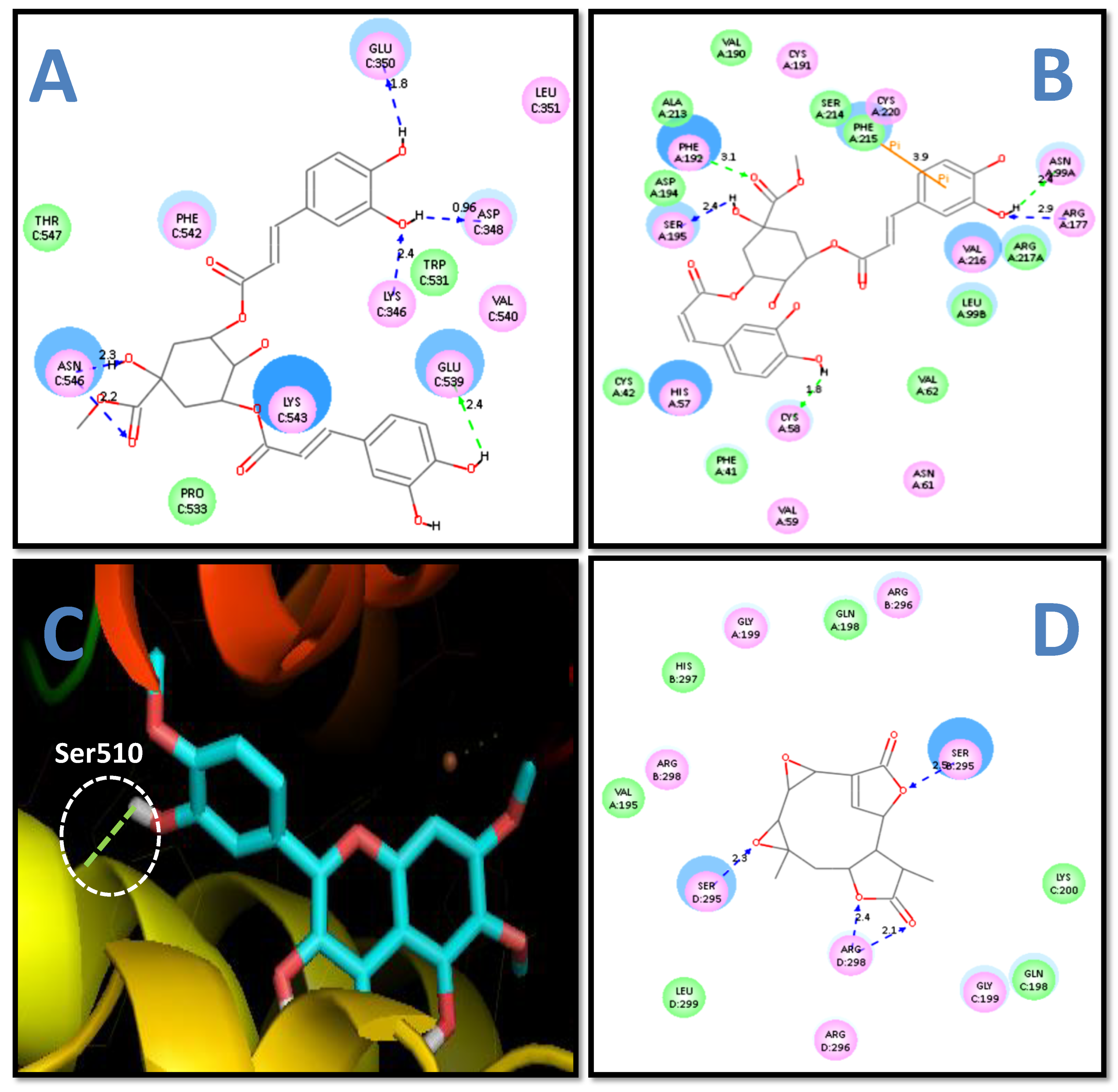
The C-docking study and free energy binding analysis (Table 5) showed that 3,5-methyl-di-O-caffeoyl quinate possesses the maximum energy interaction (−42.51 kcal/mol) with the COX 2 enzyme (as presented in Figure 2a). In contrast, psoralen revealed the least interaction energy (−15.57 kcal/mol). Moreover, eight ligands (grandifloric acid, kaurenol, o-coumaric acid, melilotoside, patuletin, 3,5-methyl-di-O-caffeoylquinate, mikanin-3-O-sulfate, and nepetin) showed interaction with the Glu539 residues of the COX 2 enzyme, as displayed in Table 5. The present results were in good conformity with our previous findings where 4-hydroxyisoleucine (4-HIL) and phytic acid (PA) showed interaction with (i) Glu539; (ii) Glu350; (iii) Asn546; and (iv) Trp531 amino acid (AA) residues of the COX 2 enzyme [16].
Stigmasterol has been described to inhibit thromboxane B2 (TXB2) production, which afterwards leads to inhibition of cyclooxygenase 1 (COX 1) activity [17]. However, no reports are available for stigmasterol’s cyclooxygenase 2 (COX 2) inhibition activity. Additionally, caryophyllene has been reported to exhibit cyclooxygenase-2 (COX-2) inhibition activity in THP-1 (human monocytic) cells [18]. Psoralen, spathulenol, syringaldehyde, and taraxerol acetate have been found to exhibit cyclooxygenase-2 (COX-2) inhibition activity [19,20,21,22,23]. All the above findings were in agreement with our results on cyclooxygenase 2 (COX 2) inhibition activity.
The HNE is an additional targeted enzyme whose docking analysis and free energy binding analysis showed that 3,5-methyl-di-O-caffeoyl quinate displayed the maximum energy of interactions (−54.66 kcal/mol), as presented in Figure 2b. Thirteen ligands (kaurenol, syringaldehyde, o-coumaric acid, melilotoside, patuletin, 3,5-methyl-di-O-caffeoyl quinate, trihydroxy-3,3′,5-trimethoxy-4′,6,7-flavone, psoralen, herniarin, 2,6-dimethoxyquinone, provincialin, mikanin-3-O-sulfate, and nepetin) exhibited interaction with Ser195 amino acid residue of HNE, as shown in Table 6. The present finding was in good agreement with our previous study, where phytic acid (PA) and 4-hydroxyisoleucine (4-HIL) demonstrated interaction with (i) Ser195; (ii) Arg147; (iii) Cys191; (iv) Phe192; (v) Gly193; (vi) Asp194; and (vii) Ser214 amino acid (AA) residues of the HNE enzyme [16].
Five sesquiterpene lactones, namely (15- (3′-Hydroxy)-methacryloyloxy-micrantholide, isobutyryloxy-15-(2′,3′-Epoxy) -micrantholide, isobutyryloxy-15-(2′-Hydroxy)-micrantholide, 4α hydroxy-1β-Acetoxy-15- eudesma-isobutyryloxy-12-8β-olide11-13-en from M. cordifolia, and Scandenolide from M. micrantha have been reported to exhibit human neutrophil elastase (HNE) inhibition activity [24]. Similarly, p-coumaric acid and di-O-caffeoyl-3,5-quinic acid, two phytochemicals, were described as possessing human neutrophil elastase (HNE) inhibition activity [25]. Both reports were in close agreement with the present findings on the human neutrophil elastase (HNE) inhibition activity.
The docking study and free binding energy analysis showed that 3,3′,5-trihydroxy-4′,6,7-trimethoxyflavone (Figure 2c) had the least binding energy (−9.71 kcal/mol) (Table 7). Moreover, five ligands (mikamicranolide, syringaldehyde, patuletin, 2,6-dimethoxyquinone, and mikanin-3-O-sulfate) exhibited interactions with the His518 amino acid residue of LOX. The current finding was in good accord with our previous study, where the compound-3e (Geranylacetophenone derivative) showed interaction with His518 amino acid residue of the LOX enzyme [26]. Similarly, our earlier study also displayed that 4-hydroxyisoleucine (4-HIL) showed interaction with (i) Ser510; (ii) His513; and (iii) Gln716 amino acid (AA) residues of the LOX enzyme [16].
Mikania micrantha (leaves and stems—ethyl acetate extract) [11], Mikania lindleyana (aerial parts of the plant—methanolic extract), and Mikania cordata (root—methanolic extract) have been described to have anti-inflammatory properties [28,29], whereas three other Mikania species (M. glomerata, M. hirsutissima, and M. laevigata) have been reported to inhibit 5- lipoxygenase (5-LOX) activity in a dose-dependent manner [30,31]. Jyothi Lakshmi [32] reported the cyclooxygenase (COX), lipoxygenase (LOX), and nitric oxide synthase (iNOS) inhibition activities of Mikania micrantha (leaf and flower extract). Similarly, (i) 6,7-dihydroxy coumarin, (ii) β- caryophyllene, and (iii) β- caryophyllene oxide have been reported to inhibit 5- lipoxygenase (5-LOX) activity [33], whereas stigmasterol has been described to inhibit 15- lipoxygenase (15-LOX) activity [34]. Kaurenoic acid has been reported to have weak lipoxygenase (LOX) inhibition activity [35]. All the above-mentioned studies are in good correlation with the current results on lipoxygenase (LOX) inhibition activity.
The docking study and binding free energy analysis with MMP 2 showed that 3,5-methyl-di-O-caffeoylquinate possessed the maximum interaction energy (−83.34 kcal/mol), and five ligands (syringaldehyde, o-coumaric acid, 3,5-methyl-di-O-caffeoylquinate, 3-O-mikanin-sulfate, and nepetin) showed interaction with the MMP2 amino acid residue Glu-202 (Table 8). This observation was in agreement with previous findings, where 4-hydroxyisoleucine (4-HIL) has shown interaction with the (i) Glu202; (ii) Ala165; and (iii) His201 amino acid (AA) residues of the MMP 2 enzyme [16].
Similarly, in the C-docking study and binding energy analysis with MMP 9, 3,5-methyl-di-O-caffeoyl quinate exhibited the maximum binding energy (−81.65 kcal/mol), and three ligands (3,5-methyl-di-O-caffeoylquinate, curcumene, and 2,6-dimethoxyquinone) displayed an interaction with His226 amino acid (AA) residue of MMP 9 (Table 9). The current result was in good correlation with our preceding study, where 3-phenyllactic acid (3-PLA) showed interaction with His226 amino acid (AA) residues of the MMP 9 enzyme [36]. Stigmasterol has been reported to reduce matrix metalloproteinase 3 (MMP 3) mRNA expression in humans and mouses, MMP 3 protein in mice, and matrix metalloproteinase 13 (MMP 13) mRNA expression in humans and mice [37]. However, in the present study, stigmasterol failed to dock with both enzymes (MMP 2 and 9).
Docking and energy binding analysis (Table 10) shows that the provincialin had maximum energy binding (−54.18 kcal/ mol) with the mPGES 2 enzyme (as illustrated in Figure 2d) and twelve ligands (syringaldehyde, o-coumaric acid, melilotoside, patuletin, 3,5-methyl-di-O-caffeoylquinate, 4′,6,7-trimethoxyflavone-3,3′,5-trihydroxy, psoralen, herniarin, provincialin, dehydrocostus lactone, mikanin-3-O-sulfate, and nepetin) had interaction with Arg298 amino acid (AA) residue of mPGES 2. Interestingly, in the present study, all 25 ligands (except for 2,6-dimethoxyquinone) showed docking and binding affinities with microsomal prostaglandin E synthase 2 (mPGES 2). Maione et al. [38] have reported that the amino acids (i) Cys110, (ii) His241, (iii) His244, (iv) Ser247, (v) Arg292, and (vi) Arg296 are the key binding residues for mPGES 2. However, there are no reports on their mPGES 2 inhibition activity.
3. Materials and Methods

3.1. Ligand (Small Molecule of Interest) Preparation

The simplified molecular input line entry specification (SMILES) of the 26 selected ligands: (i) mikamicranolide (Chemspider ID 10189069); (ii) kaurenoic acid (CID 73062); (iii) stigmasterol (CID 5280794); (iv) grandifloric acid (CID 159930); (v) kaurenol (CID 443465); (vi) spathulenol (CID 522266); (vii) caryophyllene oxide (CID 14350); (viii) syringaldehyde (CID 8655); (ix) dihydrocoumarin (CID 660); (x) o-coumaric acid (Chemspider ID 553146); (xi) taraxerol (CID 92097); (xii) melilotoside (CID 5280759); (xiii) patuletin (CID 5281678); (xiv) methyl-3,5-di-O-caffeoyl quinate (ChEBI ID 66708); (xv) 3,3′,5-trihydroxy-4′,6,7-trimethoxyflavone (Chemspider ID 4476175); (xvi) psoralen (CID 6199); (xvii) curcumene (CID 92139); (xviii) herniarin (Chemspider ID 10295); (xix) 2,6-dimethoxyquinone; (xx) bicyclogermacrene (CID 5315347); (xxi) α-bisabolol (CID 442343); (xxii) γ-elemene (CID 6432312); (xxiii) provincialin (ChEBI ID 8599); (xxiv) dehydrocostus lactone (CID 73174); (xxv) mikanin-3-O-sulfate (CID 14630674); and (xxvi) nepetin (Chemspider ID 4476172) were obtained from (i) Chemspider, (ii) PubMed, and (iii) Chemical Entities of Biological Interest. A three-dimensional structure of 2, 6-dimethoxy quinone was generated using ChemBioDraw Ultra 12.0. All the 26 ligands [above-mentioned] were sketched using Ultra 12.0 ChemBioDraw software and further MM2—molecular mechanics ligand minimization—was performed using Ultra 12.0 ChemBio3D software. Thus, these minimized energy ligands [3D images] were engaged for Autodock and in the C-docker case, and the ligand in-build preparation procedure (Accelrys, San Diego, CA, USA) was applied [16].

3.2. Protein Network Interaction Analysis

The search tool for interacting chemicals [STITCH] free web server [39] was employed to identify the interaction between ligands (26 selected phyto-constituents of Mikania species) and human proteins.

3.3. Selection of Target Protein (Enzyme) and Preparation

The 3D enzymes of (i) COX 2 (3LN1), with a resolution of 2.40 Å; (ii) HNE (1H1B [PDB number], with a resolution of 2.00 Å; (iii) LOX (1JNQ [PDB number], with a resolution of 2.10 Å; (iv) MMP 2 (1QIB [PDB number], with 2.80 angstrom (Å) resolution; (v) MMP9 (4H1Q) with 1.59 Å resolution; and (vi) mPGES 2 (1Z9H), with 2.60 Å resolution. were retrieved from the RCSB Protein Data Bank. In COX2, the C chain was processed, and mPGES 2, all chains were processed individually by eliminating the B, C, and D ligands along with the crystallographically detected water (H2O) particles. The enzymes mentioned above were primed using Chimera UCSF software for Autodocking and C-docker in-built protein preparation procedure (Accelrys, San Diego, CA, USA) was applied [16].

3.4. Physicochemical and Drug-Likeness or Bioactivity Score Analyses

The physicochemical and drug-likeness or biological activity score analyses were conducted for the selected twenty-six selected (Mikania) ligands utilizing the Mol-inspiration-free web server [16].

3.5. ADMET and TOPKAT Analyses

The ADMET and TOPKAT analyses were performed using Discovery Studio (Accelrys, San Diego, CA, USA) for the 26 selected (Mikania) ligands [16].

3.6. Docking Analysis

The docking analysis was performed for twenty-six screened compounds extracted from Mikania utilizing C-docker. The 3D structures of COX 2; MMP 2; HNE; MMP 9; and mPGES 2 were recovered from the Protein Data Bank and further processed with the C-docker procedure [40] along the protein–ligand interaction section using 3.1. Discovery Studio® (Accelrys, San Diego, CA, USA) was utilized. A model of Autodock 4.2 was used for LOX alone, where all rotatable bonds [rotb] along the twenty-six Mikania ligands were withheld for the flexible docking approach. The grid size was fixed (60 × 60 × 60) with a space of 0.375 Å between the grid points. Lamarckian Genetic Algorithm (LGA) was used to choose the good conformers. Similarly, a genetic algorithm was used to produce 100 individual docking runs for each selected Mikania ligand. In summary, the standardized Autodock step-wise docking protocol was used for the current study [16].

4. Conclusions

The present study found that 3,5-methyl-di-O-caffeoylquinate was efficient in binding with five target enzymes, whereas kaurenoic acid did not bind with the selected four targeted proteins. These two phytochemicals showed good efficacy as potential anti-inflammatory drugs of non-steroid [NSAIDs] nature. Interestingly, all 26 selected ligands (except 2, 6-dimethoxy quinone) from Mikania species showed good docking and binding to mPGES 2. Thus, the findings of this study indicate that it is possible to suppress COX 2, HNE, LOX, MMP 2 and 9, and mPGES 2 in the treatment of acute and chronic inflammatory diseases using these ligands of Mikania species.

Author Contributions

Conceptualization, software and methodology, N.R.; formal analysis and validation, V.-S.P.; data manager, M.A.W.; report documentation—initial manuscript draft preparation, N.R.; report documentation—reviewing and redrafting, S.G.K., R.V. and A.B.; supervision, R.V.; funding acquisition, A.B. All authors have read and agreed to the published version of the manuscript.

Funding

The authors express their sincere appreciation to the Researchers Supporting Project Number (RSP2023R466), King Saud University, Riyadh, Saudi Arabia.

Institutional Review Board Statement

Not applicable.

Informed Consent Statement

Not applicable.

Data Availability Statement

All of the study data details are readily available from the corresponding authors and are available upon request.

Conflicts of Interest

The authors declare no conflict of interest.

References

  1. Ismail, B.S.; Mah, L.S. Effects of Mikania micrantha HBK on germination and growth of weed species. Plant Soil 1993, 157, 107–113. [Google Scholar]
  2. Bora, A.R.; Deka, J.; Barua, I.C.; Barman, B. Intensity of Mikania micrantha in coffee and other plantations of Karbi Anglong district, Assam. Indian J. Weed Sci. 2019, 51, 95–97. [Google Scholar] [CrossRef] [Green Version]
  3. Debaprotim, D.; Suvakanta, D.; Jashabir, C. Evaluation of anticancer activity of Mikania micrantha Kunth (Asteraceae) against ehrlich ascites carcinoma in swiss albino mice. Int. J. Pharm. Res. Allied Sci. 2014, 3, 9–18. [Google Scholar]
  4. Zohmachhuana, A.; Tlaisun, M.; Mathipi, V.; Khawlhring, L.; Priya, J.S. Suppression of the RAGE gene expression in RAW 264.7 murine leukemia cell line by ethyl acetate extract of Mikania micrantha (L.) Kunth. J. Appl. Biol. Biotechnol. 2022, 10, 107–114. [Google Scholar] [CrossRef]
  5. Rahman, M.M.; Kabir, M.M.; Noman, M.A.A.; Islam, M.R.; Dash, B.K.; Akhter, S.; Uddin, M.J.; Rahman, A. Mikania cordata leaves extract promotes activity against pathogenic bacteria and anticancer activity in EAC cell-bearing swiss albino mice. J. Appl. Pharm. Sci. 2020, 10, 112–122. [Google Scholar]
  6. Da Silva, A.B.; Owiti, A.; Barbosa, W. Pharmacology of Mikania genus: A systematic review. Pharmacogn. Rev. 2018, 12, 230–237. [Google Scholar]
  7. Radhakrishnan, N.; Karthi, S.; Raghuraman, P.; Ganesan, R.; Srinivasan, K.; Edwin, E.S.; Ganesh-Kumar, S.; Mohd Esa, N.; Senthil-Nathan, S.; Vasantha-Srinivasan, P.; et al. Chemical screening and mosquitocidal activity of essential oil derived from Mikania scandens (L.) Willd. against Anopheles gambiae Giles and their non-toxicity on mosquito predators. All Life 2023, 16, 2169959. [Google Scholar] [CrossRef]
  8. Barlow, D.J.; Buriani, A.; Ehrman, T.; Bosisio, E.; Eberini, I.; Hylands, P.J. In-silico studies in Chinese herbal medicines’ research: Evaluation of in-silico methodologies and phytochemical data sources, and a review of research to date. J. Ethnopharmacol. 2012, 140, 526–534. [Google Scholar] [CrossRef] [Green Version]
  9. Prachayasittikul, V.; Worachartcheewan, A.; Shoombuatong, W.; Songtawee, N.; Simeon, S.; Prachayasittikul, V.; Nantasenamat, C. Computer-aided drug design of bioactive natural products. Curr. Top. Med. Chem. 2015, 15, 1780–1800. [Google Scholar] [CrossRef]
  10. Suyenaga, E.S.; Reche, E.; Farias, F.M.; Schapoval, E.E.S.; Chaves, C.G.M.; Henriques, A.T. Antiinflammatory investigation of some species of Mikania. Phytother. Res. 2002, 16, 519–523. [Google Scholar] [CrossRef]
  11. Perez-Amador, M.C.; Munoz Ocotero, V.; Ibarra Balcazar, R.; Garcia Jimenez, F. Phytochemical and pharmacological studies on Mikania micrantha HBK (Asteraceae). Phyton 2010, 79, 77–80. [Google Scholar]
  12. Della Pasqua, C.S.P.; Iwamoto, R.D.; Antunes, E.; Borghi, A.A.; Sawaya, A.C.H.F.; Landucci, E.C.T. Pharmacological study of anti-inflammatory activity of aqueous extracts of Mikania glomerata (Spreng.) and Mikania laevigata (Sch. Bip. ex Baker). J. Ethnopharmacol. 2019, 231, 50–56. [Google Scholar] [CrossRef] [PubMed]
  13. Kuhn, M.; von Mering, C.; Campillos, M.; Jensen, L.J.; Bork, P. STITCH: Interaction networks of chemicals and proteins. Nucleic Acids Res. 2007, 36, D684–D688. [Google Scholar] [CrossRef] [PubMed] [Green Version]
  14. Kumaraswamy, S.; Arumugam, G.; Pandurangan, A.K.; Prabhakaran, V.S.; Narayanaswamy, R. Molecular docking analysis of organic acids (OA) from honey as modulators of human ferritin, transferrin, and hepcidin. J. Microbiol. Biotechnol. Food Sci. 2023, 12, 5743. [Google Scholar] [CrossRef]
  15. Lipinski, C.A.; Lombardo, F.; Dominy, B.W.; Feeney, P.J. Experimental and computational approaches to estimate solubility and permeability in drug discovery and development settings. Adv. Drug Deliv. Rev. 2001, 46, 03–26. [Google Scholar] [CrossRef]
  16. Narayanaswamy, R.; Wai, L.K.; Esa, N.M. Molecular docking analysis of phytic acid and 4-hydroxyisoleucine as cyclooxygenase-2, microsomal prostaglandin E synthase-2, tyrosinase, human neutrophil elastase, matrix metalloproteinase-2 and-9, xanthine oxidase, squalene synthase, nitric oxide synthase, human aldose reductase, and lipoxygenase inhibitors. Pharmacogn. Mag. 2017, 13, S512–S518. [Google Scholar]
  17. Saadawi, S.; Jalil, J.; Jasamai, M.; Jantan, I. Inhibitory effects of acetylmelodorinol, chrysin and polycarpol from Mitrella kentii on prostaglandin E2 and thromboxane B2 production and platelet activating factor receptor binding. Molecules 2012, 17, 4824–4835. [Google Scholar] [CrossRef] [Green Version]
  18. Park, K.R.; Nam, D.; Yun, H.M.; Lee, S.G.; Jang, H.J.; Sethi, G.; Cho, S.K.; Ahn, K.S. β-Caryophyllene oxide inhibits growth and induces apoptosis through the suppression of PI3K/AKT/mTOR/S6K1 pathways and ROS-mediated MAPKs activation. Cancer Lett. 2011, 312, 178–188. [Google Scholar] [CrossRef]
  19. Lomarat, P.; Sripha, K.; Phanthong, P.; Kitphati, W.; Thirapanmethee, K.; Bunyapraphatsara, N. In vitro biological activities of black pepper essential oil and its major components relevant to the prevention of Alzheimer’s disease. Thai J. Pharm. Sci. 2015, 39, 94–101. [Google Scholar]
  20. Zheng, M.; Jin, W.; Son, K.H.; Chang, H.W.; Kim, H.P.; Bae, K.; Kang, S.S. The constituents isolated from Peucedanum japonicum Thunb. and their cyclooxygenase (COX) inhibitory activity. Korean J. Med. Crop Sci. 2005, 13, 75–79. [Google Scholar]
  21. Jayaprakasam, B.; Alexander-Lindo, R.L.; DeWitt, D.L.; Nair, M.G. Terpenoids from Stinking toe (Hymneae courbaril) fruits with cyclooxygenase and lipid peroxidation inhibitory activities. Food Chem. 2007, 105, 485–490. [Google Scholar] [CrossRef]
  22. Stanikunaite, R.; Khan, S.I.; Trappe, J.M.; Ross, S.A. Cyclooxygenase-2 inhibitory and antioxidant compounds from the truffle Elaphomyces granulatus. Phytother. Res. 2009, 23, 575–578. [Google Scholar] [CrossRef]
  23. Rehman, U.U.; Shah, J.; Khan, M.A.; Shah, M.R.; Khan, I. Molecular docking of taraxerol acetate as a new COX inhibitor. Bangladesh J. Pharmacol. 2013, 8, 194–197. [Google Scholar] [CrossRef] [Green Version]
  24. Siedle, B.; Cisielski, S.; Murillo, R.; Löser, B.; Castro, V.; Klaas, C.A.; Hucke, O.; Labahn, A.; Melzig, M.F.; Merfort, I. Sesquiterpene lactones as inhibitors of human neutrophil elastase. Bioorg. Med. Chem. 2002, 10, 2855–2861. [Google Scholar] [CrossRef]
  25. Siedle, B.; Hrenn, A.; Merfort, I. Natural compounds as inhibitors of human neutrophil elastase. Planta Med. 2007, 73, 401–420. [Google Scholar] [CrossRef] [PubMed] [Green Version]
  26. Ng, C.H.; Rullah, K.; Aluwi, M.F.F.M.; Abas, F.; Lam, K.W.; Ismail, I.S.; Narayanaswamy, R.; Jamaludin, F.; Shaari, K. Synthesis and docking studies of 2, 4, 6-trihydroxy-3-geranylacetophenone analogs as potential lipoxygenase inhibitor. Molecules 2014, 19, 11645–11659. [Google Scholar] [CrossRef] [Green Version]
  27. Castro, J.S.; Trzaskowski, B.; Deymier, P.A.; Bucay, J.; Adamowicz, L.; Hoying, J.B. Binding affinity of fluorochromes and fluorescent proteins to Taxol™ crystals. Mater. Sci. Eng. C 2009, 29, 1609–1615. [Google Scholar] [CrossRef]
  28. Ríos, E.V.; León, A.; Chávez, M.I.; Torres, Y.; Ramírez-Apan, M.T.; Toscano, R.A.; Bravo-Monzón, Á.E.; Espinosa-García, F.J.; Delgado, G. Sesquiterpene lactones from Mikania micrantha and Mikania cordifolia and their cytotoxic and anti-inflammatory evaluation. Fitoterapia 2014, 94, 155–163. [Google Scholar] [CrossRef]
  29. Vanderlinde, F.A.; Rocha, F.F.; Malvar, D.C.; Ferreira, R.T.; Costa, E.A.; Florentino, I.F.; Guilhon, G.; Lima, T. Anti-inflammatory and opioid-like activities in methanol extract of Mikania lindleyana, sucuriju. Rev. Bras. Farmacogn. 2012, 22, 150–156. [Google Scholar] [CrossRef] [Green Version]
  30. Kumar, S.; Bajwa, B.S.; Singh, K.; Kalia, A.N. Anti-inflammatory activity of herbal plants: A review. Int. J. Adv. Pharm. Biol. Chem. 2013, 2, 272–281. [Google Scholar]
  31. Chagas-Paula, D.A.; Oliveira, T.B.; Faleiro, D.P.; Oliveira, R.B.; Da Costa, F.B. Outstanding anti-inflammatory potential of selected Asteraceae species through the potent dual inhibition of cyclooxygenase-1 and 5-lipoxygenase. Planta Med. 2015, 81, 1296–1307. [Google Scholar] [CrossRef] [PubMed] [Green Version]
  32. Jyothilakshmi, M.; Jyothis, M.; Latha, M.S. Cyclooxygenase, Lipoxygenase, Nitric Oxide Synthase, Myeloperoxidase and Protease inhibiting activities of the leaves and flowers of Mikania micrantha Kunth. J. Complement. Med. Res. 2020, 11, 51. [Google Scholar] [CrossRef]
  33. Werz, O. Inhibition of 5-lipoxygenase product synthesis by natural compounds of plant origin. Planta Med. 2007, 73, 1331–1357. [Google Scholar] [CrossRef] [PubMed] [Green Version]
  34. El-Ahmady, S.H.; Ashour, M.L.; Wink, M. Chemical composition and anti-inflammatory activity of the essential oils of Psidium guajava fruits and leaves. J. Essent. Oil Res. 2013, 25, 475–481. [Google Scholar] [CrossRef]
  35. de Vargas, F.S.; de Almeida, P.D.; Aranha, E.S.P.; de Boleti, A.P.A.; Newton, P.; de Vasconcellos, M.C.; Junior, V.F.V.; Lima, E.S. Biological activities and cytotoxicity of diterpenes from Copaifera spp. Oleoresins. Molecules 2015, 20, 6194–6210. [Google Scholar] [CrossRef] [Green Version]
  36. Narayanaswamy, R.; Kok Wai, L.; Ismail, I.S. In silico analysis of selected honey constituents as human neutrophil elastase (HNE) and matrix metalloproteinases (MMP 2 and 9) inhibitors. Int. J. Food Prop. 2015, 18, 2155–2164. [Google Scholar] [CrossRef]
  37. Gabay, O.; Sanchez, C.; Salvat, C.; Chevy, F.; Breton, M.; Nourissat, G.; Wolf, C.; Jacques, C.; Berenbaum, F. Stigmasterol: A phytosterol with potential anti-osteoarthritic properties. Osteoarthr. Cartil. 2010, 18, 106–116. [Google Scholar] [CrossRef]
  38. Maione, F.; Minosi, P.; Di Giannuario, A.; Raucci, F.; Chini, M.G.; De Vita, S.; Bifulco, G.; Mascolo, N.; Pieretti, S. Long-lasting anti-inflammatory and antinociceptive effects of acute ammonium glycyrrhizinate administration: Pharmacological, biochemical, and docking studies. Molecules 2019, 24, 2453. [Google Scholar] [CrossRef] [Green Version]
  39. Szklarczyk, D.; Santos, A.; Von Mering, C.; Jensen, L.J.; Bork, P.; Kuhn, M. STITCH 5: Augmenting protein–chemical interaction networks with tissue and affinity data. Nucleic Acids Res. 2016, 44, D380–D384. [Google Scholar] [CrossRef] [Green Version]
  40. Wu, G.; Robertson, D.H.; Brooks, C.L.; Vieth, M. Detailed analysis of grid-based molecular docking: A case study of CDOCKER—A CHARMm-based MD docking algorithm. J. Comput. Chem. 2003, 24, 1549–1562. [Google Scholar] [CrossRef]
Figure 1. Representation of the protein network analysis (selected ligands of Mikania with human enzymes). (A) Dihydrocoumarin, (B) patuletin, (C) kaurenol, (D) psoralen, (E) curcumene, and (F) nepetin (eupafolin).
Processes 11 01722 g001
Figure 2. The two-dimensional (2D) structure of methyl-3,5-di-O-caffeoyl quinate with (A) COX 2 and (B) HNE; hydrogen atoms have been excluded in two-dimensional (2D) images for good explanation and bond distances are expressed in (Å) angstroms; (C) three-dimensional (3D) structure of 3,3′,5-trihydroxy-4′,6,7-trimethoxyflavone with LOX (docked using Autodock and analyzed using pyMOL method) and (D) two-dimensional (2D) structure of provincialin with mPGES 2.
Processes 11 01722 g002
Table 1. The physicochemical analysis of 26 (Mikania) ligands using the Mol-inspiration free web server.
Ligand Log A Natoms MW noN ●●nOH NH ◊◊Nviolations *Nrotb **
Mikamicranolide−2.1422308.37100
Kaurenoic acid4.6722302.52101
Stigmasterol7.8730412.71115
Grandifloric acid3.7523318.53201
Kaurenol4.7921288.51101
Spathulenol3.9116220.41100
Caryophyllene oxide4.1416220.41000
Syringaldehyde1.0813182.24103
Dihydrocoumarin1.7911148.22000
o-Coumaric acid1.6712164.23202
Taraxerol8.0231426.71110
Melilotoside−0.5823326.38505
Patuletin1.7024332.38502
Methyl-3,5-di-O-caffeoyl quinate2.0438530.5126310
3,3′,5-Trihydroxy-4′,6,7-trimethoxyflavone2.3126360.38304
Psoralen2.2914186.23000
Curcumene5.8215202.30014
Herniarin2.0513176.23001
2,6-Dimethoxyquinone0.5312168.24002
Bicyclogermacrene5.2915204.40010
α-Bisabolol4.6816222.41104
γ-Elemene5.4215204.40012
Provincialin1.9137518.6102111
Dehydrocostus lactone2.2917230.32000
Mikanin-3-O-sulfate0.3629424.410206
Nepetin1.9923316.37402
Note: ◊-Octanol–Water (O/W) partition coefficient; ▪-molecular weight; ■-number of non-hydrogen atoms; ◊◊- number of hydrogen bond donors [OH and NH groups]; ●● number of hydrogen bond acceptors [O and N atoms]; *- no. of rule of five violations, and ** no. of rotatable bonds (Nrotb).
Table 2. The drug-likeness or bioactivity analysis of 26 (Mikania) ligands utilized the Mol-inspiration free web server.
Ligand GPCR LigandIon-Channel Modulator Kinase InhibitorNuclear Receptor LigandProtease InhibitorEnzyme Inhibitor
Mikamicranolide0.280.030.010.660.070.56
Kaurenoic acid0.290.15−0.390.750.060.46
Stigmasterol0.12−0.08−0.490.74−0.020.53
Grandifloric acid0.210.12−0.470.780.100.43
Kaurenol0.210.10−0.210.67−0.020.44
Spathulenol−0.42−0.28−0.680.28−0.360.05
Caryophyllene oxide−0.080.14−0.860.620.000.57
Syringaldehyde−0.95−0.36−0.80−0.69−1.27−0.39
Dihydrocoumarin−0.90−0.48−1.25−0.75−1.13−0.47
o-Coumaric acid−0.64−0.37−0.98−0.25−0.90−0.21
Taraxerol0.210.02−0.200.540.000.49
Melilotoside0.17−0.03−0.130.270.040.40
Patuletin−0.14−0.340.220.13−0.350.17
3,5-Methyl-di-O-caffeoyl quinate0.11−0.07−0.060.340.070.25
3,3′,5-Trihydroxy−4′,6,7-trimethoxyflavone−0.14−0.330.200.09−0.340.14
Psoralen−0.89−0.38−1.11−1.13−1.19−0.37
Curcumene−0.47−0.12−0.80−0.24−0.72−0.14
Herniarin−1.23−0.84−1.28−1.06−1.28−0.47
2,6-Dimethoxyquinone−1.48−0.69−0.78−1.50−1.36−0.42
Bicyclogermacrene−0.75−0.69−1.11−0.65−0.88−0.16
α-Bisabolol−0.060.26−0.780.37−0.380.43
γ-Elemene−0.460.02−1.010.51−0.710.24
Provincialin0.320.23−0.150.950.070.82
Dehydrocostus lactone−0.04−0.02−0.561.00−0.220.66
Mikanin-3-O-sulfate0.08−0.300.020.010.060.45
Nepetin−0.08−0.230.220.17−0.310.16
Note: ■- G Protein-coupled receptors (GPCR).
Table 3. ADMET analysis of 26 (Mikania) ligands using Discovery Studio.
LigandHIA AS BBB aPPB **CYP2D6 ◊◊HT b
L *L **L ***Predication
Mikamicranolide043FFT
Kaurenoic acid020TFF
Stigmasterol314TFF
Grandifloric acid021TFF
Kaurenol020TFF
Spathulenol031TFT
Caryophyllene oxide020TFT
Syringaldehyde043TFF
Dihydrocoumarin031TFF
o-Coumaric acid043FFF
Taraxerol3#04TFF
Melilotoside1#44FFF
Patuletin134FFT
3,5-Methyl-di-O-caffeoyl quinate3#34FFF
3,3′,5-Trihydroxy-4′,6,7-trimethoxyflavone034TFT
Psoralen032FFT
Curcumene120TFF
Herniarin032TFT
2,6-Dimethoxyquinone043FFT
Bicyclogermacrene120TFF
α-Bisabolol020TFF
γ-Elemene120TFF
Provincialin234FFF
Dehydrocostus lactone021TFT
Mikanin-3-O-sulfate134TFT
Nepetin034TTT
Note: (■-AS—Aqueous solubility; ◊-HIA—Human intestinal absorption; ** PPB—Plasma protein binding; a-BBB—Blood–brain barrier; b HT—Hepatotoxicity; ◊◊ CYP2D6—Cytochrome P450 2D6; T—True, F—False, and L—Level). * [0—Strong. 1—Medium. 2—Weak, and 3—Very weak]; ** [0—Extremely weak, 1—Very weak, 2—Weak, 3—Strong, 4—Optimal, 5—Soluble, and 6—Warning]; *** [0—Very strong penetration, 1—Strong, 2—Moderate, 3—Low, and 4—Undefined].
Table 4. The toxicological screening of 26 (Mikania) ligands using Discovery Studio.
LigandsAB AM OI SI ◊◊Oral Toxicity *
MikamicranolideDNMII1.02
Kaurenoic acidDNMII1.53
StigmasterolDNMII1.18
Grandifloric acidDNMII1.44
KaurenolDNMII1.85
SpathulenolDNMII0.75
Caryophyllene oxideDNMII1.13
SyringaldehydeDNMII1.26
DihydrocoumarinNDNM II0.74
o-Coumaric acidDNMINI1.59
TaraxerolDNMII0.93
MelilotosideDNMINI1.32
PatuletinND MINI1.08
Methyl-3,5-di-O-caffeoyl quinateDNMINI2.37
3,3′,5-Trihydroxy-4′,6,7-trimethoxyflavoneNDMINI0.93
PsoralenDNMII0.30
CurcumeneDNMNII2.47
HerniarinDNMNII0.68
2,6-DimethoxyquinoneDNMII0.63
BicyclogermacreneDNMII0.48
α-BisabololDNMII1.65
γ-ElemeneDNMII2.00
ProvincialinDNMII3.11
Dehydrocostus lactoneDNMII1.45
Mikanin-3-O-sulfateNDNMININA **
NepetinNDNMINI0.68
Note: (AM —Ames mutagenicity, AB —Aerobic biodegradability, SI ◊◊—Skin irritancy, I —Ocular irritancy, and Oral toxicity *—Oral toxicity in rat [LD50 in g/Kg]; D—Degradable, ND—Non-degradable, M—Mutagen, NM—Non-mutagen, I—Irritant, NI—Non-irritant, and NA **—Not analyzed).
Table 5. Energy interaction analysis of twenty-six (Mikania) ligands along with cyclooxygenase 2 (COX 2) utilizing Discovery Studio.
Ligandsc-Docker Energy Interaction (-kcal/mol)Amino Acid Interaction Residue (AA)Bond Distance (Å)
Mikamicranolide22.37Asn5461.1
Kaurenoic acidF *--
Stigmasterol34.51Lys3462.5
Asp3481.2
Grandifloric acid24.17Glu5390.91
Asn5461.5 and 1.7
Kaurenol21.75Glu5390.55
Spathulenol18.08No interaction-
Caryophyllene oxide17.20No interaction-
Syringaldehyde21.57Asn5461.5
Dihydrocoumarin16.97No interaction-
o-Coumaric acid18.23Glu5392.0
Asn5461.5
Lys543 6.5
Taraxerol29.03Glu3500.96
Melilotoside32.06Lys3462.2
Asp3481.9
Glu5391.7
Asn5461.7
Lys328 6.3
Patuletin28.65Asp3480.53
Glu3502.2
Trp5311.4
Glu5392.3
Methyl-3,5-di-O-caffeoyl quinate42.51Lys3462.4
Asp3480.96
Glu3501.8
Glu5392.4
Asn5462.2 and 2.3
3,3′,5-Trihydroxy-4′,6,7-trimethoxyflavone28.20Asn5462.4
Psoralen15.57Asn5460.8 and 2.2
Lys543 5.1 and 5.8
Curcumene19.47No interaction-
Herniarin17.10Asn5462.4
2,6-Dimethoxyquinone18.20Asn5461.3
Bicyclogermacrene17.12No interaction-
α-Bisabolol23.26Asn5462.2
γ-Elemene17.41No interaction-
Provincialin37.74Lys3461.8
Dehydrocostus lactone19.95No interaction-
Mikanin-3-O-sulfate29.99Glu5392.1
Asn5462.2 and 2.5
Lys328 6.6
Nepetin32.50Asp3480.31
Glu3502.2
Trp5311.7
Glu5392.1
Note: [F *—Failed to dock; —+-π interaction].
Table 6. Energy interaction analysis of 26 ligands (Mikania) along with HNE using Discovery Studio.
LigandsEnergy Interaction of c-Docker (-kcal/mol)Amino Acid Interaction Residue (AA) Bond Distance (Å)
Mikamicranolide31.32No interaction-
Kaurenoic acidF *--
Stigmasterol34.31No interaction-
Grandifloric acid26.95Gly2192.8
Kaurenol28.05Ser1952.3
Spathulenol23.55No interaction-
Caryophyllene oxide23.54No interaction-
Syringaldehyde30.11Cys1911.8
Gly1932.6
Asp1942.8
Ser1952.8
Val2163.1
Dihydrocoumarin20.95Arg1472.2
Phe1922.8
o-Coumaric acid22.74Cys1912.0
Gly1932.8
Asp1943.1
Ser1951.9, 2.6 and 2.9
Ser2142.1
Taraxerol28.96No interaction-
Melilotoside37.79Arg1472.4
Phe1921.9 and 2.9
Gly1933.0
Ser1953.1
Gly2192.5
Patuletin35.32Cys1912.0
Gly1932.8
Asp1943.1
Ser1952.8
Ser2142.1
Gly2183.0
Gly2192.8
3,5-Methyl-di-O-caffeoyl quinate54.66Cys581.8
Asn99A2.4
Arg1772.9
Phe1923.1
Ser1952.4
Phe2153.9
4′,6,7-trimethoxy-3,3′,5-Trihydroxyflavone34.66Gly1933.0
Ser1953.1 and 3.1
Gly2192.9
Psoralen19.74Phe1923.2
Ser1952.1
Curcumene24.66No interaction-
Herniarin24.89Arg1472.9
Phe1922.8
Ser1952.1
2,6-Dimethoxyquinone22.60Gly1932.7
Ser1952.9 and 3.0
Bicyclogermacrene23.35No interaction-
α-Bisabolol25.75No interaction-
γ-Elemene18.53No interaction-
Provincialin49.20Gly1932.9
Ser1952.7 and 3.1
Gly2192.6
Val2163.1
Dehydrocostus lactone25.02No interaction-
Mikanin-3-O-sulfate40.31Cys1911.8
Phe1922.6
Gly1933.0
Ser1952.8 and 3.1
Ser2143.2
Val2161.7 and 2.7
Nepetin31.90Cys1912.1
Gly1932.9
Ser1953.0
Gly2182.8
Gly2192.6
Note: [F *—Failed to dock].
Table 7. Energy interaction analysis of twenty-six (Mikania) ligands along with LOX utilizing Autodock.
LigandsMinimal Binding Energy (-kcal/mol)Amino Acid Interaction Residue (AA) Bond Distance (Å)
Mikamicranolide8.21His5182.2
Trp5193.2
Kaurenoic acid6.50His5133.3
Gln7162.0 and 3.2
Stigmasterol7.03Ile5572.6
Grandifloric acid4.82No interaction-
Kaurenol8.37No interaction-
Spathulenol7.43No interaction-
Caryophyllene oxide8.00No interaction-
Syringaldehyde5.33Gln5142.0
His5182.7
Dihydrocoumarin5.74His5233.6
Ile5573.2
o-Coumaric acid4.44Ser5102.1
Gln5142.1
Taraxerol+11.79ND *ND *
Melilotoside6.79Ser5101.8 and 3.4
Gln5142.3
Patuletin9.32His5131.9 and 3.4
Gln5142.7
His5183.2
Arg7262.1
3,5-Methyl-di-O-caffeoyl quinate+30.23ND *ND *
4′,6,7-3,3′,5-Trihydroxy-trimethoxyflavone9.71Ser5102.1
Psoralen6.51No interaction-
Curcumene7.73No interaction-
Herniarin5.79No interaction-
2,6-Dimethoxyquinone4.93His5183.1
Bicyclogermacrene7.94No interaction-
α-Bisabolol8.11Gln7161.9 and 3.2
γ-Elemene7.54No interaction-
Provincialin+46.86ND *ND *
Dehydrocostus lactone8.26His5232.7
Mikanin-3-O-sulfate4.88Ser5101.7
His5133.2
Gln5141.8 and 2.4
His5183.5
Nepetin9.21Arg7262.1 and 3.4
Note: [+—Positive sign represents the (weak) binding energy, which may be due to an improper binding feature as demonstrated by Castro et al. [27]; ND *—Not determined].
Table 8. Energy interaction analysis of twenty-six ligands of (Mikania) MMP 2 utilizing Discovery Studio.
LigandsMinimal Binding Energy (-kcal/mol)Amino Acid Interaction Residue (AA) Bond Distance (Å)
MikamicranolideF *--
Kaurenoic acidF *--
StigmasterolF *--
Grandifloric acidF *--
KaurenolF *--
SpathulenolF *--
Caryophyllene oxideF *--
Syringaldehyde33.92Ala1672.5
Glu2022.0
Dihydrocoumarin31.67No interaction-
o-Coumaric acid38.99Glu2021.7
TaraxerolF *--
MelilotosideF *--
Patuletin47.85Gly1622.5
Leu1642.0
Ala1671.2 and 1.7
Zn501 3.6
3,5-Methyl-di-O-caffeoylquinate83.34His201 4.7
Glu2021.3
Glu2101.9
4′,6,7-trimethoxyflavone-3,3′,5-Trihydroxy48.36His1662.0
Ala1672.3
Pro2212.4
Psoralen34.40No interaction-
Curcumene34.54Zn501 3.4
Herniarin36.51No interaction-
2,6-Dimethoxyquinone33.27Leu1642.0
Ala1651.6
Bicyclogermacrene19.70No interaction-
α-Bisabolol39.85His2012.5
γ-ElemeneF *--
ProvincialinF *--
Dehydrocostus lactoneF *--
Mikanin-3-O-sulfate52.36Leu163 2.2
His1662.3
Ala1671.7
Glu2021.9
Pro2211.8
Nepetin46.27Gly1622.4
Ala1672.1
Glu2021.5
Note: [F *—Failed to dock; —+–π interaction; — π–π interaction; —Sigma–π interaction].
Table 9. Energy interaction analyzes of twenty-six ligands (Mikania) MMP 9 utilizing Discovery Studio.
LigandsEnergy Interaction of c-Docker (-kcal/mol)Amino Acid Interaction Residue (AA) Bond Distance (Å)
MikamicranolideF *--
Kaurenoic acidF *--
StigmasterolF *--
Grandifloric acidF *--
KaurenolF *--
SpathulenolF *--
Caryophyllene oxideF *--
Syringaldehyde36.26Tyr2483.2 and 3.2
Dihydrocoumarin33.03No interaction-
o-Coumaric acid40.73Ala1892.0
TaraxerolF *--
MelilotosideF *--
Patuletin43.96Leu1882.3
Methyl-3,5-di-O-caffeoyl quinate81.65His226 5.1
Gln2271.7
3,3′,5-Trihydroxy-4′,6,7-trimethoxyflavone45.60Pro1802.4
His1902.7
Psoralen33.72No interaction-
Curcumene32.74His226 3.7
Herniarin35.71Tyr2482.8
2,6-Dimethoxyquinone31.79Leu1882.9
Ala1892.7
His2263.2
BicyclogermacreneF *--
α-Bisabolol42.26No interaction-
γ-ElemeneF *--
ProvincialinF *--
Dehydrocostus lactoneF *--
Mikanin-3-O-sulfate48.76Gln2273.0
Nepetin44.73Pro1802.3 and 2.5
His1902.7
Note: [F *—Failed to dock, —π–π interaction].
Table 10. Energy interaction analyzes of twenty-six ligands (Mikania) of mPGES 2 utilizing Discovery Studio.
LigandsEnergy Interaction of c-Docker (-kcal/mol)Amino Acid Interaction Residue (AA) Bond Distance (Å)
Mikamicranolide26.05SerB2952.3 and 2.5
SerD2951.9
Kaurenoic acid22.92ArgD2922.1
SerD2951.9
ArgD2962.3
Stigmasterol32.09Lys2002.0
Grandifloric acid23.59SerD2951.9
Kaurenol22.25No interaction-
Spathulenol24.46SerD2951.3 and 1.4
Caryophyllene oxide19.57No interaction-
Syringaldehyde28.24SerD2951.8, 1.8 and 2.4
ArgD2981.7
Dihydrocoumarin23.83No interaction-
o-Coumaric acid26.40SerD2951.5
ArgB2981.7
Taraxerol29.16SerB2951.7 and 2.3
Melilotoside42.13GlnA1981.6 and 2.2
SerB2951.3 and 2.2
ArgB2981.7
Patuletin39.66GlnA1981.6
GlyA1990.96
GlyC1991.6
ArgB2981.7 and 1.8
ArgD2981.7
3,5-Methyl-di-O-caffeoyl quinate52.22GlnA1981.8
GlnC1981.9
GlyC1992.3
SerB2951.9, 2.4 and 2.5
ArgD2961.5 and 1.8
ArgD296 2.8
ArgD2981.8
6,7-Trimethoxyflavone-3,3′,5-Trihydroxy-4′39.70GlyA1991.3
GlyC1991.8
ArgB2982.0 and 2.5
ArgD2981.3
Psoralen26.93ArgB2981.5 and 1.9
Curcumene26.84No interaction-
Herniarin25.62SerD2952.3
ArgD2981.8
2,6-DimethoxyquinoneF *--
Bicyclogermacrene22.19No interaction-
α-Bisabolol28.37SerB2951.1 and 2.7
γ-Elemene20.98No interaction-
Provincialin54.18ArgB2922.2
ArgD2922.3
SerB2952.0 and 2.4
ArgB2962.4
ArgB2981.6
ArgD2981.7
Dehydrocostus lactone21.58SerB2951.8
ArgD2982.2
Mikanin-3-O-sulfate42.38SerD2952.5
ArgB2981.5
Nepetin39.41GlyA1991.4
GlyC1991.9
SerD2951.4
ArgB2981.4 and 1.7
Note: [F *—docking failed, —π–sigma interaction].
Disclaimer/Publisher’s Note: The statements, opinions and data contained in all publications are solely those of the individual author(s) and contributor(s) and not of MDPI and/or the editor(s). MDPI and/or the editor(s) disclaim responsibility for any injury to people or property resulting from any ideas, methods, instructions or products referred to in the content.