Review

Mechanical Properties of Animal Tendons: A Review and Comparative Study for the Identification of the Most Suitable Human Tendon Surrogates

1
Laboratory of Bio-Inspired Nanomechanics, Department of Structural, Geotechnical and Building Engineering, Politecnico di Torino, Corso Duca degli Abruzzi 24, 10129 Torino, Italy
2
Department of Veterinary Science, University of Turin, Largo Paolo Braccini 2-5, 10095 Grugliasco, Italy
*
Authors to whom correspondence should be addressed.
Processes 2022, 10(3), 485; https://doi.org/10.3390/pr10030485
Submission received: 24 January 2022 / Revised: 22 February 2022 / Accepted: 23 February 2022 / Published: 28 February 2022
(This article belongs to the Special Issue Mechanics of Biological Tissues and Biomaterials)

Abstract

:
The mechanical response of a tendon to load is strictly related to its complex and highly organized hierarchical structure, which ranges from the nano- to macroscale. In a broader context, the mechanical properties of tendons during tensile tests are affected by several distinct factors, due in part to tendon nature (anatomical site, age, training, injury, etc.) but also depending on the experimental setup and settings. This work aimed to present a systematic review of the mechanical properties of tendons reported in the scientific literature by considering different anatomical regions in humans and several animal species (horse, cow, swine, sheep, rabbit, dog, rat, mouse, and foal). This study was conducted according to the Preferred Reporting Items for Systematic Reviews and Meta-Analyses (PRISMA) method. The literature research was conducted via Google Scholar, PubMed, PicoPolito (Politecnico di Torino’s online catalogue), and Science Direct. Sixty studies were selected and analyzed. The structural and mechanical properties described in different animal species were reported and summarized in tables. Only the results from studies reporting the strain rate parameter were considered for the comparison with human tendons, as they were deemed more reliable. Our findings showed similarities between animal and human tendons that should be considered in biomechanical evaluation. An additional analysis of the effects of different strain rates showed the influence of this parameter.

1. Introduction

Tendon and ligament injuries are significant issues in medicine and biomedical engineering, and remain an open problem. The most commonly observed lesions in humans localize to the rotator cuff, quadriceps, patellar, and Achilles tendons, as well as to knee collateral ligaments.
In this vast research field, the Laboratory of Bio-inspired Nanomechanics at Politecnico di Torino studies the mechanical characterization of tendons and ligaments and their relative repair methods as its main research topics, evaluating innovative repair techniques. The work presented here is intended to highlight the most suitable animal subrogate model for testing traditional repair techniques (such as sutures) according to the human tendon destination, thus avoiding the use of human tissues and their related availability issues.
Indeed, the mechanical validation of novel repair techniques and materials is generally carried out on animal tendons and ligaments. Animal models are preferred in preclinical studies for two main types of research purposes: (i) the evaluation of tissue healing through different strategies (for example after growth factor and stem cell injection) and (ii) suture pattern validation.
The tendon healing process has been investigated in different species, including rats [1], mice [2], rabbits [3], and sheep [4]. Animal shoulder models are used to systematically investigate the factors influencing rotator cuff injury and repair [5]. For example, the rat model developed by Soslowsky et al. [6] is considered as the most suitable rotator cuff model due to its similarity to human anatomy (the presence of an acromial arch) and its range of motion [5]. The determination of the most suitable animal rotator cuff model allows for in vivo simulation to analyze different factors affecting the repair process, thus improving the therapeutic process [7]. Several studies dealing with tissue reconstruction via suture patterns or scaffolds used animal specimens to perform tests, especially horse [8], dog [9], and swine [10] tendons. The consistency of the experimental animal model is mandatory to obtain comparable results. Despite their wide use, by comparing the results of different studies it is evident that the reported material properties—in particular, the ultimate stress and strain values—vary, even when the same animal model is used [11]. This issue seems to be particularly evident for the estimation of the ultimate stress and strain values, which are very important parameters for the laboratory testing of tendon repair devices.
A recent study by Dominik et al. [12] compared the biomechanical properties of human semitendinosus tendons with bovine extensor and porcine flexor tendons for surgical fixation and suture technique validation. They concluded that fresh-frozen bovine extensor and porcine flexor tendons are eligible surrogates for biomechanical in vitro studies of human ligament and tendon repairs [12]. On the other hand, porcine flexor and bovine extensor tendons can be also used as grafts for anterior cruciate ligament (ACL) reconstruction [12].
Other studies have affirmed that the ultimate failure loads of modified Kessler suture repair applied on human, porcine, and ovine tendons are comparable, thus leading us to believe that these tendons are appropriate surrogates to study suturing techniques [10]. However, Hausmann et al. [10] found that sheep tendons seem to be the most suitable animal tendon model for mimicking the biomechanical behavior of human tendons. The mechanical behavior of tendons is highly anisotropic, as they show great mechanical strength only in the parallel fiber direction [13]. Indeed, the tendon tissue anisotropy can be evaluated with direction-dependent experiments, such as compression tests, as suggested by Bol et al., 2015 [14]. The biomechanical characterization of tendon can be assessed using different experimental methods; a widespread approach is based on ex vivo ultimate tensile strength testing. Other related parameters, such as load, deformation, and stiffness, can be obtained using the same method, providing information regarding the mechanical behavior of the tendon sample [11].
Several studies have also focused on the analysis of different factors such as gender, physical activity (exercise or training), and injury or disease. In addition, results can also be influenced by the experimental setup, including the environmental conditions and test protocol [11]. Indeed, it is well documented that the mechanical properties of tendons are affected by a considerable number of intrinsic and extrinsic individual factors, including anatomical site, age, and loading history [11]. Certainly, despite common features, tendons from different locations within the body show remarkable variations in terms of their morphological, molecular, and mechanical properties, which are related to their specific function [15]. For example, tendons that experience relatively high physiological stresses and have a spring function during locomotion, such as Superficial Digital Flexor Tendon (SDFT) and Deep Digital Flexor Tendon (DDFT) in horses, develop different mechanical properties from those that experience only relatively low tension stresses, such as Common Digital Extensor Tendon (CDET) [16].
Two factors were reported in the current biomechanical studies: (i) the strain rate value set during the test and (ii) the preconditioning before testing. The tendon is well-known to be a viscoelastic material, whose behavior is nonlinear and time-dependent [17]. Indeed, due to its time dependency, tendon tissue responds in different ways to strain rate variations during tensile tests; for instance, an increase in the stress and strain at failure has been observed when the strain rate increases [18].
The main purpose of this study was to analyze the biomechanical properties of tendons, such as the elastic modulus, the failure stress, and the failure strain, based on the literature considering several tendons in different animal species (ovine, bovine, swine, rabbit, rat, mouse, and equine) and compare them with those obtained from humans. Specifically, the comparison was made between animal tendons and the following human tendons:
  • Finger extensors: extensor indicis (EI) and digitorum (ED);
  • Finger flexors: digitorum superficialis (FDS), profundus (FDP), and pollicis longus (FPL);
  • Long head of biceps (LHB);
  • Anterior supraspinatus, middle supraspinatus, and posterior supraspinatus;
  • Anterior tibialis tendon (ATT) and Achilles tendon (AT).
In addition, an analysis was conducted to investigate the strain rate influence on the tendons’ mechanical properties.

2. Materials and Methods

2.1. Eligibility Criteria

The goal of this systematic review was to collect tensile testing mechanical property values as reported in peer-reviewed studies available in the scientific literature considering different animal species (equine, bovine, swine, ovine, rabbit, dog, mouse, and rat) and compare them to the mechanical behavior of human tendons. The final aim was to define the most suitable animal model.
The first step for the literature research was to establish general study inclusion and exclusion criteria. Articles were selected only if the mechanical properties of healthy tendons were reported in SI units. Furthermore, for statistical accuracy, the expression of these properties as an averaged value ± standard deviation (SD) was required. Thus, studies reporting compression and shear stress were excluded.
In specific, the following exclusion criteria were applied, removing all studies that: (i) reported tensile test results only in a graphic form or expressed them in units of measure not belonging to SI, to avoid introducing approximation errors; (ii) investigated different sutures in tendon repair technical methods; (iii) evaluated the healing of tendon injury through the application of allografts or autografts or that included the use of different kinds of scaffolds or growth factors; (iv) considered pathological or damaged tendons; (v) analyzed mechanical properties in tendons of animal species which were not considered, such as wallaby or monkey. We considered any peer-reviewed article published in English between 1965 and the current date (January 2021).

2.2. Information Sources and Search

The investigation of the published literature was performed using the Google Scholar, PubMed, ScienceDirect, and PicoPolito (Politecnico di Torino search engine) databases. The keywords “tendon”, “animal tendons”, “mechanical properties”, “elastic modulus”, and “ultimate stress” were used in separated searches and in conjunction using the Boolean operators ‘AND’ and ‘OR’. After this step, all the collected data were exported to Microsoft Excel and analyzed. The research was conducted by two of the authors (E.P. and S.P.) working independently, each of them investigating half of the number of articles analyzed and then reviewing them together one by one over three months. This study was conducted according to the Preferred Reporting Items for Systematic Reviews and Meta-Analyses (PRISMA) method.

2.3. Data Items

In this analysis the following mechanical properties were considered: elastic modulus or Young’s modulus (MPa), maximal load (N), ultimate stress (MPa), and ultimate strain (%). In addition, in regard to the experimental setup of tensile tests, we reported the preconditioning application and the strain rate value set for the tests.

2.4. Additional Analysis

Where possible, a comparison between the mechanical properties for the same species was performed to analyze the strain rate influence.

3. Results

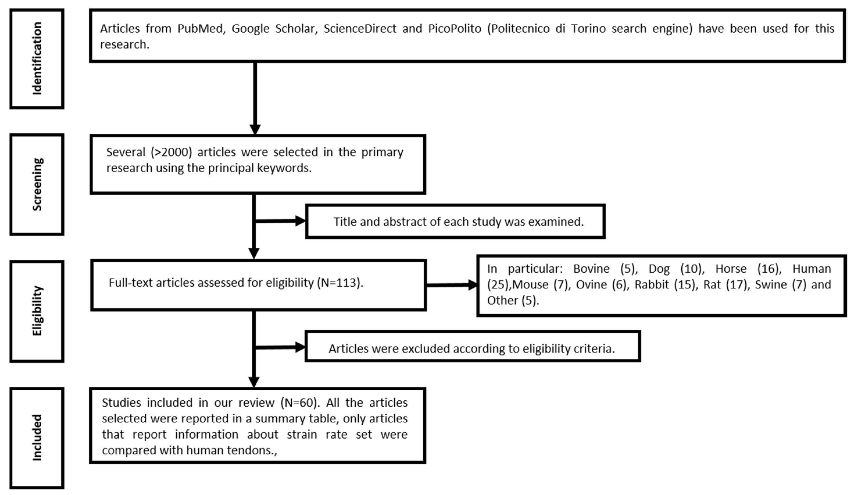
3.1. Study Selection

The search of published literature conducted via the Google Scholar, PubMed, ScienceDirect, and PicoPolito databases using the keywords tendon, mechanical properties, Young’s modulus, and ultimate stress yielded more than 2000 articles. The titles and the abstracts were analyzed to exclude fewer inherent papers, resulting in 113 articles for full-text evaluation. In the same way, a second skimming process showed that 40 out of 113 articles matched perfectly with the aim of this study, while the other 73 articles were focused on the analysis of other parameters (i.e., age; sex; healing process; suture techniques; and the insertion of allografts, autografts, and growth factors) and conditions (i.e., loading history, tendons subjected to treatments including gamma radiation, magnetic resonance imaging (MRI), atomic force microscopy (AFM), and second harmonic imaging microscopy (SHIM)), as shown in Figure 1. Since 20 out of 73 articles reported values of native tendons for reference purposes, they were also considered in this work. Finally, 60 articles were employed to obtain the values of the mechanical properties. In particular, the data were classified in animal species as follows: horse (n = 6), bovine (n = 2), swine (n = 6), ovine (n = 5), rabbit (n = 7), dog (n = 8), rat (n = 10), mouse (n = 5), human (n = 10), and 11-month-old foal (n = 1). Article summaries are illustrated in Table 1.
Table 1. All the selected articles, organized by species.
Animal SpeciesStudies
BovineDomnick et al. (2016) [12], Legerlotz et al. (2010) [11]
DogBaker et al. (2012) [19], Dejardin et al. (2001) [20], Derwin et al. (2006) [21], Derwin et al. (2007) [5], Derwin et al. (2009) [22], Balogh et al. (2016) [23], Haut et al. (1992) [24], Liu et al. (2019) [4]
HorseBatson et al. (2003) [25], Birch (2007) [15], Dowling and Dart. (2005) [26], Dowling et. al (2002) [27], Thorpe et al. (2010) [28]
MouseRigozzi et al. (2010) [29], Dyment et al. (2012) [30], Lin et al. (2005) [31], Gilday et al. (2014) [32], Mikic et al. (2008) [33]
OvineHuri et al. (2013) [34], Santoni et al. (2010) [35], Gibbons et al. (1991) [36], Rumian et al. (2009) [37], Salehpour et al. (1995) [38]
RabbitAwad et al. (2003) [3], Saber et al. (2010) [39], Young et al. (1998) [40], Juncosa-Melvin et al. (2006) [41], Trudel et al. (2007) [42], Viidik (1969) [43], Yamamoto et al. (1992) [44]
RatEliasson et al. (2007) [45], Lee et al. (2020) [46], Legerlotz et al. (2007) [47], Majewski et al. (2008) [48], Pietschmann et al. (2013) [49], Ferry et al. (2007) [50], Sahin et al. (2012) [51], Su et al. (2008) [52], Lavagnino et al. (2005) [53], Legerlotz et al. (2010) [11]
11-months-old foalCherdchutham et al. (2001) [54]
SwineDomnick et al. (2016) [12], Shadwick (1990) [16], Woo et al. (1981) [55], Smith et al. (1996) [56], Woo et al. (1980) [57], Diehl et al. (2006) [58]
HumanBirch (2007) [15], Carpenter et al. (2005) [59], Itoi et al. (1995) [60], Wren et al. (2001) [61], Lewis and Shaw (1997) [62], Butler et al. (1986) [63], Domnick et al. (2016) [12], Hashemi et al. (2005) [64], Weber et al. (2015) [65], Pring et al. (1985) [66]

3.2. Synthesis of Results

Table 1 shows the selected articles after the verification of the admission criteria, classified by animal species.

3.3. Study Characteristics

Table 2 shows the mechanical properties (loading rate, Young’s modulus, maximal load, ultimate stress, and ultimate strain) for different animal species considering strain rate and also the presence of preconditioning. Table 3 shows the same mechanical properties for different human tendons. To analyze the mechanical properties for animal tendons, two data set comparisons were considered. The first set comprised Young’s modulus, ultimate stress, and ultimate strain considering the strain rate in mm/s, while the second set dealt with the evaluation of the same parameters considering a strain rate in %/s. For standardization, data reported in mm/minutes were modified to obtain values in mm/s. Data that did not report the strain rate values were not used for graphing and analysis.
For better visualization, all data collected were arranged in different bar graphs that consider a single mechanical property according to strain rate (mm/s and %/s). In particular, to enhance the readability and recognition of the distribution of mechanical properties between the same animal tendon and tendons from different species, and to remark on the similarities between animals and humans, all species were associated with different colors: mouse (mustard-yellow), rat (yellow), dog (green), rabbit (dark green), ovine (azure), bovine (royal blue), swine (navy blue), horse and 11-month-old foal (plum). Regarding the human tendons, these were classified by type, in particular: hand flexor (gray), hand extensor (dark gray), long head of biceps (LHB; orange), and anterior and posterior supraspinatus tendons (dark orange).

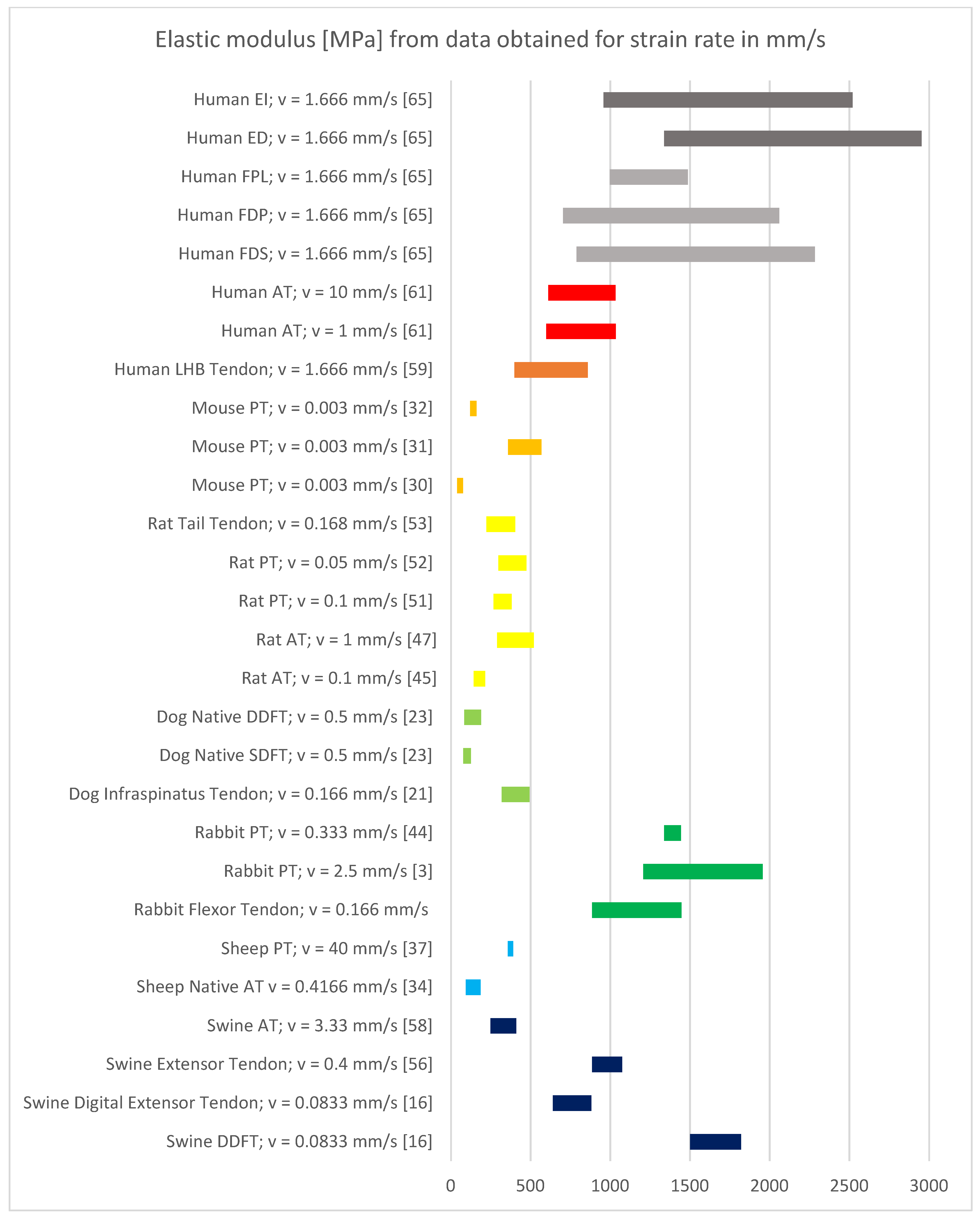
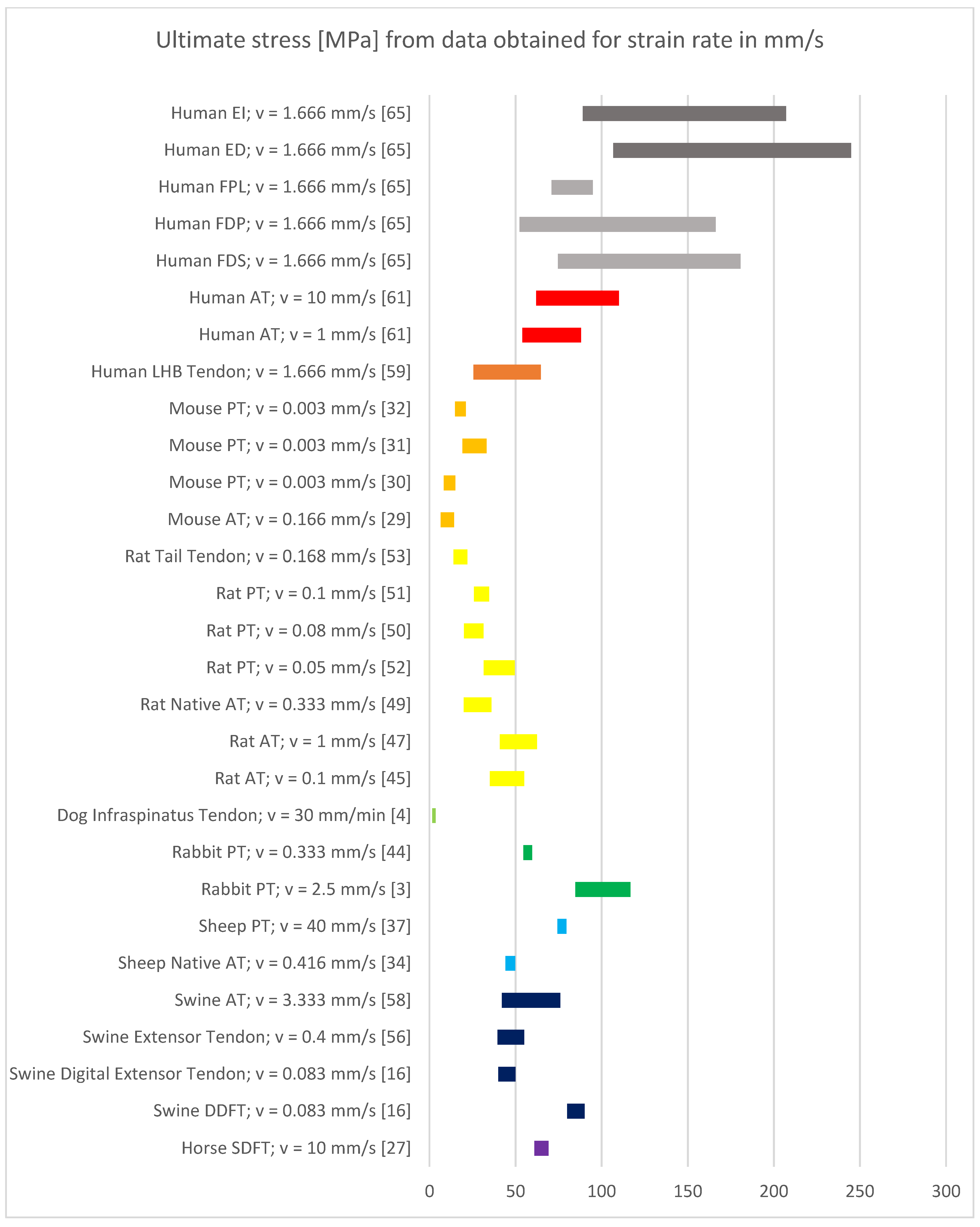
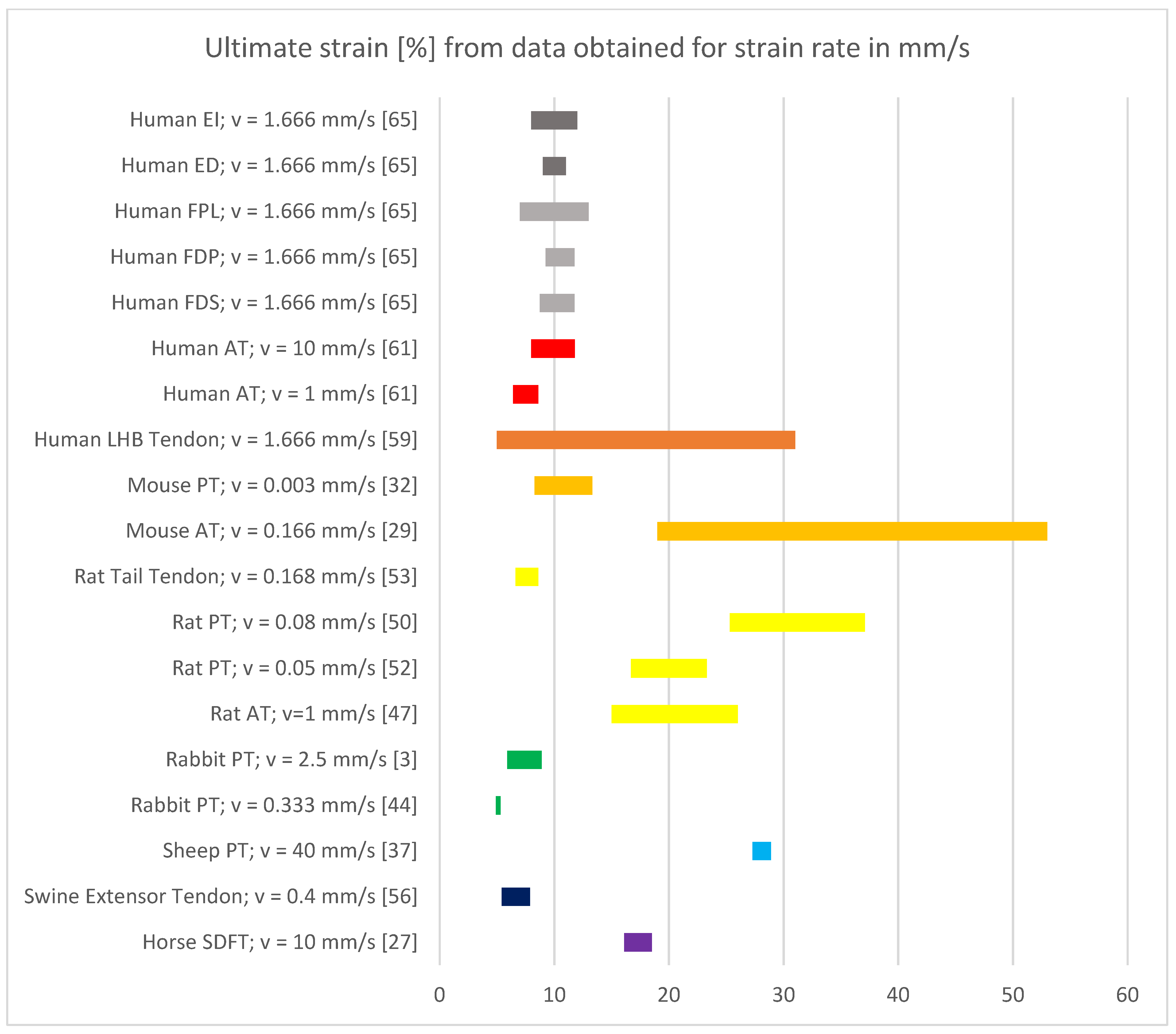
3.4. Results of Mechanical Property Evaluation in mm/s

The mechanical properties of tendons from different species showed a uniform distribution as shown for all the mechanical properties (Figure 2, Figure 3 and Figure 4). In particular, tendons belonging to different anatomical sites of the same species presented very similar results.
However, for the examined human tendons, a different distribution was shown depending on the anatomical site. Differences and similarities between human and animal tendons from different anatomical regions will be highlighted and commented upon. For each one, the best candidates will be determined; a more detailed analysis is reported in the discussion section.

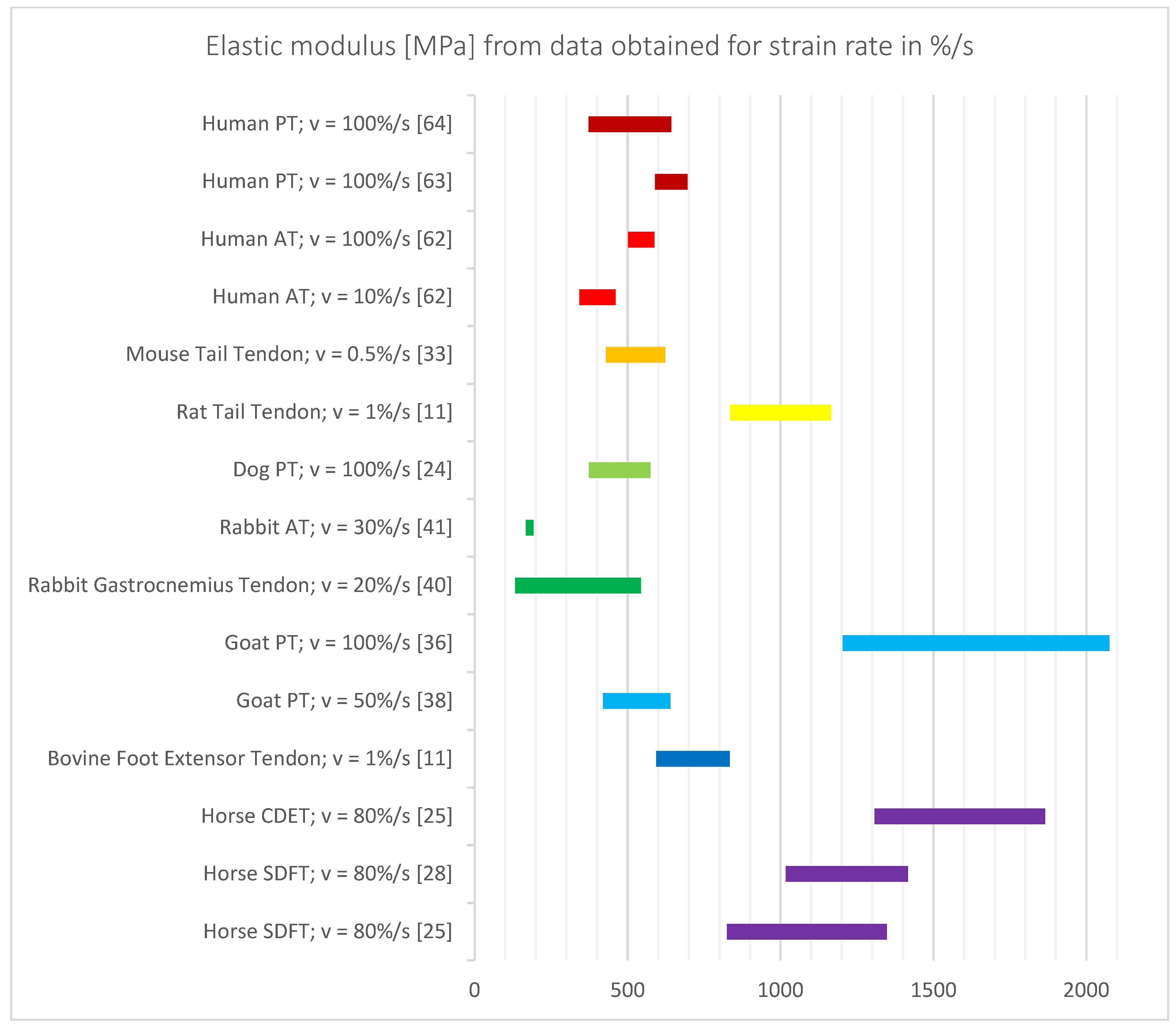
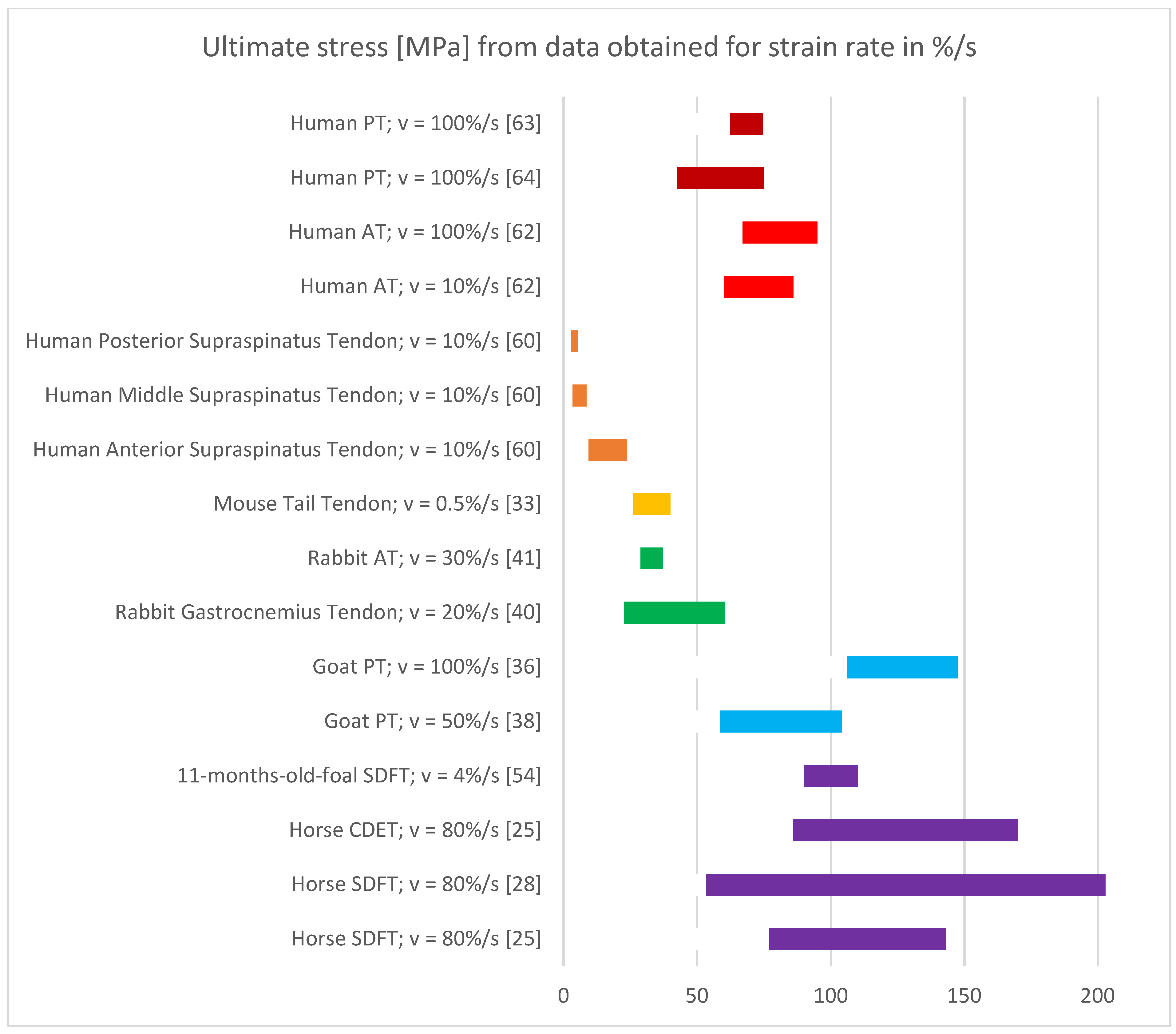
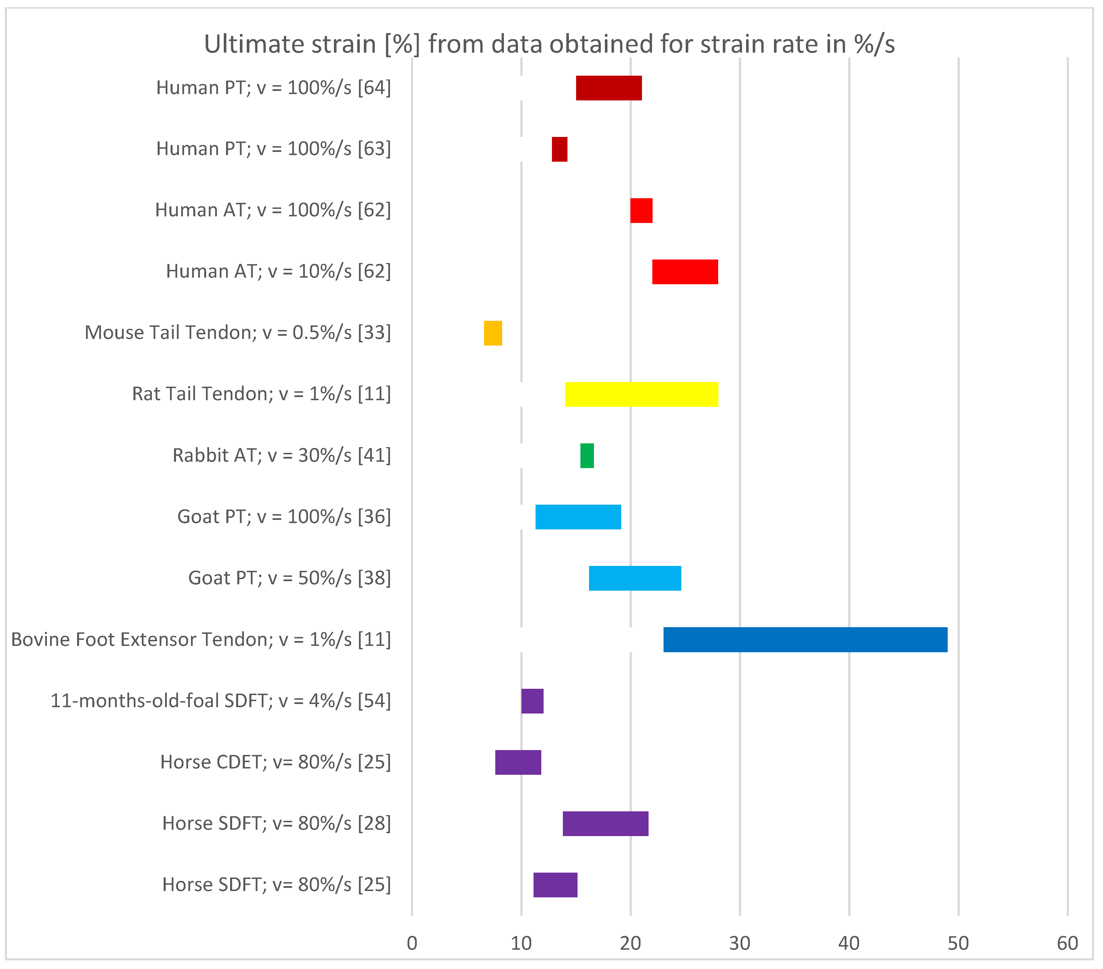
3.5. Results of Mechanical Property Evaluation in %/s

As seen in the previous section, the mechanical properties of tendons from different species showed a uniform distribution, independent of their anatomical region as shown for all the mechanical properties (Figure 5, Figure 6 and Figure 7). In the case of the mechanical properties evaluated in %/s, human tendons belonging to different anatomical sites showed different mechanical properties with respect to the same tests executed in mm/s, as described in the previous section. As before, the best candidates between human and animal tendons for different anatomical regions will be determined, and a more detailed analysis is reported in the discussion section.

3.6. Results of Additional Analysis—Strain Rate Analysis

The evaluation of the influence of strain rate on the resulting mechanical properties of the tendons was conducted on four animal species (rabbit, rat, mouse, and goat), data reported in Table 4, and the human AT, data reported in Table 5. In particular, there was no clear influence of strain rate on the results. However, changes in mechanical properties in agreement with the literature were found. The influence of strain rate on mechanical properties is discussed thoroughly below.

4. Discussion

The evaluation of the data retrieved from the published scientific literature was performed considering the strain rate with two different units (mm/s and %/s). The analysis only dealt with the comparison between human and animal tendons; thus, no comparison was performed among the mechanical properties of animal tendons.

4.1. Discussion of Results with Strain Rate in mm/s

The human tendons that included in this section are the finger flexor and extensor tendons [65,66], human LHB tendon [59], and Achilles tendon [61].
The mechanical properties of extensor and flexor finger tendons were analyzed by Pring et al., 1985 [66] and Weber et al., 2015 [65] at a strain rate of 100 mm/s. Both studies reported contrasting ultimate strain values; the difference may have been due to samples slipping during the test conducted by Pring et al. [66]. Therefore, in this study, the data from Weber et al., 2015 were used to perform the comparison [65].
The Young’s modulus and ultimate strain values obtained by Weber et al., 2015 [65] for EI and ED finger extensors showed similarities with the results obtained for rabbit PT [3]. Conversely, there were no matches for ultimate stress between all the animal species. A partial similarity could be found between swine DDFT [16], rabbit flexor tendon [39], rat tail tendon [53], and mouse PT [32].
Regarding finger flexor tendons (FPL, FDP, and FDS), the Young’s modulus and ultimate strain values obtained by Weber et al., 2015 [65] also showed similarities with data obtained for rabbit PT [3]. Moreover, there was a similarity of Young’s modulus and ultimate stress values with data reported in swine DDFT [16]; however, a comparison between ultimate strain could not be made as no data were obtained for ultimate strain for this tendon.
Wren et al., 2001 [61] analyzed the mechanical properties of the Achilles tendon with two different strain rate values. For both values, the Young’s modulus values were comparable with rabbit flexor tendon [39], swine extensor [56], and swine digital extensor [16]. Concerning the ultimate stress values for both strain rates, the results showed similarities with rabbit PT 3], sheep PT [37], swine DDFT [16], swine AT [58], and horse SDFT [27]. In addition, there was a similarity of ultimate strain for both the strain rate values with mouse PT [32] and rabbit PT [3]; however, the Achilles tendon with a strain rate of 1 mm/s also showed a match with rat tail tendon [53] and swine extensor [67]. Rat and mouse tendons are widely used in medicine to compare with the Achilles tendon in humans. Knockout mice have been used to investigate the composition variation of Achilles tendons from a histological point of view [31]. According to earlier studies evaluating mechanical behavior, contrasting results between rat/mouse and human samples have been obtained [45], which can be observed in our results.
Young’s modulus of the LHB tendon, analyzed by Carpenter et al., 2005 [59], showed clear similarities with the results obtained for swine digital extensor tendon [16], and a small similarity was noticed with mouse PT [31], rat AT [47], rat PT [52], and dog infraspinatus [21]. The LHB’s ultimate stress [59] showed similarity with results reported for rat AT [45,47,49], rat PT [50,51,52], rabbit PT [68], sheep AT [34], swine extensor tendon [56], swine digital extensor [16], and swine AT [58]. The ultimate strain results showed similarities with data for mouse PT [32], mouse AT [29], rat AT [47], rat PT [51,53], rat tail tendon [53], rabbit PT [3], sheep PT [37], swine extensor tendon [56], and horse SDFT [27]. There was also a similarity between LHB’s mechanical properties and those of the rat AT, rat PT, and swine digital extensor tendon. Indeed, rat and dog shoulder models are used to evaluate the factors that influence human rotator cuff injury and repair processes [5,6,22].
In this study, no similarities were found between human, swine, and bovine tendons; however, Dominick et al., 2016 [12] reported that swine flexor and bovine extensor tendons are eligible surrogates for human semitendinosus tendons in biomechanical studies.

4.2. Discussion of Results with Strain Rate in %/s

The human tendons included in this section are the Achilles tendon [62], patellar tendon [63,64], anterior supraspinatus, middle supraspinatus and posterior supraspinatus [60].
The Young’s modulus of the Achilles tendon obtained by Lewis et al., 1997 [62] showed similarities with mouse tail tendon [33], dog PT [24], rabbit gastrocnemius tendon [40], and goat PT [38]. In the same way, a similarity could be noticed between Achilles’ ultimate stress and results obtained for rabbit gastrocnemius [40], goat PT [38], 11-month-old foal SDFT [54], horse CDET [25], and horse SDFT [25,28]. The ultimate strain data showed similarities with rat tail tendon [11], goat PT [38], bovine foot extensor [11], and horse SDFT [28]. Essentially, the mechanical properties of the Achilles tendon [62] were comparable with those of rabbit gastrocnemius [40] and goat PT [38]. In fact, the similarities between rabbit gastrocnemius and human Achilles tendons finds confirmation by Young et al. [40] in the evaluation of Achilles tendon repair [37].
The mechanical characterization results for PT from a study conducted by Butler et al., 1986 [63] showed a value of Young’s modulus that was similar with mouse tail tendon [33], goat PT [38], and bovine foot extensor [11] values. Similar mechanical results were obtained for human PT by Hashemi et al., 2005 [64]; in this case, there were similarities with dog PT [24] and rabbit gastrocnemius [40]. The results obtained in an additional study conducted by Haut et al. [24] suggest that knee tendon and ligament reconstruction is possible with canine PT, and this finding gives significance to the mechanical evaluation that was performed as the aim of this work.
Regarding the ultimate stress, the values obtained by Butler et al. [63] showed similarities with data from goat PT [38] and horse SDFT [28], while the results of Hashemi et al. [64] showed similarities with rabbit gastrocnemius [40]. There was a clear difference in the PT ultimate strain values obtained in both articles [18,50]. The ultimate strain reported by Butler et al. [63] for PT showed similarities with rat tail tendon [11], goat PT [36], and horse SDFT [25,28]. These results were also found for PT tested by Hashemi et al. [64]; in addition, their results showed similarities with rabbit AT [41] and goat PT [38]. Essentially, the data obtained in both studies presented similarities with the mechanical properties of goat PT [38]. This result is confirmed by the fact that the goat knee model was used to evaluate patellar tendon remodeling [37]. Furthermore, Hausmann et al. [10] reported that sheep tendons seem to be the most suitable animal tendon model for mimicking human tendons; this statement has been proved valid here for PT but not for other human tendons. However, the mechanical properties achieved by PT as tested by Hashemi et al. [64] showed a similar Young’s modulus and ultimate stress to data obtained from rabbit gastrocnemius [40], with no ultimate strain data reported for this tendon. This result explains the fact that in literature, the effects of the healing process on knee ligaments and tendons are studied with rabbit knee models [3,45].
Itoi et al., 1995 [60] evaluated the ultimate stress properties of anterior supraspinatus, middle supraspinatus, and posterior supraspinatus tendons. No similarities with animal tendons were found, except for the anterior supraspinatus tendon, which shows slight similarities to rabbit gastrocnemius [40]. Several studies evaluated the healing of rotator cuff tendons in canine animal models; however, no evaluation could be performed for the results obtained in this work [4,22].

4.3. Discussion of Strain Rate Analysis

The analysis of the influence of strain rate on the mechanical properties of tendons did not show univocal results. The mechanical properties of rabbit PT obtained with a velocity of 0.333 mm/s [68] and 2.5 mm/s [3] did not show statistically significant differences for Young’s modulus (1390 ± 53 MPa vs. 1581.4 ± 374.9 MPa), while ultimate stress (57.1 ± 2.5 MPa vs. 100.7 ± 16 MPa) and ultimate strain (5.1 ± 0.2% vs. 7.4 ± 1.5%) presented statistically significant differences; in particular, an increment in the values was observed.
Rat AT mechanical properties using a strain rate of 0.1 mm/s [45] and 1 mm/s [47] presented statistically significant differences for Young’s modulus (179 ± 36 MPa vs. 405 ± 115 MPa, respectively), and this was related to an increase in tissue stiffness. On the other hand, ultimate stress (45 ± 10 MPa vs. 51.6 ± 10.8 MPa) did not present statistically significant differences when comparing the two strain rates. No definitive evaluation could be made for the ultimate strain since one article [45] did not report any value for this parameter.
The results obtained by testing rat PT with a strain rate of 0.05 mm/s [52] and 0.1 mm/s [51] showed no statistically significant differences for both Young’s modulus (386 ± 88 MPa vs. 323.88 ± 56.48 MPa) and ultimate stress (40.5 ± 8.95 MPa vs. 30.24 ± 4.41 MPa). Furthermore, no evaluation could be made regarding the ultimate strain, as [51,52] did not report any value. Thus, the increases in strain rate did not cause any changes in the mechanical properties of the tissue. Considering the study of Robinson et al., 2004 [18], the mechanical properties of mouse fascicles with modified and unmodified composition were analyzed according to the strain rate. Robinson et al., 2004 [18] showed that the mechanical properties related to the linear region are strain rate-independent over the range from 0.5%/s to 50%/s; in contrast, the failure properties are highly dependent on strain rate.
The mechanical properties of goat PT with a strain rate of 50%/s [38] and 100%/s [36] showed statistically significant differences for all mechanical properties analyzed. The strain rate increment caused an increase in Young’s modulus and ultimate stress; thus, an ultimate strain reduction is advocated, in accordance with the results obtained for rabbit PT.
Concerning human tendons, according to Wren et al., 2001 [61], the influence of strain rate increment (1 mm/s and 10 mm/s) does not cause statistically significant differences in the mechanical properties of Achilles tendons. Several authors have reported that strain rate has an important influence on the mechanical properties at failure (stress and strain); however, once again, there were no statistically significant differences even if a wide range of strain rates was evaluated. However, Abraham et al., 1967 [69] found that for the human Achilles tendon, different strain rates modified the stress magnitude and the shape of the stress–strain curve. In addition, the authors presented a double logarithmic relationship between stress magnitude and strain rate [69].
Herrick et al., 1987 [70] performed some tests on the equine flexor tendon to validate the relationship reported by Abrahams et al. [69]. They found that this relationship might be valid for low strain rate values. In conclusion, the authors affirmed that strain rate has a small and inconsistent effect on stiffness in the range from 5%/s to 50%/s [70].
The study of Lewis et al., 1997 [62] evaluated the effects of a ten times strain rate increase (10%/sec and 100%/sec) on human Achilles tendons. The data reported showed that the strain rate increase produced a statistically insignificant increase in the value of ultimate tensile strength (73 ± 13 MPa vs. 81 ± 14 MPa), a statistically insignificant decrease in the value of ultimate strain (25 ± 3% vs. 21 ± 1%), and a statistically significant increase in Young’s modulus (401 ± 59 MPa vs. 545 ± 43 MPa) [62].
In this work, we highlighted how a change in strain rate does not have a unique effect on the mechanical properties of tendons. Analyzing the results of four different tendon tissues (rabbit PT, rat AT, rat PT, and human AT) tested with a strain rate in mm/s, we observed that a strain rate increment caused a significant increase in Young’s modulus only in rat AT. A similar result was obtained for human AT by increasing the strain rate by ten times. However, this increase in Youngs’ modulus was not statistically significant. In all the other studies, there was no appreciable change in Young’s modulus. Considering the four comparisons made for strain rate values in mm/s, we observed statistically significant differences for ultimate stress and ultimate strain in rabbit PT only. In particular, the strain rate increment caused an increase in ultimate stress and a decrease in ultimate strain.
Regarding the effects of the strain rate increase in %/s, the results showed a significant increase in Young’s modulus for both goat PT and human AT. This could be due to the application of a drastic increase in the strain rate considering the measurement unit itself; as a result, setting a strain rate of 50%/s results in a tendon deformation of half its initial length in one second, e.g., a strain rate increase in goat PT from 50%/s to 100%/s produces an increment of Young’s modulus by three times.
In this context, according to Abrahams et al. [69] a low strain rate increase may have no effect. This evidence, in agreement with our findings, shows that Young’s modulus is affected only in the case of a high strain rate increment.
A 10-fold increase in strain rate in human AT [61] did not present statistically significant differences for ultimate stress and strain. However, an increase in ultimate stress and in ultimate strain could be found. Analyzing the strain rate in %/s only for goat PT [36,38] showed statistically significant differences between the results for ultimate stress and ultimate strain, while human AT [62] showed differences, but not statistically significant differences. The increment in strain rate causes an increase in ultimate stress and a decrease in ultimate strain in both tendons.
In conclusion, there is evidence that the value of strain rate can cause a change in the Young’s modulus of tendons. Nevertheless, this change in strain rate must be of a significant quantity. The data analysis showed that an increase in strain rate of ten times or greater compared to a reference strain rate value can change the properties of the tissue. The change in strain rate influences tendons’ ultimate stress and ultimate strain values. A significant strain rate increase can cause an increase in tendons’ ultimate stress and a reduction in ultimate strain.

4.4. Limitations

Despite the comprehensive search across databases, some related articles could have been missed due to the selected keywords and database limitations. This study also did not consider parameters such as animal age, sex, and lifetime activity. These parameters are factors that may influence the biomechanical characteristics of tendons. Additionally, the comparison of tendons should be conducted by evaluating their composition. Future studies should compare the influence of these parameters on the mechanical properties of animal and human tendons, which would lead to a more accurate assessment of the tendon to be used for ex vivo testing. Furthermore, important data might be contained in non-peer-reviewed studies and unpublished theses.

5. Conclusions

Most of the experimental research for human tendon injury and repair is generally performed in animal models, at least in the earliest research stages before human studies. However, animal models have several shortcomings, such as inherent biologic variability, metabolic and hormonal differences between species, and anatomical differences, introducing potential bias to the interpretation of mechanical results during preclinical experiments [31]. As a result, medical, veterinary, and bioengineering researchers must be cautious (i) in applying their results to humans and (ii) in selecting the most appropriate animal tendon for their applications.
This systematic review aimed to define the most suitable surrogate for mimicking the behavior of human tendons when subjected to uniaxial tensile tests. For this purpose, the scientific literature was reviewed extensively, searching for experimental studies involving the mechanical properties of animal tendons, such as Young’s modulus, ultimate stress, and ultimate strain. Differences and similarities with human tendons from different anatomical regions were highlighted and commented upon. For each region, the best candidates were determined and discussed.
The key findings obtained in the present review make it evident that different animals tendons show different mechanical properties. Therefore, not every animal tendon can be employed to make a direct comparison to their human counterparts (or to any other human tendon or ligament). The results showed similarities between some animal and human tendons that should be considered in the evaluation of scaffolds and sutures.
Considering the results in mm/s:
  • The flexor tendon of the hand (FPL, FDP, and FDS) shows partial similarities for Youngs’ modulus and ultimate stress with swine DDFT.
  • The extensor tendon of the hand (EI and ED) has similarities with the rabbit PT.
  • The LHB tendon shows similarities with the mechanical properties reported for the rat AT, rat PT, and swine digital extensor tendon.
Considering results the obtained for data in %/s:
  • The human PT tendon has some similarities with the results obtained for the goat PT.
  • The human AT tendon shows comparable mechanical properties with the goat PT, but there are other partial similarities.
However, these results were found only for data in %/s; no clear similarities were visible in the data in mm/s. Thus, it seems highly probable that the choice of strain rate significantly affects the results; unfortunately, different authors reported their results with different settings. Therefore, further studies will be needed to evaluate tendons from different animals and anatomical regions with the same test conditions and strain rate, in a fully comparable way. Future research may enhance the understanding of the best animal tendon species by considering (i) age, (ii) loading history, and (iii) composition.

Author Contributions

Conceptualization, V.B., M.C., M.R.R., A.B., and C.S.; methodology, V.B., and M.C.; validation, V.B., M.C., M.R.R., E.P., and S.P.; formal analysis, V.B., M.C., M.R.R., E.P., and S.P.; investigation, V.B., M.C., M.R.R., E.P., and S.P.; resources, A.B., and C.S.; data curation, V.B., M.C., M.R.R., E.P., and S.P.; writing—original draft preparation, V.B., E.P., and S.P.; writing—review and editing, M.C., A.B., and C.S.; visualization, V.B., E.P., and S.P. supervision, M.C., M.R.R., A.B., and C.S.; project administration, M.C., A.B., and C.S.; funding acquisition, C.S. All authors have read and agreed to the published version of the manuscript.

Funding

This research was partially funded by the LINKS Foundation under the Proof of Concept Instrument Programme (https://linksfoundation.com/poc/, accessed on 27 September 2021), 2019 Edition, and by the Politecnico di Torino and the Italian Ministry of Economic Development (MISE) under the Proof of Concept (PoC)–Off programme 2020.

Institutional Review Board Statement

Not applicable.

Informed Consent Statement

Not applicable.

Data Availability Statement

All data are available in the original papers.

Conflicts of Interest

The authors declare no conflict of interest.

References

  1. Chen, Y.J.; Wang, C.J.; Yang, K.D.; Kuo, Y.R.; Huang, H.C.; Huang, Y.T.; Sun, Y.C.; Wang, F.S. Extracorporeal Shock Waves Promote Healing of Collagenase-Induced Achilles Tendinitis and Increase TGF-β1 and IGF-I Expression. J. Orthop. Res. 2004, 22, 854–861. [Google Scholar] [CrossRef]
  2. Ansorge, H.L.; Beredjiklian, P.K.; Soslowsky, L.J. CD44 Deficiency Improves Healing Tendon Mechanics and Increases Matrix and Cytokine Expression in a Mouse Patellar Tendon Injury Model. J. Orthop. Res. 2009, 27, 1386–1391. [Google Scholar] [CrossRef] [PubMed] [Green Version]
  3. Awad, H.A.; Boivin, G.P.; Dressler, M.R.; Smith, F.N.L.; Young, R.G.; Butler, D.L. Repair of Patellar Tendon Injuries Using a Cell-Collagen Composite. J. Orthop. Res. 2003, 21, 420–431. [Google Scholar] [CrossRef]
  4. Liu, Q.; Yu, Y.; Reisdorf, R.L.; Qi, J.; Lu, C.K.; Berglund, L.J.; Amadio, P.C.; Moran, S.L.; Steinmann, S.P.; An, K.N.; et al. Engineered Tendon-Fibrocartilage-Bone Composite and Bone Marrow-Derived Mesenchymal Stem Cell Sheet Augmentation Promotes Rotator Cuff Healing in a Non-Weight-Bearing Canine Model. Biomaterials 2019, 192, 189–198. [Google Scholar] [CrossRef]
  5. Derwin, K.A.; Baker, A.R.; Codsi, M.J.; Iannotti, J.P. Assessment of the Canine Model of Rotator Cuff Injury and Repair. J. Shoulder Elb. Surg. 2007, 16, 140–148. [Google Scholar] [CrossRef] [PubMed] [Green Version]
  6. Soslowsky, L.J.; Carpenter, J.E.; DeBano, C.M.; Banerji, I.; Moalli, M.R. Development and Use of an Animal Model for Investigations on Rotator Cuff Disease. J. Shoulder Elb. Surg. 1996, 5, 383–392. [Google Scholar] [CrossRef]
  7. Otarodifard, K.; Wong, J.; Preston, C.F.; Tibone, J.E.; Lee, T.Q. Relative Fixation Strength of Rabbit Subscapularis Repair Is Comparable to Human Supraspinatus Repair at Time. Clin. Orthop. Relat. Res. 2014, 472, 2440–2447. [Google Scholar] [CrossRef] [Green Version]
  8. Jenson, P.W.; Lillich, J.D.; Roush, J.K.; Gaughan, E.M. Ex Vivo Strength Comparison of Bioabsorbable Tendon Plates and Bioabsorbable Suture in a 3-Loop Pulley Pattern for Repair of Transected Flexor Tendons from Horse Cadavers. Vet. Surg. 2005, 34, 565–570. [Google Scholar] [CrossRef]
  9. Duffy, D.J.; Chang, Y.-J.; Fisher, M.B.; Chambers, A.R.; Moore, G.E. Effect of Epitendinous Suture Caliber on the Tensile Strength of Repaired Canine Flexor Tendons. Am. J. Vet. Res. 2021, 82, 510–515. [Google Scholar] [CrossRef]
  10. Hausmann, J.-T.; Vekszler, G.; Bijak, M.; Benesch, T.; Vécsei, V.; Gäbler, C. Biomechanical Comparison of Modified Kessler and Running Suture Repair in 3 Different Animal Tendons and in Human Flexor Tendons. J. Hand Surg. 2009, 34, 93–101. [Google Scholar] [CrossRef]
  11. Legerlotz, K.; Riley, G.P.; Screen, H.R. Specimen Dimensions Influence the Measurement of Material Properties in Tendon Fascicles. J. Biomech. 2010, 43, 2274–2280. [Google Scholar] [CrossRef] [Green Version]
  12. Domnick, C.; Wieskötter, B.; Raschke, M.J.; Schulze, M.; Kronenberg, D.; Wefelmeier, M.; Langer, M.F.; Herbort, M. Evaluation of Biomechanical Properties: Are Porcine Flexor Tendons and Bovine Extensor Tendons Eligible Surrogates for Human Tendons in in Vitro Studies? Arch. Orthop. Trauma. Surg. 2016, 136, 1465–1471. [Google Scholar] [CrossRef]
  13. Benedict, J.V.; Walker, L.B.; Harris, E.H. Stress-Strain Characteristics and Tensile Strength of Unembalmed Human Tendon. J. Biomech. 1968, 1, 53–63. [Google Scholar] [CrossRef]
  14. Böl, M.; Ehret, A.; Leichsenring, K.; Ernst, M. Tissue-Scale Anisotropy and Compressibility of Tendon in Semi-Confined Compression Tests. J. Biomech. 2015, 48, 1092–1098. [Google Scholar] [CrossRef]
  15. Birch, H.L. Tendon Matrix Composition and Turnover in Relation to Functional Requirements. Int. J. Exp. Pathol. 2007, 88, 241–248. [Google Scholar] [CrossRef] [PubMed]
  16. Shadwick, R.E. Elastic Energy Storage in Tendons: Mechanical Differences Related to Function and Age. J. Appl. Physiol. 1990, 68, 1033–1040. [Google Scholar] [CrossRef] [Green Version]
  17. Donahue, T.L.H.; Gregersen, C.; Hull, M.L.; Howell, S. Comparison of Viscoelastic, Structural, and Material Properties of Double-Looped Anterior Cruciate Ligament Grafts Made from Bovine Digital Extensor and Human Hamstring Tendons. J. Biomech. Eng. 2000, 123, 162–169. [Google Scholar] [CrossRef] [Green Version]
  18. Robinson, P.S.; Lin, T.W.; Reynolds, P.R.; Derwin, K.A.; Iozzo, R.; Soslowsky, L.J. Strain-Rate Sensitive Mechanical Properties of Tendon Fascicles From Mice With Genetically Engineered Alterations in Collagen and Decorin. J. Biomech. Eng. 2004, 126, 252–257. [Google Scholar] [CrossRef]
  19. Baker, A.R.; McCarron, J.A.; Tan, C.D.; Iannotti, J.P.; Derwin, K.A. Does Augmentation with a Reinforced Fascia Patch Improve Rotator Cuff Repair Outcomes? Clin. Orthop. Relat. Res. 2012, 470, 2513–2521. [Google Scholar] [CrossRef] [Green Version]
  20. Dejardin, L.M.; Arnoczky, S.P.; Ewers, B.J.; Haut, R.C.; Clarke, R.B. Tissue-Engineered Rotator Cuff Tendon Using Porcine Small Intestine Submucosa. Am. J. Sports Med. 2001, 29, 175–184. [Google Scholar] [CrossRef]
  21. Derwin, K.A.; Baker, A.R.; Spragg, R.K.; Leigh, D.R.; Iannotti, J.P. Commercial Extracellular Matrix Scaffolds for Rotator Cuff Tendon Repair. J. Bone Jt. Surg. 2006, 88, 2665–2672. [Google Scholar] [CrossRef] [PubMed]
  22. Derwin, K.A.; Codsi, M.J.; Milks, R.A.; Baker, A.R.; McCarron, J.A.; Iannotti, J.P. Rotator Cuff Repair Augmentation in a Canine Model with Use of a Woven Poly-L-Lactide Device. J. Bone Jt. Surg. 2009, 91, 1159–1171. [Google Scholar] [CrossRef] [PubMed] [Green Version]
  23. Balogh, D.G.; Biskup, J.J.; O’Sullivan, M.G.; Scott, R.M.; Groschen, D.; Evans, R.B.; Conzemius, M.G. Biochemical, Histologic, and Biomechanical Characterization of Native and Decellularized Flexor Tendon Specimens Harvested from the Pelvic Limbs of Orthopedically Normal Dogs. Am. J. Vet. Res. 2016, 77, 388–394. [Google Scholar] [CrossRef] [PubMed]
  24. Haut, R.C.; Lancaster, R.L.; DeCamp, C.E. Mechanical Properties of the Canine Patellar Tendon: Some Correlations with Age and the Content of Collagen. J. Biomech. 1992, 25, 163–173. [Google Scholar] [CrossRef]
  25. Batson, E.L.; Paramour, R.J.; Smith, T.J.; Birch, H.; Patterson-Kane, J.C.; Goodship, A.E. Are the Material Properties and Matrix Composition of Equine Flexor and Extensor Tendons Determined by Their Functions? Equine Vet. J. 2010, 35, 314–318. [Google Scholar] [CrossRef]
  26. Dowling, B.; Dart, A. Mechanical and Functional Properties of the Equine Superficial Digital Flexor Tendon. Vet. J. 2005, 170, 184–192. [Google Scholar] [CrossRef]
  27. Dowling, B.A.; Dart, A.J.; Hodgson, D.R.; Rose, R.J.; Walsh, W.R. Recombinant Equine Growth Hormone Does Not Affect the in Vitro Biomechanical Properties of Equine Superficial Digital Flexor Tendon. Vet. Surg. 2002, 31, 325–330. [Google Scholar] [CrossRef]
  28. Thorpe, C.T.; Stark, R.J.F.; Goodship, A.E.; Birch, H.L. Mechanical Properties of the Equine Superficial Digital Flexor Tendon Relate to Specific Collagen Cross-Link Levels. Equine Vet. J. 2010, 42, 538–543. [Google Scholar] [CrossRef]
  29. Rigozzi, S.; Müller, R.; Snedeker, J.G. Collagen Fibril Morphology and Mechanical Properties of the Achilles Tendon in Two Inbred Mouse Strains. J. Anat. 2010, 216, 724–731. [Google Scholar] [CrossRef] [Green Version]
  30. Dyment, N.A.; Kazemi, N.; Aschbacher-Smith, L.E.; Barthelery, N.J.; Kenter, K.; Gooch, C.; Shearn, J.T.; Wylie, C.; Butler, D.L. The Relationships among Spatiotemporal Collagen Gene Expression, Histology, and Biomechanics Following Full-Length Injury in the Murine Patellar Tendon. J. Orthop. Res. 2011, 30, 28–36. [Google Scholar] [CrossRef]
  31. Lin, T.W.; Cardenas, L.; Soslowsky, L.J. Tendon Properties in Interleukin-4 and Interleukin-6 Knockout Mice. J. Biomech. 2005, 38, 99–105. [Google Scholar] [CrossRef]
  32. Gilday, S.D.; Casstevens, E.C.; Kenter, K.; Shearn, J.T.; Butler, D.L. Murine Patellar Tendon Biomechanical Properties and Regional Strain Patterns during Natural Tendon-to-Bone Healing after Acute Injury. J. Biomech. 2013, 47, 2035–2042. [Google Scholar] [CrossRef] [PubMed] [Green Version]
  33. Mikic, B.; Entwistle, R.; Rossmeier, K.; Bierwert, L. Effect of GDF-7 Deficiency on Tail Tendon Phenotype in Mice. J. Orthop. Res. 2008, 26, 834–839. [Google Scholar] [CrossRef] [PubMed]
  34. Huri, G.; Biçer, S.; Özgözen, L.; Uçar, Y.; Garbis, N.G.; Hyun, Y.S. A Novel Repair Method for the Treatment of Acute Achilles Tendon Rupture with Minimally Invasive Approach Using Button Implant: A Biomechanical Study. Foot Ankle Surg. 2013, 19, 261–266. [Google Scholar] [CrossRef]
  35. Santoni, B.G.; McGilvray, K.C.; Lyons, A.S.; Bansal, M.; Turner, A.S.; MacGillivray, J.D.; Coleman, S.H.; Puttlitz, C.M. Biomechanical Analysis of an Ovine Rotator Cuff Repair via Porous Patch Augmentation in a Chronic Rupture Model. Am. J. Sports Med. 2010, 38, 679–686. [Google Scholar] [CrossRef]
  36. Gibbons, M.J.; Butler, D.L.; Grood, E.S.; Bylski-Austrow, D.I.; Levy, M.S.; Noyes, F.R. Effects of Gamma Irradiation on the Initial Mechanical and Material Properties of Goat Bone-Patellar Tendon-Bone Allografts. J. Orthop. Res. 1991, 9, 209–218. [Google Scholar] [CrossRef]
  37. Rumian, A.P.; Draper, E.R.C.; Wallace, A.L.; Goodship, A.E. The Influence of the Mechanical Environment on Remodelling of the Patellar Tendon. J. Bone Jt. Surgery. Br. Vol. 2009, 91, 557–564. [Google Scholar] [CrossRef] [Green Version]
  38. Salehpour, A.; Butler, D.L.; Proch, F.S.; Schwartz, H.E.; Feder, S.M.; Doxey, C.M.; Ratcliffe, A. Dose-Dependent Response of Gamma Irradiation on Mechanical Properties and Related Biochemical Composition of Goat Bone-Patellar Tendon-Bone Allografts. J. Orthop. Res. 1995, 13, 898–906. [Google Scholar] [CrossRef]
  39. Saber, S.; Zhang, A.Y.; Ki, S.H.; Lindsey, D.P.; Smith, R.L.; Riboh, J.; Pham, H.; Chang, J. Flexor Tendon Tissue Engineering: Bioreactor Cyclic Strain Increases Construct Strength. Tissue Eng. Part A 2010, 16, 2085–2090. [Google Scholar] [CrossRef] [PubMed]
  40. Young, R.G.; Butler, D.L.; Weber, W.; Caplan, A.; Gordon, S.L.; Fink, D.J. Use of Mesenchymal Stem Cells in a Collagen Matrix for Achilles Tendon Repair. J. Orthop. Res. 1998, 16, 406–413. [Google Scholar] [CrossRef]
  41. Juncosa-Melvin, N.; Boivin, G.P.; Galloway, M.T.; Gooch, C.; West, J.R.; Butler, D.L. Effects of Cell-to-Collagen Ratio in Stem Cell-Seeded Constructs for Achilles Tendon Repair. Tissue Eng. 2006, 12, 681–689. [Google Scholar] [CrossRef] [PubMed] [Green Version]
  42. Trudel, G.; Koike, Y.; Ramachandran, N.; Doherty, G.; Dinh, L.; Lecompte, M.; Uhthoff, H.K. Mechanical Alterations of Rabbit Achilles’ Tendon After Immobilization Correlate with Bone Mineral Density But Not With Magnetic Resonance or Ultrasound Imaging. Arch. Phys. Med. Rehabil. 2007, 88, 1720–1726. [Google Scholar] [CrossRef] [PubMed]
  43. Viidik, A. Tensile Strength Properties of Achilles Tendon Systems in Trained and Untrained Rabbits. Acta Orthop. Scand. 1969, 40, 261–272. [Google Scholar] [CrossRef] [PubMed]
  44. Yamamoto, E.; Hayashi, K. Mechanical Properties of Collagen Fascicles from the Rabbit Patellar Tendon. J. Biomech. Eng. 1999, 121, 124–131. [Google Scholar] [CrossRef] [PubMed]
  45. Eliasson, P.; Fahlgren, A.; Pasternak, B.; Aspenberg, P. Unloaded Rat Achilles Tendons Continue to Grow, but Lose Viscoelasticity. J. Appl. Physiol. 2007, 103, 459–463. [Google Scholar] [CrossRef] [Green Version]
  46. Lee, H.W.; Wang, C.; Bae, T.S.; Yang, I.; Liu, Y.; Park, C.W.; Kim, H.N. Tendon Regeneration after Partial-Thickness Peroneus Longus Tendon Harvesting: Magnetic Resonance Imaging Evaluation and in Vivo Animal Study. Am. J. Sports Med. 2020, 48, 2499–2509. [Google Scholar] [CrossRef]
  47. Legerlotz, K.; Schjerling, P.; Langberg, H.; Brüggemann, G.-P.; Niehoff, A. The Effect of Running, Strength, and Vibration Strength Training on the Mechanical, Morphological, and Biochemical Properties of the Achilles Tendon in Rats. J. Appl. Physiol. 2007, 102, 564–572. [Google Scholar] [CrossRef] [Green Version]
  48. Majewski, M.; Betz, O.; Ochsner, P.E.; Liu, F.; Porter, R.M.; Evans, C.H. Ex Vivo Adenoviral Transfer of Bone Morphogenetic Protein 12 (BMP-12) cDNA Improves Achilles Tendon Healing in a Rat Model. Gene Ther. 2008, 15, 1139–1146. [Google Scholar] [CrossRef] [Green Version]
  49. Pietschmann, M.F.; Frankewycz, B.; Schmitz, P.; Docheva, D.; Sievers, B.; Jansson, V.; Schieker, M.; Müller, P.E. Comparison of Tenocytes and Mesenchymal Stem Cells Seeded on Biodegradable Scaffolds in a Full-Size Tendon Defect Model. J. Mater. Sci. Mater. Med. 2012, 24, 211–220. [Google Scholar] [CrossRef]
  50. Ferry, S.T.; Dahners, L.E.; Afshari, H.M.; Weinhold, P.S. The Effects of Common Anti-Inflammatory Drugs on the Healing Rat Patellar Tendon. Am. J. Sports Med. 2007, 35, 1326–1333. [Google Scholar] [CrossRef]
  51. Sahin, H.; Tholema, N.; Petersen, W.; Raschke, M.J.; Stange, R. Impaired Biomechanical Properties Correlate with Neoangiogenesis as Well as VEGF and MMP-3 Expression during Rat Patellar Tendon Healing. J. Orthop. Res. 2012, 30, 1952–1957. [Google Scholar] [CrossRef] [PubMed]
  52. Su, W.-R.; Chen, H.-H.; Luo, Z.-P. Effect of Cyclic Stretching on the Tensile Properties of Patellar Tendon and Medial Collateral Ligament in Rat. Clin. Biomech. 2008, 23, 911–917. [Google Scholar] [CrossRef] [PubMed]
  53. Lavagnino, M.; Arnoczky, S.P.; Frank, K.; Tian, T. Collagen Fibril Diameter Distribution Does Not Reflect Changes in the Mechanical Properties of in Vitro Stress-Deprived Tendons. J. Biomech. 2005, 38, 69–75. [Google Scholar] [CrossRef]
  54. Cherdchutham, W.; Meershoek, L.S.; van Weeren, P.R.; Barneveld, A. Effects of Exercise on Biomechanical Properties of the Superficial Digital Flexor Tendon in Foals. Am. J. Vet. Res. 2001, 62, 1859–1864. [Google Scholar] [CrossRef] [PubMed]
  55. Woo, S.L.-Y.; Gomez, M.A.; Amiel, D.; Ritter, M.A.; Gelberman, R.H.; Akeson, W.H. The Effects of Exercise on the Biomechanical and Biochemical Properties of Swine Digital Flexor Tendons. J. Biomech. Eng. 1981, 103, 51–56. [Google Scholar] [CrossRef] [PubMed]
  56. Smith, C.W.; Young, I.S.; Kearney, J.N. Mechanical Properties of Tendons: Changes with Sterilization and Preservation. J. Biomech. Eng. 1996, 118, 56–61. [Google Scholar] [CrossRef]
  57. Woo, S.L.-Y.; Ritter, M.A.; Amiel, D.; Sanders, T.M.; Gomez, M.A.; Kuei, S.C.; Garfin, S.R.; Akeson, W.H. The Biomechanical and Biochemical Properties of Swine Tendons—Long Term Effects of Exercise on the Digital Extensors. Connect. Tissue Res. 1980, 7, 177–183. [Google Scholar] [CrossRef]
  58. Diehl, P.; Steinhauser, E.; Gollwitzer, H.; Heister, C.; Schauwecker, J.; Milz, S.; Mittelmeier, W.; Schmitt, M. Biomechanical and Immunohistochemical Analysis of High Hydrostatic Pressure-Treated Achilles Tendons. J. Orthop. Sci. 2006, 11, 380–385. [Google Scholar] [CrossRef]
  59. Carpenter, J.E.; Wening, J.D.; Mell, A.G.; Langenderfer, J.E.; Kuhn, J.E.; Hughes, R.E. Changes in the Long Head of the Biceps Tendon in Rotator Cuff Tear Shoulders. Clin. Biomech. 2005, 20, 162–165. [Google Scholar] [CrossRef]
  60. Itoi, E.; Berglund, L.J.; Grabowski, J.J.; Schultz, F.M.; Growney, E.S.; Morrey, B.F.; An, K.-N. Tensile Properties of the Supraspinatus Tendon. J. Orthop. Res. 1995, 13, 578–584. [Google Scholar] [CrossRef]
  61. Wren, T.A.; Yerby, S.A.; Beaupre, G.; Carter, D.R. Mechanical Properties of the Human Achilles Tendon. Clin. Biomech. 2001, 16, 245–251. [Google Scholar] [CrossRef]
  62. Lewis, G.; Shaw, K.M. Tensile Properties of Human Tendo Achillis: Effect of Donor Age and Strain Rate. J. Foot Ankle Surg. 1997, 36, 435–445. [Google Scholar] [CrossRef]
  63. Butler, D.L.; Kay, M.D.; Stouffer, D.C. Comparison of Material Properties in Fascicle-Bone Units from Human Patellar Tendon and Knee Ligaments. J. Biomech. 1986, 19, 425–432. [Google Scholar] [CrossRef]
  64. Hashemi, J.; Chandrashekar, N.; Slauterbeck, J. The Mechanical Properties of the Human Patellar Tendon Are Correlated to Its Mass Density and Are Independent of Sex. Clin. Biomech. 2005, 20, 645–652. [Google Scholar] [CrossRef]
  65. Weber, J.F.; Agur, A.M.R.; Fattah, A.Y.; Gordon, K.D.; Oliver, M.L. Tensile Mechanical Properties of Human Forearm Tendons. J. Hand Surg. 2015, 40, 711–719. [Google Scholar] [CrossRef]
  66. Pring, D.J.; Amis, A.A.; Coombs, R.R.H. The Mechanical Properties of Human Flexor Tendons in Relation to Artificial Tendons. J. Hand Surg. 1985, 10, 331–336. [Google Scholar] [CrossRef]
  67. Smith, R.; Birch, H.; Goodman, S.; Heinegård, D.; Goodship, A. The Influence of Ageing and Exercise on Tendon Growth and Degeneration—Hypotheses for the Initiation and Prevention of Strain-Induced Tendinopathies. Comp. Biochem. Physiol. Part A Mol. Integr. Physiol. 2002, 133, 1039–1050. [Google Scholar] [CrossRef]
  68. Yamamoto, E.; Hayashi, K.; Yamamoto, N. Effects of Stress Shielding on the Transverse Mechanical Properties of Rabbit Patellar Tendons. J. Biomech. Eng. 2000, 122, 608–614. [Google Scholar] [CrossRef]
  69. Abrahams, M. Mechanical Behaviour of Tendon. Med. Biol. Engng. 1967, 5, 433–443. [Google Scholar] [CrossRef]
  70. Herrick, W.C.; Kingsbury, H.B.; Lou, D.Y.S. A Study of the Normal Range of Strain, Strain Rate, and Stiffness of Tendon. J. Biomed. Mater. Res. 1978, 12, 877–894. [Google Scholar] [CrossRef]
Figure 1. Flow chart of studies identified, reviewed, and excluded from the review.
Processes 10 00485 g001
Figure 2. Young’s modulus for the considered animal species (mm/s).
Processes 10 00485 g002
Figure 3. Ultimate stress for the considered animal species (mm/s).
Processes 10 00485 g003
Figure 4. Ultimate strain for the considered animal species (mm/s).
Processes 10 00485 g004
Figure 5. Young’s modulus for the considered animal species (%/s).
Processes 10 00485 g005
Figure 6. Ultimate stress for the considered animal species (%/s).
Processes 10 00485 g006
Figure 7. Ultimate strain for the considered animal species (%/s).
Processes 10 00485 g007
Table 2. Mechanical properties of animal tendons. ‘na’ indicates unavailable data.
Type of TendonSpecies/BreedPopulation
(n. of Tendons)
PreconditioningLoading Rate
(mm∙s−1; mm∙min−1; %∙s−1)
Young’s Modulus (MPa)Maximal Load (N)Ultimate Stress (MPa)Ultimate Strain (%)Reference
Bovine
Extensor
digitorum tendon
na12Yes200 mm∙min−1na1.739 ± 254nanaDomnick et al., 2016 [12]
Foot extensor tendonna5na1%∙s−1714 ± 120nana36 ± 13Legerlotz et al., 2010 [11]
Dog
Infraspinatus tendon (g)Mongrel8Yes30 mm∙min−1na1.595 ± 285nanaBaker et al., 2012 [19]
Infraspinatus tendon (b)na6na25 mm∙s−1na187 ± 31nanaDejardin et al., 2001 [20]
Infraspinatus tendon (h)na9Yes10 mm∙min−1405.3 ± 86.4nananaDerwin et al., 2006 [21]
Infraspinatus tendon (h)Mongrel8Yes6 mm∙min−1na1.349 ± 181nanaDerwin et al., 2007 [5]
Infraspinatus tendon (g)Mongrel8Yes30 mm∙min−1na1.595 ± 285nanaDerwin et al., 2009 [22]
Infraspinatus tendon (b)Mixed breed14na30 mm∙min−1na163.20 ± 61.212.60 ± 0.97naLiu et al., 2019 [4]
SDFTna6Yes0.5 mm∙s−1101.3 ± 24.01721.3 ± 729.9nanaBalogh et al., 2016 [23]
DDFTna5Yes0.5 mm∙s−1136.4 ± 52.92014.3 ± 229.5nanaBalogh et al., 2016 [23]
PTMedium and large breeds27Yes100%∙s−1474 ± 101nananaHaut et al., 1992 [24]
Horse
SDFTna26Yes80%∙s−11086 ± 261(10 ± 3) ∙103110 ± 3313.1 ± 2.0Batson et al., 2003 [25]
SDFTna12nana970.8 ± 60.410.465 ± 410115.74 ± 4.3825.98 ± 1.44Birch et al., 2007 [15]
SDFTnananana1189 ± 63(12.4 ± 1.3) ∙103109.4 ± 8.412.5 ± 1.7Dowling and Dart, 2005 [26]
SDFTStandardbred3No10 mm∙s−1na7.553 ± 88165 ± 4.117.3 ± 1.2Dowling et al., 2002 [27]
SDFTna38Yes80%∙s−11217.0 ± 199.412.379 ± 2 494128.1 ± 74.717.7 ± 3.9Thorpe et al., 2010 [28]
CDETna26Yes80%∙s−11586 ± 279(3 ± 1) ∙103128 ± 429.7 ± 2.1Batson et al., 2003 [25]
CDETna12nana1236 ± 209.63756 ± 241136.94 ± 10.4420.45 ± 1.60Birch et al., 2007 [15]
Mouse
AT (q)na10Yes10 mm∙min−1na6.0 ± 2.310.4 ± 3.936 ± 17Rigozzi et al., 2010 [29]
PT (n)na15Yes0.003 mm∙s−156.51 ± 18.294.13 ± 0.8711.68 ± 3.38naDyment et al., 2012 [30]
PT (b)na12Yes0.003 mm∙s−1462.8 ± 104.0na26.1 ± 7.0naLin et al., 2005 [31]
PT (n)na10Yes0.003 mm∙s−1140.04 ± 19.604.73 ± 1.0317.96 ± 3.0910.80 ± 2.52Gilday et al., 2014 [32]
Tail tendon (r)na20na0.5%∙s−1526 ± 97na33 ± 77.4 ± 0.81Mikic et al., 2008 [33]
Ovine
(Sheep) Native AT (b)Merino wether9na25 mm∙min−1(139.6 ± 46.7) ∙103704.5 ± 85.844.2 ± 5.6naHuri et al., 2013 [34]
(Sheep) Infraspinatus tendon (e)Rambouillet–Columbia cross9Yes0.5 %∙s−1na3516.39 ± 279.61nanaSantoni et al., 2010 [35]
(Goat) PT (f)Mixed breed24Yes100%∙s−11639.1 ± 435.91406.1 ± 363.8126.8 ± 20.815.2 ± 3.9Gibbons et al., 1991 [36]
(Sheep) PT (b)Welsh mule12Yes 40 mm∙s−1373 ± 16.7(2.92 ± 0.075) ∙10376.9 ± 2.6628.1 ± 0.80Rumian et al., 2009 [37]
(Goat) PT (b)Mixed breed21Yes50%∙s−1529.5 ± 109.71338.7 ± 463.281.4 ± 22.720.4 ± 4.2Salehpour et al., 1995 [38]
Rabbit
Flexor tendon (g)New Zealand15Yes10 mm∙min−11166 ± 28172.97 ± 14.53nanaSaber et al., 2010 [39]
Gastrocnemius tendon (h)New Zealand5na20%∙s−1337.5 ± 205.8189.0 ± 26.841.6 ± 18.9naYoung et al., 1998 [40]
AT (h)New Zealand8Yes30%∙s−1180.0 ± 12.5390.0 ± 50.033.0 ± 4.216.0 ± 0.6Juncosa-Melvin et al., 2019 [41]
AT (b)New Zealand10Yes10 mm∙s−1na768 ± 16nanaTrudel et al., 2007 [42]
AT (b)na13nanana377.4 ± 17.5nanaViidik et al., 1969 [43]
PT (i)New Zealand8na2.5 mm∙s−11.581.4 ± 374.9470.7 ± 67.2100.7 ± 16.07.4 ± 1.5Awad et al., 2003 [3]
PT (j)Japanese14Yes20 mm∙min−11.390 ± 53799 ± 4057.1 ± 2.55.1 ± 0.2Yamamoto et al., 1992 [44]
Rat
AT (k)Sprague–Dawley26Yes0.1 mm∙s−1179 ± 3663 ± 545 ± 10naEliasson et al., 2007 [45]
AT (l)Sprague–Dawley15Yes3 mm∙min−1na43.3 ± 9.6nanaLee et al., 2020 [46]
AT (m)Sprague–Dawley20Yes1 mm∙s−1405 ± 11548.6 ± 9.751.6 ± 10.820.5 ± 5.5Legerlotz et al., 2007 [47]
AT (n)Sprague–Dawley12No1000 mm∙min−1na60–80nanaMajewski et al., 2008 [48]
AT (o)Lewis6Yes20 mm∙min−1na52.6 ± 7.827.9 ± 8.0naPietschmann et al., 2013 [49]
PT (b)Sprague–Dawley141Yes0.08 mm∙s−1nana25.7 ± 5.731.2 ± 5.9Ferry et al., 2007 [50]
PT (o)Wistar8na0.1 mm∙s−1323.88 ± 56.48na30.24 ± 4.41naSahin et al., 2012 [51]
PT (p)Sprague–Dawley10Yes0.05 mm∙s−1386 ± 8855.0 ± 13.640.5 ± 8.9520.0 ± 3.3Su et al., 2008 [52]
Tail tendon (b)Sprague–Dawley6Yes0.168 mm∙s−1312.8 ± 89.5na17.95 ± 3.997.60 ± 0.99Lavagnino et al., 2005 [53]
Tail tendonna5na1%∙s−11.000 ± 165nana21 ± 7Legerlotz et al., 2010 [11]
11-month-old foal
SDFT (a)Dutch warmblood6Yes4%∙s−1nana100 ± 1011 ± 1Cherdchutham et al., 2001 [54]
Swine
Flexor digitorum
profundus tendon
na12Yes200 mm∙min−1na1795 ± 191nanaDomnick et al., 2016 [12]
DDFTna9na5 mm∙min−1(1.66 ± 0.16) ∙103na 80–90naShadwick et al., 1990 [16]
Digital flexor tendon (b)Yucatan18Yes2 cm∙min−1na(1.63 ± 0.07) ∙103nanaWoo et al., 1981 [55]
Digital extensor tendonna9na5 mm∙min−1(0.76 ± 0.12) ∙103na40–50naShadwick et al., 1990 [16]
Extensor tendon (c)na12Yes0.4 mm∙s−1(0.980 ± 0.0943) ∙103na47.29 ± 7.696.65 ± 1.23Smith et al., 1996 [56]
Medial digital extensor tendon (b)Yucatan18na2 cm∙min−1na200 ± 20nanaWoo et al., 1980 [57]
Lateral digital extensor tendon (b)Yucatan18na2 cm∙min−1na290 ± 20nanaWoo et al., 1980 [57]
AT (d)na10Yes200 mm∙min−1248–409na42–76naDiehl et al., 2006 [58]
(a) Values referred to the pastured group. (b) Values referred to control group. (c) Values referred to the fresh group. (d) Values referred to controls for tendons after HHP treatment at 600 MPa. (e) Values referred to nine intact contralateral infraspinatus tendons. (f) Values referred to the frozen control group. (g) Values referred to normal controls (NC) group. (h) Values referred to the normal group. (i) Values referred to normal central tissue. (j) Elastic modulus and ultimate stress values referred to the central portion of the patellar tendon, while stiffness, maximal load, and ultimate strain referred to the whole patellar tendon. (k) Values referred to control group for biomechanical evaluation performed after 1 week. (l) Values referred to the contralateral normal tendon for FHT (full-thickness harvesting) group. (m) Values referred to AMC (age-matched control) group. (n) Values referred to normal tendons. (o) Values referred to native tendons. (p) Values referred to the non-cyclic group. (q) Values referred to C57BL/6 (B6) group. (r) Values referred to the GDF-7 (+/+) group, i.e., wild type mice that were not deficient in GDF-7.
Table 3. Mechanical properties of the considered human tendons, as retrieved from the literature. ‘na’ indicates unavailable data.
Type of TendonPopulationPreconditioningLoading Rate
(mm∙s−1; mm∙min−1; %∙s−1)
Young’s Modulus (MPa)Maximal Load (N)Ultimate Stress (MPa)Ultimate Strain (%)Reference
Human
LHB tendon (s)7Yes100 mm∙min−1629 ± 230305 ± 96.945.1 ± 19.618 ± 13Carpenter et al., 2005 [59]
Semitendinosus tendon12Yes200 mm∙min−1na1 406 ± 216nanaDomnick et al., 2016 [12]
Anterior supraspinatus tendon11na10 %∙s−1na411.1 ± 158.816.5 ± 7.1naItoi et al., 1995 [60]
Middle supraspinatus tendon11na10 %∙s−1na152.6 ± 87.56.0 ± 2.6naItoi et al., 1995 [60]
Posterior supraspinatus
tendon
11na10 %∙s−1na88.1 ± 32.14.1 ± 1.3naItoi et al., 1995 [60]
ATTnanana426 ± 269(1.54 ± 0.17) ∙10360.60 ± 9.34naBirch et al., 2007 [15]
ATnanana212 ± 109(3.87 ± 1.61) ∙10353.53 ± 19.77naBirch et al., 2007 [15]
AT (t)11na1 mm∙s−1816 ± 2184 617 ± 1 10771 ± 177.5 ± 1.1Wren et al., 2001 [61]
AT (t)11na10 mm∙s−1822 ± 2115 579 ± 1 14386 ± 249.9 ± 1.9Wren et al., 2001 [61]
AT16Yes10 %∙s−1529.5 ± 109.7na73 ± 1325 ± 3Lewis et Shaw., 1997 [62]
AT16Yes100 %∙s−11639.1 ± 435.9na81 ± 1421 ± 1Lewis et Shaw., 1997 [62]
PT3na100 %∙s−1643.1 ± 53.0na68.5 ± 6.013.5 ± 0.7Butler et al., 1986 [63]
PT20Yes100 %∙s−1507.4 ± 135.3na58.7 ± 16.318 ± 3Hashemi et al., 2005 [64]
FDS5Yes100 mm∙min−11535.5 ± 747.5na127.65 ± 5310.25 ± 1.5Weber et al., 2015 [65]
FDP5Yes100 mm∙min−11381.5 ± 677.75na109.25 ± 5710.5 ± 1.25Weber et al., 2015 [65]
FPL5Yes100 mm∙min−11242 ± 244na82.9 ± 1210 ± 3Weber et al., 2015 [65]
ED5Yes100 mm∙min−12145.25 ± 808na175.75 ± 6910 ± 1Weber et al., 2015 [65]
EI5Yes100 mm∙min−11739 ± 781na148 ± 5910 ± 2Weber et al., 2015 [65]
(s) Failure strain value referred to tendon substance. (t) Values referred to a pooled population group.
Table 4. Influence of strain rate on mechanical properties (mm/s). ‘na’ indicates unavailable data.
TendonLoading Rate
(mm∙s−1)
Young’s Modulus (MPa)Ultimate Stress (MPa)Maximal Load (N)Ultimate Strain (%)Reference
Rabbit PT0.331390 ± 5357.1 ± 2,5799 ± 405.1 ± 0.27Yamamoto et al., 1992 [44]
Rabbit PT2.51581.4 ± 374.9100.7 ± 16470.7 ± 67.27.4 ± 1.5Awad et al., 2003 [3]
Rat AT0.1179 ± 3645 ± 1063 ± 5naEliasson et al., 2007 [45]
Rat AT1405 ± 11551.6 ± 10.848.6 ± 9.720.5 ± 5.5Legerlotz et al., 2007 [47]
Rat PT0.05386 ± 8840.5 ± 8.9555 ± 13.620.0 ± 3.3Su et al., 2008 [52]
Rat PT0.1323.88 ± 56.4830.24 ± 4.41nanaSahin et al., 2012 [51]
Human AT1816 ± 21871 ± 174617 ± 11077.5 ± 1.1Wren et al., 2001 [61]
Human AT10822 ± 21186 ± 245579 ± 11439.9 ± 1.9Wren et al., 2001 [61]
Table 5. Influence of strain rate on mechanical properties (%/s). ‘na’ indicates unavailable data.
TendonLoading Rate
(%∙s−1)
Young’s Modulus (MPa)Ultimate Stress (MPa)Maximal Load (N)Ultimate Strain (%)Reference
Goat PT50529.5 ± 109.781.4 ± 22.71338.7 ± 463.220.4 ± 4.2Salehpour et al., 1995 [38]
Goat PT1001639.1 ± 435.9126.8 ± 20.81406.1 ± 363.815.2 ± 3.9Gibbons et al., 1991 [36]
Human AT10401 ± 5973 ± 13na25 ± 3Lewis et al., 1997 [62]
Human AT100545 ± 4381 ± 14na21 ± 1Lewis et al., 1997 [62]
Publisher’s Note: MDPI stays neutral with regard to jurisdictional claims in published maps and institutional affiliations.

