Abstract
Exploring the potential usage of the acellular preparation of porcine hemoglobin (PHb) isolated from slaughterhouse blood as a cell culture media component, we have tested its effects on the functional characteristics of stromal cells of mesodermal origin. Human peripheral blood mesenchymal stromal cells (PB-MSCs) were used in this study as a primary cell model system, along with three mouse cell lines (ATDC5, MC3T3-E1, and 3T3-L1), which represent more uniform model systems. We investigated the effect of PHb at concentrations of 0.1, 1, and 10 μM on these cells’ proliferation, cycle, and clonogenic and migratory potential, and found that PHb’s effect depended on both the cell type and its concentration. At the lowest concentration used (0.1 μM), PHb showed the least evident impact on the cell growth and migration; hence, we analyzed its effect on mesenchymal cell multilineage differentiation capacity at this concentration. Even under conditions that induce a specific type of MSC differentiation (cultivation in particular differentiation media), PHb modulated chondrogenic, osteogenic, and adipogenic differentiation, making it a potential candidate for a supplement of MSC culture. Through a model of porcine hemoglobin, these findings also contribute to improving the knowledge of extracellular hemoglobin’s influence on MSCs >in vivo.
1. Introduction
As one of the best-studied macromolecules of all time, hemoglobin has attracted researchers’ attention for nearly 200 years. Nonetheless, this protein has not yet revealed all its secrets, particularly from a chemical, physiological, genetic, clinical, or biotechnological point of view, since the potential applications of hemoglobin, both from vertebrates and invertebrates, are still being actively studied [1]. The beginning of hemoglobin’s usage in biotechnology marked the exploration of its potential usage as a blood substitute, i.e., hemoglobin-based oxygen carriers, which is ongoing [2,3]. Besides human hemoglobin, porcine and bovine hemoglobin has also been thoroughly studied due to its availability and high homology with human hemoglobin [4,5]. Thus, both bovine and porcine slaughterhouse blood-derived hemoglobin has found applications in the production of functional food intended for human and animal nutrition, primarily owing to the high content of iron [6,7]. Bovine hemoglobin was demonstrated as a pH-sensitive nano-vehicle for potential cancer detection and therapy [8] and as an effective glucose biosensor in vitro [9]. Via facilitating oxygen diffusion, porcine hemoglobin isolated from slaughterhouse blood also appeared as an effective wound healing spray agent [10,11]. According to data from the literature, products based on hemoglobin, aimed to be used as a component of cell culture media, have been developed using only invertebrates’ hemoglobin so far. These preparations include hemoglobin-based products isolated from Annelida species, intended to be used as a preservation agent for transplantation organs [12,13] or as an oxygenation additive or stemness maintainer for mesenchymal stromal cell cultures [14,15]. We have previously shown that hemoglobin originating from bovine erythrocytes from slaughterhouse blood can reduce the potential of pluripotent mesenchymal cells to differentiate in the direction of chondrogenic, osteogenic, and adipogenic lineages in vitro [16]. As far as we know, data on porcine hemoglobin’s (PHb) effects on the functional properties of mesenchymal cells are lacking. Since porcine slaughterhouse blood represents a second important source of mammalian hemoglobin, isolated PHb might also act as a good model of the extracellular presence of this macromolecule, which is characteristic in many physiological or pathophysiological conditions associated with or induced by hemolysis [16]. Hence, aiming to explore the use of slaughterhouse blood-derived acellular PHb as a cell culture media constituent, we have evaluated its in vitro effects on several stromal cells of mesodermal origin.
2. Materials and Methods
2.1. Acellular Hemoglobin Preparation
Hemoglobin (Hb) was isolated from porcine erythrocytes originating from slaughterhouse blood, by a process of gradual hypotonic hemolysis, as described by Kostić and co-workers [17]. In brief, the plasma and leucocytes were removed from porcine slaughterhouse blood by centrifugation at 1800× g for 20 min, along with phosphate-buffered saline (PBS, pH 7.2–7.4) washing three times to obtain the erythrocytes. Then, 35 mM hypotonic sodium phosphate buffer for hemolysis was introduced into a beaker containing 100 mL of isolated erythrocytes (hematocrit 60%) using a peristaltic pump (Infusion pump, IP610, Biomedicine, Belgrade, Serbia) at a flow rate of 300 mL/h for 27 min. The suspension was constantly mixed in a horizontal rotational shaker (Yellow line OS 5 basic, Ika Werbe GMBH & Co., Staufen, Germany). The gradual decrease in the ionic strength of the buffer led to the gradual release of hemoglobin from the erythrocytes into the surrounding solution in the reaction beaker. After gradual hemolysis, the membranes of lysed erythrocytes were precipitated by centrifugation, at 4 °C, at 3200× g for 40 min and washed with PBS three times. The supernatants were further purified by tangential ultrafiltration through 0.2 μm and 100 kDa pore size filters (Viva Flow®50, Sartorius AG, Göttingen, Germany) [18]. Acellular porcine hemoglobin preparation was characterized by means of UV–Vis spectroscopy, photon correlation spectroscopy, SDS-PAGE, and isoelectric focusing, as reported in Drvenica et al. [18] and Stančić et al. [16]. Purified hemoglobin samples were collected at −20 °C, and the tests on cell cultures were completed for less than two years from hemoglobin isolation, since hemoglobin remains a native protein during this period [18]. Additionally, by using the ABTS (2,2’-azino-bis(3-ethylbenzothiazoline-6-sulfonic acid)) test, we confirmed the ability of the porcine hemoglobin preparation to reduce ABTS radicals in a dose-dependent manner, indicating preserved divalent iron in heme (Supplementary Material, Figure S1). ABTS radical scavenging activity was determined based on the method described in Miller et al. [19].
2.2. Cell Cultures
Peripheral blood mesenchymal stromal cells (PB-MSCs) were obtained from mononuclear cells of peripheral blood by density gradient centrifugation, according to Trivanović and co-workers [20]. In brief, mononuclear cells were cultivated at a density of 4 × 10⁵/cm2 in 25 cm2 flasks in growth medium (GM). The medium was replaced twice a week and non-adherent cells were discarded. When a colony of adherent fibroblast-like cells was noticed to be of approximate size of 5 cm2, cells were detached and cultivated in a new flask in GM. Samples were handled in compliance with the ethical standards of the regional ethical commission and the Declaration of Helsinki. PB-MSCs experiments were performed with cells of passage number ˂ 10. Cell lines ATDC5, MC3T3-E1, and 3T3-L1 were generously given by Dr. Carmelo Bernabeu (CIB, CSIC, Madrid, Spain). Both primary cells and cell lines were cultivated in growth medium (GM) consisting of Dulbecco’s modified Eagle’s medium (DMEM) (Sigma-Aldrich, Burlington, MA, USA), 10% fetal calf serum (FCS) (Capricorn-Scientific, Ebsdorfergrund, Germany), and 100 U/mL penicillin and 100 μg/mL streptomycin (PAA, Linz, Austria), at 37 °C in a humidified atmosphere of 5% CO2 and 95% air (standard conditions).
2.3. Cell Assays
The effect of porcine hemoglobin on cell proliferation was evaluated by the 3-(4,5-dimethylthiazol-2-yl)-2,5-diphenyltetrazolium bromide (MTT) test and by Hoechst 33258 staining, as described in [16]. PB-MSCs’ clonogenic capacity was determined by colony-forming unit fibroblast (CFU-F) assay [19]. Flow cytometric analysis with propidium iodide staining was applied for the assessment of PB-MSCs’ cycle progression [16]. The migratory ability of both primary PB-MSCs and mesenchymal cell lines was estimated using a scratch assay, performed as described in Stančić et al. [16]. In brief, after making a “scratch” in the cell monolayer using a pipette tip, cells were cultivated for an additional 24 h under treatment with PHb (0.1, 1, and 10 μM) in DMEM with 1% FCS, and then fixed in ice-cold methanol for 10 min and stained with 0.1% Crystal violet for 10 min. The cell migration into the scratch area was assessed by TScratch software (Computational Science and Engineering Laboratory, Swiss Federal Institute of Technology, ETH Zurich, Zurich, Switzerland), as described in Kocić et al. [21].
Chondrogenic differentiation of PB-MSCs and ADTC5 cells was induced by cultivating these cells in chondrogenic differentiation medium (CM: DMEM/5% FCS, 100 U/mL penicillin/streptomycin, 2 ng/mL of transforming growth factor-β (TGF-β) (R&D Systems, Minneapolis, MN, USA), 50 μM ascorbic acid-2-phosphate (Sigma-Aldrich, Burlington, MA, USA), and 10 nM dexamethasone (Dex) (AppliChem GmbH, Darmstadt, Germany)) for 14 and 10 days, with or without 0.1 μM porcine hemoglobin preparation (PHb). Following the culture period, chondrogenic differentiation was assessed by the presence of cartilage-specific glycosaminoglycans (GAGs) stained with Safranin O, as described by Stančić and co-workers [16].
Osteogenic differentiation of PB-MSCs and MC3T3-E1 was induced by cultivating these cells in corresponding osteogenic differentiation medium (OM) (as described in Stančić et al. [16]). OM for PB-MSCs contained DMEM/5% FCS, 100 U/mL penicillin/streptomycin, 10 mM β-glycerophosphate (Sigma-Aldrich, Burlington, MA, USA), 10 nM Dex, and 50 μM ascorbic acid-2-phosphate. Two OMs were for MC3T3-E1 to capture the effect of PHb on different osteogenesis phases of these cells: (1) OM with Dex consisted of DMEM/5% FCS, 100 U/mL penicillin/streptomycin, 10 mM β-glycerophosphate, 50 μM ascorbic acid-2-phosphate, and 10 nM Dex and (2) OM without Dex, which consisted of DMEM/5% FCS, 100 U/mL penicillin/streptomycin, 10 mM β-glycerophosphate, and 50 μM ascorbic acid-2-phosphate. In all OMs, PHb was dissolved at a concentration of 0.1 μM. Osteogenic differentiation was confirmed by BCIP/NBT (5-bromo-4-chloro-3-indolylphosphate/p-nitroblue tetrazolium chloride) (Sigma-Aldrich, Burlington, MA, USA) staining for alkaline phosphatase (ALP), performed after 7 days of cultivation, and Alizarin red staining for calcium deposition and extracellular matrix mineralization after 14 days of cultivation, as described in Stančić et al. [16].
Adipogenic differentiation medium (AM) for PB-MSCs consisted of DMEM/5% FCS, 100 U/mL penicillin/streptomycin, 100 μg/mL isobutyl-methylxanthine (IBMX) (Sigma-Aldrich, Burlington, MA, USA), 1 μM Dex, and 10 μg/mL insulin (Sigma-Aldrich, Burlington, MA, USA). To observe intracellular lipid droplets, PB-MSCs were cultivated in AM for 28 days. Adipogenic differentiation of 3T3-L1 cells was induced by cultivating them in GM until 70% confluency, and then incubating them in induction medium (DMEM with 5% FCS, 0.5 mM IBMX, 1 μM Dex, and 10 μg/mL insulin) for 3 days, insulin medium (DMEM with 5% FCS, 10 μg/mL insulin) for 4 days, and, finally, in fresh GM for 3 additional days [22]. PHb was added at a concentration of 0.1 μM in PB-MSC AM and starting from either induction or insulin medium for 3T3-L1 cells to determine its effect on different phases during adipogenesis. After the incubation period, PB-MSCs and 3T3-L1 cells were fixed with 3.7% formaldehyde and stained with 0.5% Oil Red O in isopropanol (Sigma-Aldrich, Burlington, MA, USA) to confirm the presence of intracellular lipid droplets in these cells.
The color intensity of the cells cultured in 96-well plates, stained with Safranin O, Alizarin, or BCIP/NBT, was quantified using TotalLab TL120 (TotalLab Ltd., Newcastle upon Tyne, UK). Values are expressed as relative to control (given the value 100%).
2.4. Semi-Quantitative RT-PCR Assay
Total cell RNA was isolated using TRIzol Reagent (Invitrogen, Thermo Fischer Scientific, Waltham, MA, USA). Complementary DNA (cDNA) was synthetized using the RevertAidTM H Minus First Strand cDNA Synthesis Kit (Thermo Fischer Scientific, Waltham, MA, USA) and oligo (dT) as a primer. Primer sets, corresponding annealing temperatures, and the number of amplification cycles are listed in Table S1 (human primer sets) and Table S2 (mouse primer sets) in the Supplementary Material. Primers were designed using mRNA sequences of specific genes from the Nucleotide database through the NCBI online tools and synthetized by Invitrogen (Invitrogen, Thermo Fischer Scientific, Waltham, MA, USA). As a control for the amount of cDNA in each sample, glyceraldehyde-3-Phosphate Dehydrogenase (GAPDH) was amplified. Amplicons were resolved in 1.5% agarose gel and stained with ethidium bromide. The intensity of the gel bands was quantified by densitometry. Values are expressed as relative to control (given the value 100%).
2.5. Statistical Analysis
Three independent experiments (each in triplicate) were performed to test the effect of PHb on the analyzed mesenchymal cell functions. SPSS v.25 software (SPSS Inc., Chicago, IL, USA) was used to calculate descriptive statistics of each parameter analyzed separately. All the results are given as mean ± SD of three independent experiments. To test the normal distribution of data, the Kolmogorov–Smirnov test for normality was used. This test showed a normal distribution of data for all results. The significant difference between the means of the two groups was analyzed using a two-tailed t-test [23].
3. Results
3.1. Porcine Hemoglobin Modifies Viability of Mesenchymal Cells
As expected, the cell viability of ATDC5, MC3T3-E1, 3T3-L1, and PB-MSCs when grown in GM only (spontaneous viability) increased over time (Supplementary Material, Figure S2). PHb reduced the viability of ATDC5 cells at all analyzed concentrations after 48 and 72 h cultivation and exhibited this effect after 24 h at a concentration of 10 μM only (Figure 1A). A similar inhibitory effect of PHb was observed in the culture with MC3T3-E1 cells. It decreased their viability at all three concentrations after 48 h in culture, and at a concentration of 10 μM after 72 h (Figure 1B). Contrarily, PHb stimulated the viability of 3T3-L1 cells at all tested concentrations after 48 and 72 h in culture, while it had no effect on the cell viability at concentrations of 0.1 and 1 μM after 24 h and decreased it at a concentration of 10 μM (Figure 1C).
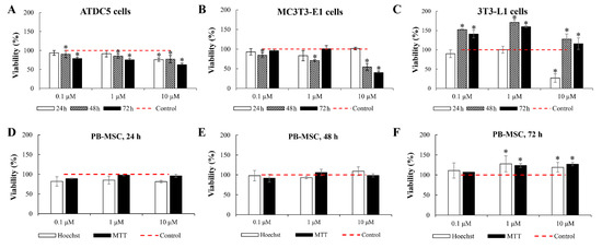
Figure 1.
Acellular porcine hemoglobin (PHb) modulates proliferation of mesenchymal cells. Viability of ATDC5, MC3T3-E1, and 3T3-L1 cells (A–C) and PB-MSCs (D–F) cultured for 24, 48, and 72 in growth medium (GM) supplemented with PHb. (A–C) The cell viability assessed by MTT assay. (D–F) The cell viability assessed by MTT assay and Hoechst labeling. Results are percentages of the corresponding control (viability of control cells cultured in GM only; set at 100%). Mean ± SD of three independent experiments is shown. * p < 0.05, compared to the corresponding control.
At all concentrations examined, PHb had no effect on PB-MSCs’ viability after 24 and 48 h in culture, as confirmed by the MTT assay and Hoechst 33258 tagging (Figure 1D,E). After 72 h, both MTT assay and Hoechst 33258 labeling showed that, at concentrations of 1 and 10 μM, PHb stimulated PB-MSCs’ viability, and that it did not affect viability at a 0.1 μM concentration (Figure 1F).
3.2. Porcine Hemoglobin Modifies PB-MSCs’ Clonogenic Potential and Cell Cycle
The number of PB-MSC colonies formed in the presence of 1 and 10 μM PHb was lower compared to the number of PB-MSCs formed in control cultures (Figure 2A,B). The observed difference was mainly due to the change in the number of large (>200 cells) PB-MSC colonies (Figure 2C).
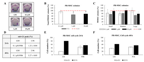
Figure 2.
Acellular porcine hemoglobin (PHb) modulates clonogenic capacity and apoptosis of PB-MSCs. (A) Micrographs of CFU-F formed in growth medium (GM) or GM supplemented with PHb (magnification 40×). (B) Clonogenic capacity of PB-MSCs after 14 days in culture with GM supplemented with PHb, evaluated by CFU-F assay. The results are percentages of the corresponding control, where the number of CFU-F formed after culture in GM only was set at 100%. * p < 0.05, PHb compared to control. (C) Number of small (50–100 cells), medium (100–200 cells), and large (more than 200 cells) CFU-F colonies formed in GM supplemented with PHb. The results are expressed as in (B). (D) PHb does not affect the number of apoptotic PB-MSCs after 24 and 48 h in culture. (E,F) PHb changes percentage of PB-MSCs in G0/G1 and G2+S phase of the cell cycle in 24 (E) and 48 h (F) cultures. All the results are given as mean ± SD of three independent experiments.
The number of apoptotic PB-MSCs (depicted by the percentage of the hypodiploid “sub-G1” peak in the DNA fluorescence histogram in Figure 2D) grown in GM only did not differ significantly from the quantity of apoptotic PB-MSCs grown in GM supplemented with PHb (Figure 2D). When PB-MSCs grew in CM only, ~20% and ~35% were in the G0/G1 phase after 24 and 48 h, respectively (Figure 2E,F). The analysis of the impact of PHb on PB-MSCs’ cell cycle progression showed that PHb induced the transient cell cycle arrest of PB-MSCs, with ~45% cells in the G0/G1 phase after 24 h (Figure 2E) and ~35% in the same stage of the cell cycle after 48 h (Figure 2F) (Supplementary Material, Figure S3).
3.3. Porcine Hemoglobin Modifies Migratory Capacity of Mesenchymal Cells
PHb acted as a weak modulator of migratory potential for all analyzed mesenchymal cells. At the concentrations of 0.1 and 1 μM, PHb slightly but significantly stimulated the migratory capacity of PB-MSCs and ATDC5 cells (Figure 3A,B). The same, significant stimulatory effect of PHb at all tested concentrations was observed in cultures of MC3T3-E1 cells (Figure 3C). Since the “scratch” in 3T3-L1 cells after 24 h in culture was almost entirely closed, PHb’s effect on the migratory capacity of these cells could not be analyzed (Figure 3D).
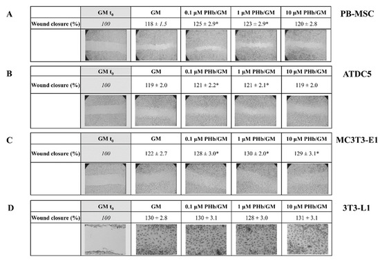
Figure 3.
Acellular porcine hemoglobin (PHb) modulates migratory capacity of mesenchymal cells. Migration of PB-MSCs (A), ATDC5 (B), MC3T3-E1 (C), and 3T3-L1 (D) cells analyzed by “scratch” assay (micrographs magnification: 40×). Upon making a “scratch” in confluent cell monolayer, cells were cultured for 24 h in the presence of growth medium (GM) or GM supplemented with PHb. The surface formed immediately after making the “scratch” was labeled “GM t0” and represented the control of the experiment (set as 100%). Results are percentages of the scratch areas covered with migrating cells, relative to GM t0. Mean ± SD of three independent is shown. * p < 0.05, compared to the corresponding control.
3.4. Porcine Hemoglobin Modifies Differentiation Capacity of Mesenchymal Cells
To evaluate the influence of PHb on the multilineage cell differentiation of mesenchymal cells, we selected the concentration of 0.1 μM, since it was the one that displayed the least prominent effect on the proliferation, migration, and clonogenic capacity of the tested cells.
The results of the analysis showed that the GAGs content in PB-MSCs and ATDC5 cells grown in the presence of 0.1 μM PHb was lower compared to that of the cells cultured in CM only (Figure 4A,B). PHb decreased the expression of chondrogenic marker SOX9 in PB-MSCs, slightly stimulated the expression of COL1A1, and increased the expression of COL2A1 after 14 days in culture (Figure 4C). Contrarily, PHb stimulated the expression levels of Sox9 in ATDC5 cells and attenuated the expression of COL11A1, while the expression product of col2a1 was not identified in these cells using RT-PCR (Figure 4D).
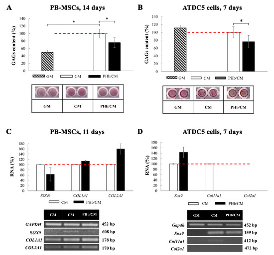
Figure 4.
Acellular porcine hemoglobin (PHb) reduces chondrogenic differentiation capacity of mesenchymal cells. Chondrogenic differentiation of PB-MSCs and ATDC5 cells assessed by Safranin O staining of GAGs in PB-MSCs after 14 days (A) and ATDC5 cells after 7 days (B) of cultivation with or without 0.1 μM PHb in chondrogenic differentiation medium (CM). Intensity of the Safranin O staining in GM supplemented with PHb is presented as relative to control (CM, set as 100%). Mean ± SD of four independent experiments is shown; * p < 0.05, compared to the corresponding control. Chondrogenic differentiation of PB-MSCs was confirmed by RT-PCR of analysis of expression of SOX9, COL1A1, and COL2A1 genes after 11 days of cultivation (C) and Sox9, Col11a, and Col2a1 genes in ATDC5 cells after 7 days of cultivation (D) in CM with or without PHb. GAPDH was used as a gel loading control. Representative RT-PCR bands are shown. The RT-PCR results are expressed as relative to corresponding control (CM, set as 100%). Mean ± SD of three independent experiments is shown.
PHb did not affect ALP activity in 7-day-cultivated PB-MSCs (Figure 5A) but it decreased the content of extracellular calcium deposits after 14 days of cultivation (Figure 5B). Following 7 days in culture with PHb, the expression levels of RUNX2 and COL1A1 were increased in PB-MSCs, while PHb decreased the expression of ALPL in these cells following 7 days of incubation (Figure 5C). After 14 days in culture, the expression levels of RUNX2, COL1A1, and ALPL markers were lower in PB-MSCs cultured in the presence of PHb than in the cells cultured in OM only (Figure 5D). The expression of the BGLAP gene in PB-MSCs was not induced after 7 or 14 days of incubation in OM (Figure 5C,D).
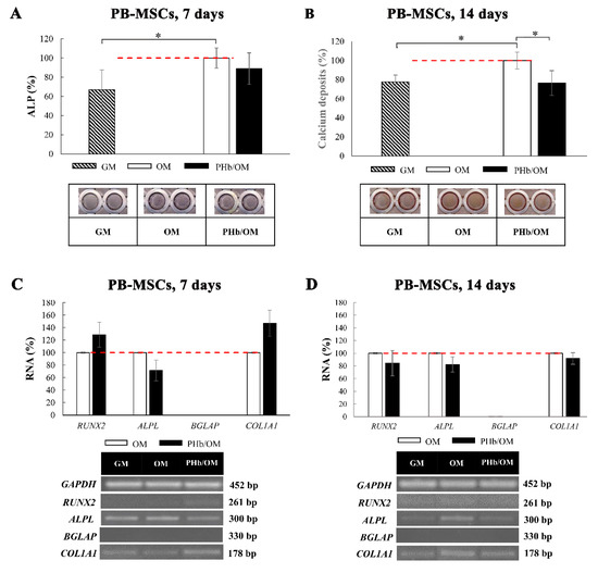
Figure 5.
Acellular porcine hemoglobin (PHb) reduces osteogenic differentiation capacity of primary PB-MSCs. Osteogenic differentiation of PB-MSCs was assessed by ALP staining (NBT/BCIP) after 7 days in culture (A) and Alizarin red staining after 14 days in culture (B) with or without 0.1 μM PHb in osteogenic differentiation medium (OM). Intensity of ALP or Alizarin red staining is presented as relative to corresponding control (OM set as 100%). Mean ± SD of four independent experiments is shown; * p < 0.05, compared to the control (OM). Osteogenic differentiation of PB-MSCs was confirmed by RT-PCR of analysis of expression of RUNX2, ALPL, BGLAP, COL1A1 genes after the cells were cultured in OM with and without PHb for 7 days (C) and 14 days (D). GAPDH was used as a gel loading control. Representative RT-PCR bands are shown. The RT-PCR results are expressed as relative to corresponding control (OM, set as 100%). Mean ± SD of three independent experiments is shown.
In MC3T3-E1 cells grown in OM without Dex, PHb decreased the activity of the ALP enzyme but had no effect on the content of extracellular calcium deposits (Figure 6A,B). On the other hand, PHb had no influence on the ALP activity or the content of extracellular calcium deposits in MC3T3-E1 cells cultured in OM with Dex (Figure 6D,E). In the OM without Dex, PHb stimulated the expression of Runx2 and Alpl, while it had no effect on the expression of the Bglap marker (Figure 6C). The expression of Runx2 in MC3T3-E1 cells cultivated in OM with Dex could not be detected (Figure 6F). In the cells grown in this medium, PHb attenuated the expression of the Alpl marker and did not affect the expression of Bglap (Figure 6F).
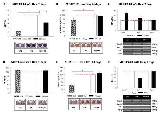
Figure 6.
Acellular porcine hemoglobin (PHb) modulates osteogenic differentiation of MC3T3-E1 cells grown in osteogenic medium with or without dexamethasone (Dex). MC3T3-E1 was cultured in osteogenic medium (OM) without (A–C) or with (E,F) added Dex. The osteogenic differentiation was assessed by ALP staining (NBT/BCIP) after 7 days in culture (A,D) and Alizarin red staining after 14 days in culture (B,E) with OM with or without PHb. Results of ALP or Alizarin red staining are presented as relative to control (OM set as 100%). Mean ± SD of four independent experiments is shown; * p < 0.05, compared to the control (OM). Osteogenic differentiation of MC3T3-E1 was confirmed by RT-PCR of analysis of expression of Runx2, Alpl, and Bglap genes after cultivating cells in OM with and without PHb for 7 days (C,F). GAPDH was used as a gel loading control. Representative RT-PCR bands are shown. RT-PCR results are expressed as relative to corresponding control (OM, set as 100%). Mean ± SD of three independent experiments is shown.
Differentiation of PB-MSCs and 3T3-L1 cells towards adipogenic lineage was confirmed by the presence of lipid droplets after Oil Red staining (Figure 7A). The expression of ADIPOQ (coding adiponectin) and LPL (coding lipoprotein lipase) was not detected in PB-MSCs after 7 or 14 days of incubation in AM (Figure 7B,C). The expression of PPARG (coding peroxisome proliferator-activated receptor gamma) was enhanced in the presence of 0.1 μM PHb after 7 days in culture (Figure 7B) and decreased after 14 days in culture with PB-MSCs (Figure 7C). PHb increased the expression of the Pparg gene when added starting from the induction medium and did not affect the expression of the Adipoq marker in 3T3-L1 cells (Figure 7D). The expression levels of both markers (Pparg and Adipoq) in 3T3-L1 cells were decreased when PHb was supplemented in insulin medium (Figure 7E).
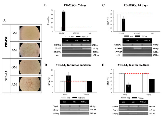
Figure 7.
Acellular porcine hemoglobin (PHb) reduces adipogenic differentiation capacity of mesenchymal cells. The adipogenic differentiation was confirmed by Oil Red staining of lipid droplets in PB-MSCs cultured for 28 days and 3T3-L1 cultured for 10 days in adipogenic medium (AM) (A). Adipogenic differentiation of PB-MSCs was confirmed by RT-PCR of analysis of expression of PPARG, ADIPOQ, and LPL genes. PB-MSCs were cultivated in adipogenic medium (AM) supplemented with or without 0.1 μM PHb for 7 days (B) and 14 days (C). Adipogenic differentiation of 3T3-L1 was confirmed by RT-PCR of analysis of Pparg and Adipoq genes. PHb was added starting from induction medium (D) or insulin medium (E). GAPDH was used as a gel loading control. Representative RT-PCR bands are shown. The RT-PCR results are expressed as relative to corresponding control (AM, set as 100%). Mean ± SD of three independent experiments is shown.
4. Discussion
In the internal milieu of erythrocytes, mammalian hemoglobin possesses a broad range of functions [24], which nowadays are well understood. However, when these biological macromolecules are present extracellularly, they exhibit many other still insufficiently elucidated biological functions [25,26,27]. This fact is increasing the added value of hemoglobin’s use for biomedical and biotechnological purposes, especially in the case of xenogeneic ones. Since mammalian erythrocytes are enucleated cells, porcine and bovine blood is of particular interest for biotechnological studies due to the fact that the manipulation and hemoglobin isolation from these cells are facilitated. Bovine and porcine slaughterhouse blood, as a natural waste derivative of the meat industry, is a very attractive, readily available source of hemoglobin. The main advantage over using slaughterhouse blood compared to outdated human blood in research is the fact that slaughterhouse blood is abundant, while a maximum of 5–10% donated human blood is not used for transfusion (becomes outdated [5] or contains anti-erythrocyte antibodies) and can be used for biotechnological purposes. Thus far, hemoglobin isolated from slaughterhouse blood has found application in studies of the interactions of proteins with ligands, molecules, and nanoparticles, and their harmfulness and safety [28,29,30,31], as a biosensor and drug carrier [29], and, to the greatest extent, as hemoglobin-based oxygen carriers [1,3]. Porcine Hb (PHb) has also been added to the portfolio of raw materials for this type of oxygen carrier [32], as a result of the restricted stock of human Hb and the potential danger of human blood-borne diseases such as hepatitis and HIV and cross-species transmission of Creutzfeldt–Jakob disease (mad cow disease) in the case of bovine blood use. PHb shows 84.4% homology to human adult hemoglobin [4]. Our studies have shown that porcine hemoglobin can be isolated from slaughterhouse blood by a simple and cost-effective process based on gradual hypotonic hemolysis [17] and tangential ultrafiltration [18]. Such a process provides a high level of protein purity, and, more importantly, isolated hemoglobin possesses physico-chemical characteristics as a native molecule [18]. Since PHb is a valuable model for the simulation of extracellular hemoglobin’s presence in vivo, in this study, it was predominantly tested on human primary cells, PB-MSCs. However, to evaluate its potential for usage as a constituent of cell culture media, besides a heterogeneous cell population of PB-MSCs, whose traits vary in many aspects (such as donors, number of passages in cell culture, etc. [33]), PHb was also tested on three cell lines of mesodermal origin—ATDC5, MC3T3-E1, and 3T3-L1—as more standardized cell systems compared to primary cells.
In this research, the effect of PHb on cell viability was dependent on its concentration and the type of the cells it was tested on. PHb had a predominantly inhibitory effect on the viability of ATDC5 and MC3T3-E1 cells, corresponding to the demonstrated effect of bovine hemoglobin (BHb) on the proliferation rate of these cells [16]. The cytotoxic effect of hemoglobin was also previously described on HUVEC cells [34], glioma cells [35], CHO-S cells [14], and rat neural cells [36,37]. The mainly stimulatory effect of PHb on 3T3-L1 cells’ viability was similar to the effect of BHb on 3T3-L1 cells’ proliferation rate [16]. Other studies documented that hemoglobin enhances the growth of human colon cancer cell lines HT-29 and Lovo, as well as the CCD-33Co normal colonic fibroblast cell line [38], human hepatoma cell line C3A [39], and primary rat astrocyte cultures [40]. The observed effect of PHb on MSCs’ viability (i.e., stimulation at higher concentrations after prolonged incubation) was in accordance with the effect of BHb on the cell proliferation rate, demonstrated using the same model systems [16].
Even though extracellular hemoglobin can be a part of the PB-MSC microenvironment, data in the literature regarding its influence on mesenchymal cells’ migratory capacity are scarce. Bo et al. [41] demonstrated that human glycated hemoglobin (HbA1c) inhibited the migration of HUVEC cells in a dose- and time-dependent manner [41]. Likewise, the peptides derived from bovine hemoglobin had the same effect on the MIAPaCa-2 cell line’s migratory capacity [42]. Contrarily, Roth et al. [35] showed that bovine hemoglobin stimulates the migration of glioma cells at a concentration of 0.5 g/L at 5% O2 [35]. This is in accordance with our results, showing that PHb slightly stimulated the migration of PB-MSCs, ATDC5, and MC3T3-E1 cells at most of the tested concentrations. However, even then, the observed changes in the migratory ability of different mesenchymal cells in the presence of PHb were within the range of ± 8% as against the related control. Since the concentration of 0.1 μM PHb minimally affected PB-MSCs’ and cell lines’ growth and migration, this concentration was chosen to further test the effects of PHb on the multilineage differentiation potential.
As expected, after induction, PB-MSCs differentiated towards osteogenic, adipogenic, and chondrogenic lineages, while the cell lines exhibited the earlier described lineage-specific differentiation. The initial data revealed equality between the cells in GM culture solely and the cells cultivated in GM with PHb added (Supplementary Material, Figure S4), so the effects of PHb were further investigated by its addition in the specific differentiation media.
The lower level of GAGs (histochemically determined by Safranin O staining) after the induction of the chondrogenic differentiation of PB-MSCs and ATCD5 in chondrogenic differentiation medium with the addition of PHb, in comparison to the conditions without PHb, indicates that PHb reduced the chondrogenic differentiation capacity of the analyzed cells. However, PHb’s impacts on the expression of genes representing markers of chondrogenic differentiation depended on the homogeneity of the cell population, as well as on the phase of chondrogenic differentiation [16,43]. Our results demonstrated that, along with the reduction in GAGs content, PHb suppressed SOX9 expression in PB-MSCs (crucial transcription factor in cartilage and marker of chondrogenesis stage II), while it mildly enhanced the expression of both collagens tested, collagen type I and collagen type II (chondrogenesis stage I and IV markers, respectively). In ATDC5 cells, PHb caused the lowering of GAGs and downregulated the expression of Col11a1; as expected, due to the genetic uniformity of this cell line, unlike PB-MSCs, expression of the late chondrogenic markers was not observed [16]. In comparison to our previous studies on bovine hemoglobin’s impact on chondrogenesis in ATDC5 cells [16], PHb revealed the same effect on the expression of Col11a1, but, contrary to bovine hemoglobin, it stimulated the expression of Sox9. Besides our recent study on bovine hemoglobin’s effects on mesenchymal stromal cells [16], to the best of the authors’ knowledge, there are no literature data on the direct influence of hemoglobin or heme on the chondrogenic differentiation of MSCs and mesenchymal cell lines. The chondrogenic capacity of MSCs derived from bone marrow was evaluated under the influence of different iron compounds [44,45], and reduced expression/shift was demonstrated for chondrogenic markers SOX9 and COL2A1. However, this could be only speculated so far, and additional research is considered necessary to confirm that the effect of hemoglobin shown in our experiments was due to heme iron.
Under an induced model of hemorrhagic anemia, mouse bone marrow MSCs have shown an increased capacity for osteogenic differentiation (and, at the same time, a reduced ability for adipogenic differentiation) [46], but the direct influence of extracellular hemoglobin has not been discussed. In this study, the results of histochemical tests after the osteogenic differentiation of PB-MSCs in the presence of PHb proved consistency with the effect of bovine hemoglobin [16]. As shown in the case of BHb [16], the activity of ALP enzymes (measured by NBT/BCIP staining) and calcium deposits (measured by Alizarin red staining) in PB-MSCs grown in the presence of PHb was lower than in the cells grown in hemoglobin-free medium. However, analysis of gene expression by RT-PCR showed slight differences in the effects of PHb on osteogenic PB-MSCs’ gene expression in comparison to bovine hemoglobin [16]. The alterations in the expression of osteogenic markers detected by RT-PCR (with the most noticeable difference in the expression of RUNX2, a crucial transcriptional agent for the dedication of MSCs to the bone-forming cells [47]), as well as differences in the results of colorimetric assays, can be attributed to the population variety of PB-MSCs, as well as to the cell storage conditions and duration [33]. The PB-MSCs used in our two studies were of the same donor and in the same passage, but the PB-MSCs used for the analysis of PHb’s effects expressed RUNX2 constitutively (although at a very low level), while the constitutive expression in PB-MSCs used for the analysis of the effect of BHb was not evident [16]. The expression of osteocalcin (encoded by the BGLAP gene) was not detected in PB-MSCs even after 14 days, which is in accordance with the accepted model of osteogenic differentiation of these cells in vitro [48]. Both PHb and BHb decreased ALPL gene expression in PB-MSCs grown for both 7 and 14 days, while they slightly reduced the expression of the type I collagen gene after 14 days in culture [16]. However, PHb increased, while BHb decreased, the expression of the type I collagen gene after 7 days in culture [16]. These variations in the effects of BHb and PHb on type I collagen gene expression could be, at least partially, explained by the diversity of the PB-MSCs population.
Guided by the already established experimental protocols to understand the delicate alterations specific to each phase of osteogenesis induced by extracellular hemoglobin [16], in this study, we also used two media for the osteogenic differentiation of MC3T3-E1 cells: OM with Dex and OM without Dex. In OM with Dex, PHb decreased the activity of the ALP enzyme but, differently from BHb (as reported in [16]), PHb did not modulate the level of calcium deposits in MC3T3-E1 cells. Both PHb and BHb in OM with Dex stimulated Alpl expression and had no significant influence on Bglap expression. However, the effects of these two hemoglobin types on Runx2 were opposite: PHb stimulated, while BHb silenced its expression. In OM with Dex, PHb exhibited effects similar to those reported for bovine hemoglobin [16]. Namely, PHb in OM with Dex did not lead to a change in the parameters detectable by histochemical staining, lowered the expression of Alpl, and did not affect Bglap expression in MC3T3-E1 cells. Furthermore, Runx2 was not detected under these conditions, suggesting the faster passage of MC3T3-E1 cells through the stages of osteogenic differentiation compared to cells grown in dexamethasone-free medium. As already speculated in the case of bovine extracellular hemoglobin’s effects on MC3T3-E1 cells [16], a possible explanation for these results could be that PHb added to culture medium causes phase shifts, i.e., a “delay” in osteogenic differentiation [48].
PPARγ, a master regulator of adipogenesis in mammals, is considered both a sufficient and necessary factor for adipogenic differentiation [49]. PHb stimulated the expression of this marker after 7 days in culture of PB-MSCs and decreased it after 14 days. Other adipogenic markers were not expressed as extended treatment was almost certainly needed to accomplish their expression. The same results were demonstrated in the case of BHb addition in PB-MSCs culture [16]. Furthermore, this study revealed that the effect of PHb on the adipogenic differentiation ability of 3T3-L1 cells was dependent on the phase of adipogenic differentiation during which hemoglobin was added, as already demonstrated for BHb [16]. After PHb addition starting from the induction medium, hemoglobin stimulated the expression of Pparγ and did not influence expression of adiponectin but inhibited the expression of these markers after addition starting from the insulin medium. It has been shown that heme stimulates the adipogenic differentiation of MSCs as well as 3T3-L1 cells [50]. Additionally, pathophysiological processes followed by increased production of denatured heme proteins are linked with an enhanced adipogenic response; this effect is mostly manifested as adipocyte hypertrophy, characterized by dysfunctional, proinflammatory adipocytes that have reduced expression of the anti-inflammatory hormone, adiponectin [51]. The inconsistencies in the effects of PHb and BHb on MSCs’ functional characteristics [16] may be attributed to differences in primary protein structure [52] or higher levels of protein organization. Moreover, our recent study showed some differences in the phospholipid content and distribution in isolated porcine and bovine hemoglobin, as an inevitable side component in hemoglobin samples due to the used preparation method [53]. Ten fatty acids were demonstrated in phospholipids originating from porcine hemoglobin and six in phospholipids presented in bovine hemoglobin samples: (1) saturated fatty acids (palmitic and stearic acid) were the most dominant fatty acids in hemoglobin preparations of both species, but the quantity of palmitic acid was higher in porcine hemoglobin samples compared to bovine ones; (2) palmitoleic, oleic, and vaccenic fatty acids were found in porcine hemoglobin preparations, while oleic acid was the only monounsaturated fatty acid detected in bovine hemoglobin samples; (3) five poly unsaturated fatty acids were detected in porcine hemoglobin samples (linolenic, γ-linolenic, α-linolenic, dihomo-γ-linolenic, arachidonic, and linoleic acid) and three (dihomo-γ-linolenic, arachidonic, and linoleic acid) in bovine hemoglobin samples [53].
There are numerous reports supporting the fact that fatty acids influence the functional characteristics of MSCs. Casado-Díaz et al. have shown that arachidonic acid induces the adipogenic differentiation of MSCs while inhibiting osteoblastogenesis [54]. In addition to stimulating MSCs’ motility [55], oleic acid can also stimulate the expression of neural markers in MSCs isolated from the human endometrium [56]. In contrast to the arachidonic effect, linolenic acid stimulates osteogenic and inhibits adipogenic MSCs’ differentiation [57]. However, there is evidence demonstrating that linolenic and α-linolenic acids reduce RUNX2 expression in MSCs and stimulate PPARG expression, as well as leading to the increased accumulation of lipid droplets in these cells in a dose-dependent manner [58]. Furthermore, an increase in the accumulation of lipid droplets in MSCs during adipogenic differentiation was obtained by Yanting et al. [59], during the cultivation of MSCs with linolenic, oleic, and palmitic acid [59]. Liu and co-workers have shown that stearic acid can stimulate MSCs’ migration and accelerate cartilage healing, as well as leading to an increase in the number of colonies of these cells [60]. According to the above-mentioned findings, the differences in the effects of PHb (porcine) hemoglobin in this study and bovine hemoglobin in our previous study [16] on the functional characteristics of mesenchymal cells could be explained, at least partially, by the differences in the appearance of individual fatty acids of phospholipid contaminants present in the porcine and bovine hemoglobin samples used. However, without additional and more detailed analysis of the influence of each of these components individually, and subsequently in synergy, the influence of trace fatty acids present in the used hemoglobin samples on the observed changes in the functional characteristics of MSCs can be speculated only.
5. Conclusions
In summary, we have reported that PHb, as xenogeneic hemoglobin, influences the proliferative, migratory, and differentiation potential of several mesenchymal cells. The effect of PHb on these cell functional characteristics depends on its concentration, cell type, and culture period. The effect of PHb on cell viability was predominantly inhibitory on ATDC5 and MC3T3-E1 cells, mainly stimulatory on 3T3-L1 cells, and stimulatory at higher concentrations after prolonged incubation on PB-MSCs. PHb slightly stimulated the migration of PB-MSCs, ATDC5, and MC3T3-E1 cells. The capacity to differentiate towards chondrogenic, osteogenic, and adipogenic lineages of PB-MSCs and corresponding cell lines (ATDC5 cells for chondrogenic differentiation, MC3T3-E1 cells for osteogenic differentiation, and 3T3-L1 cells for adipogenic differentiation) was reduced in the presence of PHb. Our results indicate that, even under conditions that induce a specific type of MSC differentiation (cultivation in particular differentiation media), PHb, similarly to BHb, has the ability to modulate multilineage differentiation, making it a potential candidate for a component of cell propagation or preservation media. Furthermore, this study discusses delicate discrepancies obtained in cells’ functional properties using porcine (PHb) and bovine (BHb) hemoglobin (two types of mammalian extracellular hemoglobin) as a consequence of the inevitable impurities of the production process, which is an essential aspect of the actual biotechnological application.
Supplementary Materials
The following are available online at https://www.mdpi.com/article/10.3390/pr10010032/s1, Figure S1: Reduction in ABTS cation radical by PHb; Figure S2: Spontaneous proliferation of (A) ATDC5, (B) MC3T3-E1, (C) 3T3-L1 cells after 24, 48, and 72 h in culture, assessed by MTT assay; Figure S3: (A) Representative flow cytometry histograms of PB-MSCs cultured in growth medium (GM) (B,D) or 0.1 PHb in GM (C,E) for 24 (B,C) or 48 h (D,E); Figure S4: Effect of porcine hemoglobin (PHb) on chondrogenic differentiation of ATDC5 cells assessed by Safranin O staining of GAGs in ATDC5 cells after 7 days of cultivation with or without 0.1 μM PHb in growth medium (GM; DMEM supplemented with 10% fetal calf serum) (A). Effect of PHb on osteogenic differentiation of PB-MSCs assessed by NBT/BCIP staining for ALP after 7 days of cultivation with or without 0.1 μM PHb in GM (B). (C) Effect of PHb on osteogenic differentiation of MC3T3-E1 cells assessed by Alizarin red staining after 14 days of cultivation with or without 0.1 μM PHb in GM (C); Table S1: Human PCR primer sets used in experiments; Table S2: Mouse PCR primer sets used in experiments.
Author Contributions
The study conception and design were performed by V.L.I.; material preparation, data collection, analysis, and interpretation were performed by A.Z.S., I.T.D. and V.L.I.; D.S.B. and B.M.B. supervised the projects; the first draft of the manuscript was written by A.Z.S. and I.T.D.; all authors commented on previous versions of the manuscript. All authors have read and agreed to the published version of the manuscript.
Funding
This work was supported by the Ministry of Education, Science and Technological Development of the Republic of Serbia (contract number 451-03-9/2021-14/200015 with Institute for Medical Research University of Belgrade, National Institute of Republic of Serbia).
Institutional Review Board Statement
Not applicable.
Informed Consent Statement
Not applicable.
Data Availability Statement
The data that support the findings of this study are available from the corresponding author (I.D.) upon reasonable request.
Acknowledgments
We would like to thank Slavko Mojsilović for performing the flow cytometry.
Conflicts of Interest
The authors declare no conflict of interest.
References
- Alayash, A. Hemoglobin-based blood substitutes: Oxygen carriers, pressor agents, or oxidants? Nat. Biotechnol. 1999, 17, 545–549. [Google Scholar] [CrossRef] [PubMed]
- Xiong, Y.; Steffen, A.; Andreas, K.; Müller, S.; Sternberg, N.; Georgieva, R.; Bäumler, H. Hemoglobin-Based Oxygen Carrier Microparticles: Synthesis, Properties, and In Vitro and In Vivo Investigations. Biomacromolecules 2012, 13, 3292–3300. [Google Scholar] [CrossRef]
- Bialas, C.; Moser, C.; Sims, C. Artificial oxygen carriers and red blood cell substitutes: A historic overview and recent developments toward military and clinical relevance. J. Trauma Acute Care Surg. 2019, 87, S48–S58. [Google Scholar] [CrossRef] [PubMed]
- Yang, Q.; Bai, S.; Li, L.; Li, S.; Zhang, Y.; Munir, M.; Qiu, H. Human Hemoglobin Subunit Beta Functions as a Pleiotropic Regulator of RIG-I/MDA5-Mediated Antiviral Innate Immune Responses. J. Virol. 2019, 93, e00718–e00719. [Google Scholar] [CrossRef] [Green Version]
- Wang, Y.; Wang, L.; Yu, W.; Gao, D.; You, G.; Li, P.; Zhang, S.; Zhang, J.; Hu, T.; Zhao, L.; et al. A PEGylated bovine hemoglobin as a potent hemoglobin-based oxygen carrier. Biotechnol. Prog. 2016, 33, 252–260. [Google Scholar] [CrossRef]
- González-Rosendo, G.; Polo, J.; Rodríguez-Jerez, J.; Puga-Díaz, R.; Reyes-Navarrete, E.; Quintero-Gutiérrez, A. Bioavailability of a Heme-Iron Concentrate Product Added to Chocolate Biscuit Filling in Adolescent Girls Living in a Rural Area of Mexico. J. Food Sci. 2010, 75, H73–H78. [Google Scholar] [CrossRef] [PubMed]
- Hoppe, M.; Brün, B.; Larsson, M.; Moraeus, L.; Hulthén, L. Heme iron-based dietary intervention for improvement of iron status in young women. Nutrition 2013, 29, 89–95. [Google Scholar] [CrossRef] [PubMed]
- Li, H.; Chen, Y.; Li, Z.; Li, X.; Jin, Q.; Ji, J. Hemoglobin as a Smart pH-Sensitive Nanocarrier To Achieve Aggregation Enhanced Tumor Retention. Biomacromolecules 2018, 19, 2007–2013. [Google Scholar] [CrossRef] [PubMed]
- Qi, W.; Yan, X.; Duan, L.; Cui, Y.; Yang, Y.; Li, J. Glucose-Sensitive Microcapsules from Glutaraldehyde Cross-Linked Hemoglobin and Glucose Oxidase. Biomacromolecules 2009, 10, 1212–1216. [Google Scholar] [CrossRef]
- Arenberger, P.; Engels, P.; Arenbergerova, M.; Gkalpakiotis, S.; García Luna Martínez, F.J.; Villarreal Anaya, A.; Jimenez Fernandez, L. Clinical results of the application of a hemoglobin spray to promote healing of chronic wounds. GMS Krankenh. Interdiszip. 2011, 6, Doc05. [Google Scholar] [CrossRef]
- Elg, F.; Hunt, S. Hemoglobin spray as adjunct therapy in complex wounds: Meta-analysis versus standard care alone in pooled data by wound type across three retrospective cohort controlled evaluations. SAGE Open Med. 2018, 6, 205031211878431. [Google Scholar] [CrossRef] [PubMed] [Green Version]
- Mallet, V.; Dutheil, D.; Polard, V.; Rousselot, M.; Leize, E.; Hauet, T.; Goujon, J.; Zal, F. Dose-Ranging Study of the Performance of the Natural Oxygen Transporter HEMO2Life in Organ Preservation. Artif. Organs 2014, 38, 691–701. [Google Scholar] [CrossRef]
- Zal, F.; Rousselot, M. Extracellular hemoglobins from Annelids, and their potential use in biotechnology. In Outstanding Marine Molecules, 1st ed.; La Barre, S., Kornprobst, J.-M., Eds.; Wiley-VCH Verlag GmbH & Co. KGaA: Weinheim, Germany, 2014; pp. 361–376. [Google Scholar] [CrossRef]
- Le Pape, F.; Bossard, M.; Dutheil, D.; Rousselot, M.; Polard, V.; Férec, C.; Leize, E.; Delépine, P.; Zal, F. Advancement in recombinant protein production using a marine oxygen carrier to enhance oxygen transfer in a CHO-S cell line. Artif. Cells Nanomed. Biotechnol. 2015, 43, 186–195. [Google Scholar] [CrossRef] [PubMed]
- Le Pape, F.; Cosnuau-Kemmat, L.; Richard, G.; Dubrana, F.; Férec, C.; Zal, F.; Leize, E.; Delépine, P. HEMOXCell, a New Oxygen Carrier Usable as an Additive for Mesenchymal Stem Cell Culture in Platelet Lysate-Supplemented Media. Artif. Organs 2017, 41, 359–371. [Google Scholar] [CrossRef]
- Stančić, A.; Drvenica, I.; Obradović, H.; Bugarski, B.; Ilić, V.; Bugarski, D. Native bovine hemoglobin reduces differentiation capacity of mesenchymal stromal cells in vitro. Int. J. Biol. Macromol. 2020, 144, 909–920. [Google Scholar] [CrossRef] [PubMed]
- Kostić, I.; Ilić, V.; Đorđević, V.; Bukara, K.; Mojsilović, S.; Nedović, V.; Bugarski, D.; Veljović, Đ.; Mišić, D.; Bugarski, B. Erythrocyte membranes from slaughterhouse blood as potential drug vehicles: Isolation by gradual hypotonic hemolysis and biochemical and morphological characterization. Colloids Surf. B Biointerfaces 2014, 122, 250–259. [Google Scholar] [CrossRef]
- Drvenica, I.; Stancic, A.; Kalusevic, A.; Markovic, S.; Dragisic-Maksimovic, J.; Nedovic, V.; Bugarski, B.; Ilic, V. Maltose-mediated long-term stabilization of freeze- and spray- dried forms of bovine and porcine hemoglobin. J. Serb. Chem. Soc. 2019, 84, 1105–1117. [Google Scholar] [CrossRef] [Green Version]
- Miller, N.J.; Rice-Evans, C.; Davies, M.J.; Gopinathan, V.; Milner, A. A novel method for measuring antioxidant capacity and its application to monitoring the antioxidant status in premature neonates. Clin. Sci. 1993, 84, 407–412. [Google Scholar] [CrossRef] [Green Version]
- Trivanovic, D.; Kocic, J.; Mojsilovic, S.; Krstic, A.; Ilic, V.; Okic-Djordjevic, I.; Santibanez, J.; Jovcic, G.; Terzic, M.; Bugarski, D. Mesenchymal stem cells isolated from peripheral blood and umbilical cord Wharton’s jelly. Srp. Arh. Celok. Lek. 2013, 141, 178–186. [Google Scholar] [CrossRef]
- Kocić, J.; Santibañez, J.F.; Krstić, A.; Mojsilović, S.; Djordević, I.O.; Trivanović, D.; Ilić, V.; Bugarski, D. Interleukin 17 inhibits myogenic and promotes osteogenic differentiation of C2C12 myoblasts by activating ERK1,2. Biochim. Biophys. Acta-Mol. Cell Res. 2012, 1823, 838–849. [Google Scholar] [CrossRef] [Green Version]
- Reed, B.; Lane, M. Insulin receptor synthesis and turnover in differentiating 3T3-L1 preadipocytes. Proc. Natl. Acad. Sci. USA 1980, 77, 285–289. [Google Scholar] [CrossRef] [PubMed] [Green Version]
- De Winter, J.C.F. Using the Student’s t-test with extremely small sample sizes. Pract. Assess. Res. Eval. 2013, 18, 10. [Google Scholar] [CrossRef]
- Bunn, H. Evolution of mammalian hemoglobin function. Blood 1981, 58, 189–197. [Google Scholar] [CrossRef] [PubMed]
- Coates, C.; Decker, H. Immunological properties of oxygen-transport proteins: Hemoglobin, hemocyanin and hemerythrin. Cell. Mol. Life Sci. 2016, 74, 293–317. [Google Scholar] [CrossRef] [PubMed] [Green Version]
- Bamm, V.; Lanthier, D.; Stephenson, E.; Smith, G.; Harauz, G. In vitro study of the direct effect of extracellular hemoglobin on myelin components. Biochim. Biophys. Acta 2015, 1852, 92–103. [Google Scholar] [CrossRef] [PubMed] [Green Version]
- Loomis, Z.; Eigenberger, P.; Redinius, K.; Lisk, C.; Karoor, V.; Nozik-Grayck, E.; Ferguson, S.; Hassell, K.; Nuss, R.; Stenmark, K.; et al. Hemoglobin induced cell trauma indirectly influences endothelial TLR9 activity resulting in pulmonary vascular smooth muscle cell activation. PLoS ONE 2017, 12, e0171219. [Google Scholar] [CrossRef] [PubMed]
- Aliakbari, F.; Haji Hosseinali, S.; Khalili Sarokhalil, Z.; Shahpasand, K.; Akbar Saboury, A.; Akhtari, K.; Falahati, M. Reactive oxygen species generated by titanium oxide nanoparticles stimulate the hemoglobin denaturation and cytotoxicity against human lymphocyte cell. J. Biomol. Struct. Dyn. 2019, 37, 4875–4881. [Google Scholar] [CrossRef]
- Kamaljeet; Bansal, S.; SenGupta, U. A Study of the Interaction of Bovine Hemoglobin with Synthetic Dyes Using Spectroscopic Techniques and Molecular Docking. Front. Chem. 2017, 4, 50. [Google Scholar] [CrossRef] [Green Version]
- Xu, L.; Liu, Z.; Liao, T.; Tuo, X. Probing the interaction between levamlodipine and hemoglobin based on spectroscopic and molecular docking methods. Spectrochim. Acta A Mol. Biomol. Spectrosc. 2019, 223, 117306. [Google Scholar] [CrossRef] [PubMed]
- Yan, X.; Liu, B.; Chong, B.; Cao, S. Interaction of Cefpiramide sodium with bovine hemoglobin and effect of the coexistent metal ion on the protein-drug association. J. Lumin. 2013, 142, 155–162. [Google Scholar] [CrossRef]
- Zhu, X.; Chu, W.; Fan, D.; Dan, N.; Chen, C.; Wang, T.; Wang, F. Variations in Dominant Antigen Determinants of Glutaraldehyde Polymerized Human, Bovine and Porcine Hemoglobin. Artif. Cells Blood Substit. Biotechnol. 2007, 35, 518–532. [Google Scholar] [CrossRef] [PubMed]
- Arnhold, S.; Elashry, M.; Klymiuk, M.; Wenisch, S. Biological macromolecules and mesenchymal stem cells: Basic research for regenerative therapies in veterinary medicine. Int. J. Biol. Macromol. 2019, 123, 889–899. [Google Scholar] [CrossRef] [PubMed]
- Fischer-Fodor, E.; Mot, A.; Deac, F.; Arkosi, M.; Silaghi-Dumitrescu, R. Towards hemerythrin-based blood substitutes: Comparative performance to hemoglobin on human leukocytes and umbilical vein endothelial cells. J. Biosci. 2011, 36, 215–221. [Google Scholar] [CrossRef] [PubMed]
- Roth, A.; Elmer, J.; Harris, D.; Huntley, J.; Palmer, A.; Nelson, T.; Johnson, J.; Xue, R.; Lannutti, J.; Viapiano, M. Hemoglobin regulates the migration of glioma cells along poly(ε-caprolactone)-aligned nanofibers. Biotechnol. Prog. 2014, 30, 1214–1220. [Google Scholar] [CrossRef] [PubMed]
- Ortegon, D.; Davis, M.; Dixon, P.; Smith, D.; Josephs, J.; Mueller, D.; Jenkins, D.; Kerby, J. The Polymerized Bovine Hemoglobin-Based Oxygen-Carrying Solution (HBOC-201) Is Not Toxic to Neural Cells in Culture. J. Trauma. 2002, 53, 1068–1072. [Google Scholar] [CrossRef]
- Lara, F.; Kahn, S.; Fonseca, A.; Bahia, C.; Pinho, J.; Graca-Souza, A.; Houzel, J.; de Oliveira, P.; Moura-Neto, V.; Oliveira, M. On the Fate of Extracellular Hemoglobin and Heme in Brain. J. Cereb. Blood Flow Metab. 2009, 29, 1109–1120. [Google Scholar] [CrossRef] [Green Version]
- Lee, R.; Kim, H.A.; Kang, B.Y.; Kim, K.H. Hemoglobin induces colon cancer cell proliferation by release of reactive oxygen species. World J. Gastroenterol. 2006, 12, 5644. [Google Scholar] [CrossRef]
- Chen, G.; Palmer, A. Hemoglobin Regulates the Metabolic, Synthetic, Detoxification, and Biotransformation Functions of Hepatoma Cells Cultured in a Hollow Fiber Bioreactor. Tissue Eng. Part A 2010, 16, 3231–3240. [Google Scholar] [CrossRef]
- Yang, Y.; Ren, J.; Sun, Y.; Xue, Y.; Zhang, Z.; Gong, A.; Wang, B.; Zhong, Z.; Cui, Z.; Xi, Z.; et al. A connexin43/YAP axis regulates astroglial-mesenchymal transition in hemoglobin induced astrocyte activation. Cell Death Differ. 2018, 25, 1870–1884. [Google Scholar] [CrossRef] [Green Version]
- Bo, J.; Guan, Y.; Guo, Y.; Xie, S.; Zhang, C.; Zhang, H.; Chen, Z.; Lu, J.; Meng, Q. Impairment of Endothelial Cell Function Induced by Hemoglobin A1c and the Potential Mechanisms. Exp. Clin. Endocrinol. Diabetes 2015, 123, 529–535. [Google Scholar] [CrossRef]
- Wang, Y.; Zhang, T.; Zhang, H.; Yang, H.; Li, Y.; Jiang, Y. Bovine Hemoglobin Derived Peptide Asn-Phe-Gly-Lys Inhibits Pancreatic Cancer Cells Metastasis by Targeting Secreted Hsp90α. J. Food Sci. 2017, 82, 3005–3012. [Google Scholar] [CrossRef] [PubMed]
- Xu, J.; Wang, W.; Ludeman, M.; Cheng, K.; Hayami, T.; Lotz, J.; Kapila, S. Chondrogenic Differentiation of Human Mesenchymal Stem Cells in Three-Dimensional Alginate Gels. Tissue Eng. Part A 2008, 14, 667–680. [Google Scholar] [CrossRef] [Green Version]
- Papadimitriou, N.; Thorfve, A.; Brantsing, C.; Junevik, K.; Baranto, A.; Barreto Henriksson, H. Cell Viability and Chondrogenic Differentiation Capability of Human Mesenchymal Stem Cells After Iron Labeling with Iron Sucrose. Stem Cells Dev. 2014, 23, 2568–2580. [Google Scholar] [CrossRef]
- Roeder, E.; Henrionnet, C.; Goebel, J.; Gambier, N.; Beuf, O.; Grenier, D.; Chen, B.; Vuissoz, P.; Gillet, P.; Pinzano, A. Dose-Response of Superparamagnetic Iron Oxide Labeling on Mesenchymal Stem Cells Chondrogenic Differentiation: A Multi-Scale In Vitro Study. PLoS ONE 2014, 9, e98451. [Google Scholar] [CrossRef] [Green Version]
- Wu, M.Q.; Zhu, R.J.; Liu, K.Y. Osteogenic and adipogenic differentiation of bone marrow multipotent mesenchymal stem cells in the mice with hemorrhagic anemia. Zhongguo Shi Yan Xue Ye Xue Za Zhi 2014, 22, 177–182. (In Chinese) [Google Scholar] [CrossRef]
- Bruderer, M.; Richards, R.; Alini, M.; Stoddart, M. Role and regulation of RUNX2 in osteogenesis. Eur. Cells Mater. 2014, 28, 269–286. [Google Scholar] [CrossRef]
- Huang, Z.; Nelson, E.; Smith, R.; Goodman, S. The Sequential Expression Profiles of Growth Factors from Osteroprogenitors to OsteoblastsIn Vitro. Tissue Eng. 2007, 13, 2311–2320. [Google Scholar] [CrossRef] [PubMed]
- Wafer, R.; Tandon, P.; Minchin, J. The Role of Peroxisome Proliferator-Activated Receptor Gamma (PPARG) in Adipogenesis: Applying Knowledge from the Fish Aquaculture Industry to Biomedical Research. Front. Endocrinol. (Lausanne) 2017, 8, 1–10. [Google Scholar] [CrossRef] [Green Version]
- Puri, N.; Sodhi, K.; Haarstad, M.; Kim, D.; Bohinc, S.; Foglio, E.; Favero, G.; Abraham, N. Heme induced oxidative stress attenuates sirtuin1 and enhances adipogenesis in mesenchymal stem cells and mouse pre-adipocytes. J. Cell. Biochem. 2012, 113, 1926–1935. [Google Scholar] [CrossRef] [PubMed] [Green Version]
- Gesta, S.; Tseng, Y.; Kahn, C. Developmental Origin of Fat: Tracking Obesity to Its Source. Cell 2007, 131, 242–256. [Google Scholar] [CrossRef] [Green Version]
- Hardison, R. Evolution of Hemoglobin and Its Genes. Cold Spring Harb. Perspect. Med. 2012, 2, a011627. [Google Scholar] [CrossRef] [PubMed] [Green Version]
- Stančić, A.; Drvenica, I.; Bugarski, B.; Ilić, V.; Bugarski, D. Extracellular xenogeneic hemoglobin suppresses the capacity for C2C12 myoblast myogenic differentiation. Arch. Biol. Sci. 2020, 72, 379–391. [Google Scholar] [CrossRef]
- Casado-Díaz, A.; Santiago-Mora, R.; Dorado, G.; Quesada-Gómez, J. The omega-6 arachidonic fatty acid, but not the omega-3 fatty acids, inhibits osteoblastogenesis and induces adipogenesis of human mesenchymal stem cells: Potential implication in osteoporosis. Osteoporos. Int. 2012, 24, 1647–1661. [Google Scholar] [CrossRef]
- Jung, Y.; Lee, S.; Oh, S.; Lee, H.; Ryu, J.; Han, H. Oleic acid enhances the motility of umbilical cord blood derived mesenchymal stem cells through EphB2-dependent F-actin formation. Biochim. Biophys. Acta Mol. Cell Res. 2015, 1853, 1905–1917. [Google Scholar] [CrossRef] [PubMed] [Green Version]
- Kojour, M.; Ebrahimi-Barough, S.; Kouchesfehani, H.; Jalali, H.; Ebrahim, M. Oleic acid promotes the expression of neural markers in differentiated human endometrial stem cells. J. Chem. Neuroanat. 2017, 79, 51–57. [Google Scholar] [CrossRef] [PubMed]
- Kim, J.; Park, Y.; Lee, S.; Park, Y. Trans-10,cis-12 Conjugated linoleic acid promotes bone formation by inhibiting adipogenesis by peroxisome proliferator activated receptor-γ-dependent mechanisms and by directly enhancing osteoblastogenesis from bone marrow mesenchymal stem cells. J. Nutr. Biochem. 2013, 24, 672–679. [Google Scholar] [CrossRef] [PubMed] [Green Version]
- Riera-Heredia, N.; Lutfi, E.; Gutiérrez, J.; Navarro, I.; Capilla, E. Fatty acids from fish or vegetable oils promote the adipogenic fate of mesenchymal stem cells derived from gilthead sea bream bone potentially through different pathways. PLoS ONE 2019, 14, e0215926. [Google Scholar] [CrossRef]
- Yanting, C.; Yang, Q.; Ma, G.; Du, M.; Harrison, J.; Block, E. Dose- and type-dependent effects of long-chain fatty acids on adipogenesis and lipogenesis of bovine adipocytes. J. Dairy Sci. 2018, 101, 1601–1615. [Google Scholar] [CrossRef] [PubMed]
- Liu, Y.; Xu, L.; Hu, L.; Chen, D.; Yu, L.; Li, X.; Chen, H.; Zhu, J.; Chen, C.; Luo, Y.; et al. Stearic acid methyl ester promotes migration of mesenchymal stem cells and accelerates cartilage defect repair. J. Orthop. Translat. 2020, 22, 81–91. [Google Scholar] [CrossRef]
Publisher’s Note: MDPI stays neutral with regard to jurisdictional claims in published maps and institutional affiliations. |
© 2021 by the authors. Licensee MDPI, Basel, Switzerland. This article is an open access article distributed under the terms and conditions of the Creative Commons Attribution (CC BY) license (https://creativecommons.org/licenses/by/4.0/).