
Facial Scanning Accuracy with Stereophotogrammetry and Smartphone Technology in Children: A Systematic Review

 ,
,  , , ,
, , , 
 and
and 
Abstract
:1. Introduction
2. Materials and Methods
2.1. Protocol, Registration and Search Strategy
2.2. Inclusion, Exclusion Criteria and Outcomes
2.3. Study Selection and Data Extraction
2.4. Risk of Bias Assessment
3. Results
3.1. Study Selection
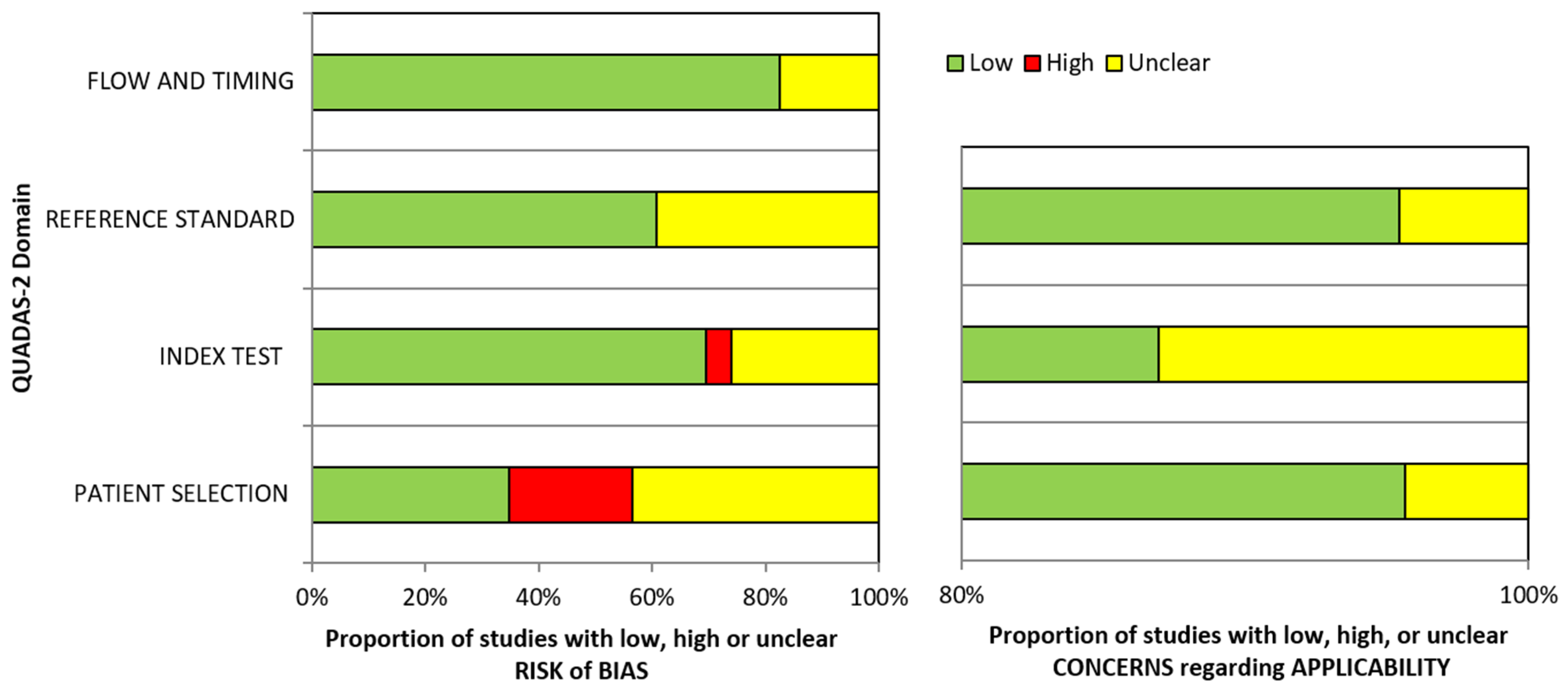
3.2. Risk of Bias and Applicability Concern
3.3. Description of the Included Studies
4. Discussion
- highly reliable, if mean accuracy is <1.0 mm,
- reliable, if mean accuracy is 1.0–1.5 mm,
- moderately reliable, if mean accuracy is 1.5–2.0 mm,
- unreliable, if mean accuracy is >2.0 mm [65].

| Study | Stereophotogrammetry (Mean ± SD) | Smartphone (Mean ± SD) | Structured Light Scanner (Mean ± SD) | Laser Scanner (Mean ± SD) | 2D Camera (Mean ± SD) |
|---|---|---|---|---|---|
| Akan et al. (2022) [39] | - | iPhoneX a: 0.753 ± 0.113 | - | - | - |
| Amornvit et al. (2019) [40] | - | iPhoneX a: NA | - | EinScan Pro: NA EinScan Pro 2X Plus: NA | - |
| Aswehlee et al. (2018) [41] | Danae1: 0.087 ± 0.002 3dMDface 1: 0.123 ± 0.007 Scanify 2: 0.849 ± 0.046 | - | - | Vivid 910: 0.068 ± 0.001 | - |
| Ayaz et al. (2020) [42] | Vectra H1 2: 0.280 * | - | - | Planmeca ProFace: 0.500 * | Nikon D800 2D camera: 0.780 * |
| Chong et al. (2021) [43] | Vectra H1 2: NA | iPad/iPhone b: NA | - | - | - |
| D’Ettorre et al. (2022) [44] | - | iPhoneXs b (Bellus3D Face App): NA iPhoneXs b (Capture App): NA | - | - | - |
| Dindaroglu et al. (2016) [45] | 3dMDface 1: NA | - | - | - | Canon EOS 40D 2D camera: NA |
| Elbashti et al. (2019) [46] | - | iPhone6 b: −24P (0.605 ± 0.124) | - | Vivid 910: 0.068 ± 0.001 | - |
| Fourie et al. (2011) [47] | Di3D 1: 0.860 ± 0.570 | - | - | Vivid 900: 0.890 ± 0.560 | - |
| Gibelli et al. (2018) [48] | Vectra M3 1: 0.650 ± 0.120 | - | - | Sense: 0.420 ± 0.170 | - |
| Gibelli et al. (2018) [49] | Vectra H1 2: 0.440 ± 0.360 Vectra M3 1: 0.220 ± 0.140 | - | - | - | - |
| Kim et al. (2018) [50] | Vectra H1 2: NA Vectra M3 1: NA | - | - | - | - |
| Liu et al. (2019) [51] | Vectra H1 2: 0.15 ± 0.015 Scanify 2: 0.740 ± 0.089 | - | - | - | - |
| Liu et al. (2021) [66] | 3dMDface 1: 0.36 ± 0.20 | Face Camera Pro Bellus c: 0.61 ± 0.47 | - | - | - |
| Modabber et al. (2016) [53] | FaceScan 3D 1: 0.523 ± 0.144 | - | Artec EVA: 0.228 ± 0.051 | - | - |
| Nightingale et al. (2020) [54] | - | iPhone8S b: −40P (0.800 ± 0.200) −60P (0.900 ± 0.400) −80P (0.800 ± 0.300) | Artec Spider: NA | - | - |
| Piedra-Cascon et al. (2020) [55] | - | Face Camera Pro Bellus c: 0.910 ± 0.320 | - | - | - |
| Ross et al. (2018) [56] | - | iPhone7 b: −30P (1.200 ± 0.300) −60P (1.200 ± 0.200) −90P (1.400 ± 0.600) | Artec Spider: NA Intel RealSense Camera SR300: 1.800 ± 0.300 | - | - |
| Rudy et al. (2020) [57] | Vectra H1 2: NA | iPhoneX a: 0.460 ± 0.010 | - | - | - |
| Wang et al. (2022) [58] | - | iPad Pro 2020 b: 1.17 ± 0.80 | ARC-7 Face Scanning System: 0.76 ± 0.61 | EinScan Pro 2X Plus: 0.69 ± 0.65 | - |
| Ye et al. (2016) [59] | 3dMDface 1: 0.620 ± 0.390 | - | 3D CaMega: 0.580 ± 0.370 | - | - |
| Zhao et al. (2017) [60] | 3dMDface 1: 0.580 ± 0.110 FaceScan3D 1: 0.570 ± 0.070 | - | - | Faro LLP: NA | - |
| Zhao et al. (2021) [61] | 3dMD face 1: NA | - | - | - | - |
5. Conclusions
Author Contributions
Funding
Institutional Review Board Statement
Informed Consent Statement
Data Availability Statement
Conflicts of Interest
References
- Launonen, A.M.; Vuollo, V.; Aarnivala, H.; Heikkinen, T.; Pirttiniemi, P.; Valkama, A.M.; Harila, V. Craniofacial asymmetry from one to three years of age: A prospective cohort study with 3d imaging. J. Clin. Med. 2020, 9, 70. [Google Scholar] [CrossRef] [PubMed]
- Lo Giudice, A.; Ortensi, L.; Farronato, M.; Lucchese, A.; Lo Castro, E.; Isola, G. The step further smile virtual planning: Milled versus prototyped mock-ups for the evaluation of the designed smile characteristics. BMC Oral Health 2020, 20, 165. [Google Scholar] [CrossRef] [PubMed]
- Giudice, A.L.; Spinuzza, P.; Rustico, L.; Messina, G.; Nucera, R. Short-term treatment effects produced by rapid maxillary expansion evaluated with computed tomography: A systematic review with meta-analysis. Korean J. Orthod. 2020, 50, 314–323. [Google Scholar] [CrossRef] [PubMed]
- Elshewy, M. Assessment of 3D Facial Scan Integration in 3D Digital Workflow Using Radiographic Markers and Iterative Closest Point Algorithm. Ph.D. Thesis, Marquette University, Milwaukee, WI, USA, 2020. [Google Scholar]
- Berssenbrügge, P.; Berlin, N.F.; Kebeck, G.; Runte, C.; Jung, S.; Kleinheinz, J.; Dirksen, D. 2D and 3D analysis methods of facial asymmetry in comparison. J. Cranio-Maxillofac. Surg. 2014, 42, e327–e334. [Google Scholar] [CrossRef]
- Chu, Y.; Yang, J.; Ma, S.; Ai, D.; Li, W.; Song, H.; Li, L.; Chen, D.; Chen, L.; Wang, Y. Registration and fusion quantification of augmented reality based nasal endoscopic surgery. Med. Image Anal. 2017, 42, 241–256. [Google Scholar] [CrossRef]
- Douglas, T.S. Image processing for craniofacial landmark identification and measurement: A review of photogrammetry and cephalometry. Comput. Med. Imaging Graph. 2004, 28, 401–409. [Google Scholar] [CrossRef]
- Mai, H.-N.; Kim, J.; Choi, Y.-H.; Lee, D.-H. Accuracy of Portable Face-Scanning Devices for Obtaining Three-Dimensional Face Models: A Systematic Review and Meta-Analysis. Int. J. Environ. Res. Public Health 2021, 18, 94. [Google Scholar] [CrossRef]
- Plooij, J.M.; Maal, T.J.; Haers, P.; Borstlap, W.A.; Kuijpers-Jagtman, A.M.; Bergé, S.J. Digital three-dimensional image fusion processes for planning and evaluating orthodontics and orthognathic surgery. A systematic review. Int. J. Oral Maxillofac. Surg. 2011, 40, 341–352. [Google Scholar] [CrossRef]
- Joda, T.; Gallucci, G.O. The virtual patient in dental medicine. Clin. Oral Implant. Res. 2015, 26, 725–726. [Google Scholar] [CrossRef]
- Cascón, W.P.; de Gopegui, J.R.; Revilla-León, M. Facially generated and additively manufactured baseplate and occlusion rim for treatment planning a complete-arch rehabilitation: A dental technique. J. Prosthet. Dent. 2019, 121, 741–745. [Google Scholar] [CrossRef]
- Kau, C.H.; Richmond, S.; Zhurov, A.; Ovsenik, M.; Tawfik, W.; Borbely, P.; English, J.D. Use of 3-dimensional surface acquisition to study facial morphology in 5 populations. Am. J. Orthod. Dentofac. Orthop. 2010, 137, S56.e51–S56.e59. [Google Scholar] [CrossRef]
- Metzler, P.; Sun, Y.; Zemann, W.; Bartella, A.; Lehner, M.; Obwegeser, J.A.; Kruse-Gujer, A.L.; Lübbers, H.-T. Validity of the 3D VECTRA photogrammetric surface imaging system for cranio-maxillofacial anthropometric measurements. Oral Maxillofac. Surg. 2014, 18, 297–304. [Google Scholar] [CrossRef] [PubMed]
- Nord, F.; Ferjencik, R.; Seifert, B.; Lanzer, M.; Gander, T.; Matthews, F.; Rücker, M.; Lübbers, H.-T. The 3dMD photogrammetric photo system in cranio-maxillofacial surgery: Validation of interexaminer variations and perceptions. J. Cranio-Maxillofac. Surg. 2015, 43, 1798–1803. [Google Scholar] [CrossRef] [PubMed]
- Verhoeven, T.; Xi, T.; Schreurs, R.; Bergé, S.; Maal, T. Quantification of facial asymmetry: A comparative study of landmark-based and surface-based registrations. J. Cranio-Maxillofac. Surg. 2016, 44, 1131–1136. [Google Scholar] [CrossRef]
- Weinberg, S.M.; Naidoo, S.; Govier, D.P.; Martin, R.A.; Kane, A.A.; Marazita, M.L. Anthropometric precision and accuracy of digital three-dimensional photogrammetry: Comparing the Genex and 3dMD imaging systems with one another and with direct anthropometry. J. Craniofacial Surg. 2006, 17, 477–483. [Google Scholar] [CrossRef]
- Weinberg, S.M.; Kolar, J.C. Three-dimensional surface imaging: Limitations and considerations from the anthropometric perspective. J. Craniofacial Surg. 2005, 16, 847–851. [Google Scholar] [CrossRef]
- Karatas, O.H.; Toy, E. Three-dimensional imaging techniques: A literature review. Eur. J. Dent. 2014, 8, 132. [Google Scholar] [CrossRef]
- Ma, L.; Xu, T.; Lin, J. Validation of a three-dimensional facial scanning system based on structured light techniques. Comput. Methods Programs Biomed. 2009, 94, 290–298. [Google Scholar] [CrossRef]
- Li, G.; Wei, J.; Wang, X.; Wu, G.; Ma, D.; Wang, B.; Liu, Y.; Feng, X. Three-dimensional facial anthropometry of unilateral cleft lip infants with a structured light scanning system. J. Plast. Reconstr. Aesthetic Surg. 2013, 66, 1109–1116. [Google Scholar] [CrossRef]
- Vlaar, S.T.; van der Zel, J.M. Accuracy of dental digitizers. Int. Dent. J. 2006, 56, 301–309. [Google Scholar] [CrossRef]
- Beuer, F.; Schweiger, J.; Edelhoff, D. Digital dentistry: An overview of recent developments for CAD/CAM generated restorations. Br. Dent. J. 2008, 204, 505–511. [Google Scholar] [CrossRef] [PubMed]
- De Felice, M.E.; Nucci, L.; Fiori, A.; Flores-Mir, C.; Perillo, L.; Grassia, V. Accuracy of interproximal enamel reduction during clear aligner treatment. Prog. Orthod. 2020, 21, 28. [Google Scholar] [CrossRef] [PubMed]
- Gwilliam, J.R.; Cunningham, S.J.; Hutton, T. Reproducibility of soft tissue landmarks on three-dimensional facial scans. Eur. J. Orthod. 2006, 28, 408–415. [Google Scholar] [CrossRef] [PubMed]
- Winder, R.; Darvann, T.A.; McKnight, W.; Magee, J.; Ramsay-Baggs, P. Technical validation of the Di3D stereophotogrammetry surface imaging system. Br. J. Oral Maxillofac. Surg. 2008, 46, 33–37. [Google Scholar] [CrossRef]
- Toma, A.M.; Zhurov, A.; Playle, R.; Richmond, S. A three-dimensional look for facial differences between males and females in a British-Caucasian sample aged 151/2 years old. Orthod. Craniofacial Res. 2008, 11, 180–185. [Google Scholar] [CrossRef]
- Sawyer, A.; See, M.; Nduka, C. Quantitative analysis of normal smile with 3D stereophotogrammetry–an aid to facial reanimation. J. Plast. Reconstr. Aesthetic Surg. 2010, 63, 65–72. [Google Scholar] [CrossRef]
- Knoops, P.G.; Beaumont, C.A.; Borghi, A.; Rodriguez-Florez, N.; Breakey, R.W.; Rodgers, W.; Angullia, F.; Jeelani, N.O.; Schievano, S.; Dunaway, D.J. Comparison of three-dimensional scanner systems for craniomaxillofacial imaging. J. Plast. Reconstr. Aesthetic Surg. 2017, 70, 441–449. [Google Scholar] [CrossRef]
- Tzou, C.-H.J.; Artner, N.M.; Pona, I.; Hold, A.; Placheta, E.; Kropatsch, W.G.; Frey, M. Comparison of three-dimensional surface-imaging systems. J. Plast. Reconstr. Aesthetic Surg. 2014, 67, 489–497. [Google Scholar] [CrossRef]
- Dekel, E.; Nucci, L.; Weill, T.; Flores-Mir, C.; Becker, A.; Perillo, L.; Chaushu, S. Impaction of maxillary canines and its effect on the position of adjacent teeth and canine development: A cone-beam computed tomography study. Am. J. Orthod. Dentofac. Orthop. 2021, 159, e135–e147. [Google Scholar] [CrossRef]
- Granata, S.; Giberti, L.; Vigolo, P.; Stellini, E.; Di Fiore, A. Incorporating a facial scanner into the digital workflow: A dental technique. J. Prosthet. Dent. 2020, 123, 781–785. [Google Scholar] [CrossRef]
- Hong, S.-J.; Noh, K. Setting the sagittal condylar inclination on a virtual articulator by using a facial and intraoral scan of the protrusive interocclusal position: A dental technique. J. Prosthet. Dent. 2020, 125, 392–395. [Google Scholar] [CrossRef] [PubMed]
- Russo, L.L.; Salamini, A.; Troiano, G.; Guida, L. Digital dentures: A protocol based on intraoral scans. J. Prosthet. Dent. 2020, 125, 597–602. [Google Scholar] [CrossRef] [PubMed]
- Revilla-León, M.; Raney, L.; Piedra-Cascón, W.; Barrington, J.; Zandinejad, A.; Özcan, M. Digital workflow for an esthetic rehabilitation using a facial and intraoral scanner and an additive manufactured silicone index: A dental technique. J. Prosthet. Dent. 2020, 123, 564–570. [Google Scholar] [CrossRef] [PubMed]
- Liu, C.-H.; Lin, I.-C.; Lu, J.-J.; Cai, D. A Smartphone App for Improving Clinical Photography in Emergency Departments: Comparative Study. JMIR Mhealth Uhealth 2019, 7, e14531. [Google Scholar] [CrossRef]
- Barbero-García, I.; Pierdicca, R.; Paolanti, M.; Felicetti, A.; Lerma, J.L. Combining machine learning and close-range photogrammetry for infant’s head 3D measurement: A smartphone-based solution. Measurement 2021, 182, 109686. [Google Scholar] [CrossRef]
- Liberati, A.; Altman, D.G.; Tetzlaff, J.; Mulrow, C.; Gøtzsche, P.C.; Ioannidis, J.P.; Clarke, M.; Devereaux, P.J.; Kleijnen, J.; Moher, D. The PRISMA statement for reporting systematic reviews and meta-analyses of studies that evaluate health care interventions: Explanation and elaboration. J. Clin. Epidemiol. 2009, 62, e1–e34. [Google Scholar] [CrossRef]
- Whiting, P.F.; Rutjes, A.W.; Westwood, M.E.; Mallett, S.; Deeks, J.J.; Reitsma, J.B.; Leeflang, M.M.; Sterne, J.A.; Bossuyt, P.M. QUADAS-2: A revised tool for the quality assessment of diagnostic accuracy studies. Ann. Intern. Med. 2011, 155, 529–536. [Google Scholar] [CrossRef]
- Akan, B.; Akan, E.; Sahan, A.O.; Kalak, M. Evaluation of 3D Face-Scan images obtained by stereophotogrammetry and smartphone camera. Int. Orthod. 2021, 19, 669–678. [Google Scholar] [CrossRef]
- Amornvit, P.; Sanohkan, S. The Accuracy of Digital Face Scans Obtained from 3D Scanners: An In Vitro Study. Int. J. Environ. Res. Public Health 2019, 16, 5061. [Google Scholar] [CrossRef]
- Aswehlee, A.M.; Elbashti, M.E.; Hattori, M.; Sumita, Y.I.; Taniguchi, H. Feasibility and Accuracy of Noncontact Three-Dimensional Digitizers for Geometric Facial Defects: An In Vitro Comparison. Int. J. Prosthodont. 2018, 31, 601–606. [Google Scholar] [CrossRef]
- Ayaz, I.; Shaheen, E.; Aly, M.; Shujaat, S.; Gallo, G.; Coucke, W.; Politis, C.; Jacobs, R. Accuracy and reliability of 2-dimensional photography versus 3-dimensional soft tissue imaging. Imaging Sci. Dent. 2020, 50, 15–22. [Google Scholar] [CrossRef] [PubMed]
- Chong, Y.M.; Liu, X.Y.; Shi, M.; Huang, J.Z.; Yu, N.Z.; Long, X. Three-dimensional facial scanner in the hands of patients: Validation of a novel application on iPad/iPhone for three-dimensional imaging. Ann. Transl. Med. 2021, 9, 1115. [Google Scholar] [CrossRef] [PubMed]
- D’Ettorre, G.; Farronato, M.; Candida, E.; Quinzi, V.; Grippaudo, C. A comparison between stereophotogrammetry and smartphone structured light technology for three-dimensional face scanning. Angle Orthod. 2022, 92, 358–363. [Google Scholar] [CrossRef]
- Dindaroglu, F.; Kutlu, P.; Duran, G.S.; Gorgulu, S.; Aslan, E. Accuracy and reliability of 3D stereophotogrammetry: A comparison to direct anthropometry and 2D photogrammetry. Angle Orthod. 2016, 86, 487–494. [Google Scholar] [CrossRef] [PubMed]
- Elbashti, M.E.; Sumita, Y.I.; Aswehlee, A.M.; Seelaus, R. Smartphone Application as a Low-Cost Alternative for Digitizing Facial Defects: Is It Accurate Enough for Clinical Application? Int. J. Prosthodont. 2019, 32, 541–543. [Google Scholar] [CrossRef] [PubMed]
- Fourie, Z.; Damstra, J.; Gerrits, P.O.; Ren, Y. Evaluation of anthropometric accuracy and reliability using different three-dimensional scanning systems. Forensic Sci. Int. 2011, 207, 127–134. [Google Scholar] [CrossRef]
- Gibelli, D.; Pucciarelli, V.; Caplova, Z.; Cappella, A.; Dolci, C.; Cattaneo, C.; Sforza, C. Validation of a low-cost laser scanner device for the assessment of three-dimensional facial anatomy in living subjects. J. Cranio-Maxillofac. Surg. 2018, 46, 1493–1499. [Google Scholar] [CrossRef]
- Gibelli, D.; Pucciarelli, V.; Cappella, A.; Dolci, C.; Sforza, C. Are portable stereophotogrammetric devices reliable in facial imaging? A validation study of VECTRA H1 device. J. Oral Maxillofac. Surg. 2018, 76, 1772–1784. [Google Scholar] [CrossRef]
- Kim, A.J.; Gu, D.; Chandiramani, R.; Linjawi, I.; Deutsch, I.C.K.; Allareddy, V.; Masoud, M.I. Accuracy and reliability of digital craniofacial measurements using a small-format, handheld 3D camera. Orthod. Craniofac. Res. 2018, 21, 132–139. [Google Scholar] [CrossRef]
- Liu, C.; Artopoulos, A. Validation of a low-cost portable 3-dimensional face scanner. Imaging Sci. Dent. 2019, 49, 35–43. [Google Scholar] [CrossRef] [Green Version]
- Liu, J.L.; Zhang, C.H.; Cai, R.L.; Yao, Y.; Zhao, Z.H.; Liao, W. Accuracy of 3-dimensional stereophotogrammetry: Comparison of the 3dMD and Bellus3D facial scanning systems with one another and with direct anthropometry. Am. J. Orthod. Dentofac. Orthop. 2021, 160, 862–871. [Google Scholar] [CrossRef] [PubMed]
- Modabber, A.; Peters, F.; Kniha, K.; Goloborodko, E.; Ghassemi, A.; Lethaus, B.; Holzle, F.; Mohlhenrich, S.C. Evaluation of the accuracy of a mobile and a stationary system for three-dimensional facial scanning. J. Cranio-Maxillofac. Surg. 2016, 44, 1719–1724. [Google Scholar] [CrossRef] [PubMed]
- Nightingale, R.C.; Ross, M.T.; Allenby, M.C.; Woodruff, M.A.; Powell, S.K. A Method for Economical Smartphone-Based Clinical 3D Facial Scanning. J. Prosthodont. -Implant. Esthet. Reconstr. Dent. 2020, 29, 818–825. [Google Scholar] [CrossRef] [PubMed]
- Piedra-Cascon, W.; Meyer, M.J.; Methani, M.M.; Revilla-Leon, M. Accuracy (trueness and precision) of a dual-structured light facial scanner and interexaminer reliability. J. Prosthet. Dent. 2020, 124, 567–574. [Google Scholar] [CrossRef] [PubMed]
- Ross, M.T.; Cruz, R.; Brooks-Richards, T.L.; Hafner, L.M.; Powell, S.K.; Woodruff, M.A. Comparison of three-dimensional surface scanning techniques for capturing the external ear. Virtual Phys. Prototyp. 2018, 13, 255–265. [Google Scholar] [CrossRef]
- Rudy, H.L.; Wake, N.; Yee, J.; Garfein, E.S.; Tepper, O.M. Three-Dimensional Facial Scanning at the Fingertips of Patients and Surgeons: Accuracy and Precision Testing of iPhone X Three-Dimensional Scanner. Plast. Reconstr. Surg. 2020, 146, 1407–1417. [Google Scholar] [CrossRef]
- Wang, C.; Shi, Y.F.; Xiong, Q.; Xie, P.J.; Wu, J.H.; Liu, W.C. Trueness of One Stationary and Two Mobile Systems for Three-Dimensional Facial Scanning. Int. J. Prosthodont. 2022, 35, 350–356. [Google Scholar] [CrossRef]
- Ye, H.Q.; Lv, L.W.; Liu, Y.S.; Liu, Y.S.; Zhou, Y.S. Evaluation of the Accuracy, Reliability, and Reproducibility of Two Different 3D Face-Scanning Systems. Int. J. Prosthodont. 2016, 29, 213–218. [Google Scholar] [CrossRef]
- Zhao, Y.J.; Xiong, Y.X.; Wang, Y. Three-Dimensional Accuracy of Facial Scan for Facial Deformities in Clinics: A New Evaluation Method for Facial Scanner Accuracy. PLoS ONE 2017, 12, e0169402. [Google Scholar] [CrossRef]
- Zhao, Z.; Xie, L.; Cao, D.; Izadikhah, I.; Gao, P.; Zhao, Y.; Yan, B. Accuracy of three-dimensional photogrammetry and cone beam computed tomography based on linear measurements in patients with facial deformities. Dentomaxillofac. Radiol. 2021, 50, 20200001. [Google Scholar] [CrossRef]
- Mai, H.-N.; Lee, D.-H. Accuracy of Mobile Device–Compatible 3D Scanners for Facial Digitization: Systematic Review and Meta-Analysis. J. Med. Internet Res. 2020, 22, e22228. [Google Scholar] [CrossRef] [PubMed]
- Volonghi, P.; Baronio, G.; Signoroni, A. 3D scanning and geometry processing techniques for customised hand orthotics: An experimental assessment. Virtual Phys. Prototyp. 2018, 13, 105–116. [Google Scholar] [CrossRef]
- Camison, L.; Bykowski, M.; Lee, W.W.; Carlson, J.C.; Roosenboom, J.; Goldstein, J.A.; Losee, J.E.; Weinberg, S.M. Validation of the Vectra H1 portable three-dimensional photogrammetry system for facial imaging. Int. J. Oral Maxillofac. Surg. 2018, 47, 403–410. [Google Scholar] [CrossRef]
- Aung, S.; Ngim, R.; Lee, S. Evaluation of the laser scanner as a surface measuring tool and its accuracy compared with direct facial anthropometric measurements. Br. J. Plast. Surg. 1995, 48, 551–558. [Google Scholar] [CrossRef]
- Liu, L.; Zhan, Q.; Zhou, J.; Kuang, Q.; Yan, X.; Zhang, X.; Shan, Y.; Li, X.; Lai, W.; Long, H. Effectiveness of an anterior mini-screw in achieving incisor intrusion and palatal root torque for anterior retraction with clear aligners. Angle Orthod. 2021, 91, 794–803. [Google Scholar] [CrossRef]
- Secher, J.J.; Darvann, T.A.; Pinholt, E.M. Accuracy and reproducibility of the DAVID SLS-2 scanner in three-dimensional facial imaging. J. Cranio-Maxillofac. Surg. 2017, 45, 1662–1670. [Google Scholar] [CrossRef]
- Lane, C.; Harrell, W., Jr. Completing the 3-dimensional picture. Am. J. Orthod. Dentofac. Orthop. 2008, 133, 612–620. [Google Scholar] [CrossRef]
- Yao, H.; Ge, C.; Xue, J.; Zheng, N. A high spatial resolution depth sensing method based on binocular structured light. Sensors 2017, 17, 805. [Google Scholar] [CrossRef]
- Sarbolandi, H.; Lefloch, D.; Kolb, A. Kinect range sensing: Structured-light versus Time-of-Flight Kinect. Comput. Vis. Image Underst. 2015, 139, 1–20. [Google Scholar] [CrossRef]
- Jia, T.; Zhou, Z.; Gao, H. Depth measurement based on infrared coded structured light. J. Sens. 2014, 2014, 852621. [Google Scholar] [CrossRef] [Green Version]
- Alfaro-Santafé, J.; Gómez-Bernal, A.; Lanuza-Cerzócimo, C.; Alfaro-Santafé, J.V.; Pérez-Morcillo, A.; Almenar-Arasanz, A.J. Three-axis measurements with a novel system for 3D plantar foot scanning: iPhone X. Footwear Sci. 2020, 12, 123–131. [Google Scholar] [CrossRef]
- Kovacs, L.; Zimmermann, A.; Brockmann, G.; Baurecht, H.; Schwenzer-Zimmerer, K.; Papadopulos, N.A.; Papadopoulos, M.A.; Sader, R.; Biemer, E.; Zeilhofer, H.-F. Accuracy and precision of the three-dimensional assessment of the facial surface using a 3-D laser scanner. IEEE Trans. Med. Imaging 2006, 25, 742–754. [Google Scholar] [CrossRef]
- Koban, K.C.; Cotofana, S.; Frank, K.; Green, J.B.; Etzel, L.; Li, Z.; Giunta, R.E.; Schenck, T.L. Precision in 3-dimensional surface imaging of the face: A handheld scanner comparison performed in a cadaveric model. Aesthetic Surg. J. 2019, 39, NP36–NP44. [Google Scholar] [CrossRef] [PubMed]
- Maués, C.; Casagrande, M.; Almeida, R.; Almeida, M.; Carvalho, F. Three-dimensional surface models of the facial soft tissues acquired with a low-cost scanner. Int. J. Oral Maxillofac. Surg. 2018, 47, 1219–1225. [Google Scholar] [CrossRef]
- Timen, C.; Speksnijder, C.M.; Van Der Heijden, F.; Beurskens, C.H.; Ingels, K.J.; Maal, T.J. Depth accuracy of the RealSense F200: Low-cost 4D facial imaging. Sci. Rep. 2017, 7, 16263. [Google Scholar]
- Koban, K.C.; Perko, P.; Etzel, L.; Li, Z.; Schenck, T.L.; Giunta, R.E. Validation of two handheld devices against a non-portable three-dimensional surface scanner and assessment of potential use for intraoperative facial imaging. J. Plast. Reconstr. Aesthetic Surg. 2020, 73, 141–148. [Google Scholar] [CrossRef]
- Bakirman, T.; Gumusay, M.U.; Reis, H.C.; Selbesoglu, M.O.; Yosmaoglu, S.; Yaras, M.C.; Seker, D.Z.; Bayram, B. Comparison of low cost 3D structured light scanners for face modeling. Appl. Opt. 2017, 56, 985–992. [Google Scholar] [CrossRef]
- Perillo, L.; Padricelli, G.; Isola, G.; Femiano, F.; Chiodini, P.; Mataresei, G. Class II malocclusion division 1: A new classification method by cephalometric analysis. Eur. J. Paediatr. Dent. 2012, 13, 192. [Google Scholar]
- Kim, P.; Chen, J.; Cho, Y.K. SLAM-driven robotic mapping and registration of 3D point clouds. Autom. Constr. 2018, 89, 38–48. [Google Scholar] [CrossRef]

| Database | Boolean Operator | Results |
|---|---|---|
| Pubmed | (“virtual patient” OR “virtual face” OR “digital facial models” OR “3D virtual facial model”) AND (“scanner” [MeSH Terms] OR “digital face scan” OR “facial scan” OR “3D face scanning” OR “facial digitalization” OR “stereophotogrammetry” OR “smartphone scanner” OR “smartphone face scanning” OR “smartphone application”) AND (“accuracy” OR “trueness” OR “precision” OR “3D comparison”). | 244 |
| Scopus | (“virtual patient” OR “virtual face” OR “digital facial models” OR “3D virtual facial model”) AND (“scanner” OR “digital face scan” OR “facial scan” OR “3D face scanning” OR “facial digitalization” OR “stereophotogrammetry” OR “smartphone scanner” OR “smartphone face scanning” OR “smartphone application”) AND (“accuracy” OR “trueness” OR “precision” OR “3D comparison”). | 215 |
| Web of Science | TS = ((virtual face OR digital facial models OR 3D virtual facial model OR digital face OR face) AND (scanner OR digital face scan OR facial scan OR 3D face scanning OR facial digitalization OR face digitalization OR stereophotogrammetry OR smartphone scanner OR smartphone face scanning OR smartphone application) AND (accuracy OR trueness OR precision OR 3D comparison)). | 320 |
| Cochrane Library | “virtual patient” OR “virtual face” OR “digital facial models” OR “3D virtual facial model” AND “scanner” OR “digital face scan” OR “facial scan” OR “3D face scanning” OR “facial digitalization” OR “stereophotogrammetry” OR “smartphone scanner” OR “smartphone face scanning” OR “smartphone application” AND “accuracy” OR “trueness” OR “precision” OR “3D comparison”. | 102 |
| LILACS | (digital facial models) AND (scanner OR stereophotogrammetry OR smartphone face scanning) AND (accuracy). | 25 |
| OpenGrey | (virtual face OR digital facial models OR 3D virtual facial model OR digital face OR face) AND (scanner OR digital face scan OR facial scan OR 3D face scanning OR facial digitalization OR face digitalization OR stereophotogrammetry OR smartphone scanner OR smartphone face scanning OR smartphone application) AND (accuracy OR trueness OR precision OR 3D comparison). | 2 |
| Total | 908 |
 High risk,
  Unclear risk,
  Low risk.
High risk, Unclear risk, Low risk.
Low risk.
High risk, Unclear risk, Low risk.| Study | Risk of Bias | Applicability Concerns | |||||||
|---|---|---|---|---|---|---|---|---|---|
| Patient selection | Index test | Reference Standard | Flow and Timing | Overall risk of bias | Patient selection | Index test | Reference Standard | Overall risk of bias | |
| Akan et al. (2021) [39] | | | | | | | | | |
| Amornvit et al. (2019) [40] | | | | | | | | | |
| Aswehlee et al.(2018) [41] | | | | | | | | | |
| Ayaz et al. (2020) [42] | | | | | | | | | |
| Chong et al. (2021) [43] | | | | | | | | | |
| D’Ettorre (2022) et al. [44] | | | | | | | | | |
| Dindaroglu et al. (2016) [45] | | | | | | | | | |
| Elbashti et al. (2019) [46] | | | | | | | | | |
| Fourie et al. (2011) [47] | | | | | | | | | |
| Gibelli et al. (2018) [48] | | | | | | | | | |
| Gibelli et al. (2018) [49] | | | | | | | | | |
| Kim et al. (2018) [50] | | | | | | | | | |
| Liu et al. (2019) [51] | | | | | | | | | |
| Liu et al. (2021) [52] | | | | | | | | | |
| Modabber et al. (2016) [53] | | | | | | | | | |
| Nightingale et al. (2020) [54] | | | | | | | | | |
| Piedra-Cascon et al. (2020) [55] | | | | | | | | | |
| Ross et al. (2018) [56] | | | | | | | | | |
| Rudy et al. (2020) [57] | | | | | | | | | |
| Wang et al. (2022) [58] | | | | | | | | | |
| Ye et al. (2016) [59] | | | | | | | | | |
| Zhao et al. (2017) [60] | | | | | | | | | |
| Zhao et al. (2021) [61] | | | | | | | | | |
| Study | Aim | Tested Method | Sample | Reference Method | N° Landmarks | Measurements (Metrics) | Conclusions |
|---|---|---|---|---|---|---|---|
| Akan et al. (2021) [39] | To analyze the accuracy of a depth sensor smartphone camera with conventional stationary stereophotogrammetry | iPhone X with depth-sensing camera b1 | 26 participants (16 F, 10 M) | 3dMD face a1 | 9 | 7 linear distances, 3 angles (mean deviations, RMSE) | Depth sensor smartphone camera may be employed for 3D facial scanning. However, complex structures visualization needs improvements. |
| Amornvit et al. (2019) [40] | To compare the facial scannings acquired through 4 scanner systems with the measurements obtained with direct anthropometry. | EinScan Pro (EP) c, EinScan Pro 2X Plus (EP+) c, iPhone X (IPX) b1, Planmeca ProMax 3D Mid (PM) e | 1 MH | Direct anthropometry | NA | Δx, Δy, and Δz (mean linear deviations) | Mean Δx and Δy measurements ranged respectively 10–50 mm and 50–120 mm. The records in the z-axis were not possible to acquire. EP+ was the most accurate, EP the intermediate, whereas IPX and PM showed less accuracy. Furthermore, EP, IPX, and PM were less accurate in measuring depths of 2 mm. |
| Aswehlee et al. (2018) [41] | To evaluate the reliability of different digitalization systems for capturing facial defects. | Vivid 910 c, Danae 100SP a1, 3dMD face a1, Scanify a2. | 1 IC | Toshiba TOSCANER-30000μCM e | 3D point clouds | Surface deviation (RMSE) | All systems were sufficiently reliable. Laser-beam, light-sectioning technology showed the best accuracy. |
| Ayaz et al. (2020) [42] | To evaluate the reliability of 2D photography and 3D scanning systems for facial digitalization. | Nikon D800 2D camera, Planmeca ProFace c, Vectra H1 a2 | 50 participants (25 M, 25 F) | Direct anthropometry | 22 | 7 linear distances, 17 angles (linear and angular mean deviation) | Stereophotogrammetry showed the best accuracy for facial digitalization compared to 2D photography and laser scanner. |
| Chong et al. (2021) [43] | To evaluate the accuracy of an iPad/iPhone application enabling patients to capture their 3D facial images compared to direct anthropometry. | iPad/iPhone camera through the application “MeinXuan” b2, Vectra H1 a2 | 20 participants | Direct anthropometry | 18 | 21 linear distances (RMSE, mean absolute difference and relative error measurement) | The measurements obtained with the iPhone/iPad application were significantly correlated to the direct anthropometric measurements. The iPhone/iPad and Vectra H1 mean RMSE were 0.08 and 0.67 mm respectively. The subnasale area needs improvement with the proposed method. |
| D’Ettorre et al. (2022) [44] | To compare the accuracy of 3D face scanning from 3D stereophotogrammetry and two different applications in smartphones supporting the TrueDepth system. | iPhone Xs equipped with Bellus3D Face or Capture applications b1 | 40 participants (27 F, 13 M) | 3dMD face a1 | 13 | Δx, Δy, and Δz (mean linear deviations) | Stationary stereophotogrammetry is a reliable and fast system. The smartphone applications showed also a good accuracy, are portable and less expensive. However, they need operator accuracy and patient compliance for the incremented time of acquisition. |
| Dindaroglu et al. (2016) [45] | To compare the stereophotogrammetry accuracy with the direct manual and digital photogrammetry methods. | Canon EOS 40D 2D camera, 3dMD face a1 | 80 participants (38 M, 42 F) | Direct anthropometry | 15 | 10 linear distances, 6 angles (mean linear and angular deviations) | 3D stereophotogrammetry performed accurate 3D facial images reliable in orthodontics. |
| Elbashti et al. (2019) [46] | To evaluate the accuracy of a smartphone application as a low-cost approach for digitizing a facial defect for 3D modeling. | Vivid 910 c, iPhone 6 b2 (24 photographs) | 1 IC | Toshiba TOSCANER-30000μCM e | 3D point clouds | Surface deviation (RMSE) | In reference to standard computed tomography imaging, data acquisition with a smartphone for 3D modeling is not as accurate as commercially available laser scanning. |
| Fourie et al. (2011) [47] | To estimate the reliability of three different 3D scanning systems namely laser surface scanning (Minolta Vivid 900), CBCT, 3D SPG (Di3D system) and to compare them to physical linear measurements. | Di3D a1, Vivid 900 c, KaVo 3D exam e | 7 cadaver heads | Direct anthropometry | 15 | 21 linear distances (mean linear deviations) | Measurements recorded by the three 3D systems appeared to be both sufficiently accurate and reliable. |
| Gibelli et al. (2018) [48] | To compare the accuracy of SPG with a low-cost laser scanner. | Sense c | 50 participants (10 M, 40 F), 1 MH | Vectra M3 a1 | 17; 3D point clouds | 14 linear distances, 12 angles/volumes/surfaces (RMSE) | The low-cost laser scanner appeared sufficiently reliable for immovable objects but it is not suitable for 3D human face scanning. |
| Gibelli et al. (2018) [49] | To validate VECTRA H1 portable SPG device to verify its applicability to 3D facial analysis. | Vectra H1 a2 | 50 participants (16 M, 34 F), 1 MH | Vectra M3 a1 | 12; 3D point clouds | 15 linear distances, 12 angles/volumes/surfaces (RMSE) | The portable Vectra H1 face scanning device proved reliable for assessing linear measurements, angles, and surface areas; conversely, the influence of involuntary facial movements on volumes and RMSE was higher compared to the stationary Vectra M3. |
| Kim et al. (2018) [50] | To evaluate the accuracy and reliability of a 3D small-format, handheld camera. | Vectra H1 a2, Vectra M3 a1 | 5 participants (NA) | Direct anthropometry | 29 | 25 linear distances (mean linear deviations) | The 3D handheld camera showed high accuracy and reliability in comparison with traditional models. |
| Liu et al. (2019) [51] | To evaluate reliability of a portable low-cost scanner (Scanify) for imaging facial casts compared to a previously validated portable digital stereophotogrammetry device (Vectra H1). | Scanify a2 | 2 IC (male) | Vectra H1 a2 | 13 | 11 linear distances (mean linear deviations) | Scanify showed to be a low-cost solution for facial digitalization but needs future improvements. |
| Liu et al. (2021) [52] | To evaluate the reliability of Bellus3D Face Camera Pro compared to 3dMDface stereophotogrammetry for face scanning. | Face Camera Pro Bellus b3 (connected to a tablet), 3dMD face a1 | 1 MH | Direct anthropometry | 20 | 8 linear distances, 5 angles (absolute mean deviations) | Both systems showed good accuracy and precision for clinical purposes. |
| Modabber et al. (2016) [53] | To evaluate the reliability of two devices for face digitalization. | Artec EVA d | 41 participants (16 M, 25 F) | FaceScan 3D a1 | 2 lego brick | Surface deviation (RMSE) | Artec EVA showed greater accuracy compared to FaceScan3D. |
| Nightingale et al. (2020) [54] | To test the use of a low-cost scanner by inexperienced operators. | iPhone 8S b2 (40, 60 or 80 photographs) | 20 participants (11 M, 9 F) | Artec Spider d | 3D point clouds | Surface deviation (RMSE) | Smartphone photogrammetry showed to be useful for novice operators for its low cost and easy learning curve. |
| Piedra-Cascon et al. (2020) [55] | To evaluate the reliability of Bellus3D Face Camera Pro for face digitalization. | Face Camera Pro Bellus b3 (connected to a tablet) | 10 participants (2 M, 8 F) | Direct anthropometry | 6 | 5 linear distances (precision and accuracy RMSE) | The new device showed to be clinically reliable for face scanning procedures. |
| Ross et al. (2018) [56] | To estimate the performance of smartphones for external ear digitalization. | iPhone 7 b2 (30, 60 or 90 photographs), Intel RealSense Camera SR300 d | 16 participants (8 M, 8 F) | Artec Spider d | 6 | Surface deviation (RMSE) | The three protocols with 30-60-90 photographs showed a good and similar accuracy. The Intel RealSense showed the worst performance and resolution. |
| Rudy et al. (2020) [57] | To compare the accuracy of portable stereophotogrammetry with iPhone X for facial digitalization. | iPhone X b1 | 16 participants (NA) | Vectra H1 a2 | 10; 3D point cloud | Landmark-to-landmark surface distances; surface deviation (RMSE) | The iPhoneX accuracy stood around 0.5 mm when compared to Vectra H1. |
| Wang et al. (2022) [58] | To compare the accuracy of two portable systems and a stationary scanner for facial digitalization. | iPad Pro 2020 b2 (Bellus 3D Dental Pro app), EinScan Pro 2X Plus (EP+) c, ARC-7 Face Scanning System d | 20 participants (5 M, 15 F) | Direct anthropometry | 12 | 14 linear distances (absolute error) | All the systems showed to be quite reliable for face digitalization. However, the iPad Pro 2020 system was the least accurate. |
| Ye et al. (2016) [59] | To assess the accuracy, reliability and reproducibility of facial digitalization through stereophotogrammetry compared to a structured light scanner. | 3D CaMega d, 3dMDfacea1 | 10 participants (5 M, 5 F) | Direct anthropometry | 16 | 21 linear distances (mean linear deviations) | Both scanners showed to be quite accurate, reliable and reproducible to create 3D facial models. |
| Zhao et al. (2017) [60] | To evaluate the accuracy of different scanning systems for face digitalization. | 3dMDfacea1, FaceScan3D a1 | 10 participants (NA) | Faro Edge LLP c | 3D point clouds | Surface deviation (RMSE) | All scanning systems showed to be sufficiently reliable for clinic purposes. |
| Zhao et al. (2021) [61] | To evaluate the accuracy of stereophotogrammetry and a CBCT system to obtain facial models with deformities and partitions. | 3dMDfacea1, NewTom 5G Inc e | 60 participants (28 M, 32 F) | Coordinate-measuring Machines (CMM) | 17 | 19 linear distances (mean linear deviations) | 3D stereophotogrammetry was more than CBCT in the acquisition of facial deformities. |
| Parameters | Stationary Stereophotogrammetry | Portable Stereophotogrammetry | Smartphones |
|---|---|---|---|
| Mean accuracy | 0.087–0.860 mm | 0.150–0.849 mm | 0.460–1.400 mm |
| Costs | 8.000–26.000$ | 1.500–10.000$ | 350–1.200$ |
| Capture time * | 1.5–600 ms | 3.5–250 ms | 2.5–350 ms |
| Approach | Different cameras placed at different angulations capture various 2D images simultaneously to create a 3D face model. | A single camera capture one image at a time. Require several acquisitions from different angles to reconstruct the 3D facial model. | A single camera capture one image at a time. Require several acquisitions from different angles to reconstruct the 3D facial model. |
| Advantages | Highly reliable system. Useful in children and uncooperative patients. | Reliable and portable system. | Comfortable, manageable, quite reliable, easy to use, inexpensive. |
| Limitations | Expensive, requires space and ambient light control. | Expensive, ambient light control, motion artifacts, greater acquisition time, require patient collaboration. | Motion artifacts, less accurate on uneven and deep surfaces, greater acquisition time, require patient collaboration. |
Publisher’s Note: MDPI stays neutral with regard to jurisdictional claims in published maps and institutional affiliations. |
© 2022 by the authors. Licensee MDPI, Basel, Switzerland. This article is an open access article distributed under the terms and conditions of the Creative Commons Attribution (CC BY) license (https://creativecommons.org/licenses/by/4.0/).
Share and Cite
Quinzi, V.; Polizzi, A.; Ronsivalle, V.; Santonocito, S.; Conforte, C.; Manenti, R.J.; Isola, G.; Lo Giudice, A. Facial Scanning Accuracy with Stereophotogrammetry and Smartphone Technology in Children: A Systematic Review. Children 2022, 9, 1390. https://doi.org/10.3390/children9091390
Quinzi V, Polizzi A, Ronsivalle V, Santonocito S, Conforte C, Manenti RJ, Isola G, Lo Giudice A. Facial Scanning Accuracy with Stereophotogrammetry and Smartphone Technology in Children: A Systematic Review. Children. 2022; 9(9):1390. https://doi.org/10.3390/children9091390
Chicago/Turabian StyleQuinzi, Vincenzo, Alessandro Polizzi, Vincenzo Ronsivalle, Simona Santonocito, Cristina Conforte, Rebecca Jewel Manenti, Gaetano Isola, and Antonino Lo Giudice. 2022. "Facial Scanning Accuracy with Stereophotogrammetry and Smartphone Technology in Children: A Systematic Review" Children 9, no. 9: 1390. https://doi.org/10.3390/children9091390
APA StyleQuinzi, V., Polizzi, A., Ronsivalle, V., Santonocito, S., Conforte, C., Manenti, R. J., Isola, G., & Lo Giudice, A. (2022). Facial Scanning Accuracy with Stereophotogrammetry and Smartphone Technology in Children: A Systematic Review. Children, 9(9), 1390. https://doi.org/10.3390/children9091390