Abstract
Background: It is necessary to analyze and monitor the facial growth of orofacial cleft patients. The documentation should therefore begin before and after primary surgeries. Technological evolution has transformed plaster models into 3D images through scanners that allow rational storage, manipulation, and rotation without the possibility of breakage or damage. Based on this fact, this narrative review aims to provide a feature on the three-dimensional tools available for the assessment of dental arches in children with orofacial cleft and mixed dentition. Material and Methods: Three databases were chosen (PubMed, ScienceDirect, and Scopus) and keywords were used to select papers. Results: During the database screening, 292 potentially relevant papers were found. After removing duplicates, titles, and abstracts, 32 papers presented qualifications for analysis. Through evaluating each document by reading it one by one, 24 papers fulfilled the eligibility criteria. Conclusions: It was concluded that digital tools—i.e., benchtop scanners which evaluate the dental arches of children with cleft lip, palate, and mixed dentition—are reproducible and reliable, without the use of ionizing radiation, allow storage, manipulation with sustainability, and help preserve the environment.
1. Introduction
Primary surgeries repair the anatomical defect but most often cause deleterious effects on facial growth, mainly related to the jaws [1,2,3,4,5,6].
As such, it is necessary to analyze and monitor the facial growth of individuals with cleft lip and palate. Facial growth documentation should begin before primary surgeries and continue after until five years of age. In addition to the documentation already included in the protocols and in plaster models, 3D photos can aid in the measurements and analyses of dental arches and facial growth.
The literature describes intraoral photos for the purposes of analyzing occlusion indexes. Plaster models are the gold standard [7] and plaster model images have been analyzed with accuracy [8]. Both intraoral photos and plaster models have proved to be reliable and reproducible [9]. Technological evolution has changed plaster models into 3D images through the use of scanners [10] that allow rational storage, manipulation, and rotation without the possibility of breakage or damage. With the use of software to carry out the evaluations, instead of using a caliper and rulers [11], more accurate linear [12] and angular measurements are obtained. In addition to these, more accurate measurements of area [6], volume [3], superimpositions [13], and occlusal contacts [14] are also obtained. All of these help to better understand what happens with the growing dental arches of patients undergoing the rehabilitation process. The software’s ability can be sufficiently precise and accurate enough to assess linear, angular, and volumetric measures, as well as surface areas and superimposition procedures [11].
We can highlight stereophotogrammetry as an aid in understanding how the facial growth and development of these patients occurs through 3D photos of the face [15,16,17], using computer programs that have linear, area, volume, and superimposition measurement tools.
Technology has become an ally in the study and observation of the craniofacial development and growth of patients with cleft lip and palate before, during, and after the rehabilitation process so that, with coherence and scientific evidence, we can improve treatment protocols. Thus, this narrative review aims to provide information on the three-dimensional tools available for the assessment of dental arches in children with cleft lip and palate at mixed dentition.
2. Materials and Methods
2.1. Search Strategy
PubMed, Scopus, and ScienceDirect were chosen as the databases reviewed. Additionally, the narrative review included papers only in the English language. The following keywords were used: Children; Cleft Lip; Cleft Palate; Imaging, Three-dimensional; and Dental Arches.
2.2. Inclusion Criteria
All studies that presented quantitative assessments, such as research, multicenter studies, randomized clinical trials, and retrospective clinical studies, were included.
2.3. Selected Sample
- -
- Maxillary dental arches of cleft lip and palate patients aged up to 12 years;
- -
- Optical devices, scanners, and stereophotogrammetry in order to reproduce 3D maxillary dental model;
- -
- Types of intervention, linear, angular, surface (area), volume measurements, and qualitative analysis of the occlusal index. Types of analysis of results, reliability, precision, repeatability (conventional vs. digital analysis), cross-sectional, and longitudinal analyses.
2.4. Exclusion Criteria
- -
- Editorials, technical notes, opinion letters, case reports, case series, systematic reviews, and congress abstracts;
- -
- Mandibular dental arches;
- -
- Adolescents and adults;
- -
- Syndromes or other craniofacial anomalies;
- -
- Magnetic resonance imaging (MRI), computed tomography (CT), cone beam computed tomography (CBCT), ultrasound, radiographs, and photographs;
- -
- Quantitative or qualitative analysis of the face;
- -
- Impacted permanent teeth, secondary bone graft surgeries, and distraction osteogenesis;
- -
- Upper airways, and/or speech–language pathology assessment.
2.5. Study Selection
According to the inclusion and exclusion criteria, two examiners independently analyzed the titles and abstracts of the articles initially selected. The full texts were read whenever the title and abstract lacked sufficient information. This procedure avoided the exclusion of relevant papers. In the absence of consensus among the examiners considering the eligibility of some documents, a third reviewer participated in the scientific discussion.
2.6. Data Extraction
The examiners collected the following information after reading the full text of each paper: title, authors, year, and device were used to acquire the 3D image. Parameters were evaluated in the dental arches, anthropometric analysis software, selected sample, and type of study (either cross-sectional or longitudinal). All data collected were stored in a table (Microsoft Word 2019, Microsoft Corporation, Redmond, DC, USA). Figure 1 presents a flowchart of the paper selection process.
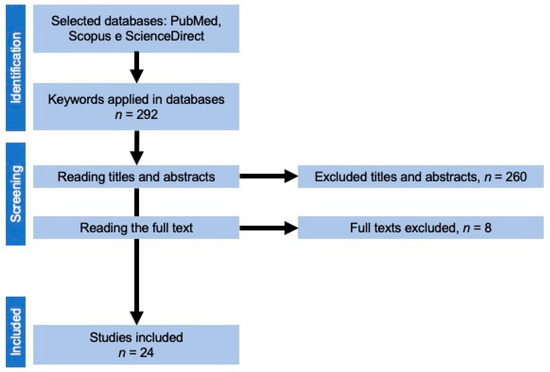
Figure 1.
Flowchart of paper selection process.
3. Results
During the database screening, 292 potentially relevant papers were found. After removing duplicates and reading the titles and abstracts, 32 papers were selected for analysis. Eight papers were excluded after carefully reading of the text. Twenty-four scientific articles were selected from between 2007 and 2022. All the studies evaluated were of participants with cleft lip and palate, 23 evaluated a UCLP patient and the other BCLP. Twelve studies were longitudinal, and the other twelve were cross-sectional (Table 1). Twenty-three studies used a scanner to obtain three-dimensional virtual dental arches, and the other used stereophotogrammetry equipment. The 3Shape Orthodontic Scanner (Copenhagen, Denmark) was the most used model (14 articles, as shown in Table 2). Fourteen different types of software were used in the studies. Mirror imaging software (Canfield Scientific Inc., Parsippany, NJ, USA) was the most used computer program (used in 5 articles). Linear measures were the most quantified (14 articles), while project palatal curve and superimposition were the least evaluated (1 article for each parameter, as shown in Table 3). Six of the selected articles were included in the reproducibility analysis (5 articles: occlusal index and 1 article: palatal surface area). Among these, one evaluated the accuracy (parameter assessed: area), while another evaluated the validity (parameter assessed: occlusal index, as shown in Table 4).

Table 1.
Studies selected for the narrative review.
Table 2.
Devices for dental arch digitalization.
Table 3.
Software and measures used in the selected studies.
Table 4.
Accuracy, validity, and reproducibility of the diagnostic tools.
4. Discussion
In the last decade, technology and innovation have also assumed a prominent position in dentistry by providing researchers with more accurate measurements in growth analysis and dental arch evaluation. The study of orofacial development and the growth of patients with cleft lip and palate is widely evaluated before and after primary surgeries [1,2,3,4,5,6,18,19,20,21,22,25,30,31,32] and for the follow-up of specific therapies [1,24]. This orofacial growth and development evaluation aims at better techniques and surgical time due to the fact that gold standard surgical protocols have not yet been described.
The image acquisition can be obtained from benchtop scanning to taking pictures. Bench scanners are the most used because they have certified technology with an affordable price. This type of equipment aims to digitize impressions, or dental models, in order to obtain 3D images, provide storage, manipulation, and the exchange in information between research centers for the purposes of cross-sectional and/or longitudinal studies as well as clinical follow-ups. However, non-dental scanners have been used as digitizers [26,27,28,29].
Another way of obtaining 3D images is through photographs, using devices such as stereophotogrammetry (Breuckmann SmartScan and Artec Eva) [23,28,29], which have the same functionality as scanners. After scanning, the images are analyzed by software that has tools capable of measuring linear distances [1,2,4,5,18,20,24,25,26,27,28,29,30,32,33,34], area [6,20,23,24,25,34], volume [3,25,29], occlusal index [19,21,22,31,33,35], angle [28,33,34] projection of palatal curve [24,34], and reproducibility [21,22,23,31,33,35]. Among the selected studies, linear measurements were the majority. The linear measurements promote the follow-up and evaluation of the anteroposterior and transversal growth of the maxilla, allowing the visualization of the malocclusion types [25,26] and arch shape [1].
The software can capture measurements of different magnitudes, including the analysis between three points (angles), between two points (area), and also three planes (volume). The analysis of the area measurements reveal the maxillary segments’ size, the arch’s total development, and the potential palate growth [23]. Volume is a broader measurement, considering the whole maxilla from the palate to the ridge and in covering all teeth. The volume is assessed from image superimposition, a relevant tool in the evaluation of craniofacial development, bone deficiency in the cleft region, and in monitoring the effect of rehabilitation protocols in patients with cleft lip and palate [3]. This technology, either intraoral or model scanning, proved to be a minimally invasive method without the use of ionizing radiation [36].
The presented technologies proved to be reliable and reproducible [21,22,23,31,33,35] for analyzing the effects of primary surgeries on dental arches [1,2,3,5,6,18,20,24,25,26,27,32], nasoalveolar devices [1,29], and the intermaxillary relationship [4,19,21,22,33,34] when comparing individuals with and without cleft lip and palate [4,28,30]. In the present study, six articles performed an analysis of accuracy, validity, and/or reliability, which corresponds to 25% of the selected articles (Table 4). All hardware and software applied in three-dimensional analysis must be tested before use in clinical cases (i.e., for diagnosis, planning, and clinical procedures) and in scientific studies. These are important criteria to guarantee the reliability of the sample, which will be evaluated in the virtual environment [37].
5. Conclusions
Based on the eligible studies of this narrative review, it is concluded that using digital tools and benchtop scanners in order to evaluate the dental arches of children with cleft lip and palate, at a mixed dentition, are reproducible, reliable, possible without the use of ionizing radiation, capable of allowing storage, allow manipulation with sustainability, and are able to assist with environment preservation.
Author Contributions
Conceptualization, P.K.J., E.C.P.A., A.L.P.F.d.A. and S.S.; methodology, P.K.J. and E.C.P.A.; formal analysis, M.A.d.A.M.M., A.L.P.F.d.A., T.M.O. and S.S.; investigation, P.K.J. and E.C.P.A.; resources, A.L.P.F.d.A., M.A.d.A.M.M., S.S. and T.M.O.; data curation, P.K.J., E.C.P.A., A.L.P.F.d.A. and S.S.; writing—original draft preparation, P.K.J., E.C.P.A., A.L.P.F.d.A., S.S., M.A.d.A.M.M. and T.M.O.; writing—review and editing, P.K.J., E.C.P.A., A.L.P.F.d.A., S.S., M.A.d.A.M.M. and T.M.O.; visualization, P.K.J., E.C.P.A., A.L.P.F.d.A., S.S., M.A.d.A.M.M. and T.M.O.; supervision, A.L.P.F.d.A., M.A.d.A.M.M., T.M.O. and S.S.; project administration, A.L.P.F.d.A., S.S., M.A.d.A.M.M. and T.M.O. All authors have read and agreed to the published version of the manuscript.
Funding
This study was financed in part by the Coordenação de Aperfeiçoamento de Pessoal de Nível Superior—Brasil (CAPES)—Finance Code 001 for publication.
Institutional Review Board Statement
Not applicable.
Informed Consent Statement
Not applicable.
Data Availability Statement
Not applicable.
Acknowledgments
The authors would like to thank CAPES (Coordenação de Aperfeiçoamento de Pessoal de Nível Superior).
Conflicts of Interest
The authors declare no conflict of interest.
References
- Jorge, P.K.; Gnoinski, W.; Vaz Laskos, K.; Felício Carvalho Carrara, C.; Gamba Garib, D.; Okada Ozawa, T.; Andrade Moreira Machado, M.A.; Pinelli Valarelli, F.; Oliveira, T.M. Comparison of Two Treatment Protocols in Children with Unilateral Complete Cleft Lip and Palate: Tridimensional Evaluation of the Maxillary Dental Arch. J. Cranio-Maxillofac. Surg. Off. Publ. Eur. Assoc. Cranio-Maxillo-fac. Surg. 2016, 44, 1117–1122. [Google Scholar] [CrossRef]
- Ambrosio, E.C.P.; Sforza, C.; De Menezes, M.; Carrara, C.F.C.; Machado, M.A.A.M.; Oliveira, T.M. Post-Surgical Effects on the Maxillary Segments of Children with Oral Clefts: New Three-Dimensional Anthropometric Analysis. J. Cranio-Maxillofac. Surg. 2018, 46, 1511–1514. [Google Scholar] [CrossRef] [PubMed]
- Ambrosio, E.C.P.; Fusco, N.D.S.; Carrara, C.F.C.; Bergamo, M.T.; Lourenço Neto, N.; Cruvinel, T.; Rios, D.; Almeida, A.L.P.F.; Soares, S.; Machado, M.A.A.M.; et al. Digital Volumetric Monitoring of Palate Growth in Children with Cleft Lip and Palate. J. Craniofac. Surg. 2022, 33, E143–E145. [Google Scholar] [CrossRef]
- Rando, G.M.; Ambrosio, E.C.P.; Jorge, P.K.; Prado, D.Z.A.; Falzoni, M.M.M.; Carrara, C.F.C.; Soares, S.; Machado, M.A.A.M.; Oliveira, T.M. Anthropometric Analysis of the Dental Arches of Five-Year-Old Children With Cleft Lip and Palate. J. Craniofac. Surg. 2018, 29, 1657–1660. [Google Scholar] [CrossRef]
- Mello, B.Z.F.; Ambrosio, E.C.P.; Jorge, P.K.; de Menezes, M.; Carrara, C.F.C.; Soares, S.; Valarelli, F.P.; Moreira Machado, M.A.A.; Oliveira, T.M. Analysis of Dental Arch in Children With Oral Cleft Before and After the Primary Surgeries. J. Craniofac. Surg. 2019, 30, 2456–2458. [Google Scholar] [CrossRef] [PubMed]
- Prado, D.Z.A.; Ambrosio, E.C.P.; Jorge, P.K.; Sforza, C.; De Menezes, M.; Soares, S.; Carrara, C.F.C.; Valarelli, F.P.; Machado, M.A.A.M.; Oliveira, T.M. Evaluation of Cheiloplasty and Palatoplasty on Palate Surface Area in Children with Oral Clefts: Longitudinal Study. Br. J. Oral Maxillofac. Surg. 2022, 60, 437–442. [Google Scholar] [CrossRef] [PubMed]
- Jones, T.; Leary, S.; Atack, N.; Chawla, O.; Ness, A.; Ireland, T.; Sandy, J. Are Photographs a Suitable Alternative to Dental Study Casts When Assessing Primary Surgical Outcome in Children Born with Unilateral Cleft Lip and Palate? Eur. J. Orthod. 2016, 38, 341–344. [Google Scholar] [CrossRef][Green Version]
- Nollet, P.J.P.M.; Katsaros, C.; van ’t Hof, M.A.; Bongaarts, C.A.M.; Semb, G.; Shaw, W.C.; Kuijpers-Jagtman, A.M. Photographs of Study Casts: An Alternative Medium for Rating Dental Arch Relationships in Unilateral Cleft Lip and Palate. Cleft Palate. Craniofac. J. 2004, 41, 646–650. [Google Scholar] [CrossRef] [PubMed]
- Alrasheed, W.A.; Owayda, A.M.; Hajeer, M.Y.; Khattab, T.Z.; Almahdi, W.H. Validity and Reliability of Intraoral and Plaster Models’ Photographs in the Assessment of Little’s Irregularity Index, Tooth Size-Arch Length Discrepancy, and Bolton’s Analysis. Cureus 2022, 14, e23067. [Google Scholar] [CrossRef] [PubMed]
- Saad, M.S.; Fata, M.; Farouk, A.; Habib, A.M.A.; Gad, M.; Tayel, M.B.; Marei, M.K. Early Progressive Maxillary Changes with Nasoalveolar Molding: Randomized Controlled Clinical Trial. JDR Clin. Transl. Res. 2020, 5, 319–331. [Google Scholar] [CrossRef] [PubMed]
- Sforza, C.; De Menezes, M.; Bresciani, E.; Cerón-Zapata, A.M.; López-Palacio, A.M.; Rodriguez-Ardila, M.J.; Berrio-Gutiérrez, L.M. Evaluation of a 3D Stereophotogrammetric Technique to Measure the Stone Casts of Patients with Unilateral Cleft Lip and Palate. Cleft Palate-Craniofacial J. Off. Publ. Am. Cleft Palate-Craniofacial Assoc. 2012, 49, 477–483. [Google Scholar] [CrossRef] [PubMed]
- Falzoni, M.M.M.; Ambrosio, E.C.P.; Jorge, P.K.; Sforza, C.; de Menezes, M.; de Carvalho Carrara, C.F.; Valarelli, F.P.; Soares, S.; Machado, M.A.A.M.; Oliveira, T.M. 3D Morphometric Evaluation of the Dental Arches in Children with Cleft Lip and Palate Submitted to Different Surgical Techniques. Clin. Oral Investig. 2021, 26, 1975–1983. [Google Scholar] [CrossRef] [PubMed]
- Ambrosio, E.C.P.; Sforza, C.; de Menezes, M.; Carrara, C.F.C.; Soares, S.; Machado, M.A.A.M.; Oliveira, T.M. Prospective Cohort 3D Study of Dental Arches in Children with Bilateral Orofacial Cleft: Assessment of Volume and Superimposition. Int. J. Paediatr. Dent. 2021, 31, 606–612. [Google Scholar] [CrossRef]
- Al-Rayes, N.Z.; Hajeer, M.Y. Evaluation of Occlusal Contacts among Different Groups of Malocclusion Using 3D Digital Models. J. Contemp. Dent. Pract. 2014, 15, 46–55. [Google Scholar] [CrossRef]
- Alazzawi, O.; Morioka, D.; Miyabe, M.; Tosa, Y.; Ohkubo, F.; Yoshimoto, S. Nasolabial Growth in Individuals With Unilateral Cleft Lip and Palate: A Preliminary Study of Longitudinal Observation Using Three-Dimensional Stereophotogrammetry. J. Craniofac. Surg. 2017, 28, e449. [Google Scholar] [CrossRef] [PubMed]
- Al-Rudainy, D.; Ju, X.; Mehendale, F.V.; Ayoub, A. Longitudinal 3D Assessment of Facial Asymmetry in Unilateral Cleft Lip and Palate. Cleft Palate. Craniofac. J. 2019, 56, 495–501. [Google Scholar] [CrossRef]
- Brons, S.; Darroudi, A.; Nada, R.; Bronkhorst, E.M.; Vreeken, R.; Berge, S.J.; Maal, T.; Kuijpers-Jagtman, A.M. Influence of Involuntary Facial Expressions on Reproducibility of 3D Stereophotogrammetry in Children with and without Complete Unilateral Cleft Lip and Palate from 3 to 18 Months of Age. Clin. Oral Investig. 2019, 23, 1041–1050. [Google Scholar] [CrossRef] [PubMed]
- Ambrosio, E.C.P.; Sforza, C.; De Menezes, M.; Gibelli, D.; Codari, M.; Carrara, C.F.C.; Machado, M.A.A.M.; Oliveira, T.M. Longitudinal Morphometric Analysis of Dental Arch of Children with Cleft Lip and Palate: 3D Stereophotogrammetry Study. Oral Surg. Oral Med. Oral Pathol. Oral Radiol. 2018, 126, 463–468. [Google Scholar] [CrossRef] [PubMed]
- Asquith, J.A.; McIntyre, G.T. Dental Arch Relationships on Three-Dimensional Digital Study Models and Conventional Plaster Study Models for Patients with Unilateral Cleft Lip and Palate. Cleft Palate. Craniofac. J. 2012, 49, 530–534. [Google Scholar] [CrossRef] [PubMed]
- Bruggink, R.; Baan, F.; Kramer, G.; Claessens, C.; Kuijpers-Jagtman, A.M.; Bronkhorst, E.M.; Maal, T.J.J.; Ongkosuwito, E. The Effect of Lip Closure on Palatal Growth in Patients with Unilateral Clefts. PeerJ 2020, 8, e9631. [Google Scholar] [CrossRef] [PubMed]
- Chawla, O.; Deacon, S.A.; Atack, N.E.; Ireland, A.J.; Sandy, J.R. The 5-Year-Olds’ Index: Determining the Optimal Format for Rating Dental Arch Relationships in Unilateral Cleft Lip and Palate. Eur. J. Orthod. 2012, 34, 768–772. [Google Scholar] [CrossRef] [PubMed]
- Chawla, O.; Atack, N.E.; Deacon, S.A.; Leary, S.D.; Ireland, A.J.; Sandy, J.R. Three-Dimensional Digital Models for Rating Dental Arch Relationships in Unilateral Cleft Lip and Palate. Cleft Palate. Craniofac. J. 2013, 50, 182–186. [Google Scholar] [CrossRef]
- De Menezes, M.; Cerón-Zapata, A.M.; López-Palacio, A.M.; Mapelli, A.; Pisoni, L.; Sforza, C. Evaluation of a Three-Dimensional Stereophotogrammetric Method to Identify and Measure the Palatal Surface Area in Children With Unilateral Cleft Lip and Palate. Cleft Palate-Craniofacial J. Off. Publ. Am. Cleft Palate-Craniofacial Assoc. 2016, 53, 16–21. [Google Scholar] [CrossRef] [PubMed]
- Eriguchi, M.; Watanabe, A.; Suga, K.; Nakano, Y.; Sakamoto, T.; Sueishi, K.; Uchiyama, T. Growth of Palate in Unilateral Cleft Lip and Palate Patients Undergoing Two-Stage Palatoplasty and Orthodontic Treatment. Bull. Tokyo Dent. Coll. 2018, 59, 183–191. [Google Scholar] [CrossRef]
- Generali, C.; Primozic, J.; Richmond, S.; Bizzarro, M.; Flores-Mir, C.; Ovsenik, M.; Perillo, L. Three-Dimensional Evaluation of the Maxillary Arch and Palate in Unilateral Cleft Lip and Palate Subjects Using Digital Dental Casts. Eur. J. Orthod. 2017, 39, 641–645. [Google Scholar] [CrossRef]
- Haque, S.; Khamis, M.F.; Alam, M.K.; Ahmad, W.M.A.W. Effects of Multiple Factors on Treatment Outcome in the Three-Dimensional Maxillary Arch Morphometry of Children With Unilateral Cleft Lip and Palate. J. Craniofac. Surg. 2020, 31, e534–e538. [Google Scholar] [CrossRef] [PubMed]
- Haque, S.; Khamis, M.F.; Alam, M.K.; Ahmad, W.M.A.W. An Investigation of Three-Dimensional Maxillary Arch Morphometry of Children With Unilateral Cleft Lip and Palate. J. Craniofac. Surg. 2021, 32, 964–966. [Google Scholar] [CrossRef] [PubMed]
- Hoffmannova, E.; Moslerová, V.; Dupej, J.; Borský, J.; Bejdová, Š.; Velemínská, J. Three-Dimensional Development of the Upper Dental Arch in Unilateral Cleft Lip and Palate Patients after Early Neonatal Cheiloplasty. Int. J. Pediatr. Otorhinolaryngol. 2018, 109, 1–6. [Google Scholar] [CrossRef] [PubMed]
- Lautner, N.; Raith, S.; Ooms, M.; Peters, F.; Hölzle, F.; Modabber, A. Three-Dimensional Evaluation of the Effect of Nasoalveolar Molding on the Volume of the Alveolar Gap in Unilateral Clefts. J. Cranio-Maxillofac. Surg. 2020, 48, 141–147. [Google Scholar] [CrossRef] [PubMed]
- Mello, B.Z.F.; Fernandes, V.M.; Carrara, C.F.C.; Machado, M.A.A.M.; Garib, D.G.; Oliveira, T.M. Evaluation of the Intercanine Distance in Newborns with Cleft Lip and Palate Using 3D Digital Casts. J. Appl. Oral Sci. 2013, 21, 437–442. [Google Scholar] [CrossRef]
- Sabelis, A.J.; Kuijpers, M.A.R.; Nada, R.M.; Chiu, Y.-T.; Bronkhorst, E.M.; Kuijpers-Jagtman, A.M.; Fudalej, P.S. Rating Dental Arch Relationships and Palatal Morphology with the EUROCRAN Index on Three Different Formats of Dental Casts in Children with Unilateral Cleft Lip and Palate. Clin. Oral Investig. 2016, 20, 943–950. [Google Scholar] [CrossRef] [PubMed][Green Version]
- Sakoda, K.L.; Jorge, P.K.; Carrara, C.F.C.; Machado, M.A.d.A.M.; Valarelli, F.P.; Pinzan, A.; Oliveira, T.M. 3D Analysis of Effects of Primary Surgeries in Cleft Lip/Palate Children during the First Two Years of Life. Braz. Oral Res. 2017, 31, e46. [Google Scholar] [CrossRef] [PubMed]
- Suzuki, A.; Yoshizaki, K.; Honda, Y.; Sasaguri, M.; Kubota, Y.; Nakamura, N.; Ohishi, M.; Oka, M.; Tashiro, H.; Katsuki, T.; et al. Retrospective Evaluation of Treatment Outcome in Japanese Children with Complete Unilateral Cleft Lip and Palate. Part 1: Five-Year-Olds’ Index for Dental Arch Relationships. Cleft Palate. Craniofac. J. 2007, 44, 434–443. [Google Scholar] [CrossRef] [PubMed]
- Wojtaszek-Slominska, A.; Renkielska, A.; Dobke, M.; Gosman, A.; Slominski, W. Orthodontic Characteristics of Maxillary Arch Deficiency in 5-Year-Old Patients Undergoing Unilateral Cleft Lip and Palate Repair with and without Early Gingivoplasty. J. Cranio-Maxillo-fac. Surg. Off. Publ. Eur. Assoc. Cranio-Maxillo-fac. Surg. 2010, 38, 155–159. [Google Scholar] [CrossRef]
- Zhu, S.; Yang, Y.; Gu, M.; Khambay, B. A Comparison of Three Viewing Media for Assessing Dental Arch Relationships in Patients with Unilateral Cleft Lip and Palate. Cleft Palate. Craniofac. J. 2016, 53, 578–583. [Google Scholar] [CrossRef]
- Hohoff, A.; Stamm, T.; Meyer, U.; Wiechmann, D.; Ehmer, U. Objective Growth Monitoring of the Maxilla in Full Term Infants. Arch. Oral Biol. 2006, 51, 222–235. [Google Scholar] [CrossRef] [PubMed]
- de Menezes, M.; Rosati, R.; Ferrario, V.F.; Sforza, C. Accuracy and Reproducibility of a 3-Dimensional Stereophotogrammetric Imaging System. J. Oral Maxillofac. Surg. Off. J. Am. Assoc. Oral Maxillofac. Surg. 2010, 68, 2129–2135. [Google Scholar] [CrossRef]
Publisher’s Note: MDPI stays neutral with regard to jurisdictional claims in published maps and institutional affiliations. |
© 2022 by the authors. Licensee MDPI, Basel, Switzerland. This article is an open access article distributed under the terms and conditions of the Creative Commons Attribution (CC BY) license (https://creativecommons.org/licenses/by/4.0/).