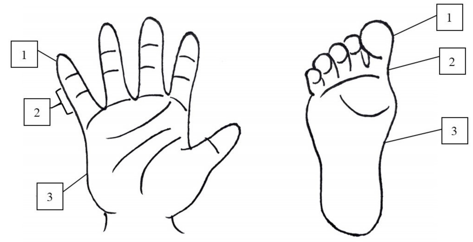
Desquamation in Kawasaki Disease
Abstract
1. Introduction
2. Materials and Methods
3. Results
4. Discussion
5. Conclusions
Author Contributions
Funding
Institutional Review Board Statement
Informed Consent Statement
Data Availability Statement
Acknowledgments
Conflicts of Interest
Abbreviations
References
- McCrindle, B.W.; Rowley, A.H.; Newburger, J.W.; Burns, J.C.; Bolger, A.F.; Gewitz, M.; Baker, A.L.; Jackson, M.A.; Takahashi, M.; Shah, P.B.; et al. Diagnosis, treatment, and long-term management of kawasaki disease: A scientific statement for health professionals from the american heart association. Circulation 2017, 135, e927–e999. [Google Scholar] [CrossRef] [PubMed]
- Rowley, A.H.; Shulman, S.T. The epidemiology and pathogenesis of kawasaki disease. Front. Pediatr. 2018, 6, 374. [Google Scholar] [CrossRef]
- Shulman, S.T.; Rowley, A.H. Kawasaki disease: Insights into pathogenesis and approaches to treatment. Nat. Rev. Rheumatol. 2015, 11, 475–482. [Google Scholar] [CrossRef] [PubMed]
- Huang, G.Y.; Ma, X.J.; Huang, M.; Chen, S.B.; Huang, M.R.; Gui, Y.H.; Ning, S.B.; Zhang, T.H.; Du, Z.D.; Yanagawa, H.; et al. Epidemiologic pictures of kawasaki disease in shanghai from 1998 through 2002. J. Epidemiol. 2006, 16, 9–14. [Google Scholar] [CrossRef] [PubMed]
- Saguil, A.; Fargo, M.; Grogan, S. Diagnosis and management of kawasaki disease. Am. Fam. Physician 2015, 91, 365–371. [Google Scholar] [PubMed]
- Wang, S.; Best, B.M.; Burns, J.C. Periungual desquamation in patients with kawasaki disease. Pediatr. Infect. Dis. J. 2009, 28, 538–539. [Google Scholar] [CrossRef] [PubMed]
- Kim, S.H.; Lee, H.J.; Lee, J.S. Clinical aspects of periungual desquamation in kawasaki disease. Iran J. Pediatr. 2018, 28, e59262. [Google Scholar]
- Milstone, L.M. Epidermal desquamation. J. Dermatol. Sci. 2004, 36, 131–140. [Google Scholar] [CrossRef]
- Oliveira, D.; Borges, A.; Simoes, M. Staphylococcus aureus toxins and their molecular activity in infectious diseases. Toxins 2018, 10, 252. [Google Scholar] [CrossRef]
- McFadden, J.P.; Baker, B.S.; Powles, A.V.; Fry, L. Psoriasis and streptococci: The natural selection of psoriasis revisited. Br. J. Dermatol. 2009, 160, 929–937. [Google Scholar] [CrossRef]
- Chong, J.H.; Aan, M.K.J. An atypical dermatologic presentation of a child with hand, foot and mouth disease caused by coxsackievirus a6. Pediatr. Infect. Dis. J. 2014, 33, 889. [Google Scholar] [CrossRef] [PubMed]
- Nag, S.S.; Dutta, A.; Mandal, R.K. Delayed cutaneous findings of hand, foot, and mouth disease. Indian Pediatr. 2016, 53, 42–44. [Google Scholar] [CrossRef] [PubMed]
- Chun, J.K.; Jeon, B.Y.; Kang, D.W.; Kim, D.S. Bacille calmette guérin (bcg) can induce kawasaki disease-like features in programmed death-1 (pd-1) gene knockout mice. Clin. Exp. Rheumatol. 2011, 29, 743–750. [Google Scholar] [PubMed]
- Chang, L.S.; Yan, J.H.; Li, J.Y.; Yeter, D.D.; Huang, Y.H.; Guo, M.M.; Lo, M.H.; Kuo, H.C. Blood mercury levels in children with kawasaki disease and disease outcome. Int. J. Environ. Res. Public Health 2020, 17, 3726. [Google Scholar] [CrossRef]
- Sireci, G.; Dieli, F.; Salerno, A. T cells recognize an immunodominant epitope of heat shock protein 65 in kawasaki disease. Mol. Med. 2000, 6, 581–590. [Google Scholar] [CrossRef]
- Gaspari, A.A.; Lotze, M.T.; Rosenberg, S.A.; Stern, J.B.; Katz, S.I. Dermatologic changes associated with interleukin 2 administration. JAMA 1987, 258, 1624–1629. [Google Scholar] [CrossRef]
- Kawasaki, T. Kawasaki disease. Proc. Jpn. Acad. Ser. B Phys. Biol. Sci. 2006, 82, 59–71. [Google Scholar] [CrossRef] [PubMed]
- Michie, C.; Kinsler, V.; Tulloh, R.; Davidson, S. Recurrent skin peeling following kawasaki disease. Arch. Dis. Child. 2000, 83, 353–355. [Google Scholar] [CrossRef][Green Version]
- Managing scarlet fever. BMJ J. 2017, 55, 102.
- Huang, P.Y.; Huang, Y.H.; Guo, M.M.; Chang, L.S.; Kuo, H.C. Kawasaki disease and allergic diseases. Front. Pediatr. 2020, 8, 614386. [Google Scholar] [CrossRef]
- Kuo, H.C.; Wang, C.L.; Liang, C.D.; Yu, H.R.; Huang, C.F.; Wang, L.; Hwang, K.P.; Yang, K.D. Association of lower eosinophil-related t helper 2 (th2) cytokines with coronary artery lesions in kawasaki disease. Pediatr. Allergy Immunol. 2009, 20, 266–272. [Google Scholar] [CrossRef] [PubMed]
- Hwang, C.Y.; Hwang, Y.Y.; Chen, Y.J.; Chen, C.C.; Lin, M.W.; Chen, T.J.; Lee, D.D.; Chang, Y.T.; Wang, W.J.; Liu, H.N. Atopic diathesis in patients with kawasaki disease. J. Pediatr. 2013, 163, 811–815. [Google Scholar] [CrossRef] [PubMed]


| Desquamation Negative | Desquamation Positive | p Value | |
|---|---|---|---|
| Total | 20 (17.9%) | 92 (82.1%) | |
| Male gender | 14/20 (70.0%) | 52/92 (56.5%) | 0.322 |
| Age | 1.1 (0.6–1.6) | 1.4 (0.8–2.6) | 0.063 |
| WBC (1000/mm3) | 12.7 (9.7–16.4) | 12.5 (10.1–14.4) | 0.536 |
| Hemoglobulin (g/dL) | 11.0 (10.3–11.9) | 11.1 (10.3–12.0) | 0.811 |
| Platelet (1000/mm3) | 333.0 (253.5–434.8) | 361.0 (285.5–435.8) | 0.416 |
| Segmented WBC (%) | 53.7 (45.0–65.6) | 61.0 (47.1–72.0) | 0.173 |
| Band WBC (%) | 0 (0–0) | 0 (0–0) | 1.000 |
| Lymphocyte (%) | 29.2 (25.4–45.1) | 29.2 (20.0–40.0) | 0.368 |
| Monocyte (%) | 7.1 (3.2–10.1) | 5.0 (3.8–8.0) | 0.277 |
| CRP (mg/L) | 35.8 (14.7–64.0) | 58.4 (28.6–116.1) | 0.078 |
| AST (IU/L) | 42.0 (32.5–62.8) | 35.0 (25.0–75.0) | 0.342 |
| ALT (IU/L) | 23.5 (16.0–93.3) | 45.0 (18.0–92.8) | 0.268 |
| Albumin (g/dL) | 4.0 (3.5–4.3) | 3.9 (3.6–4.2) | 0.852 |
| Urine WBC (/μL) | 3.0 (0–34.0) | 6.0 (0–24.0) | 0.950 |
| IVIG resistant rate | 1/20 (5.0%) | 6/92 (6.5%) | 1.000 |
| Hands’ Desquamation Negative | Hands’ DesquaMation Positive | p Value | Low-Grade Desquamation | High-Grade Desquamation | p Value | |
|---|---|---|---|---|---|---|
| Total | 21 | 91 | 77 | 14 | ||
| Male gender | 14/21 (66.7%) | 52/91 (57.1%) | 0.470 | 44/77 (57.1%) | 8/14 (57.1%) | 1.000 |
| Age | 1.0 (0.6–1.6) | 1.5 (0.7–2.6) | 0.046 * | 1.4 (0.7–2.5) | 2.2 (1.3–4.2) | 0.047 * |
| WBC (1000/mm3) | 12.4 (9.8–16.2) | 12.5 (10.1–14.4) | 0.568 | 12.5 (9.8–14.4) | 12.4 (11.4–14.8) | 0.460 |
| Hemoglobulin (g/dL) | 10.7 (10.3–11.9) | 11.1 (10.3–12) | 0.723 | 11.1 (10.2–12) | 11.2 (10.3–11.8) | 0.697 |
| Platelet (1000/mm3) | 341.0 (254.0–433.5) | 360.0 (284.0–437.0) | 0.519 | 352.0 (287.0–431.0) | 365.5 (262.3–447.0) | 0.980 |
| Segmented WBC (%) | 51.3 (45.3–65.3) | 61.0 (48.1–72.0) | 0.123 | 59.0 (46.2–70.7) | 69.8 (60.7–76.8) | 0.029 * |
| Band WBC (%) | 0 (0–0) | 0 (0–0) | 0.900 | 0 (0–0) | 0 (0–1) | 0.702 |
| Lymphocyte (%) | 29.7 (25.6–45.7) | 29.1 (20.0–40.0) | 0.236 | 30.0 (21.6–40.1) | 20.1 (14.6–31.1) | 0.030 * |
| Monocyte (%) | 7.0 (3.1–9.7) | 5.0 (3.9–8.0) | 0.412 | 5.5 (4.0–8.9) | 4.1 (2.8–4.9) | 0.006 * |
| CRP (mg/L) | 37.4 (15.3–65.0) | 58.2 (28.1–115.1) | 0.145 | 57.7 (29.0–104.5) | 101.3 (22.2–177.3) | 0.324 |
| AST (IU/L) | 42.0 (34.0–86.0) | 35.0 (25.0–69.8) | 0.214 | 35.0 (26.0–64.5) | 34.0 (25.0–178.5) | 0.923 |
| ALT (IU/L) | 24.0 (16.0–95.0) | 43.0 (18.0–88.0) | 0.395 | 39.0 (18.3–82.5) | 55.0 (15.0–237.0) | 0.935 |
| Albumin (g/dL) | 4.0 (3.5–4.3) | 3.9 (3.6–4.2) | 0.852 | 3.9 (3.5–4.2) | 4.1 (3.9–4.4) | 0.070 |
| Urine WBC (/μL) | 3.0 (0–28.5) | 6.0 (0–24.0) | 0.817 | 6.0 (0–21.0) | 6.0 (0–145.5) | 0.395 |
| IVIG resistant rate | 1/21 (4.8%) | 6/91 (6.6%) | 1.000 | 5/77(6.5%) | 1/14 (7.1%) | 1.000 |
| Feet’s Desquamation Negative | Feet’s Desquamation Positive | p Value | Low-Grade Desquamation | High-Grade Desquamation | p Value | |
|---|---|---|---|---|---|---|
| Total | 39 | 63 | 54 | 9 | ||
| Male gender | 27/39 (69.2%) | 32/63 (50.8%) | 0.067 | 27/54 (50.0%) | 5/9 (55.6%) | 1.000 |
| Age | 1.1 (0.7–1.9) | 1.6 (0.7–2.5) | 0.185 | 1.6 (0.7–2.2) | 1.4 (1.1–3.6) | 0.327 |
| WBC (1000/mm3) | 11.3 (9.2–15.2) | 12.5 (10.5–14.4) | 0.610 | 12.3 (10.2–14.0) | 14.0 (12.8–17.0) | 0.033 * |
| Hemoglobulin (g/dL) | 11.3 (10.3–12.0) | 10.9 (10.2–11.7) | 0.484 | 10.9 (10.2–11.9) | 10.7 (10.2–11.7) | 0.746 |
| Platelet (1000/mm3) | 325.0 (255.0–431.0) | 375.0 (299.0–441.0) | 0.094 | 376.5 (297.3–442.3) | 371.0 (313.0–449.5) | 0.912 |
| Segmented WBC (%) | 56.2 (44.6–68.0) | 62.0 (51.0–72.0) | 0.300 | 61.0 (49.8–69.8) | 63.0 (58.4–74.3) | 0.249 |
| Band WBC (%) | 0 (0–0) | 0 (0–0) | 0.961 | 0 (0–0) | 0 (0–0) | 0.528 |
| Lymphocyte (%) | 29.7 (20.0–44.3) | 29.0 (21.2–39.0) | 0.498 | 28.5 (21.8–39.3) | 29 (16.9–33.3) | 0.539 |
| Monocyte (%) | 7.0 (3.8–10.0) | 4.7 (3.1–7.0) | 0.070 | 5.0 (3.4–7.9) | 4.0 (3.0–4.8) | 0.098 |
| CRP (mg/L) | 39.3 (14.1–67.6) | 58.3 (32.1–122.1) | 0.023 * | 58.3 (32.1–119.8) | 76.3 (29.3–179.7) | 0.845 |
| AST (IU/L) | 42.5 (32.3–93.5) | 34.0 (25.0–68.5) | 0.045 * | 34.0 (25.5–99.5) | 25.0 (21.0–47.0) | 0.155 |
| ALT (IU/L) | 34.5 (18.0–107.3) | 38.5 (17.0–74.3) | 0.635 | 47.5 (17.8–77.3) | 23.5 (14.0–71.0) | 0.495 |
| Albumin (g/dL) | 3.9 (3.5–4.3) | 4.0 (3.6–4.2) | 0.905 | 4.0 (3.6–4.2) | 4.0 (3.6–4.4) | 0.469 |
| Urine WBC (/μL) | 3.0 (0–34.0) | 6.0 (0–24.3) | 0.763 | 6.0 (0–21.8) | 4.5 (0–142.8) | 0.931 |
| IVIG resistant rate | 2/39 (5.1%) | 4/63 (6.3%) | 1.000 | 4/54 | 0/9 | 1.000 |
| CAA a (−) | CAA (+) | p Value | |
|---|---|---|---|
| Total desquamation (+) | 59/71 (83.1%) | 33/41 (80.5%) | 0.800 |
| Hands | |||
| Desquamation (+) | 58/71 (81.7%) | 33/41 (80.5%) | 1.000 |
| High-grade | 13/58 (22.4%) | 1/33 (3.0%) | 0.016 * |
| Feet | |||
| Desquamation (+) | 42/67 (62.7%) | 21/35 (60.0%) | 0.832 |
| High-grade | 8/42 (19.0%) | 1/21 (4.5%) | 0.159 |
Publisher’s Note: MDPI stays neutral with regard to jurisdictional claims in published maps and institutional affiliations. |
© 2021 by the authors. Licensee MDPI, Basel, Switzerland. This article is an open access article distributed under the terms and conditions of the Creative Commons Attribution (CC BY) license (https://creativecommons.org/licenses/by/4.0/).
Share and Cite
Chang, L.-S.; Weng, K.-P.; Yan, J.-H.; Lo, W.-S.; Guo, M.M.-H.; Huang, Y.-H.; Kuo, H.-C. Desquamation in Kawasaki Disease. Children 2021, 8, 317. https://doi.org/10.3390/children8050317
Chang L-S, Weng K-P, Yan J-H, Lo W-S, Guo MM-H, Huang Y-H, Kuo H-C. Desquamation in Kawasaki Disease. Children. 2021; 8(5):317. https://doi.org/10.3390/children8050317
Chicago/Turabian StyleChang, Ling-Sai, Ken-Pen Weng, Jia-Huei Yan, Wan-Shan Lo, Mindy Ming-Huey Guo, Ying-Hsien Huang, and Ho-Chang Kuo. 2021. "Desquamation in Kawasaki Disease" Children 8, no. 5: 317. https://doi.org/10.3390/children8050317
APA StyleChang, L.-S., Weng, K.-P., Yan, J.-H., Lo, W.-S., Guo, M. M.-H., Huang, Y.-H., & Kuo, H.-C. (2021). Desquamation in Kawasaki Disease. Children, 8(5), 317. https://doi.org/10.3390/children8050317


