
Monitoring Expression of Balance during Therapy in Children with Postural Disorders


Abstract
1. Introduction
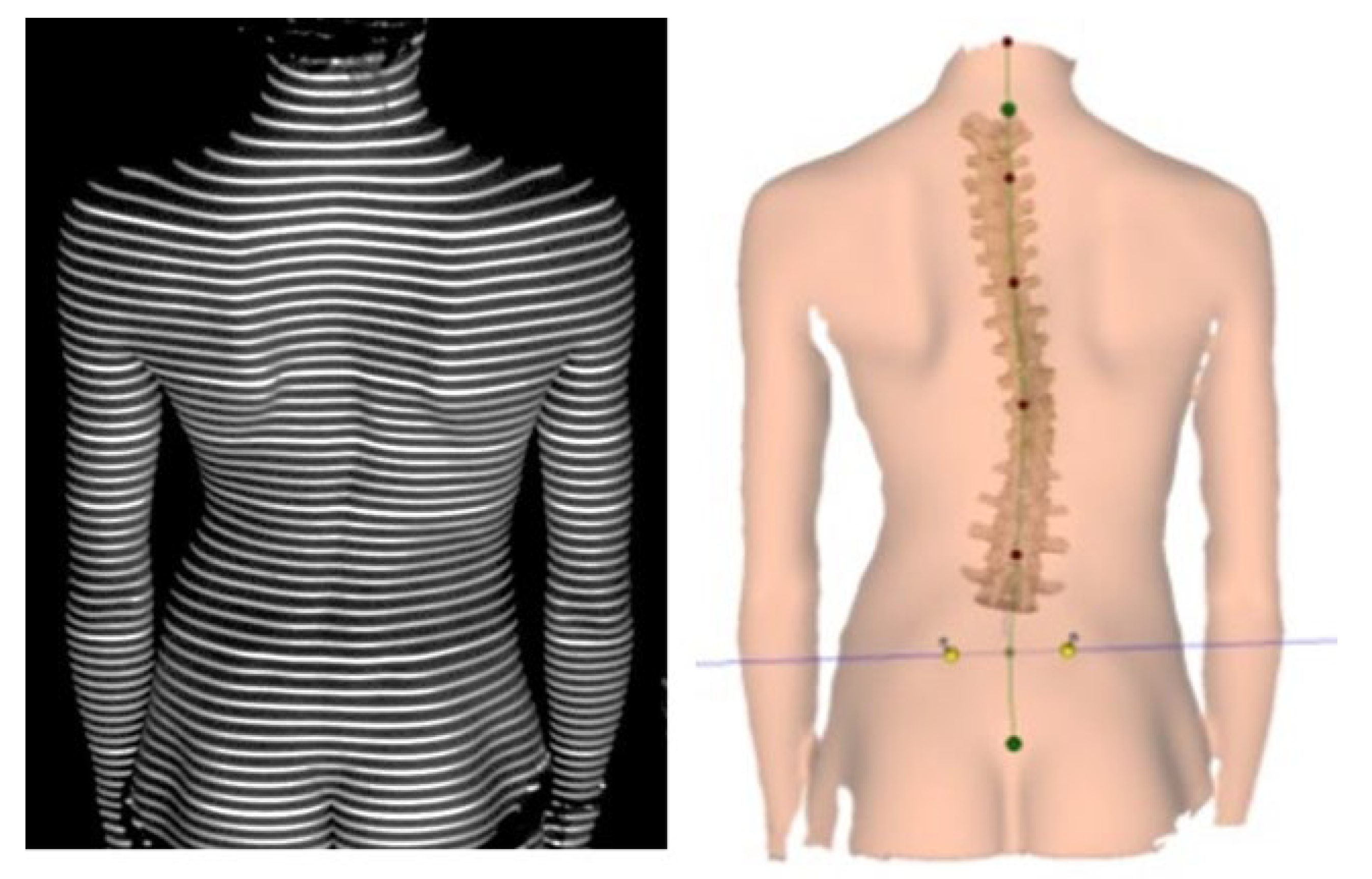
2. Materials and Methods
2.1. Subjects
- -
- Height 1.4 m (SD = 0.16);
- -
- Body weight 34.7 kg (SD = 11.84);
- -
- BMI 20.15 (SD = 2.35).
- -
- Height 1.39 m (SD = 0.13);
- -
- Body weight 38.69 (SD = 8.03);
- -
- BMI 20.02 (SD = 2.52).
- -
- Age 8–12 years;
- -
- Diagnosed shape-of-spine defect;
- -
- The legal guardian’s consent for the minor’s participation in the study.
- -
- The presence of comorbidities that may affect the shape-of-spine disorder;
- -
- Interruption or non-compliance with the orders included in the therapeutic procedure;
- -
- BMI below the 10th and above the 90th percentile.
2.2. Study Project
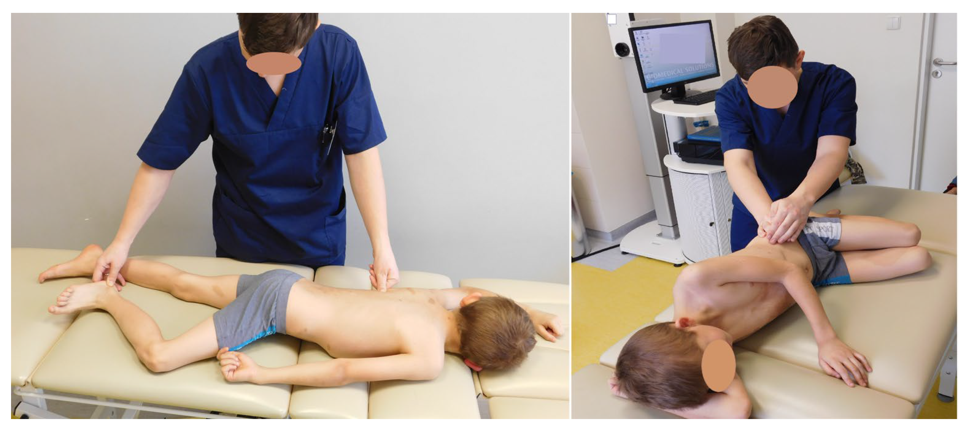
2.3. Intervention
- Maximum movement of the CoP in the frontal plane during gait—the parameter calculated in centimeters is the biggest CoP displacement in the frontal plane during gait over a distance of 16 m.
- Maximum movement of the CoP in the sagittal plane—the parameter calculated in centimeters is the biggest CoP displacement in the sagittal plane during gait over a distance of 16 m.
- Movement of the CoP in the frontal plane in static conditions—the parameter was calculated in centimeters, showing the biggest center-of-pressure displacement of the body onto the ground during 10 s in the frontal plane (to the left and right).
- Movement of the CoP in the sagittal plane in static conditions—the parameter was calculated in centimeters, exemplifying the biggest center of pressure of the body onto the ground during 10 s in the sagittal plane (forward and backward).
2.4. Statistical Methods Used
3. Results
3.1. Level of Efficiency of Equilibrium Reactions in the Test and Control Groups
3.2. Changes Occurring between Subsequent Measurements in the Study Group
4. Discussion
5. Conclusions
6. Limitations
7. Clinical Implications
Author Contributions
Funding
Institutional Review Board Statement
Informed Consent Statement
Data Availability Statement
Conflicts of Interest
References
- Lamartina, C.; Berjano, P. Classification of sagittal imbalance based on spinal alignment and compensatory me-chanisms. Eur. Spine J. 2014, 23, 1177–1189. [Google Scholar] [CrossRef]
- Pauk, J.; Daunoraviciene, K.; Ihnatouski, M.; Griskevicius, J.; Raso, J.V. Analysis of the plantar pressure distribution in children with foot deformities. Acta Bioeng. Biomech. Orig. Pap. 2010, 12, 29–34. [Google Scholar]
- Perriman, D.M.; Scarvell, J.M.; Hughes, A.R.; Lueck, C.J.; Dear, K.B.G.; Smith, P.N. Thoracic Hyperkyphosis: A Survey of Australian Physiotherapists. Physiother. Res. Int. 2012, 17, 167–178. [Google Scholar] [CrossRef] [PubMed]
- Winter, D.A. Human balance and posture control during standing and walking. Gait Posture 1995, 3, 193–214. [Google Scholar] [CrossRef]
- Lee, R.S.; Reed, D.W.; Saifuddin, A. The correlation between coronal balance and neuroaxial abnormalities detected on MRI in adolescent idiopathic scoliosis. Eur. Spine J. 2012, 21, 1106–1110. [Google Scholar] [CrossRef]
- Liao, K.; Walker, M.F.; Joshi, A.C.; Reschke, M.; Strupp, M.; Wagner, J.; Leigh, R.J. The linear vestibulo-ocular reflex, locomo-tion and falls in neurological disorders. Restor. Neurol. Neurosci. 2010, 28, 91–103. [Google Scholar] [PubMed]
- Shi, L.; Wang, D.; Chu, W.C.; Burwell, G.R.; Wong, T.T.; Heng, P.A.; Cheng, J.C. Automatic MRI segmentation and morpho-anatomy analysis of the vestibular system in adolescent idiopathic scoliosis. Neuroimage 2011, 54 (Suppl. S1), S180–S188. [Google Scholar] [CrossRef]
- Conder, R.; Zamani, R.; Akrami, M. The Biomechanics of Pregnancy: A Systematic Review. J. Funct. Morphol. Kinesiol. 2019, 4, 72. [Google Scholar] [CrossRef]
- Nault, M.-L.; Allard, P.; Hinse, S.; Le Blanc, R.; Caron, O.; Labelle, H.; Sadeghi, H. Relations Between Standing Stability and Body Posture Parameters in Adolescent Idiopathic Scoliosis. Spine 2002, 27, 1911–1917. [Google Scholar] [CrossRef]
- Campos-Mesa, M.d.C.; Rosendo, M.; Morton, K.; DelCastillo-Andrés, Ó. Effects of the Implementation of an Intervention Based on Falls Education Programmes on an Older Adult Population Practising Pilates—A Pilot Study. Int. J. Environ. Res. Public Health 2023, 20, 1246. [Google Scholar] [CrossRef]
- Hansson, E.E. Vestibular rehabilitation—For whom and how? A systematic review. Adv. Physiother. 2007, 9, 106–116. [Google Scholar] [CrossRef]
- Zhao, R.; Lu, J.; Xiao, Y.; Liu, X.; Wang, Y.; Xu, G. Effects of Gaze Stabilization Exercises on Gait, Plantar Pressure, and Balance Function in Post-Stroke Patients: A Randomized Controlled Trial. Brain Sci. 2022, 12, 1694. [Google Scholar] [CrossRef] [PubMed]
- Edyta, K.; Ewa, G.; Joanna, S.; Przemyslaw, L. Proposition of functional examination according vojta’s concept in children with scoliosis. Scoliosis 2014, 9 (Suppl. S1), O17. [Google Scholar] [CrossRef]
- Lee, B.-K. Influence of the proprioceptive neuromuscular facilitation exercise programs on idiopathic scoliosis patient in the early 20s in terms of curves and balancing abilities: Single case study. J. Exerc. Rehabil. 2016, 12, 567–574. [Google Scholar] [CrossRef] [PubMed]
- Stępień, A.; Fabian, K.; Graff, K.; Podgurniak, M.; Wit, A. An immediate effect of PNF specific mobilization on the angle of trunk rotation and the Trunk-Pelvis-Hip Angle range of motion in adolescent girls with double idio-pathic scoliosis—A pilot study. Scoliosis Spinal. Disord. 2017, 12, 29. [Google Scholar] [CrossRef] [PubMed]
- Kao, C.-L.; Hsieh, W.-L.; Wang, S.-J.; Chen, S.-J.; Wei, S.-H.; Chan, R.-C. Efficacy of a Computerized Sensor System for Evaluation and Training of Dizzy Patients. Sensors 2010, 10, 7602–7620. [Google Scholar] [CrossRef]
- Available online: https://isap.sejm.gov.pl/isap.nsf/DocDetails.xsp?id=wdu19971330883 (accessed on 24 September 2022).
- Draus, C.; Moravec, D.; Kopiec, A.; Knott, P. Comparison of Barefoot vs. Shod Gait on Spinal Dynamics Using DIERS Formetric 4D and DIERS Pedoscan Systems. Open J. Rehabil. 2015, 3, 70–76. [Google Scholar] [CrossRef][Green Version]
- Mohd Razali, N.; Bee Wah, Y. Power comparisons of Shapiro-Wilk, Kolmogorov-Smirnov, Lilliefors and Ander-son-Darling tests. J. Stat. Model. Analytics 2011, 12, 13–14. [Google Scholar]
- De Winter, J.F.C.; Dodou, D. Five-Point Likert Items: T test versus Mann-Whitney-Wilcoxon (Addendum added October 2012). Pract. Assess. Res Eval. 2010, 15, 11. [Google Scholar]
- Benavoli, A.; Corani, G.; Mangili, F. Should We Really Use Post-Hoc Tests Based on Mean-Ranks? J. Mach. Learn. Res. 2016, 17, 152–161. [Google Scholar]
- Dunn, O.J. Estimation of the Medians for Dependent Variables. Ann. Math. Stat. 1959, 30, 192–197. [Google Scholar] [CrossRef]
- Nowotny, J.; Nowotny-Czupryna, O.; Czupryna, K.; Rottermund, J. O SKOLIOZACH INACZEJ (cz. I) Podstawy fizjologiczne i fizjopatologiczne terapii skolioz. Eur. J. Clin. Exp. Med. 2021, 10, 341–350. [Google Scholar]
- Meller-Gattenyo, L. Postural control in standing among adolescents with Idiopathic Scoliosis. Scoliosis 2009, 4 (Suppl. S1), 1. [Google Scholar] [CrossRef]
- Bruyneel, A.V.; Chavet, P.; Bollini, G.; Ebermeyer, E.; Mesure, S. Idiopathic scoliosis and balance organisation in sea-ted position on a seesaw. Eur. Spine J. 2010, 19, 739–746. [Google Scholar] [CrossRef] [PubMed][Green Version]
- Rougier, P.R. Relative contribution of the pressure variations under the feet and body weight distribution over both legs in the control of upright stance. J. Biomech. 2007, 40, 2477–2482. [Google Scholar] [CrossRef]
- Roll, R.; Kavounoudias, A.; Roll, J.-P. Cutaneous afferents from human plantar sole contribute to body posture awareness. Neuroreport 2002, 13, 1957–1961. [Google Scholar] [CrossRef]
- Kavounoudias, A.; Roll, R.; Roll, J.P. Specific whole-body shifts induced by frequency-modulated vibrations of human plantar soles. Neurosci. Lett. 1999, 266, 181–184. [Google Scholar] [CrossRef]
- Carlsöö, S. The static muscle load in different work positions: An electromyographic study. Ergonomics 1961, 4, 193–211. [Google Scholar] [CrossRef]
- Radebold, A.; Cholewicki, J.; Polzhofer, G.K.; Greene, H.S. Impaired postural control of the lumbar spine is associated with delayed muscle response times in patients with chronic idiopathic low back pain. Spine 2001, 26, 724–730. [Google Scholar] [CrossRef]
- Inger, H.; Aarsland Fosdahl, M.; Arna Risberg, M.; Myklebust, G. Effect of Neuromuscular Training on Propriocep-tion, Balance, Muscle Strength, and Lower Limb Function in Female Team Handball Players Cold hypersensi-tivity in patients with severe hand injuries View project Development of a Short and Effective Shoulder Exter-nal Rotation Strength Program in Handball: A Delphi Study View project. Artic. Clin. J. Sport. Med. 2004, 14, 88–94. [Google Scholar]
- Willigenburg, N.W.; Kingma, I.; van Dieën, J.H. Center of pressure trajectories, trunk kinematics and trunk muscle activation during unstable sitting in low back pain patients. Gait Posture 2013, 38, 625–630. [Google Scholar] [CrossRef] [PubMed]
- Lê, T.T.; Kapoula, Z. Role of ocular convergence in the Romberg quotient. Gait Posture 2008, 27, 493–500. [Google Scholar] [CrossRef] [PubMed]
- Drzał-Grabiec, J.; Rachwał, M.; Podgórska-Bednarz, J.; Rykała, J.; Snela, S.; Truszczyńska, A.; Trzaskoma, Z. The effect of spinal curvature on the photogrammetric assessment on static balance in elderly women. BMC Musculoskelet. Disord. 2014, 15, 186. [Google Scholar] [CrossRef]
- Souza, J.; Pasinato, F.; Corrêa, E.; da Silva, A. Global body posture and plantar pressure distribution in individuals with and without temporomandibular disorder: A preliminary study. J. Manip. Physiol. 2014, 37, 407–414. [Google Scholar] [CrossRef] [PubMed]


| Study Group | Control Group | ||||||||
|---|---|---|---|---|---|---|---|---|---|
| Measurement | M | SD | M | SD | U | Z | p | r | |
| Maximum movement of the CoP in the frontal plane—gait (cm) | I | 10.46 | 2.50 | 9.87 | 1.92 | 9126.5 | −2.051 | 0.040 | 0.12 |
| IV | 9.80 | 1.94 | 9.87 | 1.92 | 10,650.5 | −0.007 | 0.995 | 0.00 | |
| Maximum movement of the CoP in the sagittal plane—gait (cm) | I | 20.20 | 5.83 | 16.03 | 3.23 | 5542.5 | −6.858 | <0.001 | 0.39 |
| IV | 16.38 | 3.68 | 16.03 | 3.23 | 10,198.0 | −0.614 | 0.539 | 0.03 | |
| Maximum movement of the CoP to the left—static (cm) | I | 1.03 | 1.55 | 0.56 | 0.33 | 6846.5 | −5.109 | <0.001 | 0.29 |
| IV | 0.61 | 0.41 | 0.56 | 0.33 | 9964.0 | −0.928 | 0.354 | 0.05 | |
| Maximum movement of the CoP to the right—static (cm) | I | 0.80 | 0.64 | 0.55 | 0.35 | 7445.0 | −4.307 | <0.001 | 0.24 |
| IV | 0.55 | 0.36 | 0.55 | 0.35 | 10,577.5 | −0.105 | 0.917 | 0.01 | |
| Maximum forward movement of the CoP—static (cm) | I | 1.18 | 1.01 | 0.83 | 0.41 | 7108.5 | −4.758 | <0.001 | 0.27 |
| IV | 0.85 | 0.44 | 0.83 | 0.41 | 10,273.0 | −0.513 | 0.608 | 0.03 | |
| Maximum backward movement of the CoP—static (cm) | I | 0.69 | 0.97 | 0.43 | 0.28 | 8145.5 | −3.367 | 0.001 | 0.19 |
| IV | 0.44 | 0.36 | 0.43 | 0.28 | 10,587.5 | −0.091 | 0.927 | 0.01 | |
| Measurement | M | SD | |||
|---|---|---|---|---|---|
| Maximum movement of CoP in the frontal plane—gait (cm) | I | 10.46 a | 2.50 | ||
| II | 10.38 a | 2.51 | χ2(3) = 9.67 | ||
| III | 10.15 ab | 2.30 | p = 0.022 | ||
| IV | 9.80 b | 1.94 | |||
| Maximum movement of the CoP in the sagittal plane—gait (cm) | I | 20.20 a | 5.83 | ||
| II | 18.66 b | 5.78 | χ2(3) = 42.60 | ||
| III | 18.96 ab | 4.94 | p < 0.001 | ||
| IV | 16.38 c | 3.68 | |||
| Maximum movement of the CoP to the left—static (cm) | I | 1.03 a | 1.55 | ||
| II | 1.02 a | 1.26 | χ2(3) = 38.21 | ||
| III | 1.05 a | 1.73 | p < 0.001 | ||
| IV | 0.61 b | 0.41 | |||
| Maximum movement of the CoP to the right—static (cm) | I | 0.80 a | 0.64 | ||
| II | 0.89 a | 1.35 | χ2(3) = 27.11 | ||
| III | 0.92 a | 1.16 | p < 0.001 | ||
| IV | 0.55 b | 0.36 | |||
| Maximum forward movement of the CoP—static (cm) | I | 1.18 a | 1.01 | ||
| II | 1.18 a | 1.03 | χ2(3) = 44.41 | ||
| III | 1.28 a | 1.28 | p < 0.001 | ||
| IV | 0.85 b | 0.44 | |||
| Maximum backward movement of the CoP—static (cm) | I | 0.69 a | 0.97 | ||
| II | 0.65 b | 0.96 | χ2(3) = 474.11 | ||
| III | 0.67 b | 1.14 | p < 0.001 | ||
| IV | 0.44 c | 0.36 | |||
Disclaimer/Publisher’s Note: The statements, opinions and data contained in all publications are solely those of the individual author(s) and contributor(s) and not of MDPI and/or the editor(s). MDPI and/or the editor(s) disclaim responsibility for any injury to people or property resulting from any ideas, methods, instructions or products referred to in the content. |
© 2023 by the authors. Licensee MDPI, Basel, Switzerland. This article is an open access article distributed under the terms and conditions of the Creative Commons Attribution (CC BY) license (https://creativecommons.org/licenses/by/4.0/).
Share and Cite
Żurawski, A.; Śliwiński, Z.; Kozieł, D.; Kiebzak, W. Monitoring Expression of Balance during Therapy in Children with Postural Disorders. Children 2023, 10, 974. https://doi.org/10.3390/children10060974
Żurawski A, Śliwiński Z, Kozieł D, Kiebzak W. Monitoring Expression of Balance during Therapy in Children with Postural Disorders. Children. 2023; 10(6):974. https://doi.org/10.3390/children10060974
Chicago/Turabian StyleŻurawski, Arkadiusz, Zbigniew Śliwiński, Dorota Kozieł, and Wojciech Kiebzak. 2023. "Monitoring Expression of Balance during Therapy in Children with Postural Disorders" Children 10, no. 6: 974. https://doi.org/10.3390/children10060974
APA StyleŻurawski, A., Śliwiński, Z., Kozieł, D., & Kiebzak, W. (2023). Monitoring Expression of Balance during Therapy in Children with Postural Disorders. Children, 10(6), 974. https://doi.org/10.3390/children10060974