
Centromedian–Parafascicular and Somatosensory Thalamic Deep Brain Stimulation for Treatment of Chronic Neuropathic Pain: A Contemporary Series of 40 Patients

 , and
, and
Abstract
:1. Introduction
2. Materials and Methods
2.1. Patient Selection
2.2. Surgical Procedures and Test Stimulation
2.3. DBS Programming and Follow-Up
2.4. Statistical Analysis
3. Results
4. Discussion
5. Conclusions
Author Contributions
Funding
Institutional Review Board Statement
Informed Consent Statement
Data Availability Statement
Conflicts of Interest
References
- Burchiel, K.J.; Raslan, A.M. Contemporary concepts of pain surgery. J. Neurosurg. 2019, 130, 1039–1049. [Google Scholar] [CrossRef] [PubMed]
- Rzesnitzek, L.; Hariz, M.; Krauss, J.K. The Origins of Human Functional Stereotaxis: A Reappraisal. Ster. Funct. Neurosurg. 2019, 97, 1–6. [Google Scholar] [CrossRef] [PubMed]
- Sukul, V.V.; Slavin, K.V. Deep Brain and Motor Cortex Stimulation. Curr. Pain Headache Rep. 2014, 18, 1–5. [Google Scholar] [CrossRef] [PubMed]
- Cruccu, G.; Garcia-Larrea, L.; Hansson, P.; Keindl, M.; Lefaucheur, J.-P.; Paulus, W.; Taylor, R.; Tronnier, V.; Truini, A.; Attal, N. EAN guidelines on central neurostimulation therapy in chronic pain conditions. Eur. J. Neurol. 2016, 23, 1489–1499. [Google Scholar] [CrossRef]
- Deer, T.R.; Falowski, S.; Arle, J.E.; Vesper, J.; Pilitsis, J.; Slavin, K.V.; Hancu, M.; Grider, J.S.; Mogilner, A. A Systematic Literature Review of Brain Neurostimulation Therapies for the Treatment of Pain. Pain Med. 2020, 21, 1415–1420. [Google Scholar] [CrossRef]
- Baron, R. Mechanisms of Disease: Neuropathic pain—A clinical perspective. Nat. Clin. Pract. Neurol. 2006, 2, 95–106. [Google Scholar] [CrossRef]
- Cohen, S.P.; Mao, J. Neuropathic pain: Mechanisms and their clinical implications. BMJ 2014, 348, 7656. [Google Scholar] [CrossRef] [Green Version]
- Shirvalkar, P.; Sellers, K.K.; Schmitgen, A.; Prosky, J.; Joseph, I.; Starr, P.A.; Chang, E.F. A Deep Brain Stimulation Trial Period for Treating Chronic Pain. J. Clin. Med. 2020, 9, 3155. [Google Scholar] [CrossRef]
- Slavin, K.V.; Isagulyan, E.D.; Rzaev, D.A. Deep brain stimulation for chronic pain: Time to reconsider the sceptical attitude? Brain Sci. 2020, 10, 772. [Google Scholar] [CrossRef]
- Reynolds, D.V. Surgery in the Rat during Electrical Analgesia Induced by Focal Brain Stimulation. Science 1969, 164, 444–445. [Google Scholar] [CrossRef]
- Gybels, J.; Kupers, R.; Nuttin, B. Therapeutic stereotactic procedures on the thalamus for pain. Acta Neurochir. 1993, 124, 19–22. [Google Scholar] [CrossRef]
- Richardson, D. Thalamotomy for Intractable Pain. Ster. Funct. Neurosurg. 1967, 29, 139–145. [Google Scholar] [CrossRef]
- Hitchcock, E.R.; Teixeira, M.J. A comparison of results from center-median and basal thalamotomies for pain. Surg. Neurol. 1981, 15, 341–351. [Google Scholar] [CrossRef]
- Niizuma, H.; Kwak, R.; Ikeda, S.; Ohyama, H.; Suzuki, J.; Saso, S. Follow-up results of centromedian thalamotomy for central pain. Appl. Neurophysiol. 1982, 45, 324–325. [Google Scholar]
- Lozano, A.M.; Lipsman, N.; Bergman, H.; Brown, P.; Chabardes, S.; Chang, J.W.; Matthews, K.; McIntyre, C.C.; Schlaepfer, T.E.; Schulder, M.; et al. Deep brain stimulation: Current challenges and future directions. Nat. Rev. Neurol. 2019, 15, 148–160. [Google Scholar] [CrossRef]
- Mazars, G.; Mérienne, L.; Ciolocca, C. Intermittent analgesic thalamic stimulation. Preliminary note. Rev. Neurol. 1973, 128, 273–279. [Google Scholar]
- Hosobuchi, Y.; Adams, J.E.; Rutkin, B. Chronic Thalamic Stimulation for the Control of Facial Anesthesia Dolorosa. Arch. Neurol. 1973, 29, 158–161. [Google Scholar] [CrossRef]
- Mazars, G.; Mérienne, L.; Cioloca, C. Use of thalamic stimulators in the treatment of various types of pain. Ann. Med. Interne 1975, 126, 869–871. [Google Scholar]
- Akil, H.; Richardson, D.E.; Hughes, J.; Barchas, J.D. Enkephalin-like material elevated in ventricular cerebrospinal fluid of pain patients after analgetic focal stimulation. Science 1978, 201, 463–465. [Google Scholar] [CrossRef]
- Thoden, U.; Doerr, M.; Dieckmann, G.; Krainick, J.-U. Medial thalamic permanent electrodes for pain control in man: An electrophysiological and clinical study. Electroencephalogr. Clin. Neurophysiol. 1979, 47, 582–591. [Google Scholar] [CrossRef]
- Andy, O. Parafascicular-Center Median Nuclei Stimulation for Intractable Pain and Dyskinesia (Painful-Dyskinesia). Ster. Funct. Neurosurg. 1980, 43, 133–144. [Google Scholar] [CrossRef]
- Turnbull, I.M.; Shulman, R.; Woodhurst, W.B. Thalamic stimulation for neuropathic pain. J. Neurosurg. 1980, 52, 486–493. [Google Scholar] [CrossRef]
- Ray, C.D.; Burton, C.V. Deep brain stimulation for severe, chronic pain. In Advances in Stereotactic and Functional Neurosurgery; Springer: Vienna, Austria, 1980; Volume 30, pp. 289–293. [Google Scholar]
- Hosobuchi, Y. Combined Electrical Stimulation of the Periaqueductal Gray Matter and Sensory Thalamus. Ster. Funct. Neurosurg. 1983, 46, 112–115. [Google Scholar] [CrossRef]
- Siegfried, J. Sensory Thalamic Neurostimulation for Chronic Pain. Pacing Clin. Electrophysiol. 1987, 10, 209–212. [Google Scholar] [CrossRef]
- Kumar, K.; Wyant, G.M.; Nath, R. Deep Brain Stimulation for Control of Intractable Pain in Humans, Present and Future: A Ten-Year Follow-up. Neurosurgery 1990, 26, 774–782. [Google Scholar] [CrossRef]
- Gybels, J.; Kupers, R. Deep brain stimulation in the treatment of chronic pain in man: Where and why? Neurophysiol. Clin. 1990, 20, 389–398. [Google Scholar] [CrossRef]
- Coffey, R.J. Deep Brain Stimulation for Chronic Pain: Results of Two Multicenter Trials and a Structured Review. Pain Med. 2001, 2, 183–192. [Google Scholar] [CrossRef] [Green Version]
- Coffey, R.J.; Lozano, A.M. Neurostimulation for chronic noncancer pain: An evaluation of the clinical evidence and recommendations for future trial designs. J. Neurosurg. 2006, 105, 175–189. [Google Scholar] [CrossRef] [Green Version]
- Katayama, Y.; Yamamoto, T.; Kobayashi, K.; Kasai, M.; Oshima, H.; Fukaya, C. Motor cortex stimulation for post-stroke pain: Comparison of spinal cord and thalamic stimulation. Ster. Funct. Neurosurg. 2001, 77, 183–186. [Google Scholar] [CrossRef]
- Hamani, C.; Schwalb, J.M.; Rezai, A.R.; Dostrovsky, J.O.; Davis, K.D.; Lozano, A.M. Deep brain stimulation for chronic neuropathic pain: Long-term outcome and the incidence of insertional effect. Pain 2006, 125, 188–196. [Google Scholar] [CrossRef] [PubMed]
- Yamamoto, T.; Katayama, Y.; Obuchi, T.; Kano, T.; Kobayashi, K.; Oshima, H.; Fukaya, C. Thalamic Sensory Relay Nucleus Stimulation for the Treatment of Peripheral Deafferentation Pain. Ster. Funct. Neurosurg. 2006, 84, 180–183. [Google Scholar] [CrossRef] [PubMed]
- Rasche, D.; Rinaldi, P.C.; Young, R.F.; Tronnier, V.M. Deep brain stimulation for the treatment of various chronic pain syndromes. Neurosurg. Focus 2006, 21, 1–8. [Google Scholar] [CrossRef] [PubMed]
- Plotkin, R. Results in 60 Cases of Deep Brain Stimulation for Chronic Intractable Pain. Ster. Funct. Neurosurg. 1982, 45, 173–178. [Google Scholar] [CrossRef]
- Pereira, E.A.C.; Boccard, S.G.; Linhares, P.; Chamadoira, C.; Rosas, M.J.; Abreu, P.; Rebelo, V.; Vaz, R.; Aziz, T.Z. Thalamic deep brain stimulation for neuropathic pain after amputation or brachial plexus avulsion. Neurosurg. Focus 2013, 35, E7. [Google Scholar] [CrossRef] [Green Version]
- Boccard, S.G.; Pereira, E.A.; Moir, L.; Aziz, T.Z.; Green, A.L. Long-term Outcomes of Deep Brain Stimulation for Neuropathic Pain. Neurosurgery 2013, 72, 221–231. [Google Scholar] [CrossRef]
- Hollingworth, M.; Sims-Williams, H.P.; Pickering, A.E.; Barua, N.; Patel, N.K. Single Electrode Deep Brain Stimulation with Dual Targeting at Dual Frequency for the Treatment of Chronic Pain: A Case Series and Review of the Literature. Brain Sci. 2017, 7, 9. [Google Scholar] [CrossRef] [Green Version]
- Ward, M.; Mammis, A. Deep Brain Stimulation for the Treatment of Dejerine-Roussy Syndrome. Ster. Funct. Neurosurg. 2017, 95, 298–306. [Google Scholar] [CrossRef]
- Ben-Haim, S.; Mirzadeh, Z.; Rosenberg, W.S. Deep brain stimulation for intractable neuropathic facial pain. Neurosurg. Focus 2018, 45, E15. [Google Scholar] [CrossRef]
- Hirato, M.; Miyagishima, T.; Gouda, T.; Takahashi, A.; Yoshimoto, Y. Electrical Thalamic Stimulation in the Anterior Part of the Ventral Posterolateral Nucleus for the Treatment of Patients With Central Poststroke Pain. Neuromodul. Technol. Neural Interface 2021, 24, 361–372. [Google Scholar] [CrossRef]
- Kashanian, A.; Dicesare, J.A.T.; Rohatgi, P.; Albano, L.; Krahl, S.E.; Bari, A.; De Salles, A.; Pouratian, N. Case Series: Deep Brain Stimulation for Facial Pain. Oper. Neurosurg. 2020, 19, 510–517. [Google Scholar] [CrossRef]
- Frizon, L.A.; Yamamoto, E.A.; Nagel, S.J.; Simonson, M.T.; Hogue, O.; Machado, A.G. Deep Brain Stimulation for Pain in the Modern Era: A Systematic Review. Neurosurgery 2019, 86, 191–202. [Google Scholar] [CrossRef]
- Krauss, J.K.; Pohle, T.; Weigel, R.; Kalbarczyk, A. Somatosensory thalamic stimulation versus center median-parafascicular complex stimulation in 11 patients with neuropathic pain. Stereotact Funct. Neurosurg. 2001, 77, 194. [Google Scholar]
- Krauss, J.K.; Pohle, T.; Weigel, R.; Burgunder, J.-M. Deep brain stimulation of the centre median-parafascicular complex in patients with movement disorders. J. Neurol. Neurosurg. Psychiatry 2002, 72, 546–548. [Google Scholar] [CrossRef]
- Weigel, R.; Krauss, J.K. Center Median-Parafascicular Complex and Pain Control. Ster. Funct. Neurosurg. 2004, 82, 115–126. [Google Scholar] [CrossRef]
- Weigel, R.; Capelle, H.; Schmelz, M.; Krauss, J. Selective thoracic ganglionectomy for the treatment of segmental neuropathic pain. Eur. J. Pain 2012, 16, 1398–1402. [Google Scholar] [CrossRef]
- Weigel, R.; Capelle, H.H.; Flor, H.; Krauss, J.K. Event-related cortical processing in neuropathic pain under long-term spinal cord stimulation. Pain Physician 2015, 18, 185–194. [Google Scholar]
- Schepers, I.M.; Beck, A.K.; Bräuer, S.; Schwabe, K.; Abdallat, M.; Sandmann, P.; Dengler, R.; Rieger, J.W.; Krauss, J.K. Human centromedian-parafascicular complex (CM-Pf) signals sensory cues for goal-oriented behavior selection. Neuroimage 2017, 152, 390–399. [Google Scholar] [CrossRef]
- Beck, A.-K.; Lütjens, G.; Schwabe, K.; Dengler, R.; Krauss, J.K.; Sandmann, P. Thalamic and basal ganglia regions are involved in attentional processing of behaviorally significant events: Evidence from simultaneous depth and scalp EEG. Brain Struct. Funct. 2017, 223, 461–474. [Google Scholar] [CrossRef]
- Beck, A.-K.; Sandmann, P.; Dürschmid, S.; Schwabe, K.; Saryyeva, A.; Krauss, J.K. Neuronal activation in the human centromedian-parafascicular complex predicts cortical responses to behaviorally significant auditory events. NeuroImage 2020, 211, 116583. [Google Scholar] [CrossRef]
- Young, R.F.; Chambi, V.I. Pain relief by electrical stimulation of the periaqueductal and periventricular gray matter. J. Neurosurg. 1987, 66, 364–371. [Google Scholar] [CrossRef]
- Nandi, D.; Smith, H.; Owen, S.; Joint, C.; Stein, J.; Aziz, T. Peri-ventricular grey stimulation versus motor cortex stimulation for post stroke neuropathic pain. J. Clin. Neurosci. 2002, 9, 557–561. [Google Scholar] [CrossRef] [PubMed]
- Marchand, S.; Kupers, R.; Bushnell, C.M.; Duncan, G.H. Analgesic and placebo effects of thalamic stimulation. Pain 2003, 105, 481–488. [Google Scholar] [CrossRef]
- Gray, A.M.; Pounds-Cornish, E.; Eccles, F.J.R.; Aziz, T.Z.; Green, A.L.; Scott, R.B. Deep Brain Stimulation as a Treatment for Neuropathic Pain: A Longitudinal Study Addressing Neuropsychological Outcomes. J. Pain 2014, 15, 283–292. [Google Scholar] [CrossRef] [PubMed]
- Tasker, R.; Filho, O.V. Deep Brain Stimulation for Neuropathic Pain. Ster. Funct. Neurosurg. 1995, 65, 122–124. [Google Scholar] [CrossRef]
- Levy, R.M. Deep brain stimulation for the treatment of intractable pain. Neurosurg. Clin. N. Am. 2003, 14, 389–399. [Google Scholar] [CrossRef]
- Nandi, D.; Aziz, T.Z. Deep Brain Stimulation in the Management of Neuropathic Pain and Multiple Sclerosis Tremor. J. Clin. Neurophysiol. 2004, 21, 31–39. [Google Scholar] [CrossRef]
- Bittar, R.G.; Kar-Purkayastha, I.; Owen, S.L.; Bear, R.E.; Green, A.; Wang, S.; Aziz, T.Z. Deep brain stimulation for pain relief: A meta-analysis. J. Clin. Neurosci. 2005, 12, 515–519. [Google Scholar] [CrossRef]
- Pereira, E.A.C.; Boccard, S.G.; Aziz, T.Z. Deep Brain Stimulation for Pain: Distinguishing Dorsolateral Somesthetic and Ventromedial Affective Targets. Neurosurgery 2014, 61, 175–181. [Google Scholar] [CrossRef]
- Moore, N.Z.; Lempka, S.F.; Machado, A.G. Central Neuromodulation for Refractory Pain. Neurosurg. Clin. N. Am. 2014, 25, 77–83. [Google Scholar] [CrossRef] [Green Version]
- Keifer, O.P.; Riley, J.P.; Boulis, N.M. Deep Brain Stimulation for Chronic Pain. Neurosurg. Clin. N. Am. 2014, 25, 671–692. [Google Scholar] [CrossRef] [Green Version]
- Boccard, S.G.; Pereira, E.; Aziz, T.Z. Deep brain stimulation for chronic pain. J. Clin. Neurosci. 2015, 22, 1537–1543. [Google Scholar] [CrossRef]
- Farrell, S.M.; Green, A.; Aziz, T. The Current State of Deep Brain Stimulation for Chronic Pain and Its Context in Other Forms of Neuromodulation. Brain Sci. 2018, 8, 158. [Google Scholar] [CrossRef] [Green Version]
- Fontaine, D.; Lazorthes, Y.; Mertens, P.; Blond, S.; Géraud, G.; Fabre, N.; Navez, M.; Lucas, C.; Dubois, F.; Gonfrier, S.; et al. Safety and efficacy of deep brain stimulation in refractory cluster headache: A randomized placebo-controlled double-blind trial followed by a 1-year open extension. J. Headache Pain 2009, 11, 23–31. [Google Scholar] [CrossRef] [Green Version]
- Lempka, S.F.; Malone, D.A., Jr.; Hu, B.; Baker, K.B.; Wyant, A.; Ozinga, J.G., IV; Plow, E.B.; Pandya, M.; Kubu, C.S.; Ford, P.J.; et al. Randomized clinical trial of deep brain stimulation for poststroke pain. Ann. Neurol. 2017, 81, 653–663. [Google Scholar] [CrossRef]
- Abreu, V.; Vaz, R.; Rebelo, V.; Rosas, M.J.; Chamadoira, C.; Gillies, M.J.; Aziz, T.Z.; Pereira, E.A.C. Thalamic Deep Brain Stimulation for Neuropathic Pain: Efficacy at Three Years’ Follow-Up. Neuromodul. Technol. Neural Interface 2017, 20, 504–513. [Google Scholar] [CrossRef]
- Davis, K.D.; Lozano, A.M.; Tasker, R.R.; Dostrovsky, J.O. Brain Targets for Pain Control. Ster. Funct. Neurosurg. 1998, 71, 173–179. [Google Scholar] [CrossRef]
- Boccard, S.G.; Fitzgerald, J.J.; Pereira, E.A.; Moir, L.; Van Hartevelt, T.J.; Kringelbach, M.L.; Green, A.L.; Aziz, T.Z. Targeting the affective component of chronic pain: A case series of deep brain stimulation of the anterior cingulate cortex. Neurosurgery 2014, 74, 628–635. [Google Scholar] [CrossRef] [Green Version]
- De Andrade, D.C.; Galhardoni, R.; Da Silva, V.A.; García-Larrea, L.; Dale, C.; Baptista, A.F.; Barbosa, L.M.; Menezes, L.M.B.; De Siqueira, S.R.; Valério, F.; et al. Author response: Insular and anterior cingulate cortex deep stimulation for central neuropathic pain: Disassembling the percept of pain. Neurology 2020, 94, 721–722. [Google Scholar] [CrossRef]
- Mundinger, F.; Salomao, J.F. Deep brain stimulation in mesencephalic lemniscus medialis for chronic pain. In Advances in Stereotactic and Functional Neurosurgery 4; Springer: Vienna, Austria, 1980; Volume 30, pp. 245–258. [Google Scholar]
- Son, B.-C.; Kim, D.-R.; Kim, H.-S.; Lee, S.-W. Simultaneous Trial of Deep Brain and Motor Cortex Stimulation in Chronic Intractable Neuropathic Pain. Ster. Funct. Neurosurg. 2014, 92, 218–226. [Google Scholar] [CrossRef]
- Honey, C.M.; Tronnier, V.M. Deep brain stimulation versus motor cortex stimulation for neuropathic pain: A minireview of the literature and proposal for future research. Comput. Struct. Biotechnol. J. 2016, 14, 234–237. [Google Scholar] [CrossRef] [Green Version]
- Tsubokawa, T.; Yamamoto, T.; Katayama, Y.; Moriyasu, N. Clinical Results and Physiological Basis of Thalamic Relay Nucleus Stimulation for Relief of Intractable Pain with Morphine Tolerance. Ster. Funct. Neurosurg. 1982, 45, 143–155. [Google Scholar] [CrossRef]
- Tsubokawa, T.; Yamamoto, T.; Katayama, Y.; Nishimoto, H.; Hirayama, A.; Shibuya, H. Thalamic relay nucleus stimulation for relief of intractable pain. Clinical results and beta-endorphin immunoreactivity in cerebrospinal fluid. Pain 1984, 12, 115–126. [Google Scholar] [CrossRef]
- Lenz, F.A.; Kwan, H.C.; Dostrovsky, J.O.; Tasker, R.R. Characteristics of the bursting pattern of action potentials that occurs in the thalamus of patients with central pain. Brain Res. 1989, 496, 357–360. [Google Scholar] [CrossRef]
- Sherman, S.M.; Guillery, R.W. Functional organization of thalamocortical relays. J. Neurophysiol. 1996, 76, 1367–1395. [Google Scholar] [CrossRef]
- Richardson, D.E.; Akil, H. Pain reduction by electrical brain stimulation in man. Part 1: Acute administration in periaqueductal and periventricular sites. J. Neurosurg. 1977, 47, 178–183. [Google Scholar] [CrossRef] [Green Version]
- Yezierski, R.P.; Wilcox, T.K.; Willis, W.D. The effects of serotonin antagonists on the inhibition of primate spinothalamic tract cells produced by stimulation in nucleus raphe magnus or periaqueductal gray. J. Pharmacol. Exp. Ther. 1982, 220, 266–277. [Google Scholar]
- Matsumoto, N.; Minamimoto, T.; Graybiel, A.M.; Kimura, M. Neurons in the Thalamic CM-Pf Complex Supply Striatal Neurons With Information About Behaviorally Significant Sensory Events. J. Neurophysiol. 2001, 85, 960–976. [Google Scholar] [CrossRef]
- Sadikot, A.F.; Rymar, V.V. The primate centromedian–parafascicular complex: Anatomical organization with a note on neuromodulation. Brain Res. Bull. 2009, 78, 122–130. [Google Scholar] [CrossRef]
- Smith, Y.; Galvan, A.; Ellender, T.J.; Doig, N.; Villalba, R.M.; Huerta-Ocampo, I.; Wichmann, T.; Bolam, J.P. The thalamostriatal system in normal and diseased states. Front. Syst. Neurosci. 2014, 8, 5. [Google Scholar] [CrossRef]
- Rinaldi, P.C.; Young, R.F.; Albe-Fessard, D.; Chodakiewitz, J. Spontaneous neuronal hyperactivity in the medial and intralaminar thalamic nuclei of patients with deafferentation pain. J. Neurosurg. 1991, 74, 415–421. [Google Scholar] [CrossRef] [Green Version]
- Young, R.; Vermeulen, S.; Grimm, P.; Posewitz, A.; Jacques, D.; Rand, R.; Copcutt, B. Gamma Knife Thalamotomy for the Treatment of Persistent Pain. Ster. Funct. Neurosurg. 1995, 64, 172–181. [Google Scholar] [CrossRef] [PubMed]
- Sims-Williams, H.P.; Javed, S.; Pickering, A.E.; Patel, N.K. Characterising the Analgesic Effect of Different Targets for Deep Brain Stimulation in Trigeminal Anaesthesia Dolorosa. Ster. Funct. Neurosurg. 2016, 94, 174–181. [Google Scholar] [CrossRef] [PubMed] [Green Version]
- Jeanmonod, D.; Magnin, M.; Morel, A. Low–threshold calcium spike bursts in the human thalamus. Brain 1996, 119, 363–375. [Google Scholar] [CrossRef] [PubMed] [Green Version]
- Cagnan, H.; Denison, T.; McIntyre, C.; Brown, P. Emerging technologies for improved deep brain stimulation. Nat. Biotechnol. 2019, 37, 1024–1033. [Google Scholar] [CrossRef]
- Krauss, J.K.; Lipsman, N.; Aziz, T.; Boutet, A.; Brown, P.; Chang, J.W.; Davidson, B.; Grill, W.M.; Hariz, M.I.; Horn, A.; et al. Technology of deep brain stimulation: Current status and future directions. Nat. Rev. Neurol. 2021, 17, 75–87. [Google Scholar] [CrossRef]
- Basha, D.; Dostrovsky, J.O.; Kalia, S.K.; Hodaie, M.; Lozano, A.M.; Hutchison, W.D. Gamma oscillations in the somatosensory thalamus of a patient with a phantom limb: Case report. J. Neurosurg. 2018, 129, 1048–1055. [Google Scholar] [CrossRef] [Green Version]
- Huang, Y.; Green, A.L.; Hyam, J.; Fitzgerald, J.; Aziz, T.Z.; Wang, S. Oscillatory neural representations in the sensory thalamus predict neuropathic pain relief by deep brain stimulation. Neurobiol. Dis. 2018, 109, 117–126. [Google Scholar] [CrossRef]
- Luo, H.; Huang, Y.; Xiao, X.; Dai, W.; Nie, Y.; Geng, X.; Green, A.L.; Aziz, T.Z.; Wang, S. Functional dynamics of thalamic local field potentials correlate with modulation of neuropathic pain. Eur. J. Neurosci. 2020, 51, 628–640. [Google Scholar] [CrossRef]
- Shirvalkar, P.; Veuthey, T.L.; Dawes, H.E.; Chang, E.F. Closed-Loop Deep Brain Stimulation for Refractory Chronic Pain. Front. Comput. Neurosci. 2018, 12, 18. [Google Scholar] [CrossRef] [Green Version]
- Jeanmonod, D.; Werner, B.; Morel, A.; Michels, L.; Zadicario, E.; Schiff, G.; Martin, E. Transcranial magnetic resonance imaging–guided focused ultrasound: Noninvasive central lateral thalamotomy for chronic neuropathic pain. Neurosurg. Focus 2012, 32, E1. [Google Scholar] [CrossRef] [Green Version]
- Roberts, D.G.; Pouratian, N. Stereotactic radiosurgery for the treatment of chronic intractable pain: A systematic review. Oper. Neurosurg. 2017, 13, 543–551. [Google Scholar] [CrossRef]
- UrgoSik, D.; Liscak, R. Medial Gamma Knife thalamotomy for intractable pain. J. Neurosurg. 2018, 129, 72–76. [Google Scholar] [CrossRef]


| Pre-Operatively | FU I | FU II | FU III | FU IV | |||||||||||||||||
|---|---|---|---|---|---|---|---|---|---|---|---|---|---|---|---|---|---|---|---|---|---|
| Nr | Sex | Age | Diagnosis | Etiology/Category of Pain | D/Pain | Body Part Affected | VAS Max | VAS Ave | IPG | Stim Site | VAS Max | VAS Ave | Stim Site | VAS Max | VAS Ave | Stim Site | VAS Max | VAS Ave | Stim Site | VAS Max | VAS Ave |
| 1 | M | 45 | Poststroke/central | Brainstem infarction | 2y | Face/Head | 10 | 7 | yes | CM-Pf | 0 | 0 | CM-Pf | 0 | 0 | CM-Pf | 7 | 3 | CM-Pf | 7 | 3 |
| 2 | M | 72 | Postherpetic | Herpes zoster | 2y | Head C2 L | 8 | 4 | yes | CM-Pf | 0 | 0 | CM-Pf | 0 | 0 | CM-Pf | 1 | 0 | - | - | - |
| 3 | M | 87 | Postherpetic | Herpes zoster | 2y | Thorax T3-6 R | 10 | 6 | yes | CM-Pf | 3 | 0 | - | - | - | - | - | - | - | - | - |
| 4 | F | 50 | Poststroke/ central | Multiple sclerosis | 2y | Arm L | 10 | 5 | yes | CM-Pf | 6 | 3 | CM-Pf | 6 | 3 | CM-Pf | 6 | 3 | CM-Pf | 8 | 3 |
| 5 | F | 67 | Central thalamic | Thalamic hemorrhage | 2y | Hemibody | 10 | 9 | no | - | - | - | - | - | - | - | - | - | - | ||
| 6 | M | 52 | Central thalamic | Thalamic hemorrhage | 1y | Hemibody | 8 | 6 | yes | CM-Pf | 5 | 4 | - | - | - | - | - | - | - | ||
| 7 | F | 48 | Facial pain | Trigeminal nerve surgery | 23y | Face R | 10 | 7 | yes | CM-Pf | 2 | 1 | CM-Pf | 3 | 2 | CM-Pf | 4 | 3 | CM-Pf | 6 | 4 |
| 8 | F | 40 | CRPS | Knee surgery | 10y | Leg L | 10 | 8 | yes | CM-Pf | 7 | 4 | CM-Pf | 5 | 4 | CM-Pf | 5 | 3 | CM-Pf | 4 | 3 |
| 9 | M | 73 | Postherpetic | Herpes zoster | 2y | Face R | 7 | 6 | yes | CM-Pf | 0 | 0 | CM-Pf | 0 | 0 | CM-Pf | 0 | 0 | CM-Pf | 0 | 0 |
| 10 | M | 72 | Phantom limb pain | Amputation | 35y | Leg L | 8 | 5 | yes | CM-Pf | 10 | 8 | CM-Pf | 6 | 5 | CM-Pf | 5 | 4 | - | - | - |
| 11 | F | 42 | Brachial plexus injury | Motorbike accident | 6y | Arm R | 9 | 9 | yes | CM-Pf | 2 | 2 | CM-Pf | 3 | 2 | CM-Pf | 2 | 1 | CM-Pf | 2 | 1 |
| 12 | F | 56 | Facial pain | Dental surgery | 14y | Face R | 10 | 5 | yes | CM-Pf | 7 | 4 | CM-Pf | 0 | 0 | CM-Pf | 2 | 1 | CM-Pf | 0 | 1 |
| 13 | F | 50 | Poststroke/central | Sclerodermia | 18y | Arm R | 7 | 4 | yes | CM-Pf | 3 | 1 | CM-Pf | 4 | 2 | CM-Pf | 3 | 2 | - | - | - |
| 14 | M | 54 | Central thalamic | Mesencephalic hemorrhage | 3y | Face/Arm R | 8 | 6 | no | - | - | - | - | - | - | - | - | - | - | ||
| 15 | M | 65 | Poststroke/central | Basal ganglia hemorrhage | 2y | Arm L | 9 | 9 | yes | CM-Pf | 8 | 6 | CM-Pf | 8 | 6 | CM-Pf | 7 | 5 | CM-Pf | 7 | 5 |
| 16 | M | 70 | Central thalamic | Thalamic infarction | 10y | Hemibody | 8 | 7 | no | - | - | - | - | - | - | - | - | - | - | ||
| 17 | M | 63 | Poststroke/central | Pontomesencephalic infarction | 10y | Leg R | 10 | 9 | yes | VPL | 5 | 4 | VPL | 6 | 5 | VPL | 7 | 5 | - | - | - |
| 18 | M | 34 | CRPS | Riding accident | 5y | Hand R | 10 | 9 | no | - | - | - | - | - | - | - | - | - | |||
| 19 | M | 33 | Brachial plexus injury | Motorbike accident | 14y | Arm L | 9 | 7 | yes | VPL | 3 | 2 | VPL | 3 | 2 | VPL | 7 | 4 | VPL | 2 | 2 |
| 20 | M | 52 | Spinal cord lesion | Car accident | 4y | Hand R | 8 | 7 | no | - | - | - | - | - | - | - | - | - | - | - | |
| 21 | M | 65 | Poststroke/ central | Frontoparietal infarction | 3y | Hemibody | 7 | 6 | yes | CM-Pf | 4 | 2 | CM-Pf | 4 | 2 | - | - | - | - | - | |
| 22 | F | 72 | Facial pain | Trigeminal nerve surgery | 18y | Face R | 10 | 9 | yes | VPM | 4 | 2 | VPM | 5 | 2 | VPM | 5 | 3 | VPM | 0 | 0 |
| 23 | M | 40 | Spinal cord lesion | Accident | 4y | Arm R | 10 | 8 | yes | CM-Pf | 3 | 2 | CM-Pf | 2 | 1 | CM-Pf | 2 | 1 | CM-Pf | 10 | 7 |
| 24 | F | 58 | CRPS | Ulnar nerve surgery | 2y | Arm R | 10 | 8 | yes | VPL | 4 | 2 | VPL | 4 | 2 | VPL | 4 | 2 | VPL | 5 | 4 |
| 25 | M | 47 | Facial pain | Maxillary sinus surgery | 8y | Face L | 10 | 9 | yes | VPL | 5 | 4 | VPL | 5 | 4 | VPL | 8 | 7 | VPL | 7 | 6 |
| 26 | F | 24 | CRPS | Injury of fingers | 4y | Hand R | 10 | 9 | yes | CM-Pf | 10 | 9 | VPL | 10 | 9 | CM-Pf | 10 | 9 | - | - | - |
| 27 | F | 57 | CRPS | Shoulder luxation | 2y | Arm R | 10 | 8 | yes | VPL | 3 | 2 | VPL | 3 | 2 | - | - | - | - | - | |
| 28 | F | 50 | Peripheral nerve injury | Inguinal nerve surgery | 3y | Thigh L | 9 | 6 | yes | VPL | 7 | 5 | VPL | 5 | 4 | VPL | 8 | 6 | Both | 7 | 4 |
| 29 | F | 31 | CRPS | Hand surgery | 6y | Hand R | 9 | 8 | yes | CM-Pf | 9 | 8 | CM-Pf | 9 | 8 | - | - | - | - | - | |
| 30 | F | 44 | CRPS | Ulnar nerve surgery | 10y | Arm L | 9 | 8 | yes | VPL | 2 | 2 | VPL | 10 | 9 | CM-Pf | 10 | 7 | - | - | - |
| 31 | F | 43 | Facial pain | Dental surgery | 2y | Face L | 10 | 6 | yes | VPM | 6 | 5 | VPL | 9 | 6 | VPL | 9 | 6 | VPM | 9 | 6 |
| 32 | F | 68 | Facial pain | Trigeminal nerve surgery | 3y | Face L | 8 | 8 | no | - | - | - | - | - | - | - | - | - | |||
| 33 | F | 26 | CRPS | Accident | 55y | Arm L | 9 | 8 | yes | VPL | 6 | 5 | VPL | 9 | 8 | VPL | 9 | 8 | VPL | 7 | 5 |
| 34 | M | 53 | Brachial plexus injury | Motorbike accident | 4y | Arm R | 9 | 6 | yes | CM-Pf | 10 | 5 | CM-Pf | 10 | 5 | CM-Pf | 4 | 3 | CM-Pf | 4 | 3 |
| 35 | M | 55 | Poststroke/ central | Basilary artery thrombosis | 5y | Hemibody | 7 | 4 | yes | VPL | 5 | 4 | VPL | 5 | 4 | VPL | 4 | 5 | VPL | 7 | 2 |
| 36 | M | 49 | FBSS | Spinal surgery | 5y | Leg L | 9 | 8 | yes | VPL | 8 | 6 | VPL | 8 | 5 | VPL | 4 | 2 | - | - | - |
| 37 | F | 70 | Spinal cord lesion | Dural AV fistula embolisation | 3y | Arm L | 9 | 9 | yes | VPL | 10 | 8 | VPL | 7 | 7 | - | - | - | - | - | - |
| 38 | F | 69 | Spinal cord lesion | Car accident | 10y | Legs L/R | 7 | 4 | yes | CM-Pf | 10 | 8 | - | - | - | - | - | - | - | - | - |
| 39 | M | 56 | Postherpetic | Herpes zoster | 3y | ThoraxT 10-11 | 10 | 9 | no | - | - | - | - | - | - | - | - | - | - | - | |
| 40 | F | 57 | FBSS | Spinal surgery | 9y | Leg L | 10 | 8 | yes | CM-Pf | 10 | 9 | CM-Pf | 8 | 7 | - | - | - | - | - | - |
| PreOP | FU I | FU II | FU III | FU IV | ||||||||||
|---|---|---|---|---|---|---|---|---|---|---|---|---|---|---|
| VAS | mean ± SD | n | mean ± SD | n | mean ± SD | n | mean ± SD | n | mean ± SD | n | ∆pre,I | ∆pre,II | ∆pre,III | ∆pre,IV |
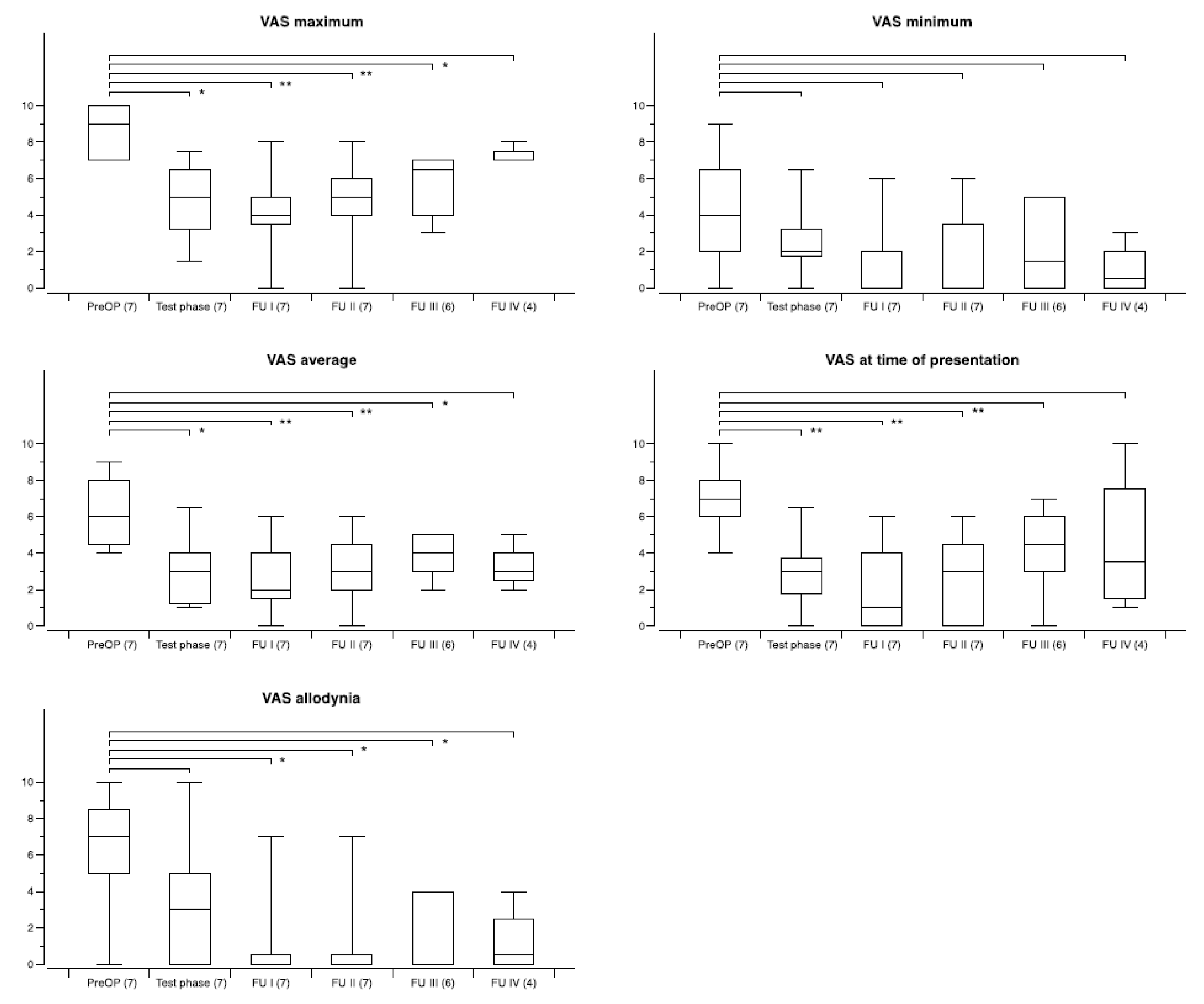
| maximum | 9.0 ± 1.1 | 40 | 5.4 ± 3.2 | 33 | 5.2 ± 3.1 | 30 | 5.3 ± 2.8 | 25 | 5.1 ± 3.2 | 18 | −40.2% | −42.0% | −41.1% | −43.4% |
| minimum | 4.8 ± 2.6 | 40 | 2.5 ± 2.6 | 33 | 2.5 ± 2.7 | 30 | 2.2 ± 2.2 | 25 | 2.1 ± 1.9 | 18 | −47.4% | −48.1% | −52.8% | −55.6% |
| average | 7.1 ± 1.6 | 40 | 3.8 ± 2.7 | 33 | 3.9 ± 2.7 | 30 | 3.7 ± 2.5 | 25 | 3.3 ± 2.1 | 18 | −46.5% | −45.5% | −47.6% | −53.8% |
| presentation | 7.5 ± 1.7 | 40 | 3.5 ± 3.0 | 33 | 3.1 ± 2.8 | 30 | 3.3 ± 2.7 | 25 | 3.0 ± 2.4 | 18 | −53.2% | −66.0% | −56.1% | −59.9% |
| allodynia | 6.9 ± 3.9 | 40 | 2.3 ± 3.5 | 33 | 2.3 ± 3.5 | 30 | 2.2 ± 3.2 | 25 | 1.8 ± 2.6 | 18 | −66.4% | −66.9% | −67.3% | −74.0% |
| FU I | FU II | FU III | FU IV | |||||
|---|---|---|---|---|---|---|---|---|
| ≥50% | ≥30% | ≥50% | ≥30% | ≥50% | ≥30% | ≥50% | ≥30% | |
| All available patients | 17/33 | 21/33 | 15/30 | 20/30 | 13/25 | 17/25 | 10/18 | 16/18 |
| Facial pain | 3/5 | 3/5 | 4/5 | 4/5 | 3/5 | 3/5 | 2/5 | 4/5 |
| CRPS | 4/7 | 5/7 | 3/7 | 3/7 | 2/5 | 2/5 | 2/3 | 3/3 |
| Poststroke/central(except thalamus) | 4/7 | 6/7 | 3/7 | 6/7 | 2/6 | 5/6 | 2/4 | 4/4 |
| Spinal cord lesion | 1/3 | 1/3 | 1/2 | 1/2 | 1/1 | 1/1 | 0/1 | 0/1 |
| Brachial plexus injury | 2/3 | 2/3 | 2/3 | 2/3 | 2/3 | 3/3 | 3/3 | 3/3 |
| Postherpetic pain | 3/3 | 3/3 | 2/2 | 2/2 | 2/2 | 2/2 | 1/1 | 1/1 |
| Central thalamic | 0/1 | 1/1 | 0/0 | 0/0 | 0/0 | 0/0 | 0/0 | 0/0 |
| Other | 0/4 | 0/4 | 0/4 | 2/4 | 1/3 | 1/3 | 0/1 | 1/1 |
| FBSS | 0/2 | 0/2 | 0/2 | 1/2 | 1/1 | 1/1 | 0/0 | 0/0 |
| Peripheral nerve | 0/1 | 0/1 | 0/1 | 1/1 | 0/1 | 0/1 | 0/1 | 1/1 |
| Phantom limb | 0/1 | 0/1 | 0/1 | 0/1 | 0/1 | 0/1 | 0/0 | 0/0 |
| Self-Rating of Overall Benefit | FU I | FU II | FU III | FU IV |
|---|---|---|---|---|
| Excellent | 6 | 5 | 6 | 3 |
| Marked | 12 | 10 | 7 | 8 |
| Moderate | 6 | 10 | 9 | 5 |
| Minor | 6 | 4 | 3 | 2 |
| None | 3 | 1 | 0 | 0 |
| Total number of patients | 33 | 30 | 25 | 18 |
Publisher’s Note: MDPI stays neutral with regard to jurisdictional claims in published maps and institutional affiliations. |
© 2021 by the authors. Licensee MDPI, Basel, Switzerland. This article is an open access article distributed under the terms and conditions of the Creative Commons Attribution (CC BY) license (https://creativecommons.org/licenses/by/4.0/).
Share and Cite
Abdallat, M.; Saryyeva, A.; Blahak, C.; Wolf, M.E.; Weigel, R.; Loher, T.J.; Runge, J.; Heissler, H.E.; Kinfe, T.M.; Krauss, J.K. Centromedian–Parafascicular and Somatosensory Thalamic Deep Brain Stimulation for Treatment of Chronic Neuropathic Pain: A Contemporary Series of 40 Patients. Biomedicines 2021, 9, 731. https://doi.org/10.3390/biomedicines9070731
Abdallat M, Saryyeva A, Blahak C, Wolf ME, Weigel R, Loher TJ, Runge J, Heissler HE, Kinfe TM, Krauss JK. Centromedian–Parafascicular and Somatosensory Thalamic Deep Brain Stimulation for Treatment of Chronic Neuropathic Pain: A Contemporary Series of 40 Patients. Biomedicines. 2021; 9(7):731. https://doi.org/10.3390/biomedicines9070731
Chicago/Turabian StyleAbdallat, Mahmoud, Assel Saryyeva, Christian Blahak, Marc E. Wolf, Ralf Weigel, Thomas J. Loher, Joachim Runge, Hans E. Heissler, Thomas M. Kinfe, and Joachim K. Krauss. 2021. "Centromedian–Parafascicular and Somatosensory Thalamic Deep Brain Stimulation for Treatment of Chronic Neuropathic Pain: A Contemporary Series of 40 Patients" Biomedicines 9, no. 7: 731. https://doi.org/10.3390/biomedicines9070731
APA StyleAbdallat, M., Saryyeva, A., Blahak, C., Wolf, M. E., Weigel, R., Loher, T. J., Runge, J., Heissler, H. E., Kinfe, T. M., & Krauss, J. K. (2021). Centromedian–Parafascicular and Somatosensory Thalamic Deep Brain Stimulation for Treatment of Chronic Neuropathic Pain: A Contemporary Series of 40 Patients. Biomedicines, 9(7), 731. https://doi.org/10.3390/biomedicines9070731