Article

The Core Stem Genes SOX2, POU5F1/OCT4, and NANOG Are Expressed in Human Parathyroid Tumors and Modulated by MEN1, YAP1, and β-catenin Pathways Activation

by
Chiara Verdelli
1,
Annamaria Morotti
2,3,
Giulia Stefania Tavanti
1,4,
Rosamaria Silipigni
5,
Silvana Guerneri
5,
Stefano Ferrero
3,4,
Leonardo Vicentini
6,
Valentina Vaira
2,3 and
Sabrina Corbetta
4,7,*
1
Laboratory of Experimental Endocrinology, IRCCS Istituto Ortopedico Galeazzi, 20161 Milan, Italy
2
Department of Pathophysiology and Organ Transplantation, University of Milan, 20122 Milan, Italy
3
Division of Pathology, Fondazione IRCCS Cà Granda Ospedale Maggiore Policlinico, 20122 Milan, Italy
4
Department of Biomedical, Surgical and Dental Sciences, University of Milan, 20122 Milan, Italy
5
Medical Genetics Laboratory, Fondazione IRCCS Cà Granda, Ospedale Maggiore Policlinico, 20122 Milan, Italy
6
Endocrine Surgery, IRCCS Istituto Auxologico, 20122 Milan, Italy
7
Endocrinology and Diabetology Service, IRCCS Istituto Ortopedico Galeazzi, 20161 Milan, Italy
*
Author to whom correspondence should be addressed.
Biomedicines 2021, 9(6), 637; https://doi.org/10.3390/biomedicines9060637
Submission received: 10 April 2021 / Revised: 28 May 2021 / Accepted: 31 May 2021 / Published: 2 June 2021
(This article belongs to the Special Issue Novelties in Parathyroid Gland Diseases)

Abstract

:
Tumors of the parathyroid glands are the second most common endocrine neoplasia. Epigenetic studies revealed an embryonic signature involved in parathyroid tumorigenesis. Here, we investigated the expression of the stem core genes SOX2, POU5F1/OCT4, and NANOG. Rare cells within normal parathyroid glands expressed POU5F1/OCT4 and NANOG, while SOX2 was undetectable. Nuclear SOX2 expression was detectable in 18% of parathyroid adenomas (PAds, n = 34) involving 5–30% of cells, while OCT4 and NANOG were expressed at the nuclear level in a more consistent subset of PAds involving 15–40% of cells. Most parathyroid carcinomas expressed the core stem genes. SOX2-expressing cells co-expressed parathormone (PTH). In PAds-derived primary cultures, silencing of the tumor suppressor gene MEN1 induced the expression of SOX2, likely through a MEN1/HAR1B/SOX2 axis, while calcium-sensing receptor activation increased SOX2 mRNA levels through YAP1 activation. In addition, inducing nuclear β-catenin accumulation in PAds-derived primary cultures by short-term incubation with lithium chloride (LiCl), SOX2 and POU5F1/OCT4 expression levels increased, while NANOG transcripts were reduced, and LiCl long-term incubation induced an opposite pattern of gene expression. In conclusion, detection of the core stem genes in parathyroid tumors supports their embryogenic signature, which is modulated by crucial genes involved in parathyroid tumorigenesis.

1. Introduction

Tumors of the parathyroid glands are the second most frequent endocrine tumors and are often associated with parathormone (PTH) hypersecretion, determining primary hyperparathyroidism (PHPT). Parathyroid tumors are mostly benign and are considered a unique condition distinct from the more aggressive histology of the parathyroid atypical adenoma and parathyroid carcinoma. Several genes have been implicated in parathyroid tumorigenesis, most of them playing a tumor suppressor role. Accumulating evidence suggests the involvement of embryonic transcription factors in parathyroid tumors development. The precise expression of these transcription factors determines differentiation and migration of the parathyroid glands during embryogenesis [1]. Parathyroid embryonic transcription factors are also expressed in parathyroid cells during adult life. Notably, their expression is aberrant in parathyroid tumors: HOX genes are deregulated in parathyroid benign tumors [2], activating variants of the glial cell missing 2 (GCM2) gene have been identified in familial and sporadic cases of PHPT [3,4,5], and the T-box transcription factor 1 (TBX1) gene is downregulated in parathyroid tumors [6,7]. Moreover, an embryonic epigenetic signature has been detected in parathyroid tumors [8]: the microRNA cluster C19MC, which is highly expressed in embryonic stem cells, is aberrantly expressed [9].
Expression of the embryonic transcription factors is often associated with the expression of the early stem core genes SOX2, POU5F1/OCT4, and NANOG. These core genes are known as the core pluripotent transcription factors and are involved in several cell and developmental processes, including maintenance of embryonic stem cells [10]. These transcription factors bind to the enhancer regions of their target genes, modulating the expression levels of differentiation and self-renewal-related genes. Downregulation of the expression of the core stem genes induces differentiation of the embryonic stem cells. Normal adult cells do not express the core stem genes, while their expression is reactivated in a variety of human cancer cells [11]. In human cancers, the expression of the core stem genes identifies cancer stem cells, which are a unique population in the tumor, comprising 2–5% of the tumor bulk. Cancer stem cells share several features with embryonic stem cells. In particular, NANOG overexpression confers stemness, unlimited self-renewal, metastasis, invasiveness, angiogenesis, and drug-resistance by activating WNT pathway, OCT4, SOX2, and other genes [12]. The expression of the core stem genes has been reported in a variety of human cancers [11] and in benign neoplasia [13,14,15]. Of note, by NanoString nCounter assay exploring the expression of 740 genes involved in the four major processes of tumor progression (angiogenesis, extracellular matrix, epithelial–mesenchymal transition, and metastasis), SOX2 gene expression has been recently detected in parathyroid cancers [7]. In addition, the core stem genes may be epigenetically modulated. For example, transcriptional activation of the C19MC cistron, which is overexpressed in a subset of parathyroid tumors [9], induces the expression of OCT4 and accelerates cellular reprogramming [16].
Based on this evidence, the present study aimed to investigate the expression of the core stem genes in human parathyroid adenomas. Moreover, we hypothesized that the known tumor suppressor menin, encoded by the MEN1 gene, whose expression is lost in multiple endocrine neoplasia (MEN1)-related parathyroid tumors [17], may be involved in the modulation of the core stem genes. Similarly, we investigated the potential role of the Hippo pathway-related YAP1 co-factor, whose nuclear expression has been recently found to be downregulated in parathyroid cancers [18], and the role of the WNT/β-catenin signaling. YAP1 [19,20,21] and active β-catenin [22] have been reported to modulate the expression of the core stem cell genes.
Here, the core stem genes profile was investigated in normal and tumor parathyroid tissues by immunohistochemistry and immunofluorescence, confirming the deregulated embryonic signature of the parathyroid neoplasia. Moreover, modulation of the core stem genes has been analyzed, providing evidence of the involvement of the tumor suppressor genes MEN1 and YAP1 and of WNT/β-catenin signaling.

2. Materials and Methods

2.1. Tissue Samples

Twenty-four PAds from patients (19 females, 5 males; age 64.1 ± 2.25 years) with sporadic PHPT were collected and analyzed by RealTime PCR experiments. A further 20 PAds samples, with clinical and biochemical features similar to those of the PAds analyzed as whole tissue, were enzymatically dispersed; cells were cultured and used for gene silencing experiments, stimulation experiments, and immunofluorescence. Additionally, formalin-fixed paraffin-embedded (FFPE) sections were obtained from 5 normal parathyroid glands involuntary removed from normocalcemic patients surgically treated for thyroid diseases, 34 PAds for SOX2 analysis, 4 PAds for OCT4 analysis, 11 PAds for NANOG analysis, 5 parathyroid atypical adenomas (PAts), 3 parathyroid carcinomas (PCas; one metastatic cancer) for SOX2 and OCT4 analysis, and 8 PCas (2 metastatic cancers) for NANOG analysis were analyzed by immunohistochemistry (IHC). This study was approved by the Institutional Ethical Committee (Ospedale San Raffaele Ethical Committee, protocol no. GPRC6A PARA, approved on 07 March 2019; CE40/2019), and patients’ informed consent was obtained from all participants.

2.2. Immunohistochemistry

All archival tissue samples were cut and stained with hematoxylin and eosin for morphological assessment before immunohistochemistry (IHC). Then, tissue sections were incubated with primary antibody to SOX2 (Clone D6D9XP® Rabbit mAb, 1:100; Cell Signaling Technology, EuroClone, Milan, Italy), OCT4 (Clone C30A3, 1:100; Cell Signaling Technology, EuroClone, Milan, Italy), NANOG (Clone D73G4, 1:100; Cell Signaling Technology, EuroClone, Milan, Italy), and PTH (BGN/1F8, sc-80924, Santa Cruz Biotechnology, DBA, Segrate, Italy). IHC was performed using Benchmark Ultra Roche Ventana Immunostainer (Roche Group, Tucson, AZ, USA) as previously described [23] and diaminobenzidine (DAB) was the chromogen. All slides were evaluated by an experienced pathologist (S.F.) and immunoreactivity was calculated as the percentage of positive cells out of the total number of parathyroid cells.

2.3. RNA Isolation and Real-Time Quantitative Reverse Transcription (qRT-PCR)

Total RNA was isolated from frozen tissues and PAds-derived primary cells using Trizol reagent (Invitrogen, ThermoFisher Scientific, Monza, Italy) following the manufacturer’s instructions. RNA was quantified by spectrophotometry at 260 nm (NANODROP ND-1000 Uv/Vis, Thermo Fisher Scientific, Whaltham, MA, USA) and DNA contamination was removed by DNase I (Life Technologies, ThermoFisher Scientific, Monza, Italy) treatment. Reverse transcription was performed using the iScript cDNA Synthesis Kit (Bio-Rad, Hercules, CA, USA) with a starting amount of 300 ng of digested RNA.
Real-Time PCR was conducted on a StepOne Plus System (ThermoFisher Scientific, Monza, Italy) using the following Taqman gene expression assays: SOX2 (Hs01053049_s1), OCT4/POU5F1 (Hs00999632_g1), NANOG (Hs02387400_g1), AXIN2 (Hs00610344_m1), DKK1 (Hs00183740_m1), ZEB1 (Hs00232783_m1), MEN1 (Hs00365720_m1), HAR1B (Hs03299152_m1), and YAP1 (Hs00371735_m1) following the manufacturer’s protocol. Gene expression was quantified using a comparative Ct method and HMBS and B2M were used as housekeeping genes (Hs00609297_m1 and Hs99999907_m1, respectively) as previously described [24].

2.4. Primary Parathyroid Adenoma Cell Isolation and Culture

Samples from 8 PAds were cut into fragments less than 1 mm3, washed with PBS, and partially digested with Collagenase type I (Worthington, Lakewood, NJ, USA) 2 mg/mL for 90 min. Tissue digestion was filtered with a cell strainer (100 μm Nylon, BD Falcon, BioScientifica, Rignano Flaminio (RM), Italy) to obtain a single cell suspension. Cells were routinely grown at 37 °C in a humidified atmosphere of 5% CO2/air in Dulbecco’s Modified Eagle’s Medium (DMEM) supplemented with 10% FBS, 2 mM glutamine, and 100 U/mL penicillin-streptomycin.

2.5. DNA Extraction and Array Comparative Genomic Hybridization (aCGH) Analysis

Genomic DNA from 17 PAds was isolated using Trizol reagent (Invitrogen). The array-CGH analysis was performed using 60-mer oligonucleotide probe technology (SurePrint G3 Human CGH 8 × 60 K, Agilent Technologies, Santa Clara, CA, USA), according to the manufacturer instructions. The Feature Extraction and Cytogenomics 3.0.4.1, with the ADM-2 algorithm (Agilent Technologies), was used for data analysis. To improve results accuracy, the Diploid Peak Centralization algorithm was also applied. To determine aberrations, we set as the threshold a minimum of five consecutive probes/regions and a minimum absolute average log ratio (MAALR) of ± 0.25. To identify lower levels of mosaicism, a second analysis was run with a MAALR of ± 0.15. Only copy number variants not already reported in the public database of genomic variants (http://projects.tcga.ca/variation/ (accessed on 10 April 2021) were listed. The GRch37/hg19 of the Human Genome Reference (March 2009) consortium was used as the reference genome. Data were partially published previously [25].

2.6. Treatment of PAds-Derived Primary Cell Preparations with Lithium Chloride

PAds-derived single cells were treated with increasing concentrations of lithium chloride (LiCl, 10–20 mM) (Sigma-Aldrich, St. Louis, MO, USA), a known inhibitor of GSK3β kinase. After 8 and 72 h of treatment, cells were harvested and used for RNA analysis.

2.7. Cell Transfection and RNA Interference

For transient RNA interference experiments, PAds-derived primary cell cultures were seeded in 6-well plates at 1.5 × 10⁵ cells/well density. Cells were transiently transfected using Lipofectamine 3000 (Invitrogen-Thermo Fisher Scientific) in Opti-MEM (Gibco, Thermo Fisher Scientific), with MEN1-directed siRNA (EHU067451, Mission EsiRNA; Sigma Aldrich) or negative control siRNA (SIC001, Mission siRNA Universal negative control; Sigma-Aldrich) for 5 h, in accordance with the manufacturer’s instruction. HAR1B silencing was obtained using Dharmafect (T-2001-01; Dharmacon) as the transfection reagent and HAR1B-directed siRNA (SASI_Hs02_00378868; Sigma-Aldrich), while YAP1 silencing was performed using Dharmafect (T-2001-01; Dharmacon) as the transfection agent and YAP1-directed siRNA (L-012200-00-005; ON-TARGET Plus siRNA SmartPool Dharmacon) or control siRNA (D-001810-10-05, ON-TARGET Non-Targeting Plus). siRNAs from Sigma Aldrich and Dharmacon are a mixture of different pre-designed siRNAs, all targeting the same mRNA sequence, minimizing the off-target effects. After 48 h, transfected cells were used for further experiments and analyzed by qRT-PCR.

2.8. Statistical Analysis

Data are presented as mean ± standard error of the mean (SEM). Non-normally distributed variables (failure of the Kolmogorov–Smirnov test) were log2 transformed. Correlations between two variables were tested with parametric or non-parametric tests as specified. Groups’ comparison was performed with one-way ANOVA adjusted for multiple comparisons. The p values less than 0.05 were considered statistically significant. All statistical analysis was performed using GraphPad Prism software v6.0 (GraphPad Inc., San Diego, CA, USA).

3. Results

3.1. The Core Stem Cell Genes SOX2, POU5F1/OCT4, and NANOG Are Expressed in Parathyroid Tumors

The IHC analysis for SOX2 failed to detect cells with specific nuclear staining in normal parathyroid glands (Figure 1A,D panel a and Supplementary Figure S1). This finding is in line with the Human Protein Atlas reports of IHC negative for SOX2 in human normal parathyroid glands (www.proteinatlas.org (accessed on 10 April 2021)) and with the recent report by Condello et al. [7]. Similarly, most of PAds samples (n = 34) were negative for SOX2 immunostaining (Figure 1A). Indeed, SOX2-expressing cells at a nuclear level were identified in 6 (18%) PAds, where the nuclear staining was detected in a proportion of cells variable from 5 to 30% of endocrine parathyroid cells (Figure 1A,D panel b) though the staining showed weak intensity. Similarly, SOX2 nuclear expression was detected in few cells in 1 out 5 PAts, while one metastatic PCa expressed SOX2 in 70% of cells (Figure 1A, black circle). Nuclei of the endothelial cells and fibroblasts in PAds were invariably negative for SOX2. In PAds, SOX2-expressing cells co-expressed PTH (Figure 1E).
At variance with SOX2, specific immunostaining for POU5F1/OCT4 showed rare nuclear OCT4-expressing cells in normal parathyroid glands (Figure 1B,D panel c), while in the FFPE sections of PAds (n = 4), IHC identified 5–20% of cells with positive nuclear OCT4 staining (Figure 1B,D panel d). OCT4 was also detectable at nuclear levels in most PAts and PCas. Staining of FFPE sections with specific anti-NANOG antibodies identified rare scattered cells expressing NANOG protein at nuclear levels in normal parathyroid glands (Figure 1C,D panel e). In PAds, IHC detected positive NANOG nuclear staining in a proportion of cells ranging 1–40% (Figure 1C,D panel f), while all PAts and PCas samples showed a consistent proportion of cells, ranging from 20 to 40%, expressing NANOG at the nuclear level.
Considering PAds with available clinical data (n = 16), we found that PAds expressing the core genes transcripts were associated with circulating ionized calcium levels lower than those associated with undetectable transcripts (1.50 ± 0.02 vs. 1.61 ± 0.03 mmol/L; p = 0.039 by a Student’s t-test). No differences were detected in circulating PTH levels, age at diagnosis, or PAds weight between the two PAds groups (Table 1). Ionized calcium is the most reliable biochemical parameters reflecting parathyroid cell function.

3.2. Modulation of Core Stem Genes Expression in Sporadic Parathyroid Tumors

3.2.1. Role of MEN1

We then analyzed the aCGH profiles in a set of 17 PAds and we correlated them with SOX2 mRNA levels. Seven PAds harbored a monosomy of the chromosome 11 (Chr.11-LOH), detectable in 35–85% of cells. PAds harboring Chr.11-LOH expressed higher mRNA levels of SOX2 compared with that of the PAds harboring biallelic Chr.11 (p = 0.025; Figure 2A). Considering that Chr.11-LOH occurs in about 40% of parathyroid adenomas and MEN1 expression is variably conserved in parathyroid tumors, we chose to silence MEN1 gene expression in order to evaluate the effect of its complete loss (Figure 2B). Transient silencing of the MEN1 gene in PAds-derived cells (n = 4; Figure 2B) increased SOX2 mRNA levels (Figure 2C), while it did not affect expression of both POU5F1/OCT4 and NANOG (data not shown).
Moreover, we previously demonstrated that MEN1 silencing increased the expression of a set of long non-coding RNAs (lncRNAs) in PAds [25]. We focused the attention on the lncRNA HAR1B, whose expression has been found to be upregulated in PAds harboring the Chr.11-LOH and has been upregulated by MEN1-silencing [25]. We tested the hypothesis that HAR1B mediates or contributes with menin to modulate the SOX2 expression in parathyroid adenomas. Interestingly, HAR1B silencing induced an increase in SOX2 (Figure 2D) and NANOG transcripts (Figure 2E), while POU5F1/OCT4 expression was unaffected in PAds-derived cell preparations (n = 3).

3.2.2. Role of CASR-Stimulated YAP1 Signaling

The transcriptional co-activator YAP1 is involved in the modulation of core stem genes [19,20,21]. A recent report showed that YAP1 accumulation at the nuclear level is induced by calcium-sensing receptor (CASR) activation in PAds-derived cells [18]. In PAds-derived cells (n = 3), R568-mediated CASR activation increased SOX2 mRNA levels, while the stimulatory effect was abolished by YAP1 silencing (Figure 2F,G).

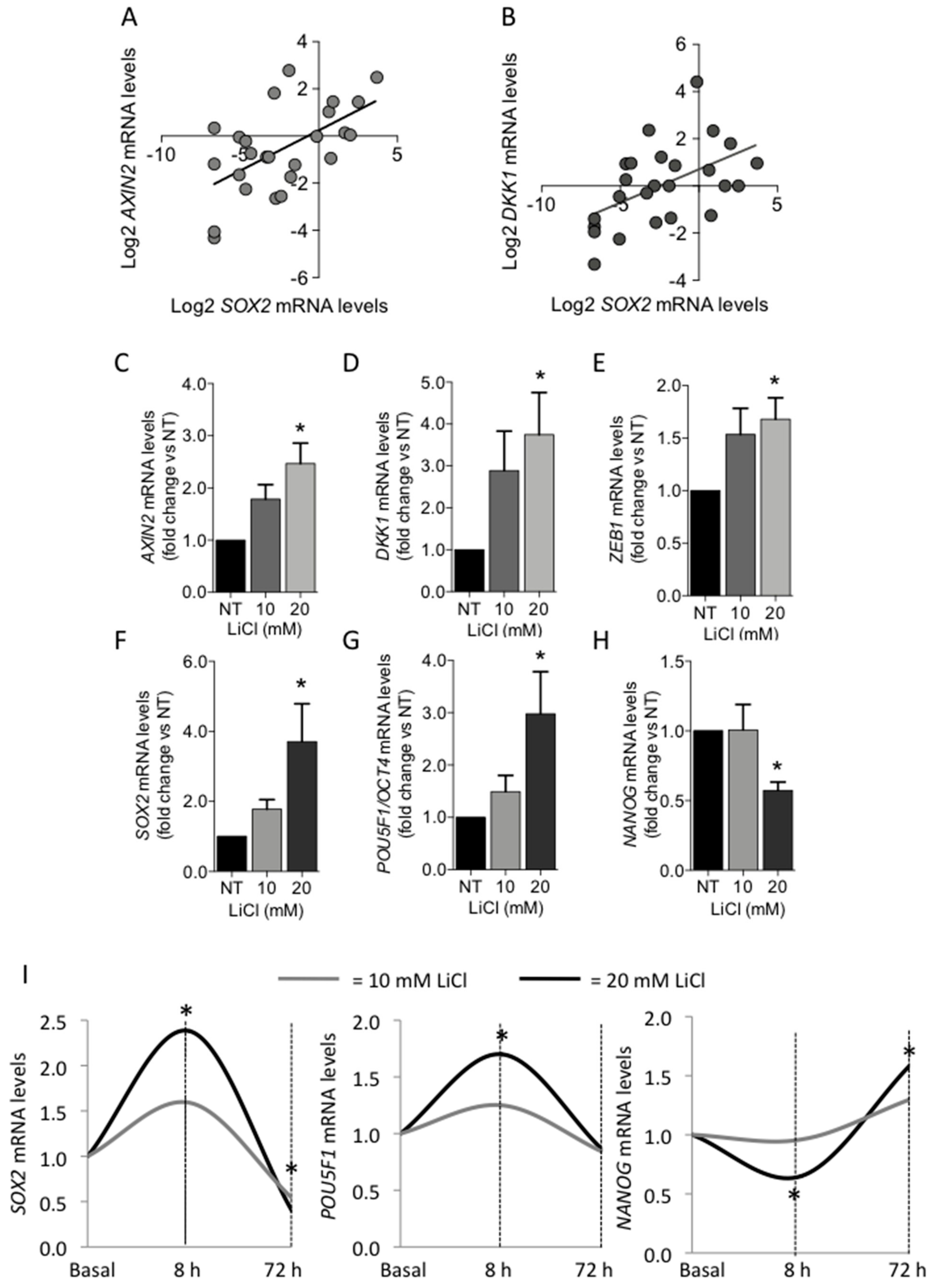
3.2.3. Role of Canonical WNT/β-Catenin Signaling Pathway

The canonical WNT/β-catenin pathway is intimately connected with pluripotency during embryogenesis [22]. Therefore, we investigated the effect of WNT/β-catenin activation on the expression of the core stem cell genes in PAds-derived cells. Positive correlations between SOX2 and AXIN2 mRNA levels (r = 0.586; p = 0.002) (Figure 3A) and between SOX2 and DKK1 mRNA levels (r = 0.519; p = 0.008) (Figure 3B) were detected.
Stimulation of PAds-derived cell cultures by increasing concentrations of lithium chloride (LiCl) (10–20 mM) for 8 h induced accumulation of active β-catenin at the nuclear level [6]. Here, we demonstrated that WNT pathway activation by LiCl for 8 h increased the β-catenin target genes AXIN2 (Figure 3C; * p = 0.001 by one-way ANOVA adjusted for multiple comparison), DKK1 (Figure 3D; * p = 0.068), and ZEB1 (Figure 3E; * p = 0.057) in PAds-derived cells (n = 6). Moreover, LiCl (10–20 mM) induced increases in SOX2 (p = 0.046; Figure 3F) and POU5F1/OCT4 transcripts (Figure 3G; * p = 0.040), while NANOG mRNA expression levels were significantly reduced (Figure 3H; * p = 0.026). In contrast, by prolonging the treatment of PAds-derived cells (n = 3) with 10–20 mM LiCl for 72 h, POU5F1/OCT4 mRNA levels returned to basal levels, while SOX2 expression levels were downregulated and NANOG transcripts were significantly increased after 20 mM LiCl stimulation (Figure 3I).

4. Discussion

The present study provides evidence supporting the aberrant embryonic gene signature of parathyroid tumors. Besides deregulation of the transcription factors TBX1 [6] and GCM2 [5], which are involved in parathyroid embryonic differentiation, and of microRNAs belonging to the early embryonic pluripotent C19MC microRNA cluster [9], parathyroid tumors harbor cells expressing the core stem genes SOX2, POU5F1/OCT4, and NANOG. Recently, SOX2 has been identified as significantly deregulated in parathyroid carcinomas [7]. In the present study, we focused our attention on the expression of SOX2 and investigation was extended to the associated core stem genes POU5F1/OCT4 and NANOG.
SOX2 is a sex-determining region of a Y-related transcription factor, which belongs to a family of DNA-binding proteins [26]. POU5F1/OCT4 is an octamer transcription factor, which binds to an eight-base pair DNA sequence and consists a Pit/Oct/Unc family of homeodomain proteins [27]. SOX2 shares a common DNA-binding site with POU5F1/OCT4 and has synergistic effects in regulation of their target genes by formation of a heterodimer. NANOG is a homeobox-containing transcription factor, and it is one of the target genes regulated by the heterodimer SOX2/OCT4. NANOG protein is expressed only in undifferentiated cells [12]. Notably, an interconnected SOX2/OCT4/NANOG regulatory circuit maintains embryonic stem cells self-renewal and pluripotency [28].
Cells expressing SOX2 could not be detected in normal parathyroid glands from normocalcemic subjects, but normal glands showed rare cells expressing OCT4 and NANOG. This observation resembles the few cells expressing core stem genes detected in normal oral epithelial cells or in cells of oral premalignant lesions [29]. Cells expressing the core stem genes were detected in a subset of parathyroid adenomas and in most parathyroid carcinomas. In line with data reported by Condello et al. [7], a metastatic parathyroid carcinoma expressed SOX2 in about 70% of cells. In PAds, SOX2-expressing cells co-expressed PTH, suggesting that re-expression of the early embryonic stem cell factor does not affect the endocrine properties of the parathyroid cells. Of note, parathyroid adenomas expressing transcripts of the three core stem genes were associated with lower circulating ionized calcium levels when compared with that of tumors with undetectable gene transcripts.
We further investigated regulation of stem core genes expression in sporadic PAds. To this aim, we tested the hypothesis that regulatory molecules critical in parathyroid tumorigenesis, namely menin, encoded by the MEN1 gene [30], calcium sensing receptor (CASR) [31], YAP1 [18], and WNT/β-catenin [6], may be involved in modulation of the stem core genes. Chromosome 11 loss of heterozygosity (LOH) can be detected in at least 40% of parathyroid tumors [32] and MEN1, which maps on chromosome 11, is an important oncosuppressor involved in parathyroid tumorigenesis [33]. In sporadic PAds, tumors with chromosome 11 LOH showed higher SOX2 transcript levels compared to that of PAds harboring biallelic chromosome 11. In line with this observation, reduction of MEN1 expression increased SOX2 mRNA expression levels in primary parathyroid adenomas cultures. MEN1 silencing is also associated with deregulation of lncRNAs, among which HAR1B is the most significantly upregulated lncRNA in PAds harboring chromosome 11 LOHs compared with that of PAds with biallelic chromosome 11 [25]. Interestingly, HAR1B silencing in PAds primary cell cultures increased SOX2 and NANOG transcripts, suggesting the existence of a MEN1-HAR1B-SOX2/NANOG regulatory axis.
Recently, it has been reported that CASR activation induces nuclear yes-associated protein 1 (YAP1) accumulation and transcription of YAP1 target genes in PAds-derived cells [18]. The transcription factor YAP1, a major effector of the tumor suppressive Hippo signaling pathway, is necessary to maintain pluripotency in embryonic stem cells. High YAP1 expression is particularly prominent in cancer stem cells. In osteosarcoma cells, YAP1 can regulate the expression of SOX2 by binding to two distinct DNA binding sites upstream and downstream of the SOX2 gene [34]. In non-small cell lung cancers, YAP1 transcriptionally induces SOX2 through a physical interaction with OCT4 [35]. In PAds-derived primary cell cultures, CASR activation by the selective agonist R568 increased the expression of SOX2, while POU5F1/OCT4 and NANOG were unaffected. Reduction of YAP1 nuclear accumulation by silencing YAP1 [18] abolished the stimulatory effect of CASR activation on SOX2 expression levels, thus suggesting that it is mediated by YAP1 signaling activation. Considering that both CASR and YAP1 feature as tumor suppressors in parathyroid adenomas, the induction of SOX2 expression may promote cell resting more than cell proliferation [36].
The WNT/β-catenin pathway plays a pivotal role in the maintenance of human embryonic stem cells pluripotency and pluripotency exit and in somatic cell reprogramming [22]. β-catenin binds POU5F1/OCT4 in embryonic stem cells [22], it stimulates human NANOG promoter activity [37] and it regulates SOX2 activity in breast cancer cells [38]. Moreover, the expression of the core stem genes SOX2, POU5F1/OCT4, and NANOG is upregulated after β-catenin nuclear accumulation in embryonic stem cells [22].
The role of β-catenin in parathyroid tumorigenesis is not clear. Parathyroid tumors did not harbor constitutively activated β-catenin pathways: Activating mutations of the CTNNB1/β-catenin gene are rare and nuclear β-catenin immunostaining has not been demonstrated so far [39,40,41,42]. However, deregulation of WNT/β-catenin signaling modulators has been detected in parathyroid tumors compared to normal glands: Promoter hypermethylation of inhibitors of the WNT signaling APC and RASSF1 [43,44], SFRP1 [45,46], and GSK3β [40] have been described in both benign and malignant parathyroid tumors, while hypermethylation of CTNNB1/β-catenin occurs in about one fourth of PAds [47]. The effect of such deregulation in parathyroid tumor cells is still far from being elucidated [40,48].
In PAds, SOX2 transcripts positively correlated with the WNT/β-catenin pathway target genes AXIN2 and DKK1. In particular, AXIN2 is a well-known target of the canonical WNT signaling, and its expression provides a reliable readout of cells responding to WNT [49,50]. Here, we showed that WNT/β-catenin activation by LiCl, demonstrated by AXIN2, DKK1, and ZEB1 increases, modulated the expression of SOX2, POU5F1/OCT4, and NANOG in PAds-derived cells. Tuning of the WNT signaling activity is of particular relevance as short-term stimulation induced an increase in SOX2 and POU5F1/OCT4 expression levels and reduced NANOG expression levels, while long-term stimulation inhibited SOX2 expression and increased NANOG expression levels.
Present data provide a new perspective in parathyroid tumorigenesis, though the study suffers from some limitations: (1) modulation of the core stem genes has been mainly investigated in terms of mRNA levels due to difficulties in detection of the low amounts of core stem gene proteins expressed by few parathyroid cells; (2) tumor parathyroid cells expressing the early embryonic stem cell genes have not been isolated and functionally characterized; (3) it has not been elucidated whether core stem genes act separately or synergistically in tumor parathyroid cells; (4) the effects of MEN1, YAP1, and HAR1B overexpression on the core stem cell genes in tumor parathyroid cells have not been investigated; (5) the functional role of the core stem genes in tumor parathyroid cells has not been investigated.

5. Conclusions

Parathyroid tumors display an embryonic signature, providing new opportunities for the development of target therapy. Crucial genes in parathyroid tumorigenesis such as MEN1, CASR, and YAP1 are regulators of the embryonic signature, suggesting new roles in parathyroid pathophysiology.

Supplementary Materials

The following are available online at https://www.mdpi.com/article/10.3390/biomedicines9060637/s1. Figure S1. IHC for SOX2. Upper panels represent normal parathyroid glands from normocalcemic patients negatively stained for SOX2. In panels a–c, nuclear positive IHC for SOX2 of sections from human non-small cell lung cancer were used as the positive control. PaN, normal parathyroid gland.

Author Contributions

Conceptualization, C.V., V.V., and S.C.; methodology, C.V., G.S.T., A.M., S.F., R.S., and S.G.; software, S.C.; validation, C.V., G.S.T., and A.M.; formal analysis, S.F. and S.C.; investigation, C.V., G.S.T., R.S., S.G., and L.V.; resources, S.F., V.V., and S.C.; data curation, S.C.; writing—original draft preparation, S.C.; writing—review and editing, V.V. and S.C.; visualization, S.C.; supervision, S.C.; project administration, S.C.; funding acquisition, V.V. and S.C. All authors have read and agreed to the published version of the manuscript.

Funding

The research was funded by grant Bando SIOMMMS 2018, assigned to Chiara Verdelli, and the Italian Ministry of Health.

Institutional Review Board Statement

This study was approved by the Institutional Ethical Committee (Ospedale San Raffaele Ethical Committee, protocol no. GPRC6A PARA, 7 March 2019; CE40/2019).

Informed Consent Statement

Informed consent was obtained from all subjects involved in the study.

Data Availability Statement

Supplementary Materials can be found at http://doi.org/10.5281/zenodo.4683021 (accessed on 10 April 2021).

Conflicts of Interest

The authors declare no conflict of interest. The funders had no role in the design of the study; in the collection, analyses, or interpretation of data; in the writing of the manuscript, or in the decision to publish the results.

References

  1. Peissig, K.; Condie, B.G.; Manley, N.R. Embryology of the parathyroid glands. Endocrinol. Metab. Clin. North. Am. 2018, 47, 733–742. [Google Scholar] [CrossRef]
  2. Shen, H.C.; Rosen, J.E.; Yang, L.M.; Savage, S.A.; Burns, A.L.; Mateo, C.M.; Agarwal, S.K.; Chandrasekharappa, S.C.; Spiegel, A.M.; Collins, F.S.; et al. Parathyroid tumor development involves deregulation of homeobox genes. Endocr. Relat. Cancer 2008, 15, 267–275. [Google Scholar] [CrossRef] [Green Version]
  3. D’Agruma, L.; Coco, M.; Guarnieri, V.; Battista, C.; Canaff, L.; Salcuni, A.S.; Corbetta, S.; Cetani, F.; Minisola, S.; Chiodini, I.; et al. Increased prevalence of the GCM2 polymorphism, Y282D, in primary hyperparathyroidism: Analysis of three Italian cohorts. J. Clin. Endocrinol. Metab. 2014, 99, E2794–E2798. [Google Scholar] [CrossRef] [PubMed] [Green Version]
  4. Guan, B.; Welch, J.M.; Sapp, J.C.; Ling, H.; Li, Y.; Johnston, J.J.; Kebebew, E.; Biesecker, L.G.; Simonds, W.F.; Marx, S.J.; et al. GCM2-activating mutations in familial isolated hyperparathyroidism. Am. J. Hum. Genet. 2016, 99, 1034–1044. [Google Scholar] [CrossRef] [Green Version]
  5. Riccardi, A.; Aspir, T.; Shen, L.; Kuo, C.L.; Brown, T.C.; Korah, R.; Murtha, T.D.; Bellizzi, J.; Parham, K.; Carling, T.; et al. Analysis of activating GCM2 sequence variants in sporadic parathyroid adenomas. J. Clin. Endocrinol. Metab. 2019, 104, 1948–1952. [Google Scholar] [CrossRef]
  6. Verdelli, C.; Avagliano, L.; Guarnieri, V.; Cetani, F.; Ferrero, S.; Vicentini, L.; Beretta, E.; Scillitani, A.; Creo, P.; Bulfamante, G.P.; et al. Expression, function, and regulation of the embryonic transcription factor TBX1 in parathyroid tumors. Lab. Investig. 2017, 97, 1488–1499. [Google Scholar] [CrossRef]
  7. Condello, V.; Cetani, F.; Denaro, M.; Torregrossa, L.; Pardi, E.; Piaggi, P.; Borsari, S.; Poma, A.M.; Muscarella, L.A.; Graziano, P.; et al. Gene expression profile in metastatic and non-metastatic parathyroid carcinoma. Endocr. Relat. Cancer 2021, 28, 111–134. [Google Scholar] [CrossRef] [PubMed]
  8. Verdelli, C.; Forno, I.; Vaira, V.; Corbetta, S. MicroRNA deregulation in parathyroid tumours suggests an embryonic signature. J. Endocrinol. Investig. 2015, 38, 383–388. [Google Scholar] [CrossRef] [PubMed]
  9. Vaira, V.; Elli, F.; Forno, I.; Guarnieri, V.; Verdelli, C.; Ferrero, S.; Scillitani, A.; Vicentini, L.; Cetani, F.; Mantovani, G.; et al. The microRNA cluster C19MC is deregulated in parathyroid tumours. J. Mol. Endocrinol. 2012, 49, 115–124. [Google Scholar] [CrossRef] [Green Version]
  10. Brafman, D.A.; Moya, N.; Allen-Soltero, S.; Fellner, T.; Robinson, M.; McMillen, Z.L.; Gaasterland, T.; Willert, K. Analysis of SOX2-expressing cell populations derived from human pluripotent stem cells. Stem Cell Rep. 2013, 1, 464–478. [Google Scholar] [CrossRef] [PubMed] [Green Version]
  11. Amini, S.; Fathi, F.; Mobalegi, J.; Sofimajidpour, H.; Ghadimi, T. The expressions of stem cell markers: Oct4, Nanog, Sox2, nucleostemin, Bmi, Zfx, Tcl1, Tbx3, Dppa4, and Esrrb in bladder, colon, and prostate cancer, and certain cancer cell lines. Anat. Cell Biol. 2014, 47, 1–11. [Google Scholar] [CrossRef] [Green Version]
  12. Najafzadeh, B.; Asadzadeh, Z.; Motafakker, A.R.; Mokhtarzadeh, A.; Baghbanzadeh, A.; Alemohammad, H.; Abdoli Shadbad, M.; Vasefifar, P.; Najafi, S.; Baradaran, B. The oncogenic potential of NANOG: An important cancer induction mediator. J. Cell Physiol. 2021, 236, 2443–2458. [Google Scholar] [CrossRef] [PubMed]
  13. Amaya, C.N.; Bryan, B.A. Enrichment of the embryonic stem cell reprogramming factors Oct4, Nanog, Myc, and Sox2 in benign and malignant vascular tumors. BMC Clin. Pathol. 2015, 15, 18. [Google Scholar] [CrossRef] [Green Version]
  14. Chang, C.V.; Araujo, R.V.; Cirqueira, C.S.; Cani, C.M.; Matushita, H.; Cescato, V.A.; Fragoso, M.C.; Bronstein, M.D.; Zerbini, M.C.; Mendonca, B.B.; et al. Differential expression of stem cell markers in human adamantinomatous craniopharyngioma and pituitary adenoma. Neuroendocrinology 2017, 104, 183–193. [Google Scholar] [CrossRef]
  15. Peverelli, E.; Giardino, E.; Treppiedi, D.; Meregalli, M.; Belicchi, M.; Vaira, V.; Corbetta, S.; Verdelli, C.; Verrua, E.; Serban, A.L.; et al. Dopamine receptor type 2 (DRD2) and somatostatin receptor type 2 (SSTR2) agonists are effective in inhibiting proliferation of progenitor/stem-like cells isolated from nonfunctioning pituitary tumors. Int. J. Cancer 2017, 140, 1870–1880. [Google Scholar] [CrossRef]
  16. Mong, E.F.; Yang, Y.; Akat, K.M.; Canfield, J.; VanWye, J.; Lockhart, J.; Tsibris, J.C.M.; Schatz, F.; Lockwood, C.J.; Tuschl, T.; et al. Chromosome 19 microRNA cluster enhances cell reprogramming by inhibiting epithelial-to-mesenchymal transition. Sci. Rep. 2020, 10, 3029. [Google Scholar] [CrossRef] [Green Version]
  17. Bhuiyan, M.M.; Sato, M.; Murao, K.; Imachi, H.; Namihira, H.; Takahara, J. Expression of menin in parathyroid tumors. J. Clin. Endocrinol. Metab. 2000, 85, 2615–2619. [Google Scholar] [CrossRef]
  18. Tavanti, G.S.; Verdelli, C.; Morotti, A.; Maroni, P.; Guarnieri, V.; Scillitani, A.; Silipigni, R.; Guerneri, S.; Maggiore, R.; Mari, G.; et al. Yes-Associated protein 1 is a novel calcium sensing receptor target in human parathyroid tumors. Int. J. Mol. Sci. 2021, 22, 2016. [Google Scholar] [CrossRef]
  19. Chung, H.; Lee, B.K.; Uprety, N.; Shen, W.; Lee, J.; Kim, J. Yap1 is dispensable for self-renewal but required for proper differentiation of mouse embryonic stem (ES) cells. EMBO Rep. 2016, 17, 519–529. [Google Scholar] [CrossRef] [PubMed] [Green Version]
  20. Jin, W.; Wang, L.; Zhu, F.; Tan, W.; Lin, W.; Chen, D.; Sun, Q.; Xia, Z. Critical POU domain residues confer Oct4 uniqueness in somatic cell reprogramming. Sci. Rep. 2016, 6, 20818. [Google Scholar] [CrossRef]
  21. Sun, X.; Ren, Z.; Cun, Y.; Zhao, C.; Huang, X.; Zhou, J.; Hu, R.; Su, X.; Ji, L.; Li, P.; et al. Hippo-YAP signaling controls lineage differentiation of mouse embryonic stem cells through modulating the formation of super-enhancers. Nucleic Acids Res. 2020, 48, 7182–7196. [Google Scholar] [CrossRef]
  22. Takao, Y.; Yokota, T.; Koide, H. β-catenin up-regulates Nanog expression through interaction with Oct-3/4 in embryonic stem cells. Biochem. Biophyisical Res. Commun. 2007, 353, 699–705. [Google Scholar] [CrossRef] [Green Version]
  23. Forno, I.; Ferrero, S.; Russo, M.V.; Gazzano, G.; Giangiobbe, S.; Montanari, E.; Del Nero, A.; Rocco, B.; Albo, G.; Languino, L.R.; et al. Deregulation of MiR-34b/Sox2 predicts prostate cancer progression. PLoS ONE 2015, 10, e0310060. [Google Scholar] [CrossRef]
  24. Verdelli, C.; Avagliano, L.; Creo, P.; Guarnieri, V.; Scillitani, A.; Vicentini, L.; Steffano, G.B.; Beretta, E.; Soldati, L.; Costa, E.; et al. Tumour-associated fibroblasts contribute to neoangiogenesis in human parathyroid neoplasia. Endocr. Relat. Cancer. 2015, 22, 87–98. [Google Scholar] [CrossRef] [Green Version]
  25. Morotti, A.; Forno, I.; Verdelli, C.; Guarnieri, V.; Cetani, F.; Terrasi, A.; Silipigni, R.; Guerneri, S.; Andrè, V.; Scillitani, A.; et al. The oncosuppressors MEN1 and CDC73 are involved in lncRNA deregulation in human parathyroid tumors. J. Bone Miner. Res. 2020, 35, 2423–2431. [Google Scholar] [CrossRef]
  26. Novak, D.; Hüser, L.; Elton, J.J.; Umansky, V.; Altevogt, P.; Utikal, J. SOX2 in development and cancer biology. Semin. Cancer Biol. 2020, 67, 74–82. [Google Scholar] [CrossRef]
  27. She, S.; Wei, Q.; Kang, B.; Wang, Y.J. Cell cycle and pluripotency: Convergence on octamer-binding transcription factor 4 (Review). Mol. Med. Rep. 2017, 16, 6459–6466. [Google Scholar] [CrossRef] [PubMed] [Green Version]
  28. Kashyap, V.; Rezende, N.C.; Scotland, K.B.; Shaffer, S.M.; Persson, J.L.; Gudas, L.J.; Mongan, N.P. Regulation of stem cell pluripotency and differentiation involves a mutual regulatory circuit of the NANOG, OCT4, and SOX2 pluripotency transcription factors with polycomb repressive complexes and stem cell microRNAs. Stem Cells Dev. 2009, 18, 1093–1108. [Google Scholar] [CrossRef]
  29. Swain, N.; Thakur, M.; Pathak, J.; Swain, B. SOX2, OCT4 and NANOG: The core embryonic stem cell pluripotency regulators in oral carcinogenesis. J. Oral Maxillofac. Pathol. 2020, 24, 368–373. [Google Scholar] [CrossRef]
  30. Brewer, K.; Costa-Guda, J.; Arnold, A. Molecular genetic insights into sporadic primary hyperparathyroidism. Endocr. Relat. Cancer 2019, 26, R53–R72. [Google Scholar] [CrossRef]
  31. Mingione, A.; Verdelli, C.; Terranegra, A.; Soldati, L.; Corbetta, S. Molecular and clinical aspects of the target therapy with the calcimimetic cinacalcet in the treatment of parathyroid tumors. Curr. Cancer Drug Targets 2015, 15, 563–574. [Google Scholar] [CrossRef]
  32. Dwight, T.; Twigg, S.; Delbridge, L.; Wong, F.K.; Farnebo, F.; Richardson, A.L.; Nelson, A.; Zedenius, J.; Philips, J.; Larsson, C.; et al. Loss of heterozygosity in sporadic parathyroid tumours: Involvement of chromosome 1 and the MEN1 gene locus in 11q13. Clin. Endocrinol. (Oxf) 2000, 53, 85–92. [Google Scholar] [CrossRef]
  33. Falchetti, A. Genetics of parathyroids disorders: Overview. Best Pract. Res. Clin. Endocrinol. Metab. 2018, 32, 781–790. [Google Scholar] [CrossRef]
  34. Verma, N.K.; Gadi, A.; Maurizi, G.; Roy, U.B.; Mansukhani, A.; Basilico, C. Myeloid zinc finger 1 and GA Binding Protein co-operate with Sox2 in regulating the expression of Yes-Associated Protein 1 in cancer cells. Stem Cells 2017, 35, 2340–2350. [Google Scholar] [CrossRef] [Green Version]
  35. Bora-Singhal, N.; Nguyen, J.; Schaal, C.; Perumal, D.; Singh, S.; Coppola, D.; Chellappan, S. YAP1 regulates OCT4 activity and SOX2 expression to facilitate self-renewal and vascular mimicry of stem-like cells. Stem Cells 2015, 33, 1705–1718. [Google Scholar] [CrossRef] [Green Version]
  36. Metz, E.P.; Rizzino, A. Sox2 dosage: A critical determinant in the functions of Sox2 in both normal and tumor cells. J. Cell Physiol. 2019, 234, 19298–19306. [Google Scholar] [CrossRef]
  37. Kim, C.G.; Chung, I.Y.; Lim, Y.; Lee, Y.H.; Shin, S.Y. A Tcf/Lef element within the enhancer region of the human NANOG gene plays a role in promoter activation. Biochem. Biophys. Res. Commun. 2011, 410, 637–642. [Google Scholar] [CrossRef]
  38. Ye, X.; Wu, F.; Wu, C.; Wang, P.; Jung, K.; Gopal, K.; Ma, Y.; Li, L.; Lai, R. β-catenin, a Sox2 binding partner, regulates the DNA binding and transcriptional activity of Sox2 in breast cancer cells. Cell. Signal. 2014, 26, 492–501. [Google Scholar] [CrossRef]
  39. Ikeda, S.; Ishizaki, Y.; Shimizu, Y.; Fujimori, M.; Ojima, Y.; Okajima, M.; Sugino, K.; Asahara, T. Immunohistochemistry of cyclin D1 and beta-catenin, and mutational analysis of exon 3 of beta-catenin gene in parathyroid adenomas. Int. J. Oncol. 2002, 20, 463–466. [Google Scholar]
  40. Juhlin, C.C.; Haglund, F.; Villablanca, A.; Forsberg, L.; Sandelin, K.; Bränström, R.; Larsson, C.; Höög, A. Loss of expression for the Wnt pathway components adenomatous polyposis coli and glycogen synthase kinase 3-beta in parathyroid carcinomas. Int. J. Oncol. 2009, 34, 481–492. [Google Scholar]
  41. Cetani, F.; Pardi, E.; Banti, C.; Collecchi, P.; Viacava, P.; Borsari, S.; Fanelli, G.; Naccarato, A.G.; Saponaro, F.; Berti, P.; et al. Beta-catenin activation is not involved in sporadic parathyroid carcinomas and adenomas. Endocr. Relat. Cancer 2010, 17, 1–6. [Google Scholar] [CrossRef] [Green Version]
  42. Guarnieri, V.; Baorda, F.; Battista, C.; Bisceglia, M.; Balsamo, T.; Gruppioni, E.; Fiorentino, M.; Muscarella, L.A.; Coco, M.; Barbano, R.; et al. A rare S33C mutation of CTNNB1 encoding β-catenin in a parathyroid adenoma found in an Italian primary hyperparathyroid cohort. Endocrine 2012, 41, 152–155. [Google Scholar] [CrossRef]
  43. Juhlin, C.C.; Kiss, N.B.; Villablanca, A.; Haglund, F.; Nordenström, J.; Höög, A.; Larsson, C. Frequent promoter hypermethylation of the APC and RASSF1A tumour suppressors in parathyroid tumours. PLoS ONE 2010, 5, e9472. [Google Scholar] [CrossRef] [Green Version]
  44. Sulaiman, L.; Juhlin, C.C.; Nilsson, I.L.; Fotouhi, O.; Larsson, C.; Hashemi, J. Global and gene-specific promoter methylation analysis in primary hyperparathyroidism. Epigenetics 2013, 8, 646–655. [Google Scholar] [CrossRef] [Green Version]
  45. Dahl, E.; Wiesmann, F.; Woenckhaus, M.; Stoehr, R.; Wild, P.J.; Veeck, J.; Knüchel, R.; Klopocki, E.; Sauter, G.; Simon, R.; et al. Frequent loss of SFRP1 expression in multiple human solid tumours: Association with aberrant promoter methylation in renal cell carcinoma. Oncogene 2007, 26, 5680–5691. [Google Scholar] [CrossRef] [Green Version]
  46. Starker, L.F.; Svedlund, J.; Udelsman, R.; Dralle, H.; Åkerström, G.; Westin, G.; Lufton, R.P.; Björklund, P.; Carling, T. The DNA methylome of benign and malignant parathyroid tumors. Genes Chromosomes Cancer 2011, 50, 735–745. [Google Scholar] [CrossRef] [PubMed]
  47. Svedlund, J.; Aurén, M.; Sundström, M.; Dralle, H.; Åkerström, G.; Björklund, P.; Westin, G. Aberrant WNT/β-catenin signaling in parathyroid carcinoma. Mol. Cancer 2010, 9, 924. [Google Scholar] [CrossRef] [Green Version]
  48. Sveldlund, J.; Barazeghi, E.; Stålbrg, P.; Hellman, P.; Åkerström, G.; Björklund, P.; Westin, G. The histone methyltransferase EZH2, an oncogene common to benign and malignant parathyroid tumors. Endocr. Relat. Cancer 2014, 21, 231–239. [Google Scholar] [CrossRef] [Green Version]
  49. Clevers, H.; Loh, K.M.; Nusse, R. Stem cell signaling. An integral program for tissue renewal and regeneration: Wnt signaling and stem cell control. Science 2014, 346, 1248012. [Google Scholar] [CrossRef]
  50. Lustig, B.; Jerchow, B.; Sachs, M.; Weiler, S.; Pietsch, T.; Karsten, U.; van de Wetering, M.; Clevers, H.; Schlag, P.M.; Birchmeier, W.; et al. Negative feedback loop of Wnt signaling through upregulation of conductin/axin2 in colorectal and liver tumors. Mol. Cell Biol. 2002, 22, 1184–1193. [Google Scholar] [CrossRef] [Green Version]
Figure 1. Expression of core stem genes in human parathyroid tissues. (A) Percentage of cells expressing SOX2 detected by immunohistochemistry (IHC) in parathyroid tissues; black circle represents a metastatic PCa. PaNs, normal parathyroid glands; PAds, parathyroid adenomas; PAts, parathyroid atypical adenomas; PCas, parathyroid carcinomas. (B) Percentage of cells expressing POU5F1/OCT4 detected by IHC in parathyroid tissues. (C) Percentage of cells expressing NANOG detected by IHC in parathyroid tissues; * p = 0.005 vs. PAds by Kruskall–Wallis test adjusted for multiple comparison. (D) Normal parathyroid cells did not show specific nuclear immunostaining for SOX2 (a); PAds FFPE sections showed a subset of cells with weakly positive nuclear staining for SOX2 (b). Nuclear staining for POU5F1/OCT4 was detected in rare cells in normal parathyroid glands (c) (white heads of arrow), while POU5F1/OCT4-expressing cells were detected in cell clusters in PAds (d). Nuclear staining for NANOG was detected in rare cells in normal parathyroid glands (e) (white heads of arrow), and in a few PAds cells (f). All representative sections were acquired at 40× magnification and scale bars represent 200 nm. Inserts show specific nuclear staining. (E) Immunostaining of contiguous sections showing SOX2-expressing cells (left, brown) and the corresponding PTH staining (right, red); scale bars represent 200 nm.
Biomedicines 09 00637 g001
Figure 2. Effect of MEN1 gene silencing and the CASR/YAP1 activated pathway on the expression of the core stem genes in PAds-derived cells. (A) SOX2 gene expression levels according to PAds genetic background. Data are presented as log2 fold change with respect of the mean gene expression levels in normal parathyroid glands; bars represent mean and SEM; Chr 11-WT, PAds harboring both copies of chromosome 11; Chr 11-LOH, PAds harboring one copy of the chromosome 11; * p = 0.025. (B) MEN1 gene silencing by smart pool siRNAs induced significant reduction of menin protein expression; (C) SOX2 mRNA levels after MEN1 silencing in PAds-derived cells; * p = 0.041 by one-way ANOVA adjusted for multiple comparison; data presented as mean ± SEM of four independent experiments. (D) Effect of HAR1B silencing on SOX2 mRNA expression levels in PAds-derived cells (n = 3); (E) HAR1B silencing induced significant increases in NANOG mRNA levels. (F) YAP1 gene silencing by smart pool siRNAs induced significant reduction of the YAP1 protein expression; (G) Effect of CASR activation on SOX2 expression after 0.5 μM R568 treatment, with or without YAP1 transient silencing in PAds-derived cells (n = 3). C-, PAds-derived cells treated with negative control siRNA; MEN1 siRNA, PAds-derived cells treated with specific siRNA against MEN1; HAR1B siRNA, PAds-derived cells treated with specific siRNA against HAR1B; YAP1 siRNA, PAds-derived cells treated with specific siRNA against YAP1; * p = 0.036; # p = 0.050.
Biomedicines 09 00637 g002
Figure 3. Effect of WNT/β-catenin activation on core stem genes expression in PAds-derived cells. (A) Positive correlation between SOX2 and AXIN2 mRNA levels. (B) Positive correlation between SOX2 and DKK1 mRNA levels; data are log2 transformed. Effects of LiCl treatment (10–20 mM) for 8 h in PAds-derived cells on AXIN2 (C), DKK1 (D), ZEB1 (E), SOX2 (F), POU5F1/OCT4 (G), and NANOG (H) mRNA levels. (I) Effects of LiCl treatment (light grey lines, 10 mM; black lines, 20 mM) in PAds-derived cells on SOX2, POU5F1/OCT4, and NANOG mRNA levels after 8 and 72 h; * p < 0.05 by one-way ANOVA adjusted for multiple comparisons.
Biomedicines 09 00637 g003
Table 1. Comparison of clinical and biochemical parameters between parathyroid adenomas co-expressing core stem genes and parathyroid adenomas with undetectable core stem genes. SOX2, POU5F1/OCT4, and NANOG transcripts were detected by qRT-PCR in total RNA from tumor samples; data are log2 transformed and presented as mean ± SEM. Comparisons were analyzed by Student’s t-test. PAds, parathyroid adenomas; n, number; Ca2+, plasma ionized calcium; Ca, serum total calcium; PTH, plasma parathormone.
ParametersPAds Co-Expressing
Core Stem Genes mRNAs
PAds with Undetectable
Core Stem Genes mRNAs
p
n610
SOX2 mRNA levels5.964 ± 1.8360.024 ± 0.0230.0008
POU5F1/OCT4 mRNA levels2.842 ± 0.527−0.2920 ± 0.227<0.0001
NANOG mRNA levels3.727 ± 0.4530.065 ± 0.070<0.0001
Age (years)51.0 ± 6.159.0 ± 2.80.197
Sex (female/male)6/07/30.250
Plasma Ca2+ (mmol/L)1.51 ± 0.021.61 ± 0.030.039
Serum total Ca (mg/dl)11.5 ± 0.111.6 ± 0.30.977
Serum PTH (pg/ml)231.2 ± 44.8228.5 ± 47.80.971
Tumor size (cm)1.70 ± 0.152.24 ± 0.400.312
Publisher’s Note: MDPI stays neutral with regard to jurisdictional claims in published maps and institutional affiliations.

