Article

Unprecedented Monoterpenoid Polyprenylated Acylphloroglucinols with a Rare 6/6/5/4 Tetracyclic Core, Enhanced MCF-7 Cells’ Sensitivity to Camptothecin by Inhibiting the DNA Damage Response

Fujian Provincial Key Laboratory of Innovative Drug Target, School of Pharmaceutical Sciences, Xiamen University, Xiamen 361102, China
*
Authors to whom correspondence should be addressed.
These authors contributed equally to this work.
Biomedicines 2021, 9(10), 1473; https://doi.org/10.3390/biomedicines9101473
Submission received: 18 September 2021 / Revised: 8 October 2021 / Accepted: 9 October 2021 / Published: 14 October 2021
(This article belongs to the Topic Compounds with Medicinal Value)

Abstract

:
(±)-Hypersines A–C (13), the three pairs of enantiomerically pure monoterpenoid polyprenylated acylphloroglucinols with an unprecedented 6/6/5/4 fused ring system, were isolated from Hypericum elodeoides. Their structures, including absolute configurations, were elucidated by comprehensive spectroscopic data, single-crystal X-ray diffraction, and quantum chemical calculations. The plausible, biosynthetic pathway of 13 was proposed. Moreover, the bioactivity evaluation indicated that 1a might be a novel DNA damage response inhibitor, and could enhance MCF-7 cell sensitivity to the anticancer agent, camptothecin.

1. Introduction

Polycyclic polyprenylated acylphloroglucinols (PPAPs) have been a research hotspot among the chemical community due to their fascinating structures and diversiform pharmacological activities [1]. Hitherto, more than 800 PPAPs have been reported as isolated from the plants of the Guttiferae family, especially the genera Hypericum and Garcinia [2,3,4]. Monoterpenoid polyprenylated acylphloroglucinols (MTPAPs), a special type of structurally diverse PPAPs, are generally decorated with a geranyl or a cyclic monoterpene fragment at C-3 of the phloroglucinol ring [5,6]. To date, approximately 110 MTPAPs (Table S1) have been identified, and these can be divided into several types due to their different ring systems, including the uncyclized MTPAPs [7,8] and cyclized MTPAPs with 6/5, 6/6, 6/5/6, 6/6/6, and 6/7/5 ring systems (Figure S1) [6,9,10,11]. The MTPAPs exhibit a broad range of biological activities including cytotoxicity, anti-HIV, anti-inflammatory, anthelmintic, and antibacterial activities [12,13,14,15,16,17]. The intriguing structures, as well as the potential bioactivities of MTPAPs, have garnered our attention.
Hypericum elodeoides, a medicinal herb belonging to the genera Hypericum, is traditionally used in China for the treatment of stomatitis, infantile pneumonia, and mastitis. Previous phytochemical investigations of H. elodeoides have led to several PPAPs derivatives with various skeletons, xanthones, and flavonoids [6,18,19,20,21,22]. In this study, three pairs of novel MTPAPs with unprecedented 6/6/5/4 tetracyclic scaffolds, (±)-Hypersines A–C (13), were isolated and identified from H. elodeoides (Figure 1). All featured an unusual, four-membered carbocycle with large tension, concurrently fused with five- and six-membered rings in the monoterpene moiety, these fused with the methylated acylphloroglucinol core and led to a new pattern of ring systems. A four-membered carbocycle existed in several PPAPs, such as hyperjapones H–I [23] and hyperjapones B–E [24], however, the four-membered ring in 13 possessed a rather different parallel and biosynthetic pathway. Interestingly, compounds 1 and 2 were obtained as C-2’ epimers, and both existed as enantiomers. The epimers and racemic mixtures could be successfully separated by the chiral column. Herein, we report the isolation, structural elucidation, proposed biosynthetic pathway, and biological evaluations of (±)-Hypersines A–C.

2. Materials and Methods

2.1. General Methods

UV spectra were detected on a Shimadzu UV-2600 UV-visible spectrophotometer (Shimadzu, Tokyo, Japan). IR spectra were recorded on a Bruker ALPHA FT-IR spectrometer (Bruker, Rheinstetten, Germany), with KBr pellets. Optical rotations were measured with a Rudolph Autopol IV/IV-T automatic polarimeter (Rudolph Research Analytical, NJ, USA). Extensive NMR spectra were acquired on Bruker Avance 600 Ⅲ spectrometers (Bruker, Rheinstetten, Germany), using chloroform-d as a solvent. HRESIMS experiments were conducted on a Thermo Scientific Q Exactive Quadrupole–Orbitrap mass spectrometer (Thermo Fisher Scientific Inc., Waltham, MA, USA). ECD spectra were obtained on a MOS-500 circular dichroism spectrometer (BioLogic, Seyssinet-Pariset, France) in methanol. Silica gel (200–300 mesh, Qingdao Marine Chemical Inc., Qingdao, China), MCI gel CHP 20P/P120 (75–150 μm, Mitsubishi Chemical Industries, Tokyo, Japan), and ODS (50 μm, YMC Co. Ltd., Kyoto, Japan) were applied as packing for column chromatography. Semi-preparative HPLC was performed on a Shimadzu LC-16P pump system (Shimadzu, Tokyo, Japan), with a UV detector and a YMC-Pack ODS-A (5 μm, 10 × 250 mm) (YMC Co. Ltd., Kyoto, Japan) or a COSMOSIL 5C18-AR-Ⅱ (5 μm, 10 × 250 mm)( Nacalai Tesque, Kyoto, Japan) column. The enantioseparation was carried out by using CHIRALPAK® IG column (5 μm, 10 × 250 mm, Daicel Chiral Technologies Co. Tokyo, Japan). Fractions were monitored by TLC (Qingdao Marine Chemical Inc., Qingdao, China), and visualized under a UV lamp at either 254 nm or 365 nm.

2.2. Plant Material

The whole plants of H. elodeoides were collected from Yunnan Province, People’s Republic of China, in August 2016. The plant was authenticated by Prof. Zhen-Ji Li (College of the Environment and Ecology, Xiamen University), and a voucher specimen (ID HE201608) deposited at the School of Pharmaceutical Sciences, Xiamen University.

2.3. Extraction and Isolation

The air-dried and powered herbs of H. elodeoides (7500.0 g) were soaked in methanol (3 × 75 L) at room temperature three times. The crude extract (800.0 g) was subjected to the silica gel column chromatography, eluting with CH2Cl2, EtOAc, and methanol to afford three fractions (Fr. A–C). Then Fr. A (69.5 g) was divided into six parts (Fr. A1–6) by MCI gel column (MeOH−H2O, from 8:2 to 10:0, v/v). Fr. A4 (2.55 g) was fractionated by silica gel with a petroleum ether—ethyl acetate gradient elution system (100:0 to 10:1, v/v) to provide six fractions: Fr. A4.1–4.6. Fr. A4.3 (0.6606 g) was further separated by pre-HPLC (ACN–H2O, 90:10, v/v) to obtain Fr. A4.3.1–6. The fraction, Fr. A4.3.6 (0.0148 g), was purified by COSMOSIL 5C18-AR-Ⅱ (5 μm, 10 × 250 mm) column (MeOH-H2O, 85:15, v/v) to yield the mixtures of compounds 1 and 2. Fr. A4.3.5 (0.0392 g) was separated by COSMOSIL 5C18-AR-Ⅱ (5 μm, 10 × 250 mm) column (MeOH-H2O, 80:20, v/v) to yield compound 3. The chiral resolution of 1, 2, and 3 were carried out by CHIRALPAK® IG column (MeOH-H2O, 80:20 and 90:10, v/v) to yield 1a (2.2 mg), 1b (2.8 mg), 2a (2.5 mg), 2b (2.1 mg), 3a (1.2 mg), and 3b (1.0 mg).

2.3.1. Hypersine A

UV (CH3OH) λmax (log ε) = 203 (3.87), 226 (3.60), 251 (3.56), 276 (3.46), 332 (3.73) nm; IR νmax = 3314, 2955, 1657, 1558, 1460, 1419, 1346, 1192, 1133 cm−1; see Table 1 for 1H NMR (600 MHz) and 13C NMR (150 MHz) data; HRESIMS [M + H]+ m/z 373.2375 (calculated for C23H33O4+ 373.2373).
1a: Light brown oil [ α ] D 28 + 71.0 (c 0.18, CH3OH); ECD (CH3OH) λε) 211 (−12.15), 239 (−2.42), 255 (−4.17), 329 (+5.85) nm.
1b: Light brown oil, [ α ] D 28 − 67.0 (c 0.23, CH3OH); ECD (CH3OH) λε) 207 (+7.66), 239 (+0.53), 253 (+1.54), 330 (−4.07) nm.

2.3.2. Hypersine B

UV (CH3OH) λmax (log ε) = 203 (3.91), 226 (3.61), 251 (3.59), 276 (3.49), 332 (3.73) nm; IR νmax = 3291, 2963, 1693, 1656, 1615, 1525, 1459, 1415, 1189, 1134 cm−1; see Table 1 for 1H NMR (600 MHz) and 13C NMR (150 MHz) data; HRESIMS [M + H]+ m/z 373.2375 (calculated for C23H33O4+ 373.2373).
2a: Light brown oil, [ α ] D 28 + 88.5 (c 0.21, CH3OH); ECD (CH3OH) λε) 214 (−7.16), 240 (−1.18), 254 (−2.31), 333 (+3.77) nm.
2b: Light brown oil, [ α ] D 28 − 97.1 (c 0.18, CH3OH); ECD (CH3OH) λε) 210 (+8.06), 238 (+0.55), 253 (+1.94), 333 (−4.74) nm.

2.3.3. Hypersine C

UV (CH3OH) λmax (log ε) = 203 (3.56), 227 (3.35), 250 (3.33), 278 (3.22), 332 (3.39) nm; IR νmax = 2976, 2881, 1653, 1533, 1458, 1372, 1200, 1117 cm−1; see Table 1 for 1H NMR (600 MHz) and 13C NMR (150 MHz) data; HRESIMS [M + H]+ m/z 359.2212 (calculated for C22H31O4+ 359.2217).
3a: Light brown oil, [ α ] D 28 + 24.0 (c 0.10, CH3OH); ECD (CH3OH) λε) 210 (−4.62), 241 (−0.77), 254 (−1.09), 326 (+2.63) nm.
3b: Light brown oil, [ α ] D 28 − 33.7 (c 0.08, CH3OH); ECD (CH3OH) λε) 213 (+4.67), 237 (+1.04), 253 (+1.33), 328 (−2.73) nm.

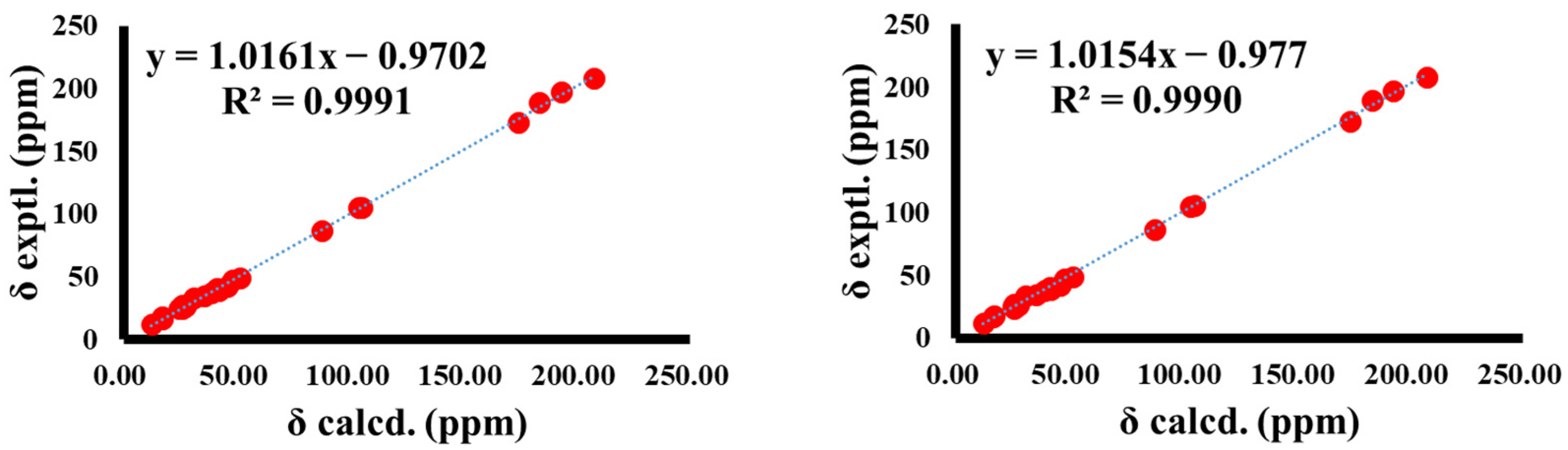
2.4. Quantum Chemical Calculation of 13C NMR

All structures were optimized at the b3lyp/6-31+g (d, p) level for all conformations using Gaussian09 [25]. The 13C NMR shielding constants were computed using the Gauge Independent Atomic Orbital (GIAO) method [26] at the mpw1pw91/6-311+g (2d, p) level, with SMD in chloroform using Gaussian09. The final, calculated 13C NMR chemical shifts were obtained by averaging the 13C NMR chemical shifts of the optimized conformers according to the Boltzmann distribution theory and their relative Gibbs free energy (∆G).

2.5. Quantum Chemical Calculation of ECD Spectra

Monte Carlo conformational searches were carried out by means of the Spartan’s 10 software, using the Merck Molecular Force Field (MMFF). The conformers with a Boltzmann population of over 2% were chosen for ECD calculations, and were then initially optimized at B3LYP/6-311g (2d, p) level in MeOH using the CPCM polarizable conductor calculation model. The theoretical calculation of ECD was conducted in MeOH using time-dependent density-functional theory (TDDFT) at the B3LYP/6-311g (2d, p) level, for all conformers. The rotatory strengths for 60 excited states were calculated. ECD spectra were generated using the SpecDis 1.7.1 program (University of Würzburg, Würzburg, Germany) and GraphPad Prism 5 (University of California San Diego, CA, USA) from dipole-length rotational strengths, and by applying Gaussian band shapes with sigma = 0.3 eV.

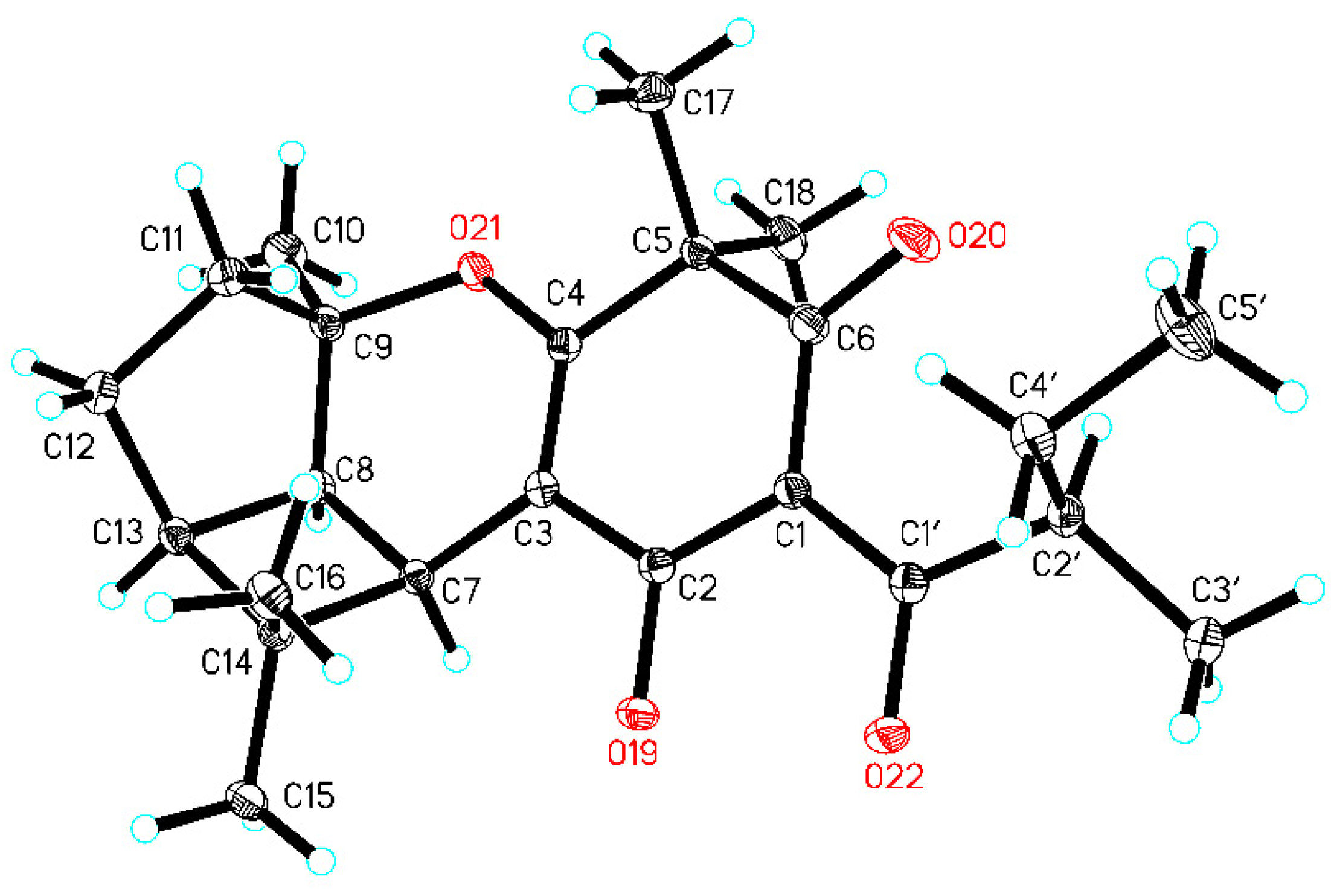
2.6. Single-Crystal X-ray Diffraction Analysis of 2b

Crystal data for 2b: C23H31O4 (M = 371.48 g/mol), orthorhombic, space group P212121 (no. 19), a = 9.25724(11) Å, b = 10.10414(12) Å, c = 21.6403(3) Å, V = 2024.16(4) Å3, Z = 4, T = 149.99(10) K, μ(Cu Kα) = 0.653 mm1, Dcalc = 1.219 g/cm3, 11,032 reflections measured (8.172° ≤ 2Θ ≤ 147.862°), 4009 unique (Rint = 0.0246, Rsigma = 0.0261), which were used in all calculations. The final R1 was 0.0332 (I > 2σ(I)), and wR2 was 0.0876 (all data). The goodness of fit for F2 was 1.053. Flack/Hooft parameter = 0.01(7)/0.02(7).

2.7. Cell Culture

The MCF-7 cell line was obtained from the National Collection of Authenticated cell cultures (Shanghai, China). The cells were cultured in Dulbecco’s Modified Eagle’s Medium (DMEM, BasalMedia, Shanghai, China), containing 10% FBS (Thermo Fisher Scientific, Waltham, MA, USA) at 37 °C, and 5% CO2.

2.8. MTT Assay

The cell viability was evaluated by the MTT assay. MCF-7 cells were seeded in a 96-well plate, at a density of 1 × 10⁴ cells/well. The cells were grown overnight at 37 °C with humidified 5% CO2, and then treated with compounds 1a3b (20 μM) for 48 h. The supernatants were discarded and 15 μL MTT reagent, as well as 60 μL DMEM, were added. After incubating for 4 h, the supernatants were removed and DMSO was added. The absorbances were measured at 490 nm by the Thermo Multiskan MK3 (Thermo Scientific, Waltham, MA, USA). The cell viability was analyzed as follows:
Growth Rate (%) = [(ODsample-ODblank)/(ODcontrol-ODblank)] × 100%

2.9. Western Blotting

Western blotting was conducted as described [27]. After exposure to compounds and CPT for 48 h, cells were lysed by the RIPA buffer with a protease inhibitor and a phosphatase inhibitor (MCE). Cell lysates were separated by sodium dodecyl sulfate polyacrylamide gel electrophoresis (SDS-PAGE) and transferred into the PVDF membrane. The membranes were blocked in no-fat milk with TBST and incubated with primary antibody at 4 °C overnight. Then, the membranes were washed 3 times in TBST and incubated with secondary antibody at room temperature. After being washed, the target proteins on the membranes were detected by chemi-luminescence reagent (Advansta, San Jose, CA, USA).

2.10. Cell Cycle Analysis

The MCF-7 cells were seeded in 6 cm dishes. After exposure to CPT with or without compound 1a for 48 h, the cells were harvested by trypsin with no EDTA, and fixed in cold 75% ethanol overnight. Following centrifuge (1000 rpm, 5 min room temperature) and removing the supernatant, the cells were incubated with 10 μg/mL RNase in PBS at 37 °C for 30 min, and with PI staining buffer at room temperature for 5 min in the dark. The cell cycle distribution was analyzed by flow cytometry (CytoFlex, Beckman Coulter, Los Angeles, CA, USA).

2.11. Statistical Analysis

The data were shown as mean ± SD, and the differences between samples were evaluated statistically with ANOVA analysis and Tukey’s posteriori comparison through GraphPad Prism 6.0. Differences were statistically significant at p < 0.05.

3. Results

3.1. Isolation of Compounds 13

The whole plants of H. elodeoides were extracted with methanol at room temperature. The crude extract was subjected to the silica gel column chromatography, eluting with CH2Cl2, EtOAc, and methanol. Compounds 13 were isolated from the CH2Cl2 fraction by a series of column chromatography. Then, three pairs of enantiomers, (±)-Hypersines A–C, were purified by chiral HPLC separation with CHIRALPAK® IG column.

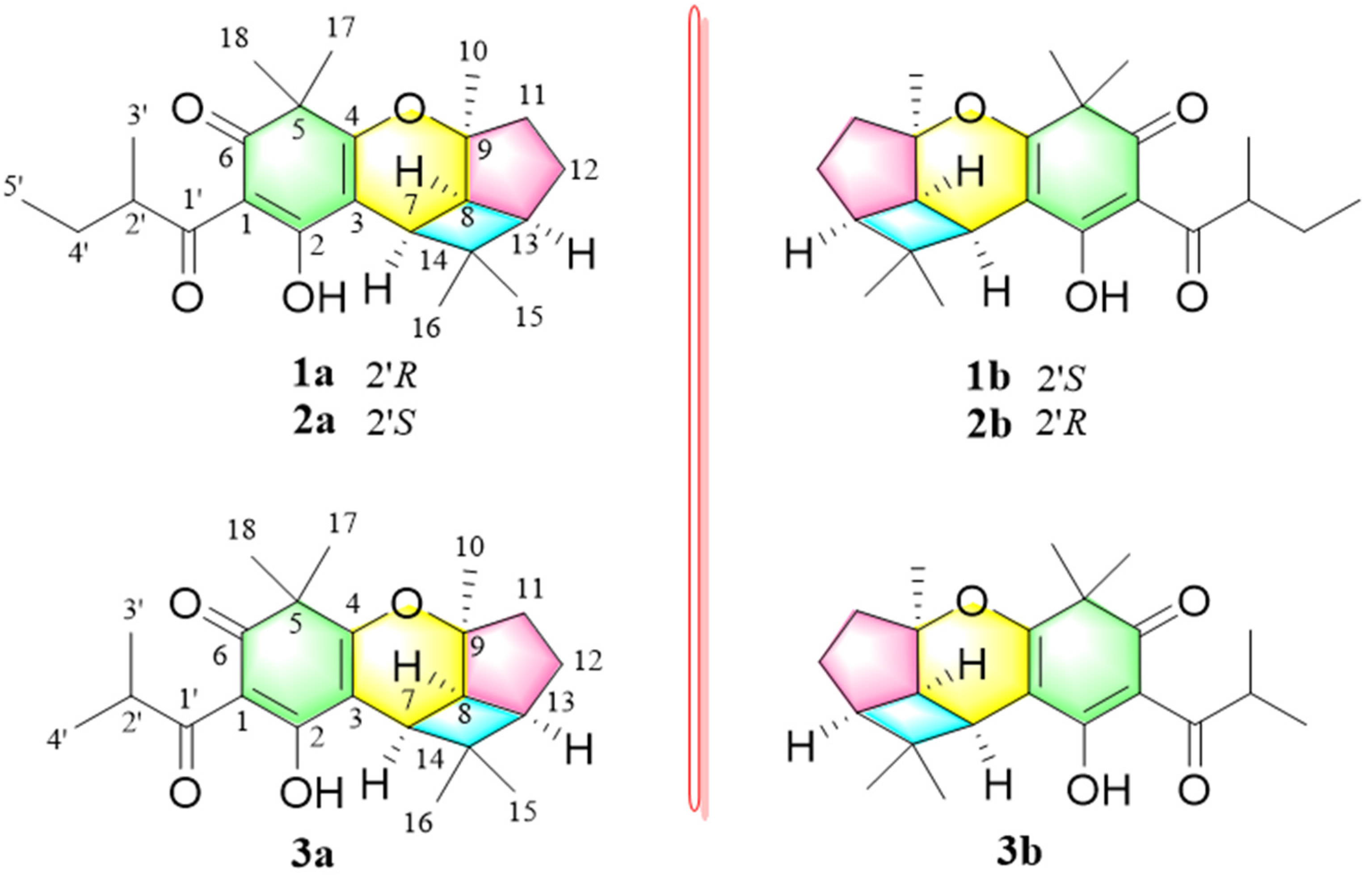
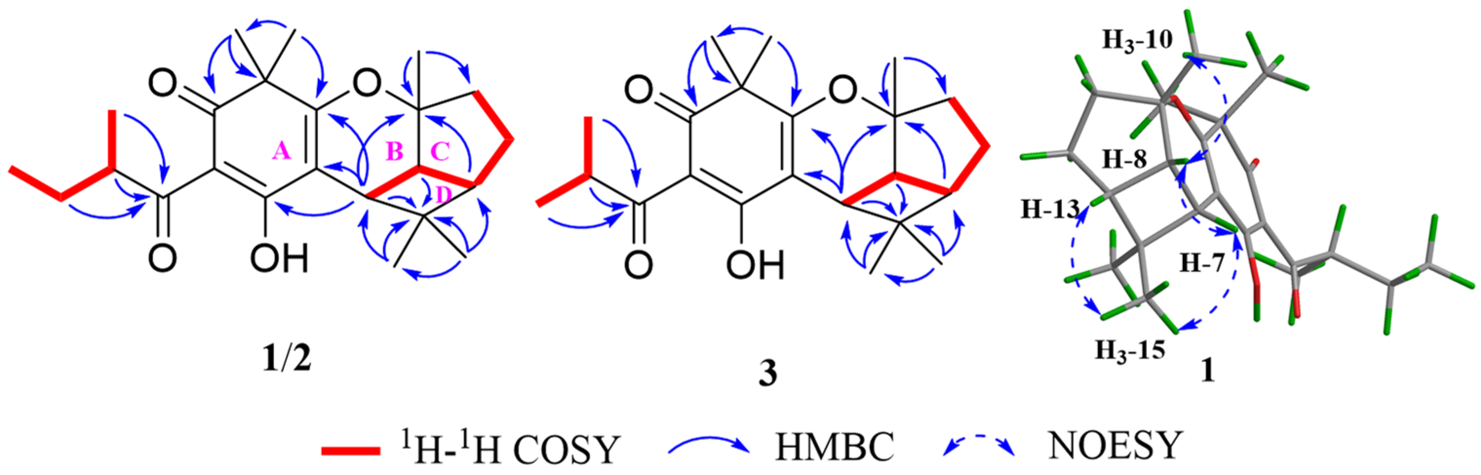
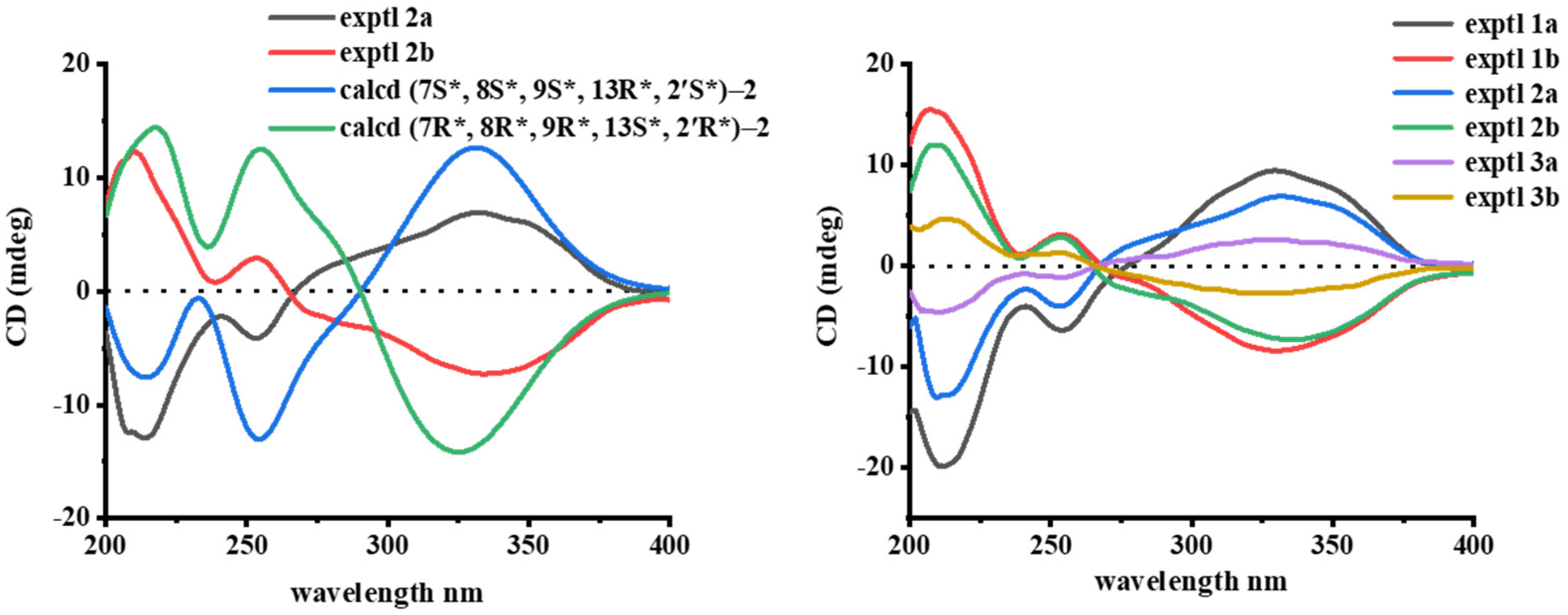
3.2. Structural Elucidation of Compounds 13

The structures, including absolute configurations of isolated new compounds (±)-hypersines A–C, were elucidated by comprehensive spectroscopic data, single-crystal X-ray diffraction, and quantum chemical calculations.
(±)-Hypersine A (1a/1b) and (±)-Hypersine B (2a/2b) were obtained as two pairs of C-2′ epimers. The mixtures of 1a/1b and 2a/2b performed a single chromatographic peak in the conventional RP-18 column, but could be separated into four peaks by chiral columns (Figure S2). The correspondence of the two pairs of enantiomers, 1a/1b and 2a/2b, were ensured by comparing their 1H NMR data (Figure S3). The molecular formulae of 1a/1b and 2a/2b were assigned as C23H32O4, on the basis of the HR-ESI-MS molecular ion peak at m/z 373.2375 [M + H]+ (calculated for C23H33O4+, 373.2379) (1a/1b) and m/z 373.2374 [M + H]+ (calculated for C23H33O4+, 373.2379) (2a/2b), indicating eight indices of hydrogen deficiency. The characteristic absorption bands of UV spectra (203, 226, 251, 276, and 332 nm) and IR spectra (3314 and 1657 cm−1) deduced the presence of an enolic 1,3-diketone moiety in 1 [28,29,30]. The 1H NMR and HSQC spectra of 1 showed the signals assignable to five singlet methyl groups (δH 1.40 (s), 1.36 (s), 1.34 (s), 1.32 (s) and 0.80 (s)), one doublet methyl (δH 1.14 (d, J = 7.2)), and one triplet methyl (δH 0.90 (t, J = 7.2)) The 13C NMR and DEPT-135 afforded twenty-three carbon resonances, including seven methyls, three methylenes, four methines, and nine quaternary carbons. The downfield signals at δC 207.9 (C-1’), 197.3 (C-6), 189.4 (C-2), and 105.0 (C-3) suggested the existence of an enol-β-triketone moiety [22,25,26]. The above data indicated that 1 was similar to (±)-Hyperjaponol A [30], with a filicinic acid core, and proposed to be a derivative of MTPAPs.
The planning structure of 1 was constructed by analyzing its 2D NMR data (Figure 2). The spin systems of H-7/H-8/H-13/H2-12/H2-11 and H3-2’/H-3’/H2-4’/H3-5’ in the 1H-1H COSY spectrum indicated the existence of two fragments: CH-7–CH-8–CH-13–CH2-12–CH2-11 and CH3-3’–CH-2’–CH-4’–CH3-5’. The HMBC correlations from CH3-17 and CH3-18 to C-4, C-5, and C-6, and from H-7 to C-3, C-4, and C-9 confirmed the presence of the filicinic acid core (ring A), and that the oxygen heterocycles (ring B) were fused to ring A. By referring to (±)-Hyperjaponol A, the 2-methylbutanoyl group was connected to the C-1 of ring A according to the HMBC correlations from H-2’, H-3’, and H-4’ to C-1’. In addition, the HBMC correlations from H-8 to C-11, from H-13 to C-9, and from CH3-10 to C-8, C-9, and C-11 revealed the existence of the five-membered ring C. Furthermore, the HMBC interactions from H-7 to C-14 and from CH3-15 and CH3-16 to C-7, C-13, and C-14 established the infrequent four-membered ring (ring D).
The relative configuration of 1 was illustrated by analyzing the NOESY interactions (Figure 2). The cross-peaks of H-7/H-8, H-7/CH3-15, CH3-15/H-13, and H-8/CH3-10 indicated that these protons were cofacial. However, the relative configuration of C-2’ could not be determined due to the absence of conclusive evidence. By meticulously comparing the 1D and 2D NMR data of 1 and 2, almost identical chemical shifts and correlation signals were observed, except for the tiny difference presented at C-2′ and its surrounding signals. Herein, 1 and 2 were deduced to have the same planning structure as well as the same relative configuration in the 6/6/5/4 core, and were proposed to be a pair of C-2′ epimers (Figure 1).
To further confirm the novel 6/6/5/4 tetracyclic architecture in 1 and 2, as well as try to determine the relative configuration of C-2′, the quantum chemical calculation of the 13C NMR data of two isomers, (7R*, 8R*, 9R*, 13S*, 2′S*)-1 and (7R*, 8R*, 9R*, 13S*, 2′R*)-2, were performed. The results showed that both (7R*, 8R*, 9R*, 13S*, 2′S*)-1 and (7R*, 8R*, 9R*, 13S*, 2′R*)-2 have a good correlation coefficient (R2 ≥ 0.9990) with the experimental 13C NMR data of 1 and 2, which solidified the deduced skeleton of 1 and 2 (Figure 3). Furthermore, the results confirmed that the change in configuration at C-2′ had a slight effect on the NMR data. Thus, the configuration of C-2’ in 2-methylbutanoyl fragment was difficult to assign by NMR calculations.
The application of single-crystal X-ray crystallography technique is an unambiguous method to determine the configuration of C-2’ in 2-methylbutanoyl fragment. Fortunately, a high-quality single crystal of 2b was successfully obtained in a mixed solvent (acetone–water, 9:1) for X-ray diffraction. The results afforded fatal evidence for the absolute configuration of 2b at C-2′, with a Flack parameter of 0.01(7) (CCDC 2080724) (Figure 4). Therefore, the absolute stereochemistry of 2b was explicitly assigned as 7R, 8R, 9R, 13S, 2′R. The absolute configuration of 2a could be accordingly assigned as 7S, 8S, 9S, 13R, 2′S, since 2a/2b were a pair of enantiomers. Moreover, the quantum chemical ECD calculation method was used to further confirm the absolute configuration of 2a and 2b. The theoretically calculated ECD data of (7S*, 8S*, 9S*, 13R*, 2′S*)-2 and (7R*, 8R*, 9R*, 13S*, 2′R*)-2 were in good agreement with the experimental ECD data of 2a and 2b, which supported the assignment of the absolute configurations of 2a and 2b as 7S, 8S, 9S, 13R, 2′S and 7R, 8R, 9R, 13S, 2′R, respectively (Figure 5). Accordingly, the absolute configurations of 1a/1b were respectively assigned as 7S, 8S, 9S, 13R, 2′R and 7R, 8R, 9R, 13S, 2′S by comparing their experimental ECD curves and NMR data with the C-2′ epimers 2a/2b (Figure 5).
(±)-Hypersines C (3a/3b) were also obtained as a pair of enantiomers with the molecular formula of C22H30O4, based on the HR-ESI-MS data (m/z 359.2212, [M + H]+) (calculated for C22H31O4+, 359.2217). Extensive analysis of the 1H NMR and 13C NMR data of 3 and 2 suggested that 3 was the homologue of 2, except for the absence of a methylene group. The 1H-1H COSY correlations of H-2’/H-3’/H-4’, together with the HMBC correlations from H-2’ to C-1’, from H-3’ to C-1’, and from H-4’ to C-1’, indicated the presence of an isobutyryl in 3 instead of a 2-methylbutanoyl group in 2. The relative configuration of 3 was deduced by analyzing of the NOESY spectrum. The NOESY interactions of H-7/H-8, H-7/CH3-15, CH3-15/H-13, and H-8/CH3-10 suggested that the relative configuration of 3 was identical to that of 2. The enantiomers 3a and 3b were successfully separated by chiral column (Figure S2). The overall pattern of the experimental ECD curve of 3a and 3b, correspondingly, have the same cotton effects with 2a and 2b, suggesting that they have the same absolute configurations. Meanwhile, the ECD data of 3b were consistent with 2b. Thus, the absolute configurations of 3a and 3b were assigned as 7S, 8S, 9S, 13R and 7R, 8R, 9R, 13S, respectively (Figure 5).

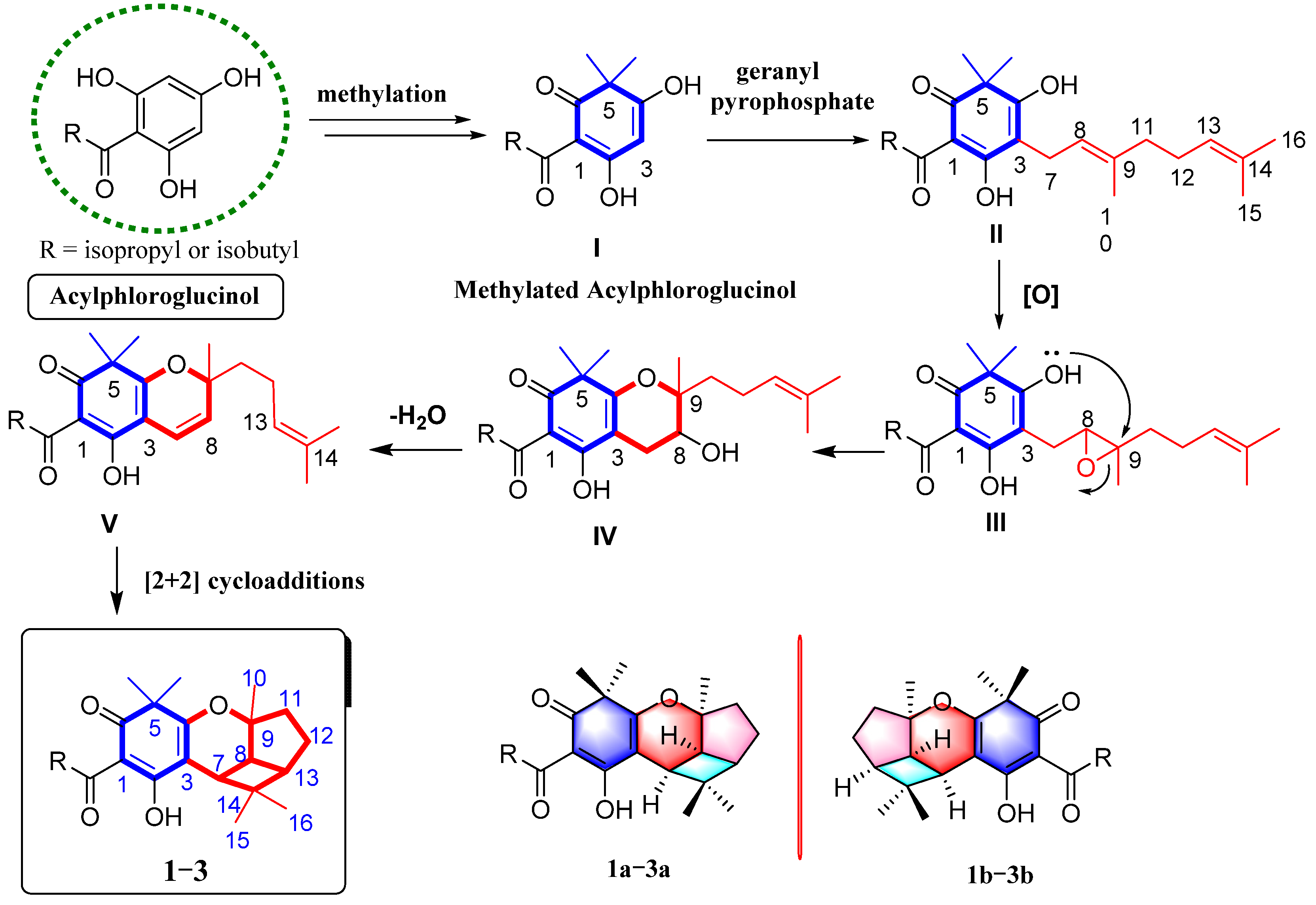
3.3. Plausible Biosynthetic Pathway to 13

(±)-Hypersines A–C represent the first example of MTPAPs with a rare 6/6/5/4 ring system, which is quite different from the known MTPAPs. The plausible biosynthetic pathway for 13 was proposed (Scheme 1). Acylphloroglucinol was considered as the biogenetic precursor to PPAPs, which had undergone methylation reaction to obtain intermediate I with a filicinic acid core. Subsequently, intermediate II could be formed by the enzyme-catalyzed addition of geranyl pyrophosphate to the filicinic core. Then, epoxidation occurred at Δ [8] to generate an epoxypropane intermediate III. Next, the six-membered oxygen ring was formed and generated intermediate IV. After the dehydration of OH-8, the unsaturated pyranoid ring was generated. Finally, the 6/6/5/4 tetracyclic skeleton was connected by [2 + 2] cycloadditions [9,19].

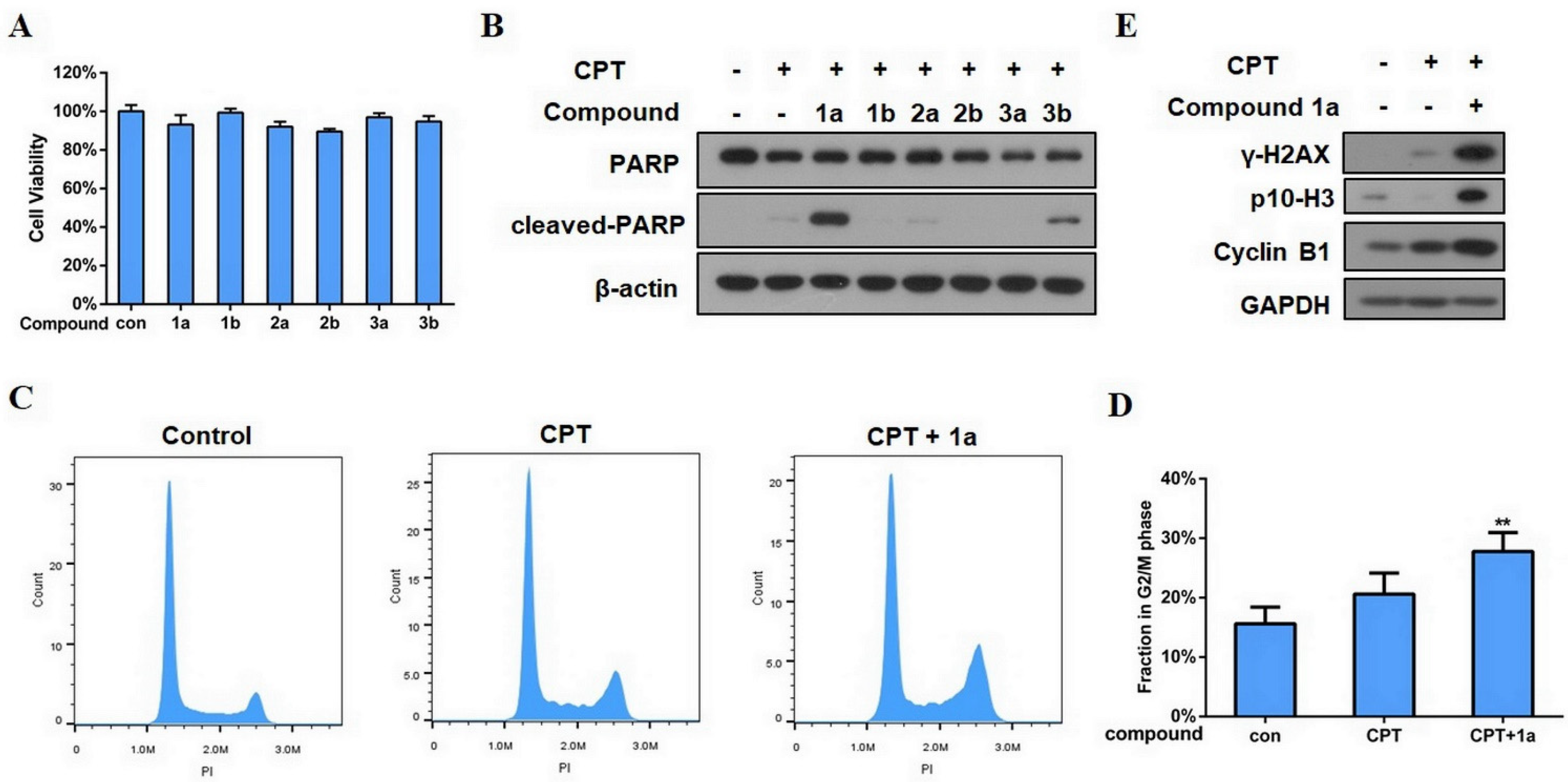
3.4. Biological Activity Evaluation of the Isolated Compounds

The biological activities of the isolated compounds, (±)-Hypersines A–C (1a3b), were evaluated. In our previous studies, several PPAPs were found to show an inhibitory effects on the proliferation of human breast cancer cells (MCF-7) [6,21]. In this study, the MTT assay was used to measure the viability of MCF-7 cells after exposure to the isolated compounds. The results showed that none of the compounds could suppress the proliferation of MCF-7 cells in 20 μM (Figure 6A). Poly ADP-ribose polymerase (PARP) is a marker protein of apoptosis. Interestingly, when the compounds were co-treated with the anticancer agent, camptothecin (CPT), in a low concentration, 1a (20 μM) could visibly trigger the cleavage of PARP combined with CPT (50 nM), while there was no PARP cleavage when treated with CPT (50 nM) alone (Figure 6B). As known, CPT causes cell death by suppressing DNA synthesis and eliciting DNA double-strand break (DSB) [31]. The above result indicated that 1a could reinforce the CPT-induced apoptosis of MCF-7 cells. Further research exhibited that the percentage of MCF-7 cells in the G2/M phase increased after the co-treatment of CPT and 1a (Figure 6C or Figure 6D). Meanwhile, the combination of CPT and 1a obviously augmented the expression of γ-H2AX (the biomarker of DSB) (Figure 6E) [32]. Furthermore, the CPT restrained the phosphorylation of the Ser 10 of histone H3 (pS10-H3, the mitotic biomarker) [33]. This may be owing to CPT-induced DSB causing DNA damage response (DDR), which means that cells with damaged DNA cannot prematurely undergo mitosis [34]. However, synergy between CPT and 1a could strongly elevate the level of pS10-H3, indicating that the majority of cells might enter M phase, which is demonstrated by the increasing level of Cyclin B1 (Figure 6E) [33]. Furthermore, the flow cytometry analysis indicated that the cells were arrested at G2/M phase with the co-treatment of CPT and 1a (Figure 6C or Figure 6D). The above data indicated that 1a might urge MCF-7 cells with CPT-induced DSB to progress into mitosis by restricting DDR and by rendering damaged DNA to remain in M phase. Compromised DNA entering into mitosis can lead to the apoptosis of cells [35]. This may be the reason why 1a potentiated the CPT-induced apoptosis of MCF-7 cells, but specific mechanisms remained to be investigated. DDR inhibitors are usually used as sensitizers of antitumor agents to play a role in tumor treatment [36]. Therefore, 1a might be considered as a novel inhibitor of DDR, and could enhance the sensitivity of MCF-7 cells to CPT.

4. Conclusions

In summary, (±)-hypersines A–C (13), the three pairs of unprecedented MPAPs with a rare 6/6/5/4 tetracyclic scaffold, were obtained from H. elodeoides. As far as we know, hypersines A–C are the first reported MTPAPs characterized by a peculiar 6/6/5/4 tetracyclic core, formed by a rare, tensional four-membered carbocycle concurrently fused with five- and six-membered rings. Furthermore, the possible biosynthetic pathway of 13 was proposed, which might provide a reference for the organic synthesis of these compounds. The biological activity results showed that the combination of 1a and a low concentration of CPT could visibly trigger the cleavage of PARP, and could up-regulate the expression level of γ-H2AX, pS10-H3, and Cyclin B1, indicating that 1a might enhance MCF-7 cell sensitivity to camptothecin by inhibiting the DNA damage response. This is the first report of MTPAPs as potential sensitizers of anti-tumor drugs. In general, this study enriches the structural diversity and biological activity of MTPAPs, and provides novel insight for further understanding MTPAPs. However, among these compounds, only 1a exhibited potent activity. In future research, more analogues need to be obtained to further investigate the structure–activity relationship. A more detailed mechanism that 1a enhances MCF-7 cell sensitivity to CPT needs to be further explored. This research suggests that the co-administration of 1a and chemotherapeutic drugs could be an effective strategy for overcoming tumor MDR, and this deserves further exploration.

Supplementary Materials

The following are available online at https://www.mdpi.com/article/10.3390/biomedicines9101473/s1, Figure S1: MTPAPs derivatives reported previously.

Author Contributions

Conceptualization, G.-H.W. and T.L.; methodology, X.-Z.L. and M.Z.; formal analysis, X.-Z.L. and M.Z.; investigation, C.-C.D., H.-H.Z., X.L. and S.-L.H.; resources, W.-J.T. and H.-F.C.; data curation, C.-C.D., H.-H.Z., X.L. and S.-L.H.; writing—original draft preparation, X.-Z.L. and M.Z.; writing—review and editing, X.-Z.L. and M.Z.; visualization, W.-J.T.; supervision, W.-J.T. and H.-F.C.; project administration, W.-J.T. and H.-F.C.; funding acquisition, W.-J.T. and H.-F.C. All authors have read and agreed to the published version of the manuscript.

Funding

This study was supported by grants from the National Natural Science Foundation of China (NSFC) (No.81602988), the Fundamental Research Funds for the Central Universities (No.20720190079), and the Natural Science Foundation of Fujian Province (No.2019J007).

Institutional Review Board Statement

Not applicable.

Informed Consent Statement

Not applicable.

Conflicts of Interest

The authors declare no conflict of interest.

References

  1. Tian, W.J.; Qiu, Y.Q.; Yao, X.J.; Chen, H.F.; Dai, Y.; Zhang, X.K.; Yao, X.S. Dioxasampsones A and B, two polycyclic polyprenylated acylphloroglucinols with unusual epoxy-ring-fused skeleton from Hypericum sampsonii. Org. Lett. 2014, 16, 6346–6349. [Google Scholar] [CrossRef] [PubMed]
  2. Yang, X.W.; Grossman, R.B.; Xu, G. Research progress of polycyclic polyprenylated acylphloroglucinols. Chem. Rev. 2018, 118, 3508–3558. [Google Scholar] [CrossRef] [PubMed]
  3. Lou, H.Y.; Li, Y.N.; Yi, P.; Jian, J.Y.; Hu, Z.X.; Gu, W.; Huang, L.J.; Li, Y.M.; Yuan, C.M.; Hao, X.J. Hyperfols A and B: Two highly modified polycyclic polyprenylated acylphloroglucinols from hypericum perforatum. Org. Lett. 2020, 22, 6903–6906. [Google Scholar] [CrossRef] [PubMed]
  4. Duan, Y.; Xie, S.; Bu, P.; Guo, Y.; Shi, Z.; Guo, Y.; Cao, Y.; Sun, W.; Qi, C.; Zhang, Y. Hypaluton A, an immunosuppressive 3,4-nor-polycyclic polyprenylated acylphloroglucinol from hypericum patulum. J. Org. Chem. 2021, 86, 6478–6485. [Google Scholar] [CrossRef]
  5. Li, Y.R.; Xu, W.J.; Wei, S.S.; Lu, W.J.; Luo, J.; Kong, L.Y. Hyperbeanols F-Q, diverse monoterpenoid polyprenylated acylphloroglucinols from the flowers of hypericum beanie. Phytochemistry 2019, 159, 56–64. [Google Scholar] [CrossRef]
  6. Qiu, D.; Zhou, M.; Chen, J.; Wang, G.; Lin, T.; Huang, Y.; Yu, F.; Ding, R.; Sun, C.; Tian, W.; et al. Hyperelodiones A-C, monoterpenoid polyprenylated acylphoroglucinols from hypericum elodeoides, induce cancer cells apoptosis by targeting RXRα. Phytochemistry 2020, 170, 112216. [Google Scholar] [CrossRef]
  7. Zhang, Z.; Elsohly, H.N.; Jacob, M.R.; Pasco, D.S.; Walker, L.A.; Clark, A.M. Natural products inhibiting candida albicans secreted aspartic proteases from tovomita krukovii. Planta Medica 2002, 68, 49–54. [Google Scholar] [CrossRef]
  8. Crockett, S.L.; Wenzig, E.M.; Kunert, O.; Bauer, R. Anti-inflammatory phloroglucinol derivatives from hypericum empetrifolium. Phytochem Lett. 2008, 1, 37–43. [Google Scholar] [CrossRef] [Green Version]
  9. Hu, L.; Xue, Y.; Zhang, J.; Zhu, H.; Chen, C.; Li, X.N.; Liu, J.; Wang, Z.; Zhang, Y.; Zhang, Y. (+/-)-Japonicols A-D, acylphloroglucinol-based meroterpenoid enantiomers with anti-KSHV activities from hypericum japonicum. J. Nat. Prod. 2016, 79, 1322–1328. [Google Scholar] [CrossRef]
  10. Ao, Z.; Liu, Y.Y.; Lin, Y.L.; Chen, X.l.; Chen, K.; Kong, L.Y.; Luo, J.G. Hyperpatulones A and B, two new peroxide polyprenylated acylphloroglucinols from the leaves of hypericum patulum. Tetrahedron Lett. 2020, 61, 151385. [Google Scholar] [CrossRef]
  11. Zhou, X.; Xu, W.; Li, Y.; Zhang, M.; Tang, P.; Lu, W.; Li, Q.; Zhang, H.; Luo, J.; Kong, L. Anti-inflammatory, antioxidant, and anti-nonalcoholic steatohepatitis acylphloroglucinol meroterpenoids from hypericum bellum flowers. J. Agric. Food Chem. 2021, 69, 646–654. [Google Scholar] [CrossRef]
  12. Mamemura, T.; Tanaka, N.; Shibazaki, A.; Gonoi, T.; Kobayashi, J.I. Yojironins A–D, meroterpenoids and prenylated acylphloroglucinols from Hypericum yojiroanum. Tetrahedron Lett. 2011, 52, 3575–3578. [Google Scholar] [CrossRef]
  13. Schmidt, S.; Jurgenliemk, G.; Schmidt, T.J.; Skaltsa, H.; Heilmann, J. Bi-, tri-, and polycyclic acylphloroglucinols from hypericum empetrifolium. J. Nat. Prod. 2012, 75, 1697–1705. [Google Scholar] [CrossRef] [PubMed]
  14. Fobofou, S.A.; Franke, K.; Sanna, G.; Porzel, A.; Bullita, E.; la Colla, P.; Wessjohann, L.A. Isolation and anticancer, anthelminthic, and antiviral (HIV) activity of acylphloroglucinols, and regioselective synthesis of empetrifranzinans from hypericum roeperianum. Bioorg. Med. Chem. 2015, 23, 6327–6334. [Google Scholar] [CrossRef]
  15. Zhu, H.; Chen, C.; Liu, J.; Sun, B.; Wei, G.; Li, Y.; Zhang, J.; Yao, G.; Luo, Z.; Xue, Y.; et al. Hyperascyrones A-H, polyprenylated spirocyclic acylphloroglucinol derivatives from hypericum ascyron linn. Phytochemistry 2015, 115, 222–230. [Google Scholar] [CrossRef]
  16. Liu, Y.Y.; Ao, Z.; Xue, G.M.; Wang, X.B.; Luo, J.G.; Kong, L.Y. Hypatulone A, a Homoadamantane-type acylphloroglucinol with an intricately caged core from hypericum patulum. Org. Lett. 2018, 20, 7953–7956. [Google Scholar] [CrossRef] [PubMed]
  17. Wu, Z.-N.; Niu, Q.-W.; Zhang, Y.-B.; Luo, D.; Li, Q.-G.; Li, Y.-Y.; Kuang, G.-K.; He, L.-J.; Wang, G.-C.; Li, Y.-L. Hyperpatulones A–F, polycyclic polyprenylated acylphloroglucinols from hypericum patulum and their cytotoxic activities. RSC Adv. 2019, 9, 7961–7966. [Google Scholar] [CrossRef] [Green Version]
  18. Qiu, D.; Zhou, M.; Lin, T.; Chen, J.; Wang, G.; Huang, Y.; Jiang, X.; Tian, W.; Chen, H. Cytotoxic components from hypericum elodeoides targeting RXRα and inducing HeLa cell apoptosis through caspase-8 activation and PARP cleavage. J. Nat. Prod. 2019, 82, 1072–1080. [Google Scholar] [CrossRef]
  19. Li, Q.J.; Tang, P.F.; Zhou, X.; Lu, W.J.; Xu, W.J.; Luo, J.; Kong, L.Y. Dimethylated acylphloroglucinol meroterpenoids with anti-oral-bacterial and anti-inflammatory activities from hypericum elodeoides. Bioorg. Chem. 2020, 104, 104275. [Google Scholar] [CrossRef]
  20. Li, Q.J.; Tang, P.F.; Zhou, X.; Lu, W.J.; Xu, W.J.; Luo, J.; Kong, L.Y. Elodeoidins A–H, acylphloroglucinol meroterpenoids possessing diverse rearranged skeletons from hypericum elodeoides. Org. Chem. Front. 2021, 8, 1409–1414. [Google Scholar] [CrossRef]
  21. Qiu, D.R.; Zhou, M.; Liu, X.Z.; Chen, J.J.; Wang, G.H.; Lin, T.; Yu, F.R.; Ding, R.; Sun, C.L.; Tian, W.J.; et al. Cytotoxic polyprenylated phloroglucinol derivatives from hypericum elodeoides choisy modulating the transactivation of RXRα. Bioorganic Chem. 2021, 107, 104578. [Google Scholar] [CrossRef]
  22. Hashida, C.; Tanaka, N.; Kawazoe, K.; Murakami, K.; Sun, H.D.; Takaishi, Y.; Kashiwada, Y. Hypelodins A and B, polyprenylated benzophenones from hypericum elodeoides. J. Nat. Med. 2014, 68, 737–742. [Google Scholar] [CrossRef]
  23. Li, Y.P.; Yang, X.W.; Xia, F.; Yan, H.; Ma, W.G.; Xu, G. Hyperjapones F–I, terpenoid polymethylated acylphloroglucinols from hypericum japonicum. Tetrahedron Lett. 2016, 57, 5868–5871. [Google Scholar] [CrossRef]
  24. Yang, X.W.; Li, Y.P.; Su, J.; Ma, W.G.; Xu, G. Hyperjapones A-E, terpenoid polymethylated acylphloroglucinols from hypericum japonicum. Org. Lett. 2016, 18, 1876–1879. [Google Scholar] [CrossRef] [PubMed]
  25. Frisch, M.J.; Trucks, G.W.; Schlegel, H.B.; Scuseria, G.E.; Robb, M.A.; Cheeseman, J.R.; Scalmani, G.; Barone, V.; Mennucci, B.; Petersson, G.A.; et al. Gaussian 09, Rev. C 01, Gaussian, Inc.: Wallingford, CT, USA, 2009.
  26. Wolinski, K.; Hinton, J.F.; Pulay, P. 17O NMR chemical shifts of polyoxides in gas phase and in solution. J. Am. Chem. Soc. 1990, 112, 8251–8260. [Google Scholar] [CrossRef]
  27. Li, F.; Kozono, D.; Deraska, P.; Branigan, T.; Dunn, C.; Zheng, X.F.; Parmar, K.; Nguyen, H.; DeCaprio, J.; Shapiro, G.I. CHK1 inhibitor blocks phosphorylation of FAM122A and promotes replication stress. Mol. Cell 2020, 80, 410–422.e6. [Google Scholar] [CrossRef] [PubMed]
  28. Lshiguro, K.; Yamak, M.; Kashihara, M.; Takagi, S.; Isoi, K. Sarothralin G: A new antimicrobial compound from hypericum japonicum. Planta Med. 1990, 56, 274–276. [Google Scholar] [CrossRef]
  29. Ishiguro, K.; Nagat, S.; Fukumoto, H.; Yamaki, M.; Isoi, K. Phloroglucinol derivatives from hypericum japonicum. Phytochemistry 1994, 35, 469–471. [Google Scholar] [CrossRef]
  30. Hu, L.; Zhang, Y.; Zhu, H.; Liu, J.; Li, H.; Li, X.N.; Sun, W.; Zeng, J.; Xue, Y.; Zhang, Y. Filicinic acid based meroterpenoids with anti-epstein-barr virus activities from hypericum japonicum. Org. Lett. 2016, 18, 2272–2275. [Google Scholar] [CrossRef]
  31. Hsiang, Y.H.; Hertzberg, R.; Hecht, S.; Liu, L.F. Camptothecin induces protein-linked DNA breaks via mammalian DNA topoisomerase I. J. Biol. Chem. 1985, 260, 14873–14878. [Google Scholar] [CrossRef]
  32. Chopra, S.S.; Jenney, A.; Palmer, A.; Niepel, M.; Chung, M.; Mills, C.; Sivakumaren, S.C.; Liu, Q.; Chen, J.Y.; Yapp, C.; et al. Torin2 exploits replication and checkpoint vulnerabilities to cause death of pi3k-activated triple-negative breast cancer cells. Cell Syst. 2020, 10, 66–81.e11. [Google Scholar] [CrossRef] [PubMed] [Green Version]
  33. Xie, G.; Zhou, Y.; Tu, X.; Ye, X.; Xu, L.; Xiao, Z.; Wang, Q.; Wang, X.; Du, M.; Chen, Z.; et al. Centrosomal localization of rxralpha promotes plk1 activation and mitotic progression and constitutes a tumor vulnerability. Dev. Cell 2020, 55, 707–722.e9. [Google Scholar] [CrossRef] [PubMed]
  34. Lanz, M.C.; Dibitetto, D.; Smolka, M.B. DNA damage kinase signaling: Checkpoint and repair at 30 years. EMBO J. 2019, 38, e101801. [Google Scholar] [CrossRef] [PubMed]
  35. Kim, H.; Xu, H.; George, E.; Hallberg, D.; Kumar, S.; Jagannathan, V.; Medvedev, S.; Kinose, Y.; Devins, K.; Verma, P.; et al. Combining PARP with ATR inhibition overcomes PARP inhibitor and platinum resistance in ovarian cancer models. Nat. Commun. 2020, 11, 3726. [Google Scholar] [CrossRef] [PubMed]
  36. Carrassa, L.; Damia, G. DNA damage response inhibitors: Mechanisms and potential applications in cancer therapy. Cancer Treat Rev. 2017, 60, 139–151. [Google Scholar] [CrossRef]
Figure 1. Structures of compounds 13.
Biomedicines 09 01473 g001
Figure 2. Key 2D NMR correlations of 13.
Biomedicines 09 01473 g002
Figure 3. Regression analysis of the experimental (exptl.) versus calculated (calcd.) 13C NMR chemical shifts of 1 (left) and 2 (right).
Biomedicines 09 01473 g003
Figure 4. X-ray crystallographic structure of 2b.
Biomedicines 09 01473 g004
Figure 5. The experimental (exptl) and calculated (calcd) ECD spectra of 1a/b3a/b.
Biomedicines 09 01473 g005
Scheme 1. Plausible Biosynthetic Pathway to 13.
Biomedicines 09 01473 sch001
Figure 6. (A) MCF-7 cells were exposed to compounds (1a3b) for 48 h. The cells viability was measured via MTT assay. The group for con means MCF-7 cells only treated with DMSO. (B) MCF-7 cells were incubated with 50 nM CPT, with or without 20 μM compounds, for 48 h, western blotting was used to analyze the level of cleaved-PARP and PARP. (C) After treatment with CPT plus 1a for 48 h, the cell cycle of MCF-7 cells was determined through flow cytometry. (D) The percentages of G2/M phase. The group for con means MCF-7 cells only exposed to DMSO. ** p < 0.01vs Con group. (E) MCF-7 cells were treated with CPT and 1a in combination for 48 h, the levels of relative protein (p10-H3, cyclin B1, γ-H2AX) were investigated by western blotting.
Biomedicines 09 01473 g006
Table 1. 1H NMR (600 MHz, in CDCl3) and 13C NMR (150 MHz, in CDCl3) data for 13.
No.1a/1b2a/2b3a/3b
δH (mult, J, Hz)δCδH (mult, J, Hz)δCδH (mult, J, Hz)δC
1 105.6 105.5 104.9
2 189.4 189.3 189.3
3 105.0 105.0 104.8
4 172.7 172.7 172.8
5 48.8 48.8 48.7
6 197.3 197.3 197.2
72.90 d (9.6)34.82.90 d (9.6)34.82.90 d (9.6)34.8
82.55 dd (9.6, 7.2)37.62.55 t (7.8)37.62.55 t (7.2)37.5
9 86.7 86.7 86.7
101.40 s27.41.40 s27.41.40 s27.4
11α 1.77 m40.4α 1.76 m40.4α 1.76 m40.3
β 1.88 m β 1.88 m β 1.88 m
12α 1.61 m26.2α 1.61 m26.2α 1.61 m26.1
β 1.70 m β 1.70 m β 1.68 m
132.42 t (7.8)47.12.42 t (7.2)47.12.43 t (7.8)47.0
14 38.8 38.8 38.8
151.34 s33.51.35 s33.51.34 s33.5
160.80 s18.10.80 s18.00.80 s18.0
171.32 s24.51.32 s24.11.32 s24.3
181.36 s25.21.36 s25.61.36 s25.5
1’ 207.9 208.0 208.6
2’3.84 m42.13.83 m42.23.97 m35.9
3’1.14 d (7.2)16.71.12 d (7.2)16.71.13 d (7.2)19.1
4’α 1.40 overlap26.9α 1.41 overlap26.81.15 d (7.2)19.2
β 1.73 m β 1.74 m
5’0.90 t (7.2)11.90.93 t (7.2)12.0
Publisher’s Note: MDPI stays neutral with regard to jurisdictional claims in published maps and institutional affiliations.

