1. Introduction
Thrombotic cardiovascular diseases, including myocardial infarction, ischemic stroke, coronary artery disease, and pulmonary embolism, remain among the leading causes of morbidity and mortality worldwide [
1]. The formation of pathological thrombi can obstruct blood flow, induce local ischemia and oxidative injury, and eventually lead to severe organ dysfunction [
2,
3]. During thrombogenesis, vascular endothelial injury exposes subendothelial matrix components and promotes platelet adhesion, activation, and aggregation. Activated platelets subsequently release multiple pro-thrombotic mediators, recruit additional platelets, and cooperate with fibrin networks to stabilize thrombotic structures [
4].
Current pharmacological strategies for thrombotic diseases include anticoagulants, antiplatelet agents, and thrombolytic drugs [
5], while their applications are restricted by poor thrombus specificity; moreover, the ability of conventional antithrombotic agents to modulate the pathological thrombus microenvironment is relatively poor [
5,
6,
7]. These challenges highlight the need for targeted therapeutic systems capable of localizing at thrombotic sites and improving the local pathological microenvironment.
The thrombus microenvironment is characterized by abundant fibrin, activated platelets, and elevated levels of reactive oxygen species (ROS) [
3,
8,
9]. H
2O
2 is a stable ROS in the vascular system, which plays an important role in platelet activation, platelet–endothelial interactions, and inflammatory responses. Excessive H
2O
2 can aggravate endothelial injury, amplify platelet activation, and promote the secretion of inflammatory cytokines, thereby contributing to the progression of thrombotic vascular diseases [
10,
11,
12]. Recent megakaryocyte transcriptomic analysis further demonstrated age- and sex-specific changes in programs related to platelet activation, inflammation, mitochondrial function, and oxidative stress, underscoring the biological heterogeneity connecting redox regulation with thrombotic potential [
13]. Therefore, scavenging H
2O
2 and attenuating oxidative stress may be an effective strategy for suppressing platelet activation and alleviating thrombosis-associated oxidative and inflammatory injury.
Nanomedicine-based drug delivery systems, including polymeric micelles, liposomes, and other nanoscale carriers, can improve drug solubility, prolong systemic circulation, and reduce off-target toxicity [
14,
15]. Moreover, the surface functionalization of nanocarriers with thrombus-targeting ligands enables selective accumulation at thrombotic sites [
15]. Among various thrombus-associated targets, fibrin is particularly attractive owing to its abundance, structural importance within thrombi, and extensive distribution throughout the clot network [
16,
17]. Peptide ligands are well-suited for nanocarrier functionalization due to their small molecular size, low immunogenicity, ease of synthesis, and flexible chemical modifiability [
18,
19]. Nevertheless, the practical application of many thrombus-targeting peptides remains challenged by suboptimal proteolytic stability, binding affinity, or in vivo targeting performance [
20,
21]. In this study, hexapeptides, due to their practical balance of structural simplicity, synthetic feasibility, and sufficient side-chain diversity for fibrin-pocket recognition, were selected as an initial focus for CADD-guided screening [
22,
23]. All-trans retinoic acid (atRA), a biologically active metabolite of vitamin A, has been reported to exhibit anti-inflammatory, antioxidant, antiplatelet, fibrinolytic, and anti-atherosclerotic functions, indicating its potential value in the treatment of thrombotic diseases [
24,
25]. However, the clinical translation of atRA for cardiovascular applications is restricted by its poor aqueous solubility, limited stability, and unsatisfactory bioavailability [
26]. Prodrug and nanocarrier strategies may mitigate these limitations by improving its physicochemical properties and enabling controlled release at pathological sites. Boronate ester linkages, which can be selectively oxidized by H
2O
2 in ROS-rich environments, are widely employed as H
2O
2-responsive chemical motifs [
27,
28]. Incorporating a boronate ester structure into an atRA-based prodrug may enable H
2O
2-triggered drug release while simultaneously consuming excessive H
2O
2. Furthermore, polymeric micelles were selected due to their amphiphilic core–shell structure, which is suitable for enhancing aqueous solubilization of poorly water-soluble atRA and its hydrophobic prodrug BORA [
29,
30]. More broadly, the recent nanotechnology literature has emphasized that surface engineering and stimulus-responsive designs can improve drug localization and controlled release, while also highlighting the need for rigorous evaluation of biodistribution, efficacy, and safety before clinical translation [
31].
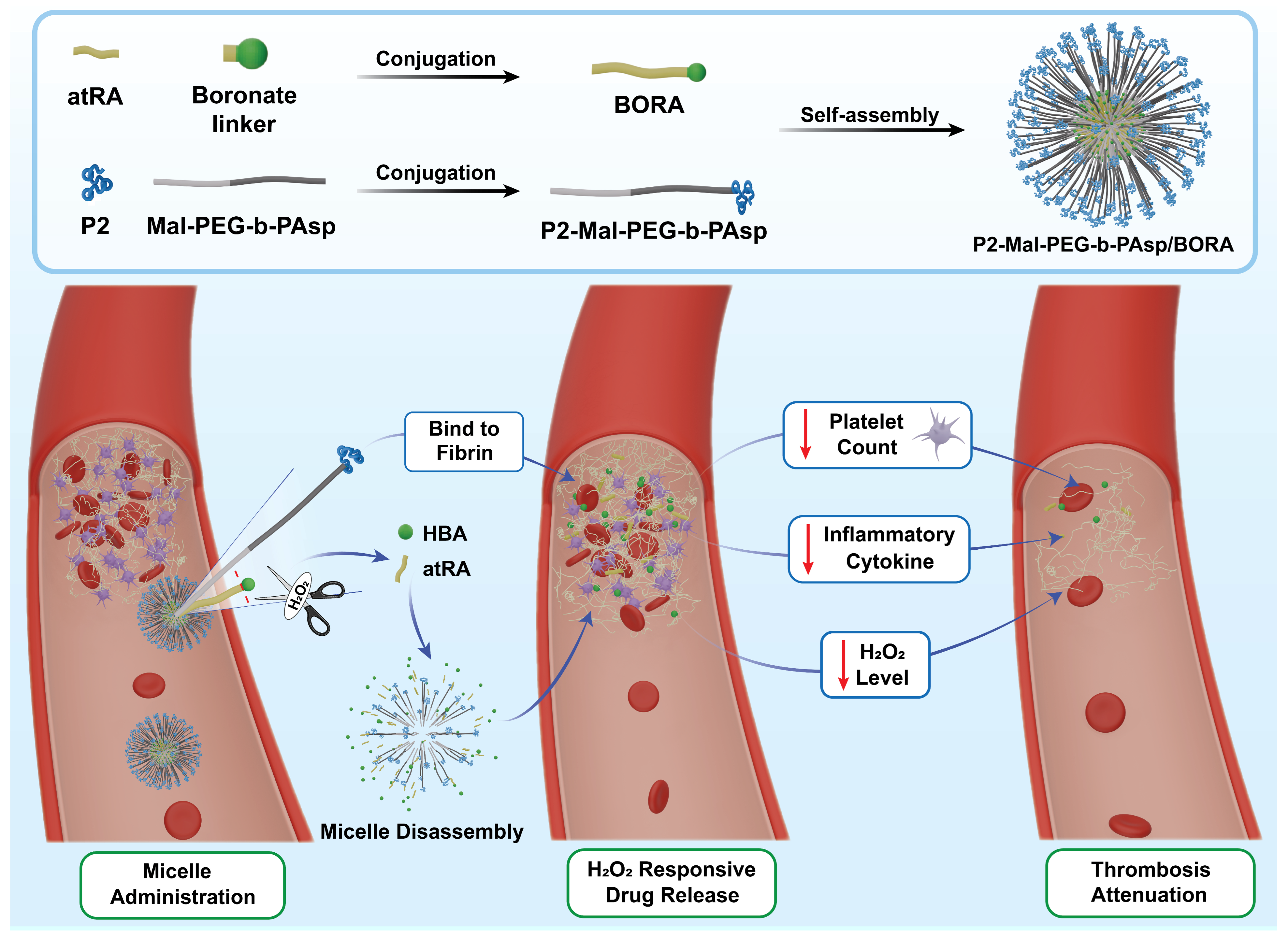
Herein, we developed a P2-functionalized and H
2O
2-responsive polymeric micelle for thrombus microenvironment modulation. A candidate fibrin-binding peptide, P2 (VTFIKC), was first screened using computer-aided drug design (CADD), and its fibrin-binding and thrombus-association properties were supported by in vitro assays. An atRA-based boronate ester prodrug, termed BORA, was then synthesized by conjugating atRA to an H
2O
2-responsive boronate linker, with the aim of enabling H
2O
2-dependent cleavage and the release of atRA and p-hydroxybenzyl alcohol (HBA) under oxidative conditions. The peptide-modified amphiphilic polymer P2-Mal-PEG-b-PAsp was further constructed and co-assembled with BORA to form P2-Mal-PEG-b-PAsp/BORA micelles. The micelles were designed to combine P2-mediated fibrin-binding potential with H
2O
2-responsive release and H
2O
2 scavenging, thereby modulating oxidative, inflammatory, and platelet-related changes associated with thrombosis (
Figure 1). The physicochemical properties, H
2O
2 responsiveness, in vitro cytoprotective and anti-inflammatory effects, together with preliminary in vivo effects of the micelles in a thrombosis model, were systematically evaluated.
2. Materials and Methods
2.1. Chemical Reagents
Tetrahydrofuran (THF) and Cyanine 5.5 (Cy5.5) was purchased from Shanghai Aladdin Bio-Chem Technology Co., Ltd. (Shanghai, China). n-Hexane, N,N-dimethylformamide (DMF), anhydrous diethyl ether, anhydrous ethanol, dichloromethane, ethyl acetate, methanol, dichloroethane, anhydrous sodium sulfate, hydrogen peroxide (H2O2), calcium chloride (CaCl2), acetonitrile, phosphotungstic acid, potassium bromide, concentrated nitric acid, and acetone were purchased from Sinopharm Chemical Reagent Co., Ltd. (Shanghai, China). Triphosgene, benzyl-L-aspartate, all-trans retinoic acid, 4-(bromomethyl)phenylboronic acid, trimethylolethane, cesium carbonate, dichloroacetic acid, hydrogen bromide/acetic acid solution, and deuterated dimethyl sulfoxide were obtained from Shanghai Bide Pharmaceutical Technology Co., Ltd. (Shanghai, China). Dimethyl sulfoxide (DMSO) for polymer synthesis was purchased from Sinopharm Chemical Reagent Co., Ltd., while DMSO used for cell experiments was obtained from Nanjing Chemical Reagent Co., Ltd. (Nanjing, China). Thrombin and Tween-20 were purchased from Sigma-Aldrich, Shanghai, China. Maleimide-poly(ethylene glycol)-amine (Mal-PEG-NH2) was purchased from Xi’an Ruixi Biological Technology Co., Ltd. (Xi’an, China). The retinoic acid boronate ester prodrug BORA, Mal-PEG-b-PAsp, P2-Mal-PEG-b-PAsp, blank P2-Mal-PEG-b-PAsp micelles, and BORA-loaded P2-Mal-PEG-b-PAsp micelles were synthesized or prepared in-house as described in the experimental procedures.
2.2. Biological and Cell Culture Reagents
Fibrin, fibrinogen, and D-dimer were purchased from Sigma-Aldrich, Shanghai, China. The Monolith RED-NHS protein labelling kit was obtained from NanoTemper Technologies, Munich, Germany. Phosphate-buffered saline (PBS, pH 7.4), DMEM medium, penicillin-streptomycin, the reactive oxygen species (ROS) detection kit, and PBS buffer (pH 7.4) were obtained from Nanjing KeyGen Biotech Co., Ltd., Nanjing, China. Fetal bovine serum (FBS) was purchased from Gibco, Waltham, MA, USA. Trypsin was obtained from Nanjing Wisent Biotechnology Co., Ltd., Nanjing, China. Peptides designed by our research group including MRFIEC, VTFIKC, MRFVKS, MGFIKG, and MTLVKS were synthesized by Sangon Biotech Co., Ltd., Shanghai, China. The CCK-8 assay kit was purchased from Beyotime Biotechnology Co., Ltd., Shanghai, China. TNF-α and IL-1β ELISA kits were purchased from R&D Systems, Minneapolis, MN, USA. Ferric chloride hexahydrate (FeCl3·H2O, AR) and chloral hydrate were purchased from Shanghai Aladdin Bio-Chem Technology Co., Ltd. Paraformaldehyde was obtained from iFangBio, Changsha, China. Heparin sodium anticoagulant tubes and EDTA anticoagulant tubes were purchased from Jiangsu Kangjian Medical Apparatus Co., Ltd., Taizhou, China.
2.3. Cell Lines
Human umbilical vein endothelial cells (HUVECs) were obtained from the Shanghai Institutes for Biological Sciences, Chinese Academy of Sciences. Cells were cultured at 37 °C in a humidified atmosphere containing 5% CO2 using DMEM complete medium supplemented with 10% FBS.
2.4. Animals and Ethical Approval
Female Sprague–Dawley (SD) rats aged 6 to 8 weeks with body weights ranging from 180 to 220 g were purchased from Jiangsu Qinglongshan Biotechnology Co., Ltd. (Zhenjiang, China, License No.: SCXK (Su) 2024-0001) and were used consistently throughout this preliminary study to reduce sex-related biological heterogeneity within the limited cohort and to maintain consistency across experimental groups. Fresh rat whole blood, plasma, and red blood cells were used for the in vitro thrombus adhesion and hemolysis assays. The rats were reared in specific pathogen-free (SPF) animal rooms using individual ventilated cages (IVC). The housing conditions were set at a temperature of (22 ± 2) °C, relative humidity of (50 ± 10)%, and a 12 h light/dark cycle, with free access to standard diet and drinking water. All animal experiments were performed in strict accordance with the Laboratory Animal Management Regulations of China Pharmaceutical University and approved by the Animal Ethics Committee of China Pharmaceutical University (Ethics Approval No.: 2025-03-002; Date: 3rd March 2025). The 3R principles (Reduction, Replacement, Refinement) were fully implemented throughout the experiments to minimize animal suffering. Carbon dioxide inhalation was adopted for euthanasia in compliance with ethical standards.
2.5. Computer-Aided Design and Screening of Fibrin-Binding Peptides
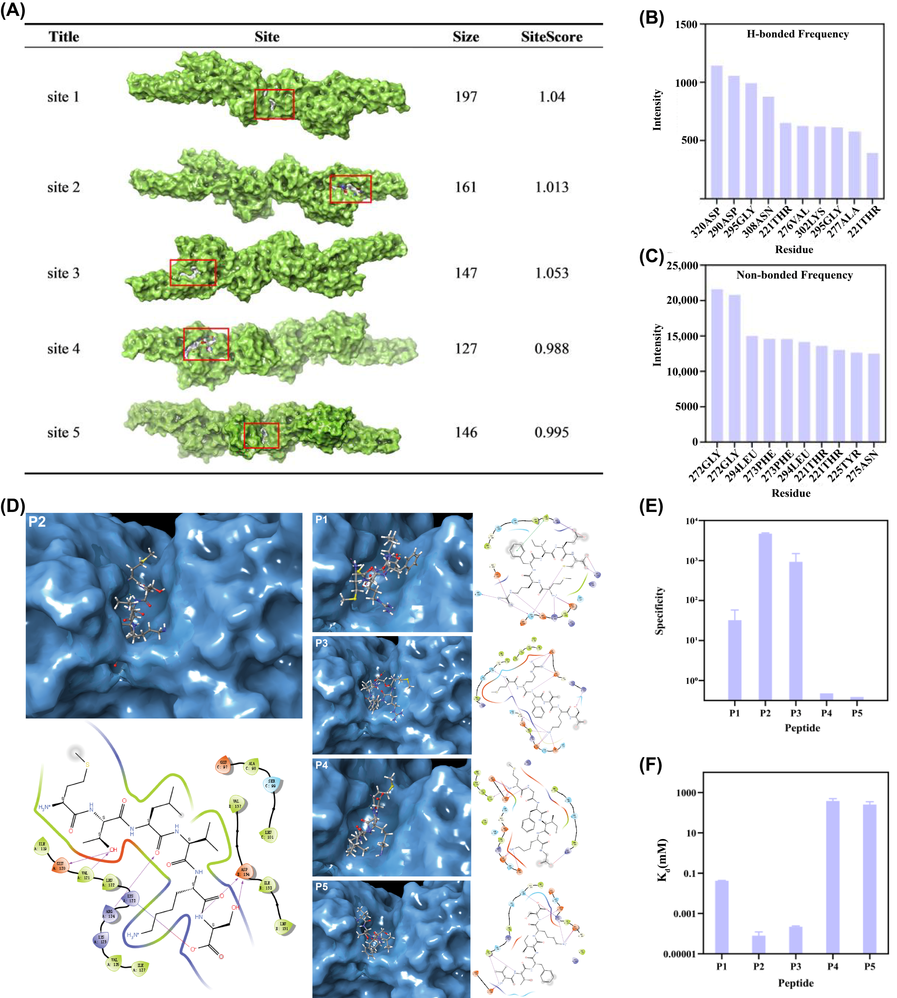
The crystal structure of fibrin was obtained from the RCSB Protein Data Bank (PDB ID: 1FZC) and imported into Schrödinger 2021. The structure was prepared using the Protein Preparation Wizard by adding hydrogens, removing water molecules, assigning protonation states, completing missing side chains, optimizing the hydrogen-bond network, and performing energy minimization with the OPLS3 force field. Potential binding pockets were predicted using the SiteMap (2021-3) module and evaluated based on pocket size, SiteScore, and hydrophilic/hydrophobic characteristics. The optimized fibrin structure was also analysed using the FT-map server to identify binding hot spots and high-frequency residues involved in hydrogen-bonding and non-bonded interactions. Based on the combined SiteMap and FT-map results, the major binding region and key interacting residues were selected for peptide library construction and molecular docking.
A peptide library was constructed according to the Mekler–Idlis complementary peptide theory. Key residues in the selected fibrin-binding region were used to design sense peptide sequences, and the corresponding antisense peptide library was generated using Python 3.5. The peptide library was then imported into Schrödinger 2021 for docking-based screening. Peptide docking was performed using the Peptide Docking workflow, with the selected fibrin-binding pocket defined as the docking region. Binding conformations and interaction patterns were evaluated by Glide docking, followed by MM-GBSA calculations to estimate peptide–fibrin binding affinity. Candidate peptides were ranked based on docking score, predicted binding energy, binding orientation, and interactions with key residues, including hydrogen-bonding, hydrophobic, and π–π interactions. Five hexapeptides were selected for further experimental validation: MRFIEC (P1), VTFIKC (P2), MRFVKS (P3), MGFIKG (P4), and MTLVKS (P5).
2.6. Stability Evaluation of Candidate Peptides
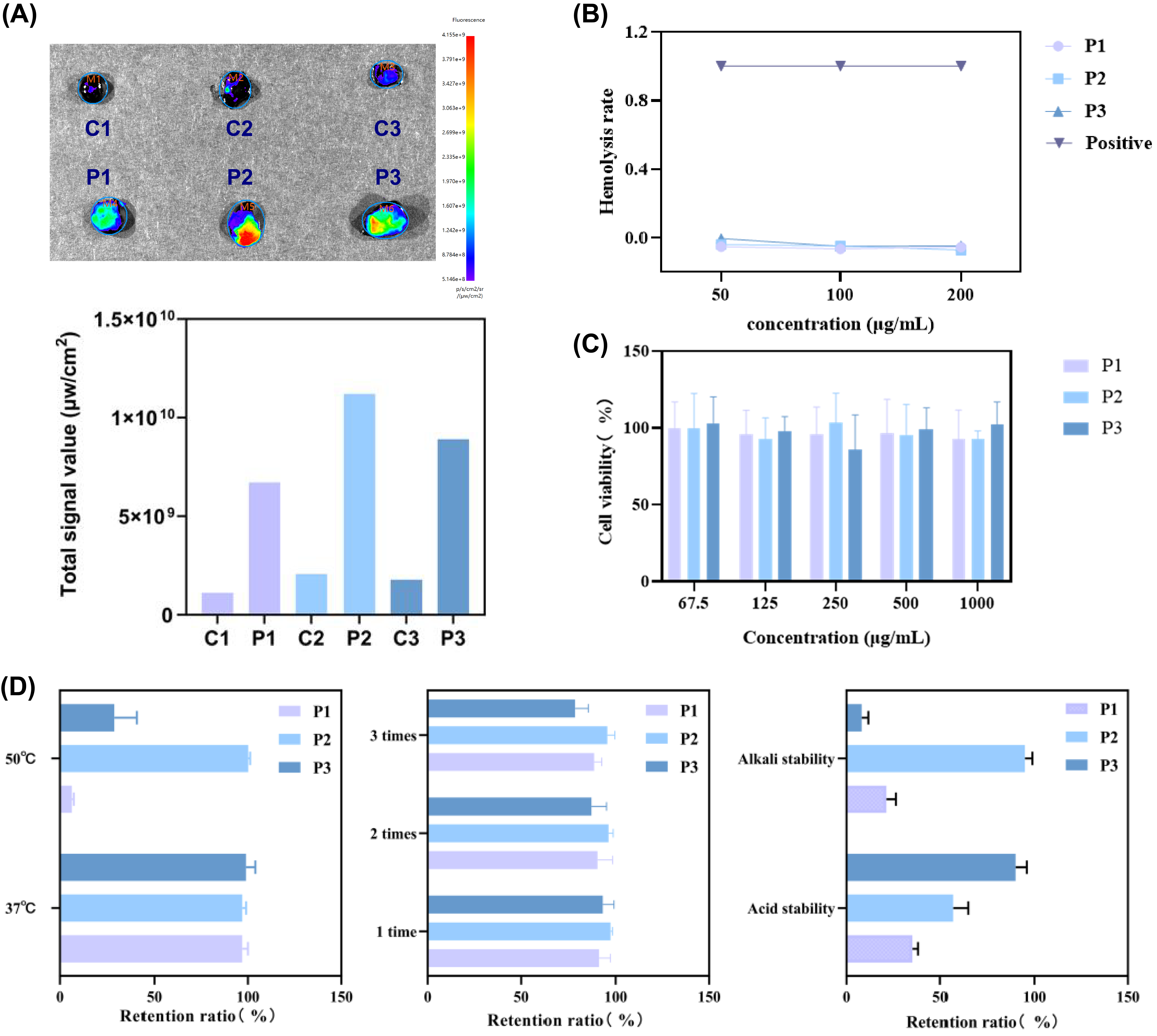
The chemical stability of the candidate peptides was evaluated under different stress conditions, including acidic, alkaline, high-temperature, and repeated freeze–thaw treatments. Briefly, peptide solutions were subjected to acidic conditions (pH 2), alkaline conditions (pH 12), high-temperature treatment (50 °C), or repeated freeze–thaw cycles. Untreated peptide solutions were used as controls. After treatment, the remaining peptide content was analysed by high-performance liquid chromatography (HPLC). Peptide degradation was evaluated by comparing the peak area of each treated sample with that of the untreated control at the same retention time.
HPLC analysis was performed using a mobile phase consisting of acetonitrile and water (10:90, v/v). The column temperature was maintained at 25 °C, the detection wavelength was set at 214 nm, and the flow rate was 1 mL/min.
2.7. In Vitro Fibrin-Binding and Thrombus-Association Assays
The binding affinity of the candidate peptides toward fibrin was evaluated by microscale thermophoresis (MST). Before MST analysis, fibrin was fluorescently labelled using a Monolith RED-NHS protein labelling kit. The buffer-exchange column was placed in a 1.5 mL centrifuge tube and centrifuged at 1500× g for 1 min to remove the storage solution. NHS buffer (300 μL) was added to the column and centrifuged at 1500× g for 1 min; this washing step was repeated three times. The target protein solution was then loaded onto the column and centrifuged at 1500× g for 2 min to collect the buffer-exchanged protein. RED-NHS dye was dissolved in DMSO to 600 μM and diluted with NHS labelling buffer to 300 μM. Subsequently, 90 μL of buffer-exchanged protein solution and 10 μL of dye solution were mixed in a 1.5 mL centrifuge tube and incubated in the dark for 30 min. After labelling, the mixture was transferred to an equilibrated purification column, and 550 μL of binding reaction buffer was added to collect the fluorescently labelled protein.
For MST analysis, peptide solutions were serially diluted with PBS containing 0.05% Tween-20 over a concentration range of 1 mM to 50 μM. Equal volumes of peptide solution and fluorescently labelled protein solution were mixed in PCR tubes and loaded into MST capillaries. Binding affinity was measured using a Monolith NT.115 instrument according to the manufacturer’s instructions, and the dissociation constant (
Kd) was calculated. Fibrinogen and D-dimer were used as control proteins to assess binding specificity. The
Kd and specificity values were obtained from three independent measurements and expressed as mean ± standard deviation. The specificity was calculated according to the following equation:
The association of the candidate peptides with in vitro-formed thrombi was evaluated using a thrombus adhesion assay. Fresh rat blood was collected by cardiac puncture into heparin sodium anticoagulant tubes and centrifuged at 2500× g for 10 min at 4 °C to obtain plasma. Thrombus formation was induced by adding thrombin (1 U) and CaCl2 (20 mM) to the plasma. After formation, the thrombi were washed three times with PBS to remove residual soluble components. Fluorescently labelled peptides were added to the thrombus samples and incubated for 30 min. The thrombi were then washed three times with PBS to remove unbound peptides. Fluorescence signals were detected using an in vivo imaging system, and fluorescence intensity was quantified and normalized to thrombus area to compare thrombus association among different candidate peptides.
2.8. Protection Against H2O2-Induced Endothelial Injury
The protective effect of micelles against H2O2-induced endothelial injury was evaluated using a CCK-8 assay. HUVECs in the logarithmic growth phase were digested, resuspended in complete DMEM medium, counted using a hemocytometer, and seeded into 96-well plates at 10,000 cells per well in 100 μL medium. After incubation at 37 °C in a humidified 5% CO2 atmosphere for 24 h, the culture medium was removed.
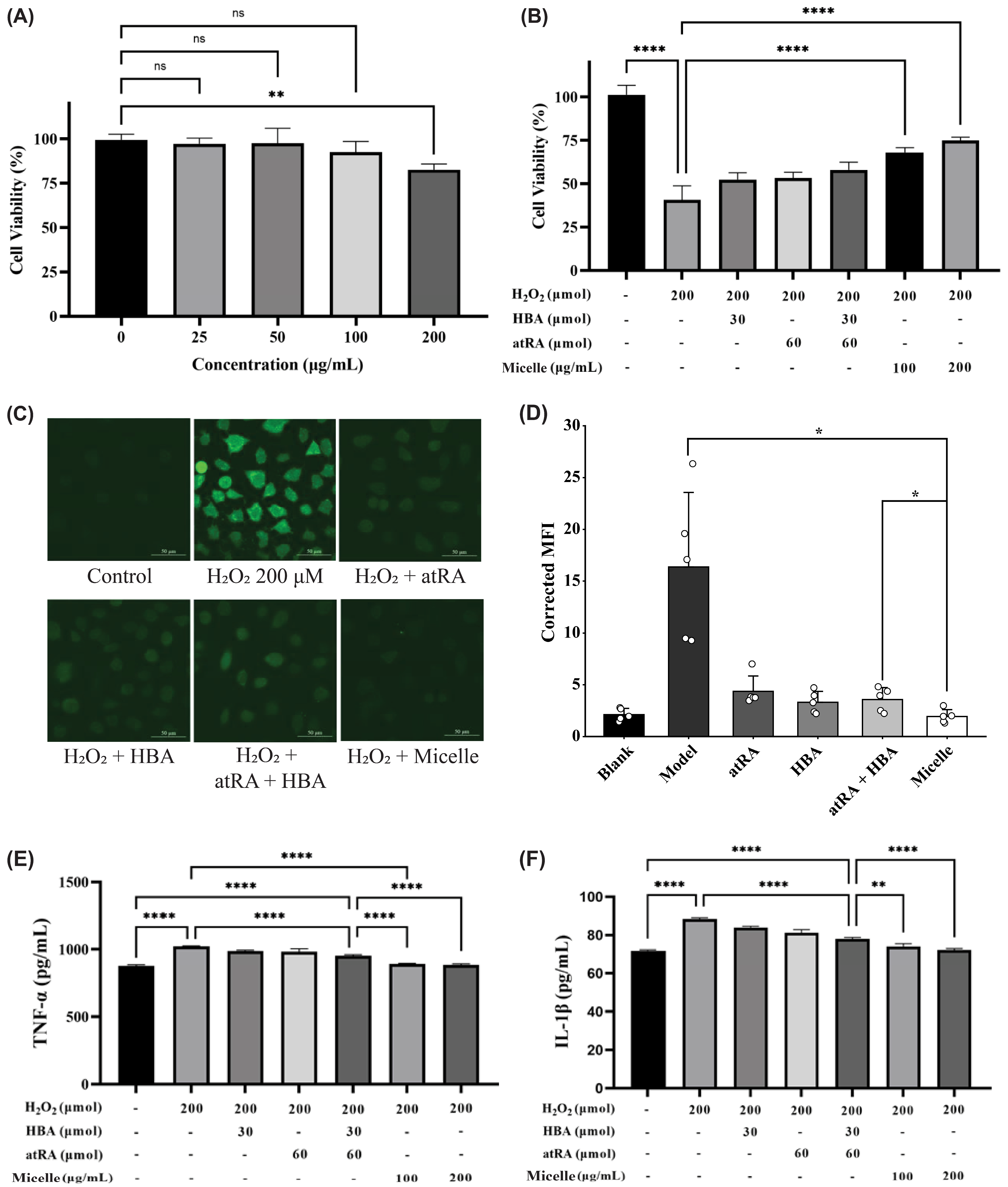
Cells were treated with free HBA (30 μg/mL), free atRA (60 μg/mL), HBA plus atRA at the same concentrations, or BORA-loaded P2-Mal-PEG-b-PAsp micelles (100 and 200 μg/mL). After 4 h of incubation, 100 μL of H2O2 solution (200 μM) was added to each well, followed by further incubation for 24 h. Untreated cells served as the normal control, and cells treated with H2O2 alone served as the oxidative injury model group.
After H2O2 stimulation, 20 μL of CCK-8 solution was added to each well and incubated for 4 h at 37 °C. After shaking for 10 min, absorbance was measured at 450 nm using a microplate reader. Cell viability was calculated relative to the negative control group, and the cytoprotective effect was evaluated by comparing treated groups with the H2O2 model group.
2.9. Synthesis and Characterization of Components for Micelle Construction
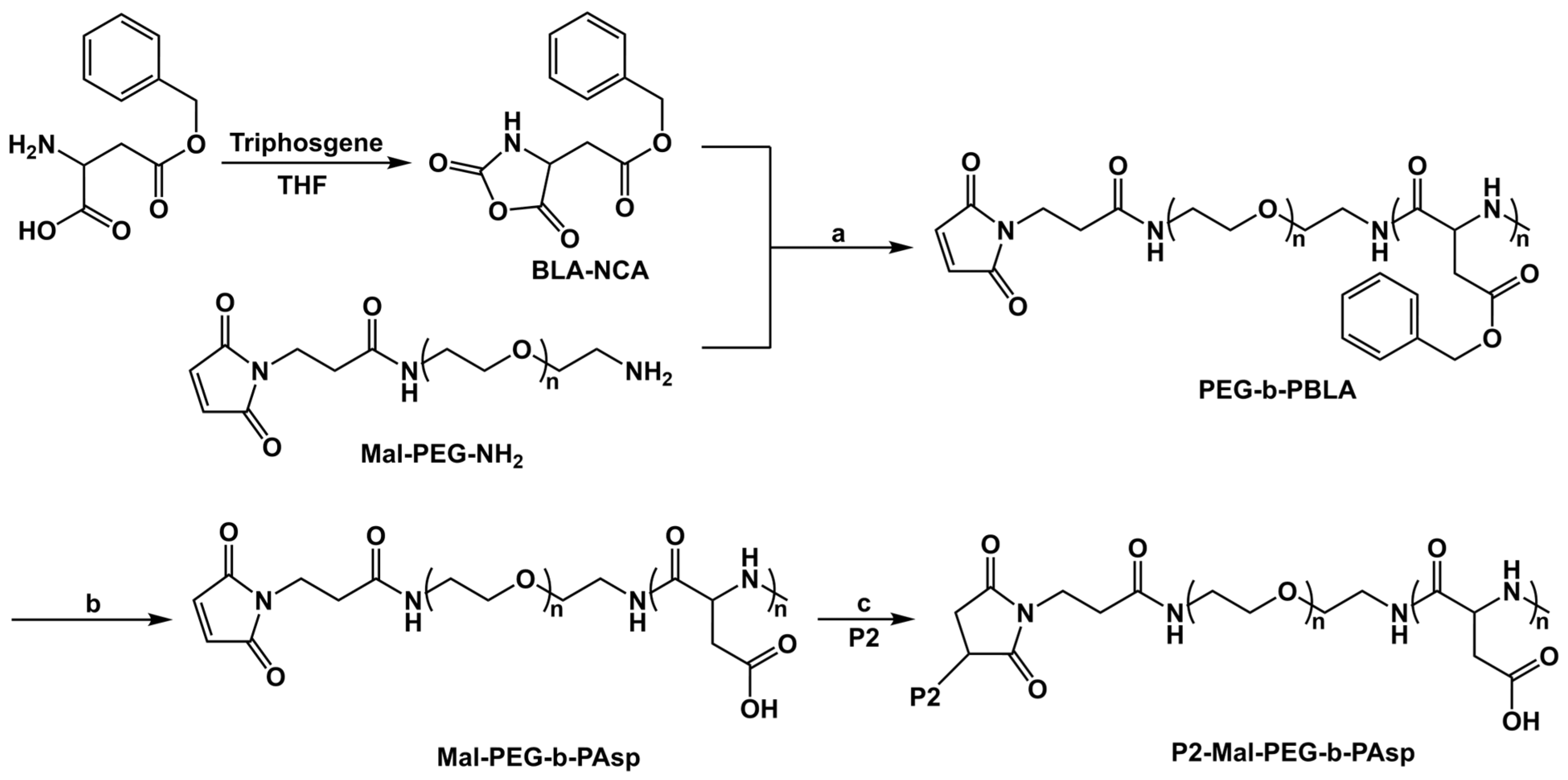
The synthetic routes of BORA, Mal-PEG-b-PAsp, and P2-Mal-PEG-b-PAsp are shown in
Figure 2 and
Figure 3, and detailed synthetic procedures and characterization data are provided in the
Supporting Information.
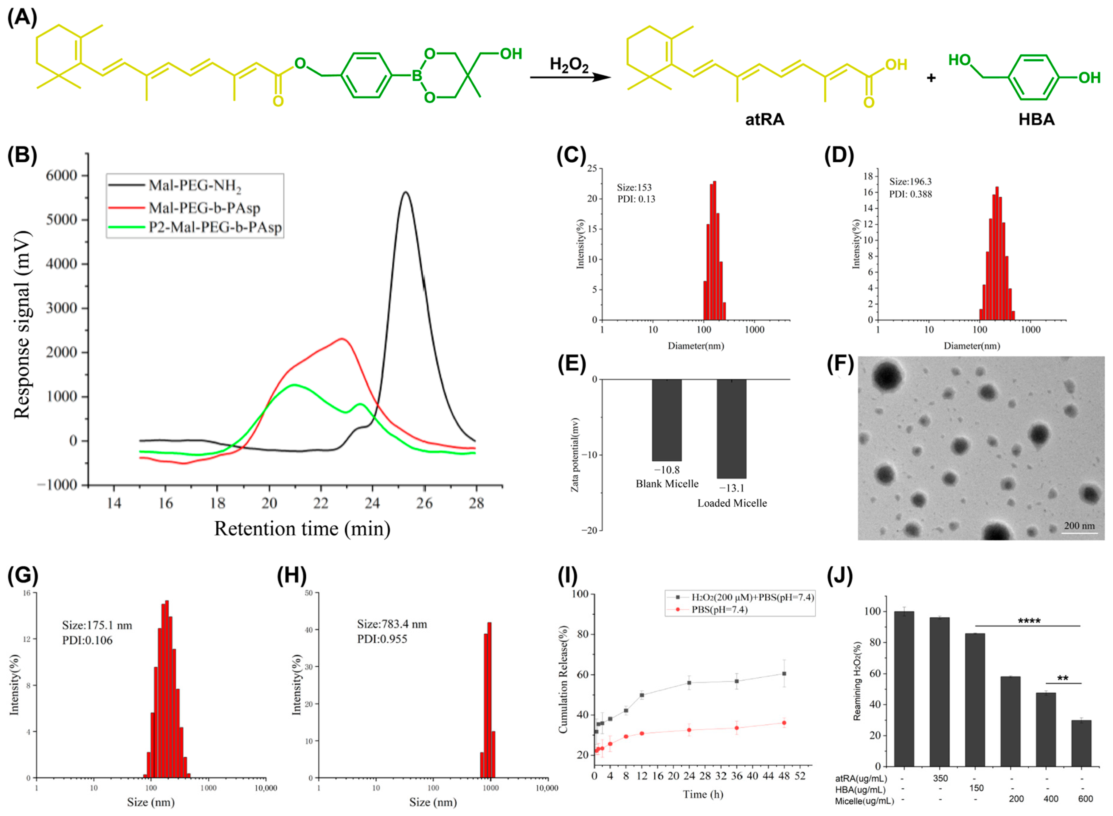
Briefly, a boronate ester-containing intermediate, hereafter referred to as the boronate linker, was first prepared from 4-(bromomethyl)phenylboronic acid and trimethylolethane. The boronate linker was subsequently reacted with all-trans retinoic acid (atRA) in the presence of cesium carbonate to afford the H2O2-responsive prodrug BORA. After extraction and chromatographic purification, the structure of BORA was confirmed by 1H NMR and ESI-MS.
Mal-PEG-b-PAsp was synthesized through N-carboxyanhydride ring-opening polymerization. β-Benzyl-L-aspartate N-carboxyanhydride (BLA-NCA) was first prepared from benzyl-L-aspartate and triphosgene and then polymerized using Mal-PEG-NH2 as the macroinitiator to obtain Mal-PEG-b-PBLA. The benzyl protecting groups were subsequently removed by acidolysis to afford Mal-PEG-b-PAsp. Polymers with different block ratios were prepared by adjusting the feed molar ratio of Mal-PEG-NH2 to BLA-NCA. The polymer structures and block ratios were characterized by 1H NMR.
Using P2 as the selected candidate fibrin-binding peptide, P2-Mal-PEG-b-PAsp was synthesized through a thiol-maleimide Michael addition reaction between the terminal cysteine residue of P2 and the maleimide group of Mal-PEG-b-PAsp. After the reaction, the product was purified by dialysis against deionized water and collected by lyophilization. Successful conjugation of P2 to Mal-PEG-b-PAsp was confirmed by the disappearance of the maleimide proton signal in the 1H NMR spectrum and by gel permeation chromatography (GPC).
2.10. Preparation of P2-Mal-PEG-b-PAsp/BORA Micelles
Blank and BORA-loaded P2-Mal-PEG-b-PAsp micelles were prepared using a solvent exchange method. For the preparation of blank micelles, P2-Mal-PEG-b-PAsp polymer (10 mg) was dissolved in DMF (5 mL) under magnetic stirring. Subsequently, ultrapure water (10 mL) was slowly added dropwise to the polymer solution, and the mixture was stirred at room temperature for 2 h. The resulting solution was transferred into a dialysis bag and dialyzed against ultrapure water for 24 h to remove DMF, yielding blank P2-Mal-PEG-b-PAsp micelles. For the preparation of BORA-loaded micelles, P2-Mal-PEG-b-PAsp polymer (10 mg) and BORA prodrug were co-dissolved in DMF (5 mL) under magnetic stirring. Ultrapure water (10 mL) was then slowly added dropwise to the mixed solution, followed by stirring at room temperature for 2 h to allow micelle self-assembly. The solution was subsequently transferred into a dialysis bag and dialyzed against ultrapure water for 24 h to remove DMF and unencapsulated components, affording BORA-loaded P2-Mal-PEG-b-PAsp micelles.
2.11. Characterizations of P2-Mal-PEG-b-PAsp/BORA Micelles
After dialysis, blank P2-Mal-PEG-b-PAsp micelles and BORA-loaded P2-Mal-PEG-b-PAsp micelles were characterized for hydrodynamic diameter, particle size distribution, polydispersity index (PDI), and zeta potential by dynamic light scattering (DLS). Before measurement, the micelle suspensions were appropriately diluted with ultrapure water to avoid multiple scattering. Each sample was equilibrated at room temperature and measured in triplicate, and results were expressed as mean ± standard deviation. The triplicate measurements represented repeated instrument readings of the same sample rather than independently prepared formulation batches.
The morphology of BORA-loaded micelles was observed by transmission electron microscopy (TEM). Briefly, a drop of diluted micelle suspension was placed onto a carbon-coated copper grid and allowed to adsorb for several minutes. Excess liquid was removed with filter paper, followed by negative staining with phosphotungstic acid solution. After air-drying at room temperature, the samples were observed by TEM to evaluate micellar morphology and nanoscale structure.
The BORA loading content was determined by inductively coupled plasma-based boron quantification. A known amount of lyophilized BORA-loaded micelles was digested with concentrated nitric acid until a clear solution was obtained. The digest was diluted to a fixed volume with ultrapure water, and the boron concentration was quantified using a standard calibration curve. Since each BORA molecule contains one boron atom, the amount of BORA incorporated into the micelles was calculated from the measured boron content and the theoretical boron content of BORA. Blank micelles were processed in the same manner and used as background controls. The denominator was the total mass of the lyophilized BORA-loaded micellar formulation, including both polymer and BORA. The apparent drug-loading content was calculated according to the following equation. The reported value was derived from one independently prepared micelle batch; recovery, encapsulation efficiency, and batch-to-batch variability were not independently determined. Drug loading content was calculated according to the following equation:
2.12. H2O2 Responsiveness Assay
BORA-loaded micelles were prepared as described above and transferred into a dialysis bag. The dialysis bag was immersed in PBS containing 1 mM H2O2 and incubated at 37 °C in a shaking incubator for 4 h. After incubation, the micelle solution was collected, and the hydrodynamic diameter and polydispersity index were measured by dynamic light scattering (DLS) to assess H2O2-induced micellar structural changes.
2.13. In Vitro Drug Release Assay
P2-Mal-PEG-b-PAsp polymer and BORA prodrug were dissolved in DMF and slowly added dropwise into water under stirring to allow micelle formation. The resulting micelle solution was transferred into a dialysis bag with a molecular weight cut-off of 3000 Da. PBS containing 200 μM H2O2 (pH 7.4) was used as the external release medium to simulate an oxidative physiological environment. The dialysis system was incubated at 37 °C in a shaking incubator. At 1, 2, 4, 6, 10, 12, 24, and 48 h after initiation of the release assay, 1 mL of the external medium was collected and replaced with an equal volume of fresh release medium to maintain a constant total volume of 60 mL. The absorbance of released all-trans retinoic acid was measured at 350 nm using a UV–vis spectrophotometer. A standard curve of atRA was established by UV–vis spectrophotometry, and the cumulative release profile was calculated accordingly. As UV–vis measurement at this wavelength may not unambiguously distinguish free atRA from intact BORA or other retinoid-containing species, the results were expressed as apparent atRA-equivalent release rather than definitive release of free atRA, and used only as a simple and preliminary reference for monitoring changes associated with the H2O2-triggered reaction.
2.14. H2O2 Scavenging Assay
The H
2O
2-scavenging capacity of the micelles was determined using a commercial H
2O
2 detection kit based on titanium sulfate colorimetry, a method previously applied to evaluate H
2O
2 consumption by nanomaterial systems [
32,
33]. BORA-loaded micelles at different concentrations (200, 400, and 600 μg/mL, 200 μL) were added to H
2O
2 solution (200 μM) and incubated at 37 °C for 2 h. After incubation, the titanium sulfate reagent and alkaline solution from the assay kit were added, and the mixture was allowed to stand at room temperature for 5 min. The samples were then centrifuged at 500×
g for 10 min, and the supernatant was discarded. The resulting precipitate was washed twice with cold acetone and dissolved for absorbance measurement at 412 nm using a microplate reader. Free atRA and HBA, the degradation products of BORA after ROS responsiveness, were used as control groups. The H
2O
2-scavenging efficiency was calculated from the residual H
2O
2-associated absorbance.
2.15. Hemocompatibility Assays
Fresh rat whole blood was collected into heparin sodium anticoagulant tubes and centrifuged at 300×
g for 20 min at room temperature. After centrifugation, the supernatant was discarded, and the red blood cells were washed several times with PBS. The purified red blood cells were then diluted to 10% with normal saline. Subsequently, 100 μL of the red blood cell suspension was transferred into 1.5 mL centrifuge tubes and incubated with peptide samples at different concentrations (50, 100, and 200 μg/mL). Deionized water and PBS were used as the positive and negative controls, respectively. After incubation at 37 °C for 4 h, the samples were centrifuged at 300×
g for 5 min. The supernatant was collected, and the absorbance at 541 nm was measured using a microplate reader. The hemolysis rate was calculated according to the following equation:
2.16. Cytocompatibility Assays
HUVECs in the logarithmic growth phase were digested, resuspended in complete culture medium, and counted using a hemocytometer. The cells were seeded into 96-well plates at a density of 5000 cells per well and incubated at 37 °C in a humidified atmosphere containing 5% CO
2 for 24 h. The culture medium was then replaced with fresh medium containing different concentrations of candidate peptides (67.5, 125, 250, 500, and 1000 μg/mL), followed by further incubation for 72 h. After incubation, 20 μL of CCK-8 solution was added to each well, and the cells were incubated for another 4 h. The absorbance at 450 nm was measured using a microplate reader. Cell viability was calculated using the following equation:
The cytocompatibility of BORA-loaded P2-Mal-PEG-b-PAsp micelles was further assessed in HUVECs using a CCK-8 assay. HUVECs were seeded into 96-well plates and incubated overnight to allow cell attachment. The cells were then treated with BORA-loaded micelles at different concentrations and incubated under standard cell culture conditions. After treatment, CCK-8 solution was added to each well and incubated to allow formazan formation. The absorbance was measured using a microplate reader, and cell viability was calculated relative to the untreated control group. The assay was used to evaluate the concentration-dependent cytotoxicity of the micelles and to determine a suitable concentration range for subsequent in vitro efficacy studies.
2.17. Intracellular ROS Detection in HUVECs
Intracellular ROS levels were detected using DCFH-DA as a fluorescent probe. HUVECs were seeded into confocal dishes at 1 × 10⁵ cells per dish and incubated at 37 °C in a humidified 5% CO2 atmosphere for 24 h. After cell attachment, the medium was removed, and cells were treated with free atRA, free HBA, or BORA-loaded P2-Mal-PEG-b-PAsp micelles for 3 h. H2O2 was then added at a final concentration of 200 μM, followed by incubation for 24 h to induce intracellular oxidative stress.
After treatment, the medium was discarded, and cells were washed three times with PBS. DCFH-DA solution (10 μM, 1 mL) was added to each confocal dish and incubated at 37 °C for 20 min. The DCFH-DA solution was then removed, and cells were washed with PBS to eliminate extracellular probe. Finally, 200 μL of PBS was added to each dish, and samples were protected from light before imaging. Green DCF fluorescence was visualized using an LSM 800 confocal laser scanning microscope (Carl Zeiss Microscopy GmbH, Jena, Germany), and fluorescence intensity was used to compare intracellular ROS accumulation among different groups.
2.18. Anti-Inflammatory Activity in HUVECs
The anti-inflammatory activity of BORA-loaded P2-Mal-PEG-b-PAsp micelles was evaluated by measuring TNF-α and IL-1β levels in H2O2-stimulated HUVECs using ELISA. HUVECs were seeded into 6-well plates at 3 × 10⁵ cells per well and incubated at 37 °C in a humidified 5% CO2 atmosphere for 24 h. After cell attachment, the medium was removed, and cells were treated with free HBA, free atRA, HBA plus atRA, or BORA-loaded P2-Mal-PEG-b-PAsp micelles for 3 h. H2O2 was then added at a concentration of 200 μM, followed by incubation for 24 h to induce inflammatory activation. Untreated cells served as the normal control, and cells treated with H2O2 alone served as the inflammatory model group.
After treatment, culture supernatants were collected and centrifuged at 100× g for 5 min to remove cell debris. TNF-α and IL-1β levels in the supernatants were quantified using the corresponding ELISA kits according to the manufacturers’ instructions. Standards and samples were added to ELISA plates, with duplicate wells for each standard and triplicate wells for each sample. Absorbance was measured using a microplate reader, and cytokine concentrations were calculated from the corresponding standard curves. The anti-inflammatory activity of each formulation was evaluated by comparing TNF-α and IL-1β levels among different treatment groups.
The reported n values for cell viability, intracellular ROS and ELISA assays represent independent biological experiments.
2.19. Preliminary In Vivo Evaluation in FeCl3-Induced Thrombosis
The preliminary in vivo effects of BORA-loaded P2-Mal-PEG-b-PAsp micelles were evaluated in a FeCl3-induced carotid artery thrombosis model in female SD rats. Rats were anesthetized by intraperitoneal injection of 10% chloral hydrate at 40 mg/kg, fixed in the supine position, and shaved in the neck region. A longitudinal incision was made along the neck midline, and the surrounding muscles were bluntly separated with hemostatic forceps to expose the left carotid artery. Body temperature was maintained at 37 °C using a heating pad during surgery.
To induce thrombosis, filter paper (1 cm × 2 cm) soaked in 10% FeCl3 aqueous solution was wrapped around the exposed left carotid artery for 5 min and covered with plastic film to prevent nonspecific injury to adjacent tissues. After FeCl3 treatment, the plastic film and filter paper were removed, and the injured vascular area was gently rinsed with PBS to remove residual FeCl3.
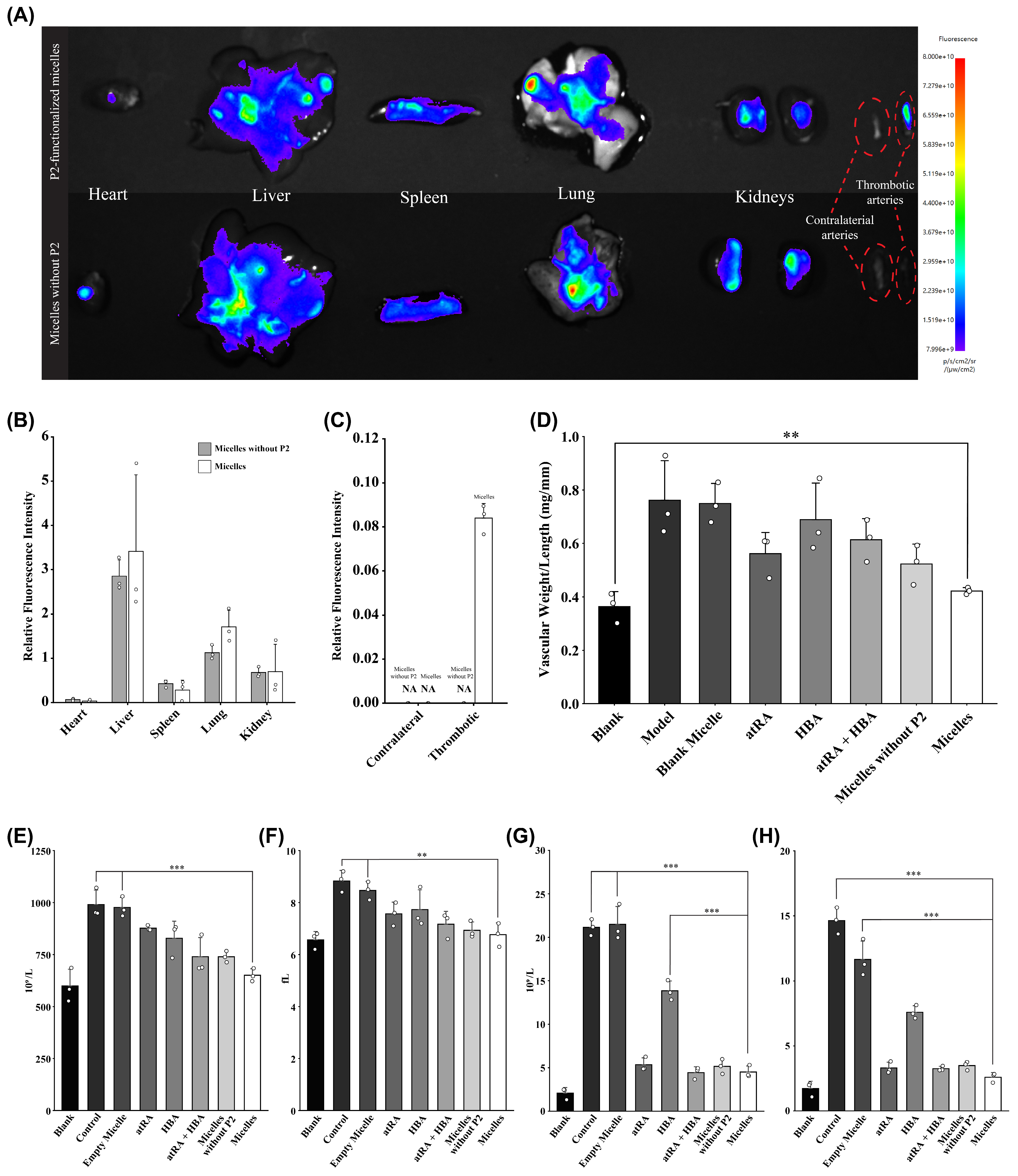
To evaluate the ex vivo biodistribution of the micelles, Cy5.5 was physically encapsulated within the hydrophobic micellar core during micelle preparation. Cy5.5-loaded micelles without P2 and Cy5.5-loaded complete P2-functionalized micelles were administered via the tail vein at an equivalent Cy5.5 dose of 100 μg/kg. At 2 h [
17] after administration and following blood collection, the rats were perfused with PBS through the cardiac apex. The heart, liver, spleen, lungs, kidneys, contralateral carotid artery, and thrombotic carotid artery were then excised and subjected to ex vivo fluorescence imaging. Relative fluorescence intensity was quantified using the analysis software supplied with the fluorescence imaging system.
For the evaluation of in vivo antithrombotic effects, different formulations were administered immediately via tail vein injection. The blank group received normal saline without FeCl3 treatment, while the model group received normal saline after FeCl3-induced thrombosis. Treatment groups received free atRA, free HBA, atRA plus HBA, drug-free micelles, micelles without P2 or BORA-loaded P2-Mal-PEG-b-PAsp micelles at 10 mg/kg in an injection volume of 200 μL. Animals were randomly assigned to the experimental groups. No animals or measurements were excluded from the final analysis. Formal allocation concealment and investigator blinding were not implemented during treatment administration or outcome assessment.
At 2 h after administration, whole blood was collected by cardiac puncture into EDTA anticoagulant tubes. The tubes were gently inverted to mix the blood with anticoagulant and stored at 4 °C before analysis. Platelet count, mean platelet volume, white blood cell count, and lymphocyte count were measured using an automated hematology analyzer.
The reported n values in this experiment correspond to independent biological replicates.
2.20. Statistical Analysis
All statistical analyses were performed using R software (version 4.6.1). Comparisons involving multiple treatment groups or dose levels were analyzed using one-way or two-way analysis of variance (ANOVA), as appropriate for the experimental design, followed by multiplicity-adjusted post hoc comparisons. Unpaired two-tailed Student’s t-tests were retained only for direct comparisons between two independent groups. Data are presented as mean ± standard deviation (SD). Statistical significance was determined using multiplicity-adjusted p values where applicable: ns, adjusted p > 0.05; * adjusted p < 0.05; ** adjusted p < 0.01; *** adjusted p < 0.001; and **** adjusted p < 0.0001.
4. Conclusions
In this study, a P2-functionalized, H2O2-responsive retinoic acid prodrug micellar system was developed as a candidate formulation for attenuating thrombosis-associated oxidative and inflammatory responses. The candidate fibrin-binding peptide P2 (VTFIKC) was identified through computer-aided peptide screening, and its fibrin-binding and in vitro thrombus-association properties were supported by in vitro assays. Based on its binding affinity, low hemolytic activity, cytocompatibility, and stability under the tested conditions, P2 was selected as the ligand for micelle construction.
The H2O2-responsive prodrug BORA was synthesized and co-assembled with P2-Mal-PEG-b-PAsp to form P2-functionalized polymeric micelles. The resulting P2-Mal-PEG-b-PAsp/BORA micelles showed suitable nanoscale size, negative surface charge, defined morphology, and substantial apparent prodrug-loading content. Upon H2O2 exposure, the micelles showed changes in particle size distribution, accelerated retinoic acid release, and concentration-dependent H2O2 consumption, supporting their H2O2-responsive formulation design.
In vitro, micelle treatment was associated with improved HUVEC viability following H2O2 exposure, lower intracellular ROS-associated fluorescence, and reduced TNF-α and IL-1β concentrations. In a FeCl3-induced carotid artery thrombosis model, systemic administration of the micelles was associated with changes in platelet- and leukocyte-related hematological indices, and the micelles showed preferential accumulation in the thrombotic carotid artery.
Overall, in line with advances in targeted and stimuli-responsive nanodrug delivery [
49,
50], this study combined a newly screened candidate fibrin-binding peptide, an H
2O
2-responsive atRA prodrug, and a polymeric micellar carrier into a single formulation. Nowadays, mechanism-oriented vascular intervention and formulation-enabled cardiovascular drug delivery are increasingly being explored as complementary therapeutic strategies [
51,
52]. Unlike conventional antithrombotic strategies that mainly interfere with coagulation, platelet aggregation, or fibrinolysis systemically [
5,
53,
54], this micelle modulated thrombosis-associated oxidative and inflammatory responses through P2-mediated fibrin-binding potential, H
2O
2-dependent release behavior, and H
2O
2 consumption. The current findings provide formulation and biological proof-of-concept for further investigation of this platform, while the small animal cohort, limited mechanistic endpoints, and incomplete in vivo distribution, pharmacokinetic, and safety characterization preclude definitive conclusions regarding thrombus-specific delivery or therapeutic efficacy.