1. Introduction
Globally, endometrial cancer accounts for approximately 7% of all malignancies and represents the fourth most common cancer in women [
1]. In Turkey, the age-standardized incidence rate was reported as 11.3 per 100,000 in 2020, ranking fifth among all cancers [
2]. Notably, it is the sole malignancy that has displayed declining survival rates over the last four decades. Mortality rates remain substantially elevated in advanced-stage and high-histological-grade tumor types [
3]. Obesity, metabolic syndrome, diabetes mellitus, increased life expectancy, and an aging population are widely accepted as the primary factors driving this escalation. While the prognosis is favorable for a significant proportion of cases diagnosed at an early stage, certain patients display an unexpectedly aggressive clinical course, demonstrating that classifications relying solely on histopathological evaluation have several limitations [
4].
For decades, endometrial cancers have been classified into Type I and Type II tumors according to the dualistic model defined by Bokhman. Within this framework, endometrioid tumors are generally considered estrogen-related and low-grade neoplasms with favorable prognoses. Conversely, serous tumors are aggressive, high-grade malignancies with poor outcomes. However, tumors with identical histology often follow markedly different clinical trajectories, highlighting the biological heterogeneity of endometrial cancer and the need for molecular classification [
5].
The genomic classification established by The Cancer Genome Atlas (TCGA) project represents a landmark milestone in the molecular-based evaluation of endometrial cancer. Within this framework, tumors are categorized into four distinct molecular subgroups:
POLE-ultramutated, mismatch repair-deficient/microsatellite instability-high (MMRd/MSI-H), copy-number low (NSMP), and copy-number high/p53-abnormal. Subsequent studies have demonstrated that these molecular subgroups display profound differences not only in terms of genomic features but also regarding prognosis, recurrence risk, and therapeutic response [
6,
7].
The increasing clinical significance of TCGA-inspired molecular classification has resulted in the development of algorithms such as ProMisE (Proactive Molecular Risk Classifier for Endometrial Cancer) aimed at adapting this approach into routine pathology practice. Currently, molecular classification utilizing
POLE mutation, MMR protein deficiency, and p53 status has emerged as a critical component of risk assessment in endometrial cancers. Indeed, molecular classification was formally incorporated for the first time into the staging system updated by the International Federation of Obstetrics and Gynecology (FIGO) in 2023, underscoring the pivotal role of molecular data in guiding patient management [
8].
Advances in next-generation sequencing (NGS) technologies have enabled a more detailed examination of the molecular architecture of endometrial cancer. In addition to well-known driver genes such as
TP53,
PTEN,
PIK3CA,
KRAS,
CTNNB1, and
POLE, less-studied genomic alterations and co-mutation patterns can be identified. Notably, NGS-based analyses are particularly beneficial for identifying interacting molecular pathways during tumor progression and uncovering novel biomarkers [
9].
While numerous studies have addressed the molecular classification of endometrial cancer, comprehensive evaluations that simultaneously assess menopausal status, histological subtype, tumor grade, co-mutation patterns, and TCGA-inspired molecular subgroups within the same patient cohort remain limited. Furthermore, studies establishing the relationship between histopathological features and molecular data derived from targeted NGS panels utilized in routine clinical practice are relatively scarce [
5].
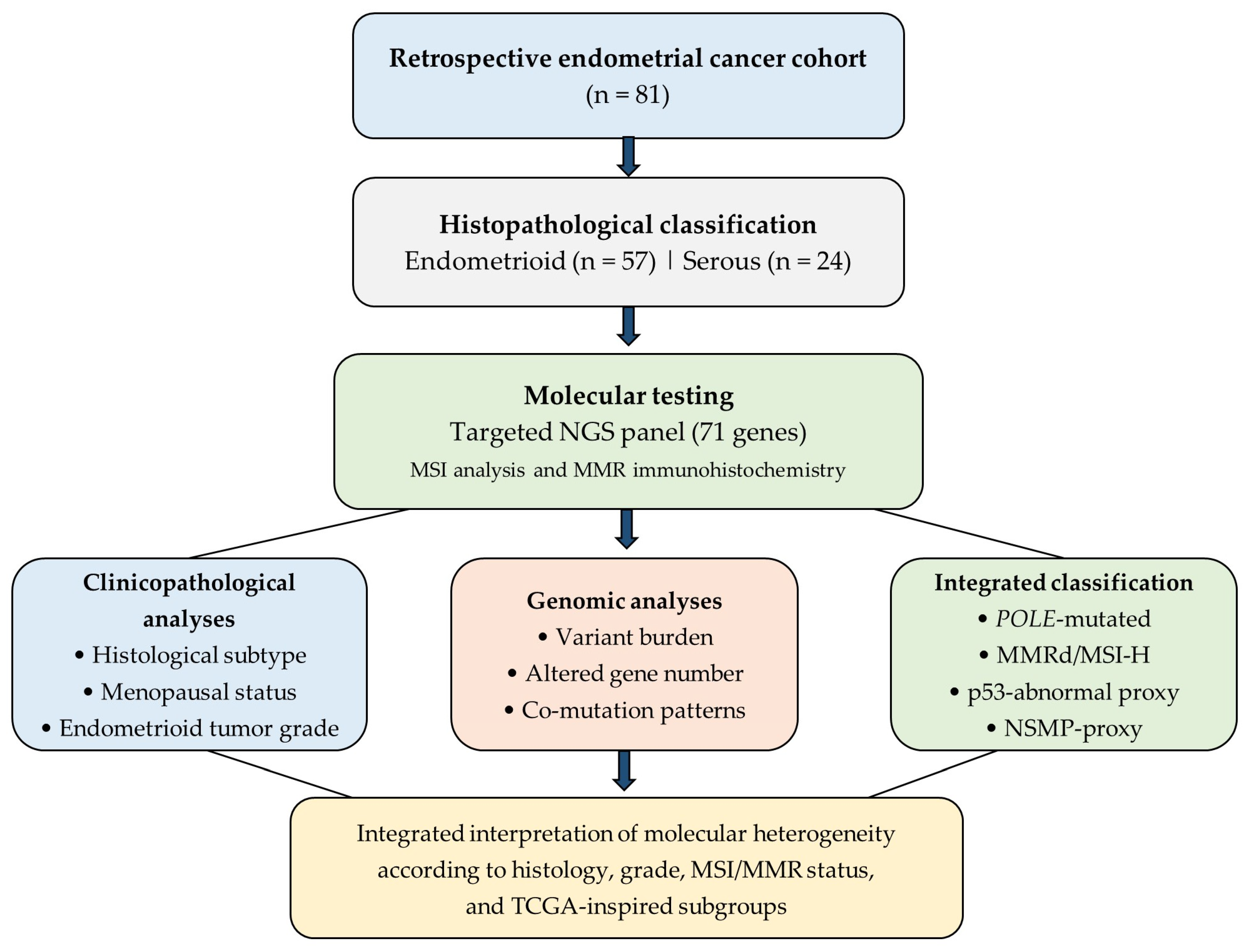
The objective of this study is to evaluate next-generation sequencing data from cases diagnosed with endometrial cancer. Molecular variations were investigated within the framework of menopausal status, histological subtype, tumor grade, mismatch repair/MSI status, co-mutation patterns, and TCGA-inspired molecular classification, providing novel data regarding the molecular heterogeneity of endometrial cancer. Accordingly, the current study applied a surrogate TCGA-inspired molecular classification based on targeted sequencing, MSI-PCR, and MMR immunohistochemistry.
3. Results
3.1. Clinicopathological Characteristics of the Cohort
A total of 81 endometrial cancer cases were evaluated in this study. Regarding histological subtypes, 57 cases (70.4%) were classified as endometrioid carcinoma, whereas 24 cases (29.6%) were categorized as serous carcinoma. When endometrioid carcinomas were evaluated according to tumor grade, Grade 1, Grade 2, and Grade 3 tumors accounted for 15.8%, 59.6%, and 24.6% of the cases, respectively. In terms of menopausal status, 8 patients (9.9%) were premenopausal and 73 patients (90.1%) were postmenopausal. The mean age was found to be 44.9 ± 5.5 years in the premenopausal group and 65.3 ± 7.4 years in the postmenopausal group.
A total of 756 variant records were examined across the entire cohort. The mean number of variants per case was 9.12 ± 5.34 in endometrioid carcinomas and 9.83 ± 6.68 in serous carcinomas (p = 0.284). Similarly, the mean number of mutated genes per case was calculated as 6.79 ± 4.01 in endometrioid carcinomas and 7.88 ± 4.86 in serous carcinomas (p = 0.331).
When endometrioid carcinomas were evaluated according to tumor grade, the mean number of variants was found to be 4.44 ± 2.19 in Grade 1 tumors, 9.68 ± 5.37 in Grade 2 tumors, and 11.42 ± 6.11 in Grade 3 tumors (p = 0.006). The number of mutated genes per case was 3.78 ± 1.39, 7.41 ± 3.84, and 8.17 ± 4.72, respectively (p = 0.021). No significant difference was observed between the histological subtypes in terms of total variant load and the number of mutated genes per case.
Regarding menopausal status, the number of variants per case was 8.75 ± 4.92 in the premenopausal group and 9.40 ± 5.84 in the postmenopausal group. Similarly, the number of mutated genes per case was found to be 6.00 ± 3.02 in the premenopausal group and 7.11 ± 4.29 in the postmenopausal group. No significant differences were detected between the two groups based on menopausal status regarding total variant load and the number of mutated genes per case (p = 0.726 and p = 0.479, respectively).
In our molecular classification analysis, the NSMP-proxy subgroup was identified as the most frequent category, which was followed by the
POLE-mutated proxy, p53-abnormal proxy, and MMRd/MSI-H subgroups. In the MMR/MSI evaluation, the loss of MLH1 and PMS2 expressions along with the MMRd/MSI-H phenotype was observed in a distinct portion of the cohort, a finding consistent with the molecular heterogeneity of endometrial cancer. The clinicopathological and molecular characteristics of the patients are summarized in
Table 1.
3.2. Histological Subtype-Based Molecular Findings
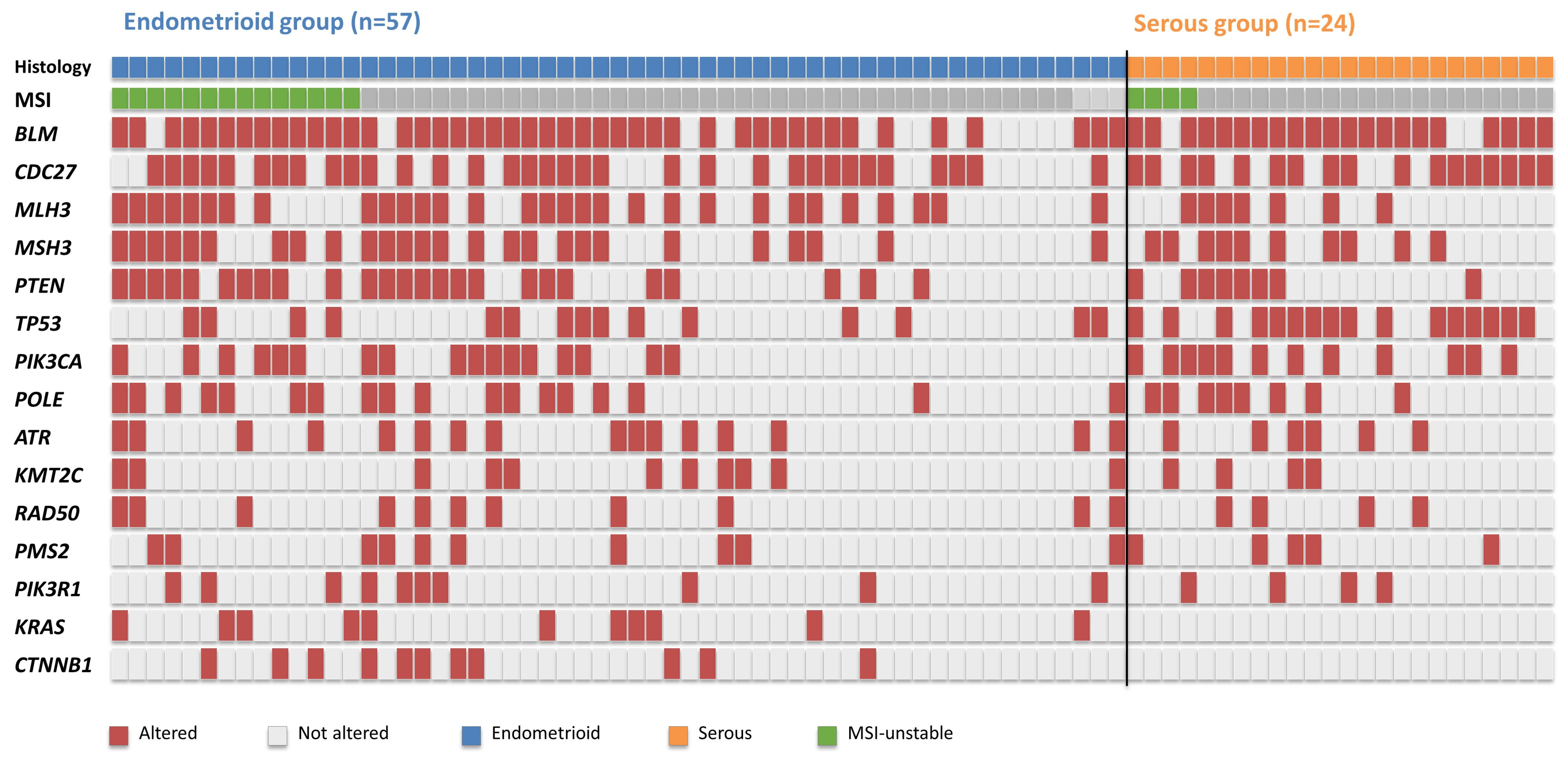
In endometrioid carcinomas, BLM variations were most frequently identified (77.2%), which were followed by CDC27 (57.9%), MLH3 (52.6%), MSH3 (45.6%), PTEN (43.9%), POLE (31.6%), PIK3CA (29.8%), and TP53 (26.3%). In serous carcinomas, BLM (87.5%), CDC27 (70.8%), TP53 (66.7%), PIK3CA (50.0%), MSH3 (41.7%), PTEN (33.3%), POLE (33.3%), and MLH3 (29.2%) variations were prominent.
TP53 alterations remained significantly more frequent in serous carcinomas than in endometrioid carcinomas after BH-FDR correction (raw p = 0.001, BH-FDR q = 0.015). In contrast, the nominal associations observed for CTNNB1 and KRAS did not remain statistically significant after multiple-testing correction (both q = 0.140). No significant differences were observed between the two histological subtypes regarding PTEN, PIK3CA, POLE, BLM, CDC27, MLH3, and MSH3 variations.
Detailed information on all unique pathogenic and likely pathogenic variants identified in the study cohort is presented in
Supplementary Table S1. A complete oncoprint including all 81 endometrial cancer cases and all pathogenic or likely pathogenic alterations identified in the study cohort is provided in
Supplementary Figure S1.
When evaluated in terms of MMR/MSI status, MLH1 loss, PMS2 loss, and MSI were detected in 24.6%, 22.8%, and 24.6% of endometrioid carcinomas, respectively. In serous carcinomas, MSI instability was observed at a rate of 16.7%. Although the MMRd/MSI-H rate appeared higher in endometrioid tumors, this difference was not statistically significant (
p = 0.564) (
Table 2 and
Figure 2).
3.3. Molecular Alterations According to Menopausal Status
When evaluated at the gene level, BLM variations were most frequently observed in the premenopausal group and were detected in all cases (8/8, 100%). This was followed by CDC27 (62.5%), MLH3 (50.0%), ATR (37.5%), MSH3, PTEN, POLE, CTNNB1, and KMT2C (each 25.0%). In the postmenopausal group, however, BLM variations were identified in 57 cases (78.1%), CDC27 in 45 cases (61.6%), MSH3 in 34 cases (46.6%), MLH3 in 33 cases (45.2%), PTEN in 31 cases (42.5%), TP53 in 30 cases (41.1%), PIK3CA in 28 cases (38.4%), and POLE in 24 cases (32.9%).
When comparing gene frequencies, no statistically significant differences were observed between the groups. Nevertheless, TP53 and PIK3CA variations were found to occur at higher rates in the postmenopausal group. Specifically, TP53 variations were detected in 12.5% of the premenopausal group and 41.1% of the postmenopausal group (p = 0.145), while PIK3CA variations were observed at rates of 12.5% and 38.4%, respectively (p = 0.248).
Upon evaluating MMR immunohistochemistry and MSI findings, MLH1 loss was identified in 2 cases (25.0%) in the premenopausal group and 16 cases (21.9%) in the postmenopausal group (p = 1.000). Similarly, PMS2 loss was observed at rates of 25.0% and 20.5%, respectively (p = 0.672). In contrast, no loss of MSH2 or MSH6 expression was observed in the premenopausal group. MSI was detected in 25.0% of premenopausal cases and 21.9% of postmenopausal cases, demonstrating no significant difference between the groups (p = 1.000). After BH-FDR correction, no gene-level associations remained statistically significant. Given the small number of premenopausal patients (n = 8), these subgroup comparisons should be regarded as exploratory and interpreted with caution.
3.4. Grade-Specific Molecular Alterations in Endometrioid Carcinomas
When endometrioid carcinomas were evaluated according to tumor grade, an increase in genomic alteration burden was observed with increasing grade. The mean number of variants was found to be 4.44 in Grade 1 tumors, 9.68 in Grade 2 tumors, and 11.42 in Grade 3 tumors (p = 0.006). Similarly, a significant increase was detected in the number of mutated genes per case (p = 0.021).
BLM variations were observed at rates of 33.3%, 85.3%, and 83.3% in Grade 1, Grade 2, and Grade 3 tumors, respectively (p = 0.004). CDC27 variations were found at rates of 22.2%, 64.7%, and 75.0%, respectively (p = 0.034). While MLH3 variations were detected in 11.1%, 64.7%, and 58.3% of the cases (p = 0.016), MSH3 variations were observed at rates of 11.1%, 50.0%, and 66.7%, in the same order (p = 0.036). After BH-FDR correction, only the association between BLM alterations and tumor grade remained statistically significant (BH-FDR q = 0.030). The nominal associations observed for CDC27, MLH3, and MSH3 did not retain statistical significance after multiple-testing correction (q > 0.05).
Although PTEN and PIK3CA variations were observed to occur more frequently in high-grade tumors, these differences did not reach statistical significance (p = 0.125 and p = 0.154, respectively). In contrast, TP53 variations were found not to be associated with histological grade in endometrioid tumors (p = 0.901).
The MMRd/MSI-H rate was found to be 11.1% in Grade 1 tumors, 26.5% in Grade 2 tumors, and 28.6% in Grade 3 tumors. Although a trend toward an increase in the MMRd/MSI-H rate was observed with increasing grade, the difference between the groups was not statistically significant (
p = 0.417) (
Table 3).
3.5. Co-Mutation Analysis
Co-mutation analysis demonstrated the presence of a BLM-centered co-occurrence pattern across the entire cohort. The most frequent co-occurrence was observed between BLM and CDC27, which was detected in 45 cases (55.6%). This was followed by BLM–MLH3 (42.0%), BLM–MSH3 (40.7%), CDC27–MSH3 (37.0%), CDC27–MLH3 (35.8%), BLM–PTEN (34.6%), and MLH3–MSH3 (33.3%) co-occurrences.
In endometrioid carcinomas, the most frequent co-mutations were BLM–CDC27 (52.6%), BLM–MLH3 (47.4%), BLM–MSH3 (42.1%), CDC27–MLH3 (42.1%), and CDC27–MSH3 (38.6%). In serous carcinomas, BLM–CDC27 (62.5%), BLM–TP53 (54.2%), CDC27–TP53 (45.8%), TP53–PIK3CA (41.7%), and BLM–PIK3CA (37.5%) were the most frequent co-mutation pairs.
Histology-specific comparisons showed that BLM–TP53 co-mutation was significantly more frequent in serous carcinomas than in endometrioid carcinomas (54.2% vs. 22.8%, raw p = 0.009). Similarly, TP53–PIK3CA (41.7% vs. 10.5%, raw p = 0.004) and CDC27–TP53 (45.8% vs. 17.5%, raw p = 0.012) co-mutations were enriched in serous carcinomas.
In endometrioid carcinomas, an increase in the number of co-mutations was observed with increasing tumor grade. While BLM–CDC27 was the dominant co-mutation pair in Grade 1 tumors, BLM–MLH3, BLM–MSH3, and CDC27–MSH3 became more prominent in Grade 2 tumors. In Grade 3 tumors, BLM–PTEN, BLM–PIK3CA, and MLH3–MSH3 combinations appeared relatively more frequent. Concurrent alterations involving DNA repair genes and the PI3K/PTEN pathway were particularly noticeable in higher-grade tumors.
Following BH-FDR correction applied across the 11 predefined co-mutation comparisons presented in
Table 4,
BLM–TP53,
TP53–PIK3CA, and
CDC27–TP53 remained statistically significant (all adjusted q = 0.044), whereas the remaining co-mutation pairs did not retain statistical significance after multiple-testing correction.
3.6. TCGA-Inspired Molecular Classification Results
In the TCGA-inspired molecular classification, the NSMP-proxy subgroup was identified as the most frequent category across the entire cohort (34.6%). This was followed by the POLE-mutated proxy (32.1%), p53-abnormal proxy (21.0%), and MMRd/MSI-H (12.3%) groups.
When evaluated in terms of histological subtypes, the NSMP-proxy group was dominant in endometrioid carcinomas (40.4%). Within the endometrioid cases, 31.6% fell into the POLE-mutated proxy group, 15.8% into the p53-abnormal proxy group, and 12.3% into the MMRd/MSI-H group. In contrast, the p53-abnormal proxy group was observed to be represented at a prominently higher rate in serous carcinomas (41.7%). In these serous cases, the POLE-mutated, NSMP-proxy, and MMRd/MSI-H groups were detected at rates of 33.3%, 16.7%, and 8.3%, respectively.
When endometrioid carcinomas were examined according to tumor grade, the vast majority of Grade 1 tumors fell into the NSMP-proxy group (66.7%). In Grade 2 tumors, the NSMP-proxy and POLE-mutated groups were represented at similar rates. In Grade 3 tumors, however, a relative increase was observed in the p53-abnormal proxy and MMRd/MSI-H subgroups. The distribution of TCGA- inspired molecular subgroups differed significantly between endometrioid and serous carcinomas (Pear-son’s χ
2 test,
p = 0.012, q = 0.012), with p53-abnormal tumors being more frequent in serous carcinomas and NSMP tumors being more frequent in endometrioid carcino-mas. No statistically significant difference was observed in the distribution of TCGA-inspired molecular subgroups across endometrioid tumor grades (raw
p = 0.084, BH-FDR q = 0.084). (
Table 5).
Detailed
POLE variant characteristics are provided in
Supplementary Table S2, whereas variant-level information for
POLE,
TP53,
BLM,
CDC27,
MLH3, and
MSH3, including VAF and sequencing coverage values, is presented in
Supplementary Table S3.
The overlap between molecular classifiers before hierarchical subgroup assignment is presented in
Supplementary Table S4.
The mean variant burden according to the TCGA-inspired molecular subgroups is summarized in
Supplementary Table S5.
4. Discussion
In this study, next-generation sequencing data from 81 endometrial cancer cases were evaluated within the framework of histological subtype, endometrioid tumor grade, menopausal status, co-mutation patterns, and TCGA-inspired molecular classification. Our findings demonstrated that there was no prominent molecular segregation according to menopausal status; in contrast, more significant molecular differences emerged regarding histological subtype and tumor grade. Particularly, the marked elevation of TP53 variations in serous carcinomas, the presence of CTNNB1 and KRAS alterations exclusively within the endometrioid group, and the increase in variant burden with increasing grade in endometrioid tumors appeared consistent with current molecular classification approaches. Accordingly, the molecular subgroups reported in this study represent surrogate categories derived from targeted sequencing, MSI-PCR, and MMR immunohistochemistry rather than the original TCGA genomic framework.
The most significant shift in endometrial cancer management over the past decade has been the integration of molecular classification systems into traditional histopathological classification. The original groups defined by the TCGA have subsequently been adopted in clinical practice as the
POLE-mutated proxy, MMRd, p53-abnormal, and NSMP subgroups, which offer enhanced clinical applicability. The ProMisE approach has also integrated this molecular classification into the practical pathology workflow, significantly increasing the importance of
POLE mutation, MMR protein loss, and p53 status in risk stratification [
7,
13]. Indeed, the inclusion of molecular subgroups in staging and risk assessment processes in the 2023 FIGO update clearly demonstrates that this approach has become a standard component in management strategies [
7,
8,
13].
In the current cohort, the identification of the NSMP-proxy and POLE-mutated proxy groups as the most frequent molecular categories further supports the molecular heterogeneity of endometrial cancer. However, the molecular subgroup assignment in the present study was based on a targeted 71-gene NGS panel combined with MSI-PCR and MMR immunohistochemistry rather than whole-exome or whole-genome sequencing. Therefore, the molecular classification should be interpreted as a TCGA-inspired classification. Although this integrated approach is practical and applicable in routine clinical practice, the targeted panel is not designed to detect genome-wide copy-number alterations or comprehensive mutational signatures. Therefore, all molecular subgroups identified in this study (POLE-mutated proxy, MMRd/MSI-H, p53-abnormal, and NSMP) should be interpreted as surrogate molecular categories rather than direct TCGA molecular subgroups derived from genome-wide genomic profiling.
TP53 alterations were more frequent in serous carcinomas than in endometrioid carcinomas, and this association remained significant after BH-FDR correction, supporting the well-established role of
TP53 abnormalities in serous endometrial carcinoma. Serous endometrial cancers are classically characterized as high-grade, aggressive tumors that largely correspond to the p53-abnormal/copy-number high molecular subgroup. In current molecular classification studies, the p53-abnormal group is reported to be particularly associated with serous histology and high-grade endometrioid tumors, as well as being linked to poorer clinical outcomes [
14,
15,
16]. In this study, the detection of
TP53 variations at a rate of 66.7% in serous carcinomas and 26.3% in endometrioid carcinomas—with this difference being statistically significant—further supports the p53-dominant molecular profile of serous carcinomas.
CTNNB1 and
KRAS alterations were detected exclusively in endometrioid carcinomas. However, these associations did not remain statistically significant after BH-FDR correction for multiple testing and should therefore be interpreted as exploratory findings requiring validation in larger independent cohorts. Nevertheless, the distribution of these alterations is consistent with the established molecular characteristics of endometrioid endometrial carcinoma.
CTNNB1, a key component of the Wnt/β-catenin signaling pathway, is among the most frequently altered genes in low-grade, early-stage endometrioid tumors and has recently been recognized as an additional prognostic marker within the NSMP molecular subgroup, where it has been associated with an increased risk of recurrence. Likewise,
KRAS alterations are well-recognized drivers of PI3K/RTK/RAS pathway activation and are predominantly observed in endometrioid carcinomas. Although the associations identified in the present study did not retain statistical significance after multiple-testing correction, the complete absence of
CTNNB1 and
KRAS alterations in serous carcinomas supports the concept that endometrioid and serous endometrial cancers arise through distinct molecular pathways, consistent with previous genomic and molecular classification studies [
17,
18].
When MMR/MSI findings were evaluated, the MMRd/MSI-H rate in endometrioid carcinomas appeared higher relative to serous carcinomas; nevertheless, this difference did not reach statistical significance. In endometrial cancers, MMR defect is particularly associated with endometrioid histology, and the MMRd/MSI-H phenotype is reported in nearly one-third of cases across many series. MMRd tumors carry clinical significance both for Lynch syndrome screening and for immunotherapy response [
9,
19,
20]. In this study, the detection of MSI at a rate of 24.6% in endometrioid carcinomas and 16.7% in serous carcinomas is consistent with the direction of the literature, though it did not produce a significant difference due to sample size.
The increase in the mean number of variants and mutated genes with increasing grade in endometrioid carcinomas suggests a potential relationship between tumor progression and genomic complexity. While the mean number of variants was 4.44 in Grade 1 tumors, it increased to 11.42 in Grade 3 tumors. Similarly, the number of mutated genes showed an upward trend from Grade 1 to Grade 3. This finding demonstrates that high-grade endometrioid tumors harbor a more complex structure not only morphologically but also molecularly. It is reported in the literature that the p53-abnormal subgroup and indicators of genomic instability can be observed more frequently in high-grade endometrioid tumors [
15,
16]. In our data,
TP53 did not show a significant difference among endometrioid grade groups; in contrast,
BLM,
CDC27,
MLH3, and
MSH3 variations demonstrated a marked increase in Grade 2 and Grade 3 tumors.
Among the grade-associated alterations, only the association for BLM remained significant after BH-FDR correction, whereas the associations observed for CDC27, MLH3, and MSH3 should be regarded as nominal findings that require validation in larger cohorts.
The
BLM gene is a member of the RecQ helicase family and is involved in DNA replication, homologous recombination, and double-strand break repair. Loss of
BLM function has been associated with chromosomal instability and cancer susceptibility; furthermore, it was reported that
BLM expression and alterations might carry significance within the context of genomic instability in various tumor types [
21,
22].
CDC27, on the other hand, is a component of the anaphase-promoting complex/cyclosome structure and plays a role in mitotic progression, chromosome segregation, and cell cycle regulation. The role of
CDC27 in cancer biology is not yet as clearly defined as that of classic driver genes; nevertheless, it was indicated that disruptions in the APC/C complex might be linked to mitotic checkpoint failure and aneuploidy [
23,
24]. In this study, the observation of the
BLM–CDC27 co-mutation as the most frequent combination across the entire cohort suggests that the cell cycle and genome stability axis becomes prominent in this series.
The increase in
MLH3 and
MSH3 variations, particularly in high-grade endometrioid tumors, is also noteworthy. Although
MLH3 and
MSH3 are not utilized in routine clinical classification as extensively as the classic MMR proteins (
MLH1,
MSH2,
MSH6, and
PMS2), they function as accessory components of the mismatch repair mechanism. It has been discussed in the literature that
MSH3 alterations in endometrial cancers can be observed particularly in MSI-positive tumors, and in some cases, whether they are the cause or the consequence of MSI remains a subject of debate [
25,
26]. Similarly, while the role of
MLH3 in endometrial cancer has been investigated, its pathogenic effect and clinical utility are not as clearly established as those of the classic MMR genes [
27,
28].
In conclusion, the more frequent detection of BLM, CDC27, MLH3, and MSH3 variations in high-grade tumors in our study further suggests that DNA repair mechanisms and cell cycle control become progressively disrupted during tumor progression. These findings support the premise that, alongside well-defined driver genes such as TP53, PTEN, and PIK3CA, additional molecular pathways involved in maintaining genome integrity may also contribute to the biological heterogeneity of endometrial cancer. Nevertheless, the findings regarding BLM, CDC27, MLH3, and MSH3 do not possess the same level of clinical establishment as well-known endometrial cancer driver genes like PTEN, PIK3CA, ARID1A, TP53, and CTNNB1. Therefore, the findings regarding BLM, CDC27, MLH3, and MSH3 should be regarded as exploratory and hypothesis-generating rather than confirmatory. Their biological and clinical significance requires validation in larger independent cohorts and functional studies before definitive conclusions can be drawn.
In the analysis performed according to menopausal status, no statistically significant differences were detected regarding total variant burden, number of mutated genes, MSI/MMR status, or major gene frequencies. The numerically higher prevalence of TP53 and PIK3CA variations in the postmenopausal group may represent a biologically plausible trend. However, the inclusion of only eight premenopausal patients substantially limits the statistical power of these comparisons. Therefore, all findings related to menopausal status should be considered exploratory and interpreted with caution rather than as evidence of definitive molecular segregation.
Co-mutation analysis revealed distinct co-occurrence patterns across histological subtypes. Co-mutations involving BLM–CDC27, BLM–MLH3, BLM–MSH3, and CDC27–MLH3 were more frequently observed in endometrioid carcinomas, whereas BLM–TP53, CDC27–TP53, and TP53–PIK3CA co-mutations were more prominent in serous carcinomas. This distribution suggests that genomic instability in serous carcinomas may be driven predominantly by TP53-related molecular events, whereas endometrioid carcinomas appear to harbor a more complex co-mutation network involving DNA damage repair and cell-cycle regulatory pathways. Furthermore, the increasing diversity of co-mutation patterns observed with higher tumor grade in endometrioid carcinomas parallels the progressive increase in variant burden identified in these tumors. After BH-FDR correction, BLM–TP53, TP53–PIK3CA, and CDC27–TP53 co-mutations remained statistically significant, whereas the remaining co-mutation pairs did not retain statistical significance.
The most significant strength of this study is the comprehensive evaluation of NGS data alongside histological subtype, grade, MSI/MMR status, menopausal status, co-mutation patterns, and TCGA-inspired molecular classification within a single-center, real-world cohort. Particularly, the increasing variant burden with increasing grade in endometrioid carcinomas and the TP53-dominant profile in serous carcinomas stand out as robust findings consistent with the literature and biologically meaningful. Furthermore, the recurrent alterations within the BLM, CDC27, MLH3, and MSH3 axis provide novel observations that may warrant further investigation in prospective studies.
Nevertheless, several limitations of this study should be acknowledged. First, this was a retrospective, single-center study with a relatively limited sample size. In particular, the small number of premenopausal patients (n = 8) and the limited number of Grade 1 endometrioid tumors reduced the statistical power of subgroup analyses. Therefore, comparisons based on menopausal status and certain histological or grade subgroups should be considered exploratory, and negative findings should be interpreted with caution. Second, menopausal status was determined according to age rather than documented clinical menopausal information because these data were not consistently available in the retrospective cohort. Although this approach has been used in previous retrospective studies, some degree of patient misclassification cannot be excluded. Third, because of the relatively small sample size and the low frequency of several genetic alterations, multivariable analyses were not performed, as reliable model estimation could not be achieved. In addition, several nominally significant gene-level and co-mutation associations lost statistical significance after correction for multiple comparisons, indicating that these findings require validation in larger independent cohorts. Fourth, molecular subgroup classification was established using a targeted 71-gene NGS panel together with MSI-PCR and MMR immunohistochemistry rather than whole-exome or whole-genome sequencing. Consequently, genome-wide copy-number alterations and comprehensive mutational signatures could not be evaluated, and the molecular subgroup assignment should be regarded as a TCGA-inspired surrogate classification rather than a complete TCGA classification. Accordingly, the molecular heterogeneity described in this study reflected targeted panel-based profiling rather than comprehensive genomic characterization. Fifth, although variant interpretation was performed using ACMG criteria and QCI™ annotation, matched normal tissue or peripheral blood samples were not available for paired analysis. Therefore, definitive discrimination between somatic and germline variants could not be achieved for all detected variants. Sixth, the functional significance of recurrent alterations, particularly in BLM, CDC27, MLH3, and MSH3, was not experimentally validated. Moreover, these variants could not be confirmed by an independent molecular method such as Sanger sequencing or droplet digital PCR because of the limited availability of residual FFPE tissue and DNA. Finally, comprehensive clinicopathological outcome data, including FIGO stage, adjuvant treatment, recurrence, survival, treatment response, and long-term follow-up, were not available for all patients. Consequently, the prognostic significance of the identified TCGA-inspired molecular subgroups could not be evaluated. Future multicenter prospective studies integrating comprehensive clinical outcome data, functional validation, and broader genomic profiling will be essential to confirm and extend the present findings.




 ,
,