The Immunopathology of Preeclampsia

 , , and
Abstract
1. Introduction
2. Methodology
3. Innate Immunity in Preeclampsia
3.1. Complement
3.2. Neutrophils and Eosinophils
3.3. Mast Cells
3.4. Macrophages
3.5. Dendritic Cells (DCs)
3.6. Myeloid-Derived Suppressor Cells (MDSCs)
3.7. Natural Killer (NK) Cells
3.8. NKT Cells
3.9. T Gamma-Delta Lymphocytes (Tγδ)
4. Adaptive Immunity in Preeclampsia
4.1. T Lymphocytes
4.2. B Lymphocytes and Preeclampsia
5. Angiogenesis and Preeclampsia
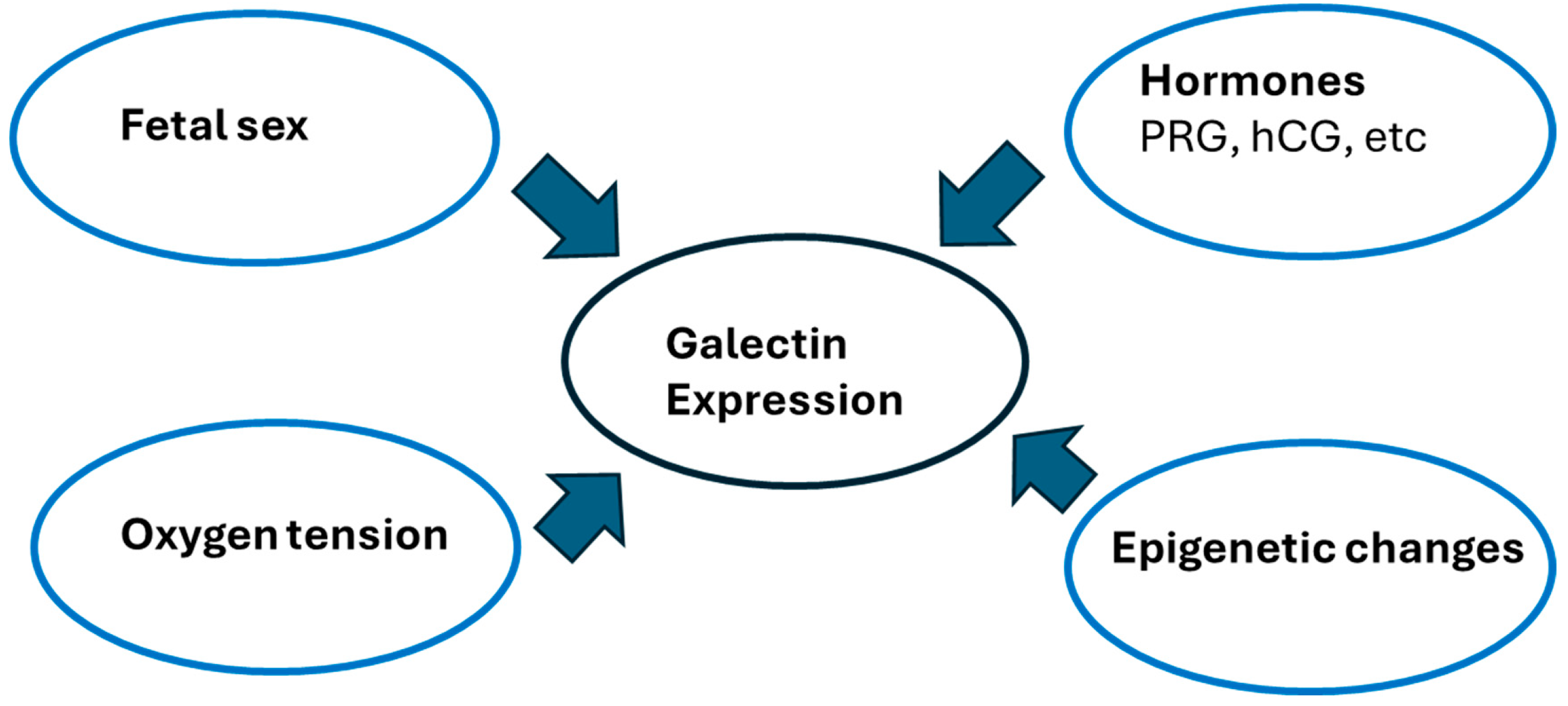
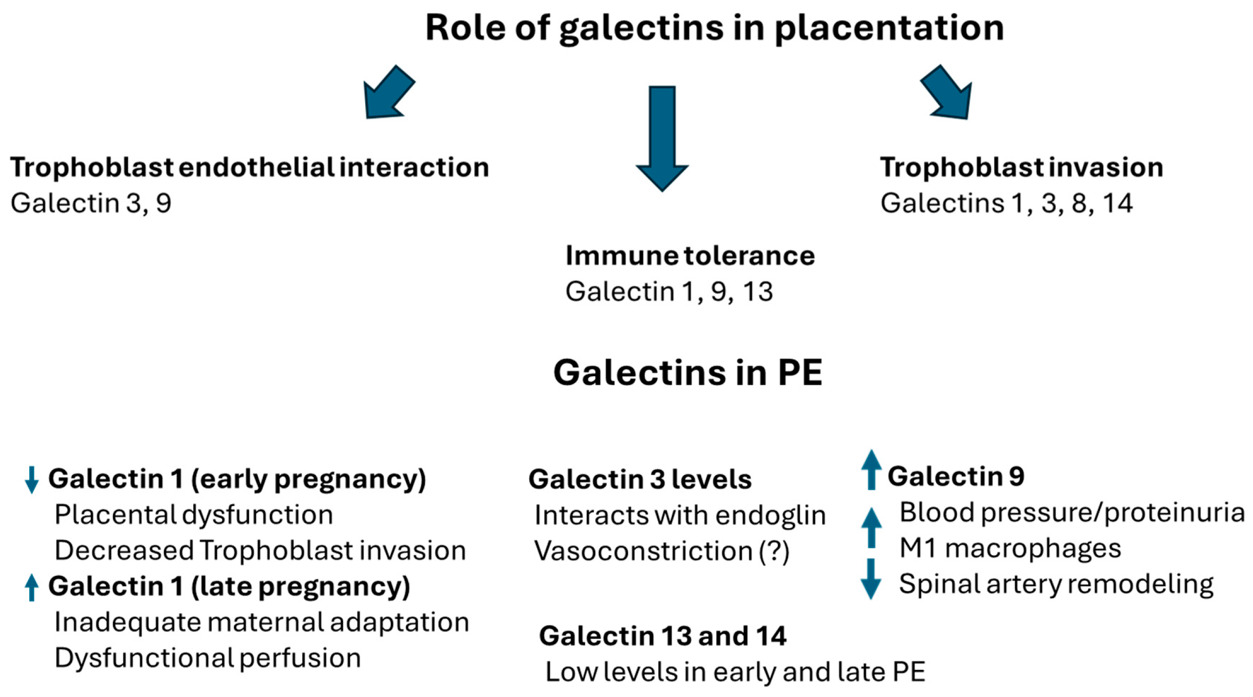
6. Galectins in Preeclampsia
7. Immunogenetics and Preeclampsia
7.1. The Role of Human Leukocyte Antigens (HLA) in Preeclampsia
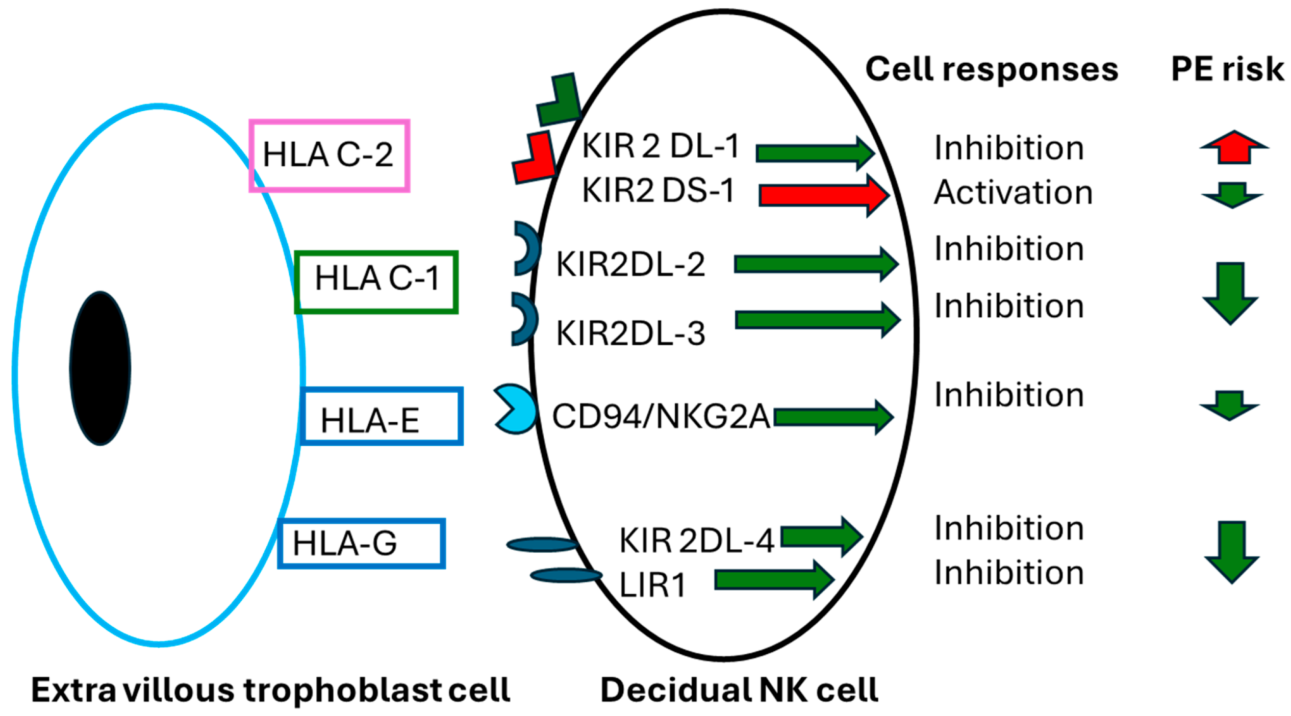
7.2. Role of Killer Immunoglobulin-like Receptors (KIR) in Preeclampsia
8. Immune Checkpoints in Preeclampsia
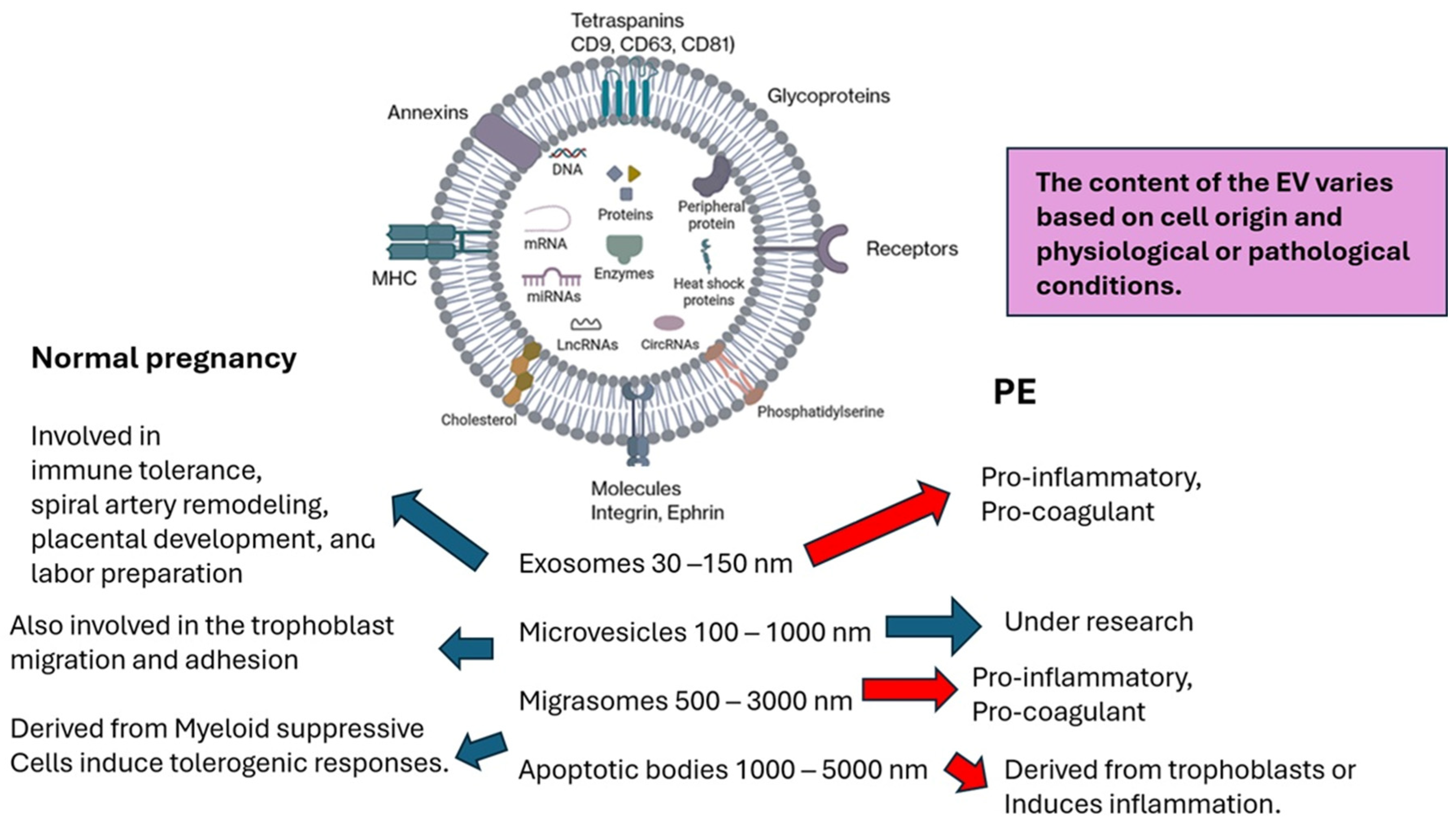
9. Extracellular Vesicles in Preeclampsia
10. MicroRNAs (miRNAs) and Preeclampsia

| miRNA Increased | Target | Observation |
|---|---|---|
| miR-155 | (1) Alters SMAD2 and SMAD5 members of the TGFβ signaling pathway. (2) Decrease T regulatory cells, downregulate CTLA4 transcription and function. (3) Regulates Vascular Peroxidase 1 and oxidative stress and mitochondrial glucose metabolism via skeletal muscle. | Cysteine-rich angiogenic inducer 61 (CYR61)/miR-155 Ratio has been proposed as a biomarker for Diagnosis and severity of PE |
| miR-125b | Downregulates the Kv1.1 voltage-gated potassium channel and glypican 1, a cell surface heparan sulfate proteoglycan. It inhibits cytotrophoblast invasion and disrupts endothelial cell function. | Predictive marker and a potential therapeutic target. |
| miR-181a-5p | Downregulates genes involved in the MAPK/ERK signal pathway. | Associated with the worst outcomes in patients with E. |
| miR-29b | It inhibits Vascular Endothelial Growth Factor A (VEGFA) and reduces anti-apoptotic proteins like Myeloid Cell Leukemia Sequence 1 (MCL1), while downregulating essential enzymes (MMP2 and MMP9) required for placental cell invasion of the uterine wall. | Marker of PE severity |
| miR-206 | Regulates genes in Muscle Development and Regeneration, tumor suppression, proliferation, neurodevelopment, and glucose-6-phosphate dehydrogenase. | Proposed as a diagnostic marker |
| miR-495 | Impedes normal placental development by repressing Histone deacetylase 2 (HDAC2), accelerating cell proliferation, invasion, and migration, while decreasing apoptosis via the P53/PUMA pathway. | Proposed as a diagnostic marker |
| miR-125b | It inhibits trophoblast Invasion and function. Placental exosomes transfer miR-125b into endothelial cells, disrupting the endothelial barrier. It induces the secretion of pro-inflammatory cytokines. | Proposed as a diagnostic marker |
| miR-206 | Inhibits trophoblast invasion and promotes placental inflammation. Suppresses the AGTR1 (Angiotensin II Receptor Type 1) gene. Downregulates VEGFA, | Diagnostic biomarker |
| MiR-517 and miR-526 | Trophoblast dysfunction, hypoxia, and angiogenic imbalance. Endothelial damage. | Promising diagnostic marker of PE |
| miR-17 | Suppresses key angiogenesis genes (like VEGFA, Hypoxia-inducing factor 1A (HIF1A), and the Ephrin system). | Proposed as a biomarker. |
| miRNA decreased | The decrease impairs | Observation |
| miR-21 | (1) Trophoblast invasion and function are decreased, and apoptosis is increased, (2) poor spiral artery remodeling and chronic hypoxia | Lower miR-21 levels in the first trimester are associated with the future onset and severity of preeclampsia and serve as a marker for early detection. |
| miR-146a | (1) Proliferation, reduced invasion, and increased apoptosis of trophoblast cells, (2) Decreased spiral artery remodeling, and (3) overactivation of NFkB inflammatory pathways. | Proposed as a diagnostic marker and for PE therapeutics. |
| miR-126 | (1) Angiogenesis, decreasing placental vasculogenesis, (2) trophoblast invasion and placental development, and (3) endothelial and trophoblast viability. | Proposed as a diagnostic marker and for PE therapeutics. |
| miR-195 | (1) The healthy invasion and migration of trophoblast cells, (2) reduces VEGF production and angiogenesis, and (3) increases the hypoxia-induced damage and oxidative stress. | Proposed as a diagnostic marker and for PE therapeutics. |
| miR-363 | (1) Poor placentation, including inadequate trophoblast migration and impaired spiral artery remodeling. (2) Decrease VEGFA secretion. | Proposed as an early-stage marker of PE. |
11. Cytokines and Leptin in Preeclampsia
12. Prostaglandins, Leukotrienes, Thromboxanes, and Resolvins
13. Radicals and Preeclampsia
13.1. Oxygen Radicals
13.2. Nitric Oxide, Peroxynitrite, and Protein Modifications
14. Cell Death in Preeclampsia

15. Infectious Diseases and Preeclampsia

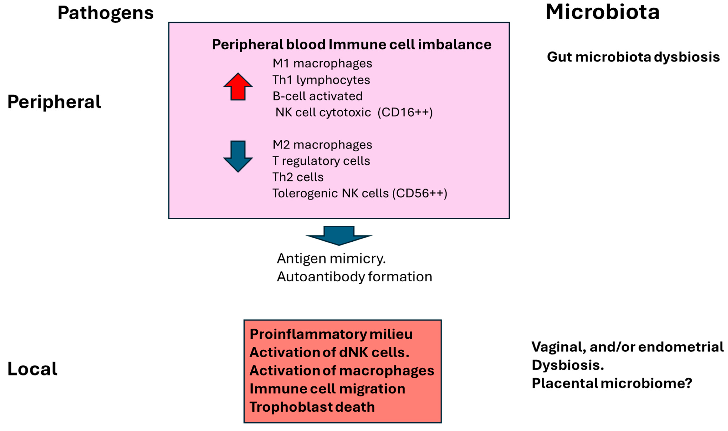
16. Microbiota and Preeclampsia
17. Autoimmune Diseases and Preeclampsia
18. Atopic Dermatitis (AD) and Preeclampsia
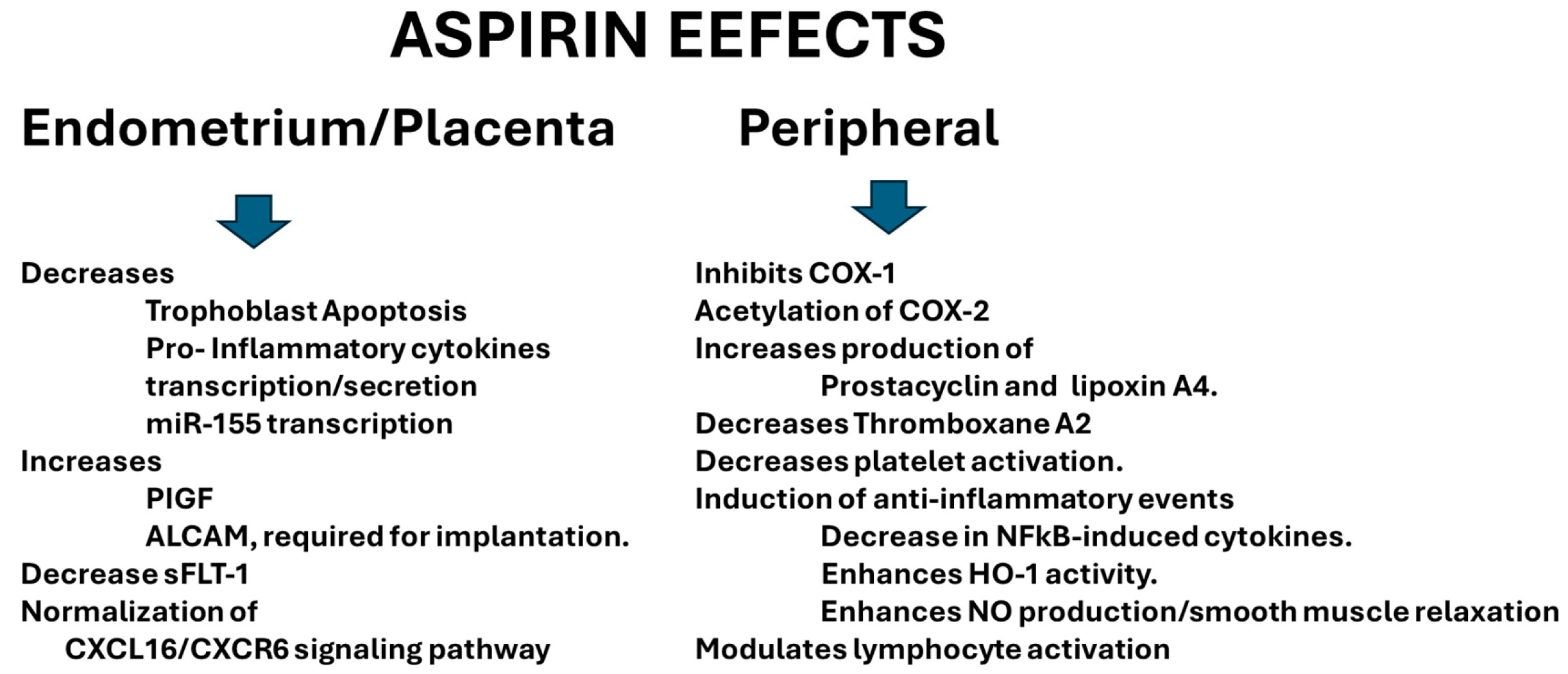
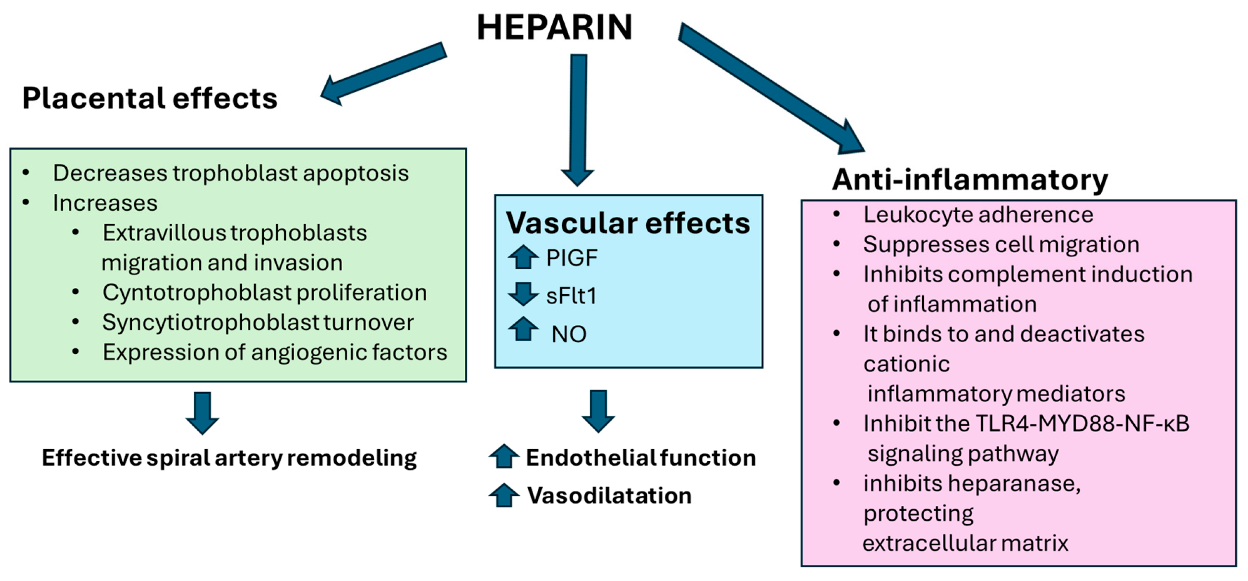
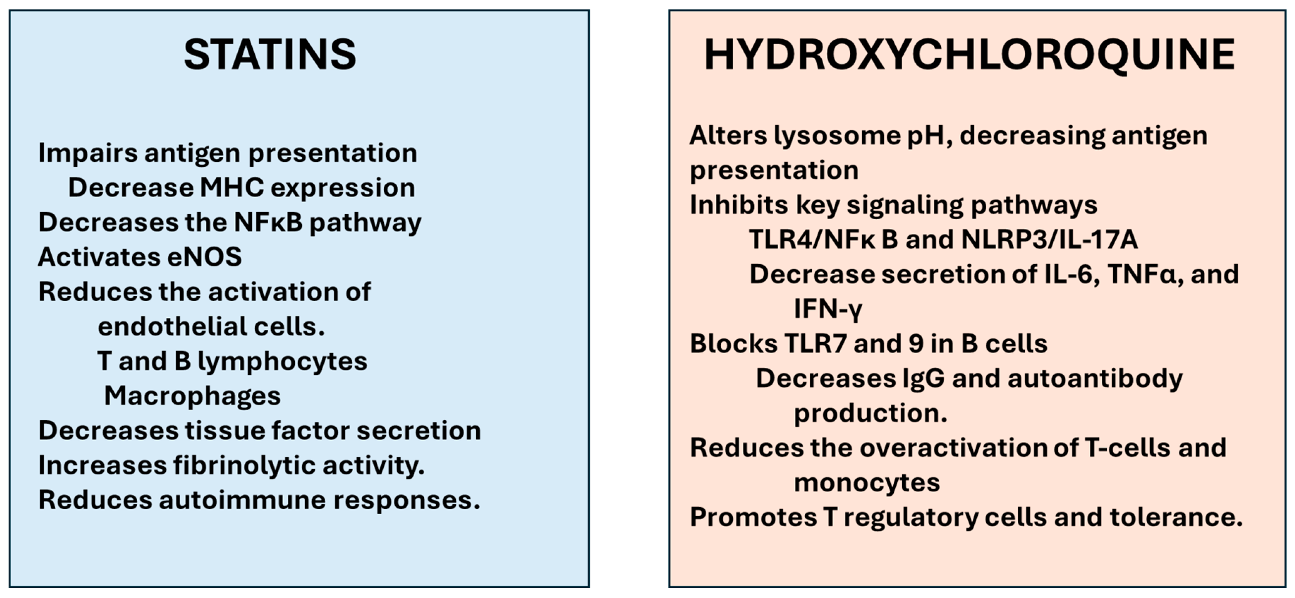
19. Immunological Treatments
20. Future Perspectives

21. Conclusions
Author Contributions
Funding
Data Availability Statement
Conflicts of Interest
References
- Gestational Hypertension and Preeclampsia: ACOG Practice Bulletin, Number 222. Obstet. Gynecol. 2020, 135, e237–e260. [CrossRef] [PubMed]
- Braunthal, S.; Brateanu, A. Hypertension in pregnancy: Pathophysiology and treatment. SAGE Open Med. 2019, 7, 2050312119843700. [Google Scholar] [CrossRef] [PubMed]
- Ryan, K.; McGrath, L.; Brookfield, K. Hypertension Management in Pregnancy. Annu. Rev. Med. 2025, 76, 315–326. [Google Scholar] [CrossRef] [PubMed]
- Harmon, A.C.; Cornelius, D.C.; Amaral, L.M.; Faulkner, J.L.; Cunningham, M.W., Jr.; Wallace, K.; LaMarca, B. The role of inflammation in the pathology of preeclampsia. Clin. Sci. 2016, 130, 409–419. [Google Scholar] [CrossRef] [PubMed]
- Borzychowski, A.M.; Sargent, I.L.; Redman, C.W. Inflammation and pre-eclampsia. Semin. Fetal Neonatal Med. 2006, 11, 309–316. [Google Scholar] [CrossRef] [PubMed]
- Blakey, H.; Sun, R.; Xie, L.; Russell, R.; Sarween, N.; Hodson, J.; Hargitai, B.; Marton, T.; Neil, D.A.H.; Wong, E.; et al. Pre-eclampsia is associated with complement pathway activation in the maternal and fetal circulation and placental tissue. Pregnancy Hypertens. 2023, 32, 43–49. [Google Scholar] [CrossRef] [PubMed]
- Burwick, R.M.; Java, A.; Regal, J.F. The role of complement in normal pregnancy and preeclampsia. Front. Immunol. 2025, 16, 1643896. [Google Scholar] [CrossRef] [PubMed]
- Burwick, R.M.; Feinberg, B.B. Complement activation and regulation in preeclampsia and hemolysis, elevated liver enzymes, and low platelet count syndrome. Am. J. Obstet. Gynecol. 2022, 226, S1059–S1070. [Google Scholar] [CrossRef] [PubMed]
- Canzoneri, B.J.; Lewis, D.F.; Groome, L.; Wang, Y. Increased neutrophil numbers account for leukocytosis in women with preeclampsia. Am. J. Perinatol. 2009, 26, 729–732. [Google Scholar] [CrossRef] [PubMed]
- Giaglis, S.; Stoikou, M.; Grimolizzi, F.; Subramanian, B.Y.; van Breda, S.V.; Hoesli, I.; Lapaire, O.; Hasler, P.; Than, N.G.; Hahn, S. Neutrophil migration into the placenta: Good, bad or deadly? Cell Adhes. Migr. 2016, 10, 208–225. [Google Scholar] [CrossRef] [PubMed]
- Qin, Z.; Long, Y. Immunological dysregulation in preeclampsia: Pathogenesis and clinical implications. Int. Immunopharmacol. 2025, 163, 115314. [Google Scholar] [CrossRef] [PubMed]
- Deer, E.; Herrock, O.; Campbell, N.; Cornelius, D.; Fitzgerald, S.; Amaral, L.M.; LaMarca, B. The role of immune cells and mediators in preeclampsia. Nat. Rev. Nephrol. 2023, 19, 257–270. [Google Scholar] [CrossRef] [PubMed]
- Ribatti, D. Endogenous inhibitors of angiogenesis: A historical review. Leuk. Res. 2009, 33, 638–644. [Google Scholar] [CrossRef] [PubMed]
- Zeisler, H.; Llurba, E.; Chantraine, F.; Vatish, M.; Staff, A.C.; Sennström, M.; Olovsson, M.; Brennecke, S.P.; Stepan, H.; Allegranza, D.; et al. Predictive Value of the sFlt-1:PlGF Ratio in Women with Suspected Preeclampsia. N. Engl. J. Med. 2016, 374, 13–22. [Google Scholar] [CrossRef] [PubMed]
- Rada, B. Neutrophil Extracellular Traps. Methods Mol. Biol. 2019, 1982, 517–528. [Google Scholar] [CrossRef] [PubMed]
- Hernández González, L.L.; Pérez-Campos Mayoral, L.; Hernández-Huerta, M.T.; Mayoral Andrade, G.; Martínez Cruz, M.; Ramos-Martínez, E. Targeting Neutrophil Extracellular Trap Formation: Exploring Promising Pharmacological Strategies for the Treatment of Preeclampsia. Pharmaceuticals 2024, 17, 605. [Google Scholar] [CrossRef] [PubMed]
- Kong, X.; Guo, Z.; Dong, J.; Hao, B.; Jiao, Y.; Wang, J.; Wu, Y.; Kang, S. Association of first-trimester peripheral blood count-derived immune markers with the risk of incident hypertensive disorders of pregnancy: A retrospective cohort study. Hypertens. Res. 2026, 49, 1182–1192. [Google Scholar] [CrossRef] [PubMed]
- Gelaw, Y.; Asrie, F.; Walle, M.; Getaneh, Z. The value of eosinophil count in the diagnosis of preeclampsia among pregnant women attending the University of Gondar Comprehensive Specialized Hospital, Northwest Ethiopia, 2021. BMC Pregnancy Childbirth 2022, 22, 557. [Google Scholar] [CrossRef] [PubMed]
- Chavez, B.; Kiaris, H. Insights on the role of the chemokine CCL8 in pathology. Cell. Signal. 2025, 134, 111951. [Google Scholar] [CrossRef] [PubMed]
- Purcell, W.M. Human placental mast cells: A role in pre-eclampsia? Med. Hypotheses 1992, 39, 281–283. [Google Scholar] [CrossRef] [PubMed]
- Wang, Y.; Gu, Y.; Lewis, D.F.; Alexander, J.S.; Granger, D.N. Elevated plasma chymotrypsin-like protease (chymase) activity in women with preeclampsia. Hypertens. Pregnancy 2010, 29, 253–261. [Google Scholar] [CrossRef] [PubMed]
- Faas, M.M.; De Vos, P. Innate immune cells in the placental bed in healthy pregnancy and preeclampsia. Placenta 2018, 69, 125–133. [Google Scholar] [CrossRef] [PubMed]
- Derbala, Y.; Elazzamy, H.; Bilal, M.; Reed, R.; Salazar Garcia, M.D.; Skariah, A.; Skariah, A.; Dambaeva, S.; Fernandez, E.; Germain, A.; et al. Mast cell-induced immunopathology in recurrent pregnancy losses. Am. J. Reprod. Immunol. 2019, 82, e13128. [Google Scholar] [CrossRef] [PubMed]
- Elieh Ali Komi, D.; Shafaghat, F.; Haidl, G. Significance of mast cells in spermatogenesis, implantation, pregnancy, and abortion: Cross talk and molecular mechanisms. Am. J. Reprod. Immunol. 2020, 83, e13228. [Google Scholar] [CrossRef] [PubMed]
- Staff, A.C.; Fjeldstad, H.E.; Fosheim, I.K.; Moe, K.; Turowski, G.; Johnsen, G.M.; Alnaes-Katjavivi, P.; Alnaes-Katjavivi, P.; Sugulle, M. Failure of physiological transformation and spiral artery atherosis: Their roles in preeclampsia. Am. J. Obstet. Gynecol. 2022, 226, S895–S906. [Google Scholar] [CrossRef] [PubMed]
- Broekhuizen, M.; Hitzerd, E.; van den Bosch, T.P.P.; Dumas, J.; Verdijk, R.M.; van Rijn, B.B.; Danser, A.H.J.; van Eijck, C.H.J.; Reiss, I.K.M.; Mustafa, D.A.M. The Placental Innate Immune System Is Altered in Early-Onset Preeclampsia, but Not in Late-Onset Preeclampsia. Front. Immunol. 2021, 12, 780043. [Google Scholar] [CrossRef] [PubMed]
- Kumaraswami, S.; Farkas, G. Management of a Parturient with Mast Cell Activation Syndrome: An Anesthesiologist’s Experience. Case Rep. Anesthesiol. 2018, 2018, 8920921. [Google Scholar] [CrossRef] [PubMed]
- Rudra, C.B.; Williams, M.A.; Frederick, I.O.; Luthy, D.A. Maternal asthma and risk of preeclampsia: A case-control study. J. Reprod. Med. 2006, 51, 94–100. [Google Scholar] [PubMed]
- Mirzakhani, H.; Carey, V.J.; McElrath, T.F.; Laranjo, N.; O’Connor, G.; Iverson, R.E.; Lee-Parritz, A.; Strunk, R.C.; Bacharier, L.B.; Macones, G.A.; et al. The Association of Maternal Asthma and Early Pregnancy Vitamin D with Risk of Preeclampsia: An Observation From Vitamin D Antenatal Asthma Reduction Trial (VDAART). J. Allergy Clin. Immunol. Pract. 2018, 6, 600–608.e2. [Google Scholar] [CrossRef] [PubMed]
- Dash, S.P.; Gupta, S.; Sarangi, P.P. Monocytes and macrophages: Origin, homing, differentiation, and functionality during inflammation. Heliyon 2024, 10, e29686. [Google Scholar] [CrossRef] [PubMed]
- Bezemer, R.E.; Faas, M.M.; van Goor, H.; Gordijn, S.J.; Prins, J.R. Decidual macrophages and Hofbauer cells in fetal growth restriction. Front. Immunol. 2024, 15, 1379537. [Google Scholar] [CrossRef] [PubMed]
- Mercnik, M.H.; Schliefsteiner, C.; Fluhr, H.; Wadsack, C. Placental macrophages present distinct polarization pattern and effector functions depending on clinical onset of preeclampsia. Front. Immunol. 2023, 13, 1095879. [Google Scholar] [CrossRef] [PubMed]
- Wei, Y.; Su, Y.; Liu, C.; Ma, X.; Ling, Z.; Wang, Y.; Qiao, R. Macrophage and Preeclampsia: Macrophage Polarization Imbalance at the Maternal-Fetal Interface. J. Clin. Lab. Anal. 2025, 39, e70046. [Google Scholar] [CrossRef] [PubMed]
- Wei, R.; Lai, N.; Zhao, L.; Zhang, Z.; Zhu, X.; Guo, Q.; Chu, C.; Fu, X.; Li, X. Dendritic cells in pregnancy and pregnancy-associated diseases. Biomed. Pharmacother. 2021, 133, 110921. [Google Scholar] [CrossRef] [PubMed]
- Miller, D.; Motomura, K.; Galaz, J.; Gershater, M.; Lee, E.D.; Romero, R.; Gomez-Lopez, N. Cellular immune responses in the pathophysiology of preeclampsia. J. Leukoc. Biol. 2022, 111, 237–260. [Google Scholar] [CrossRef] [PubMed]
- Wei, X.; Yang, X. The central role of natural killer cells in preeclampsia. Front. Immunol. 2023, 14, 1009867. [Google Scholar] [CrossRef] [PubMed]
- Köstlin-Gille, N.; Gille, C. Myeloid-Derived Suppressor Cells in Pregnancy and the Neonatal Period. Front. Immunol. 2020, 11, 584712. [Google Scholar] [CrossRef] [PubMed]
- Sundararajan, A.; Vora, K.; Natesan, S. Assessment of Quantitative and Functional Aspects of Dendritic Cell Subsets in Early Onset Pre-Eclampsia Patients. ARJGO 2021, 4, 1–14. [Google Scholar]
- Wang, Y.; Liu, Y.; Shu, C.; Wan, J.; Shan, Y.; Zhi, X.; Sun, L.; Yi, H.; Yang, Y.G.; He, J. Inhibition of pregnancy-associated granulocytic myeloid-derived suppressor cell expansion and arginase-1 production in preeclampsia. J. Reprod. Immunol. 2018, 127, 48–54. [Google Scholar] [CrossRef] [PubMed]
- Dong, S.; Shah, N.K.; He, J.; Han, S.; Xie, M.; Wang, Y.; Cheng, T.; Liu, Z.; Shu, C. The abnormal expression of Tim-3 is involved in the regulation of myeloid-derived suppressor cells and its correlation with preeclampsia. Placenta 2021, 114, 108–114. [Google Scholar] [CrossRef] [PubMed]
- Mace, E.M. Human natural killer cells: Form, function, and development. J. Allergy Clin. Immunol. 2023, 151, 371–385. [Google Scholar] [CrossRef] [PubMed]
- Chen, S.; Zhu, H.; Jounaidi, Y. Comprehensive snapshots of natural killer cells functions, signaling, molecular mechanisms and clinical utilization. Signal Transduct. Target. Ther. 2024, 9, 302. [Google Scholar] [CrossRef] [PubMed]
- Faas, M.M.; de Vos, P. Uterine NK cells and macrophages in pregnancy. Placenta 2017, 56, 44–52. [Google Scholar] [CrossRef] [PubMed]
- Garmendia, J.V.; De Sanctis, J.B. A Brief Analysis of Tissue-Resident NK Cells in Pregnancy and Endometrial Diseases: The Importance of Pharmacologic Modulation. Immuno 2021, 1, 174–193. [Google Scholar] [CrossRef]
- Garmendia, J.V.; De Sanctis, C.V.; Hajdúch, M.; De Sanctis, J.B. Exploring the Immunological Aspects and Treatments of Recurrent Pregnancy Loss and Recurrent Implantation Failure. Int. J. Mol. Sci. 2025, 26, 1295. [Google Scholar] [CrossRef] [PubMed]
- Li, Q.; Sharkey, A.; Sheridan, M.; Magistrati, E.; Arutyunyan, A.; Huhn, O.; Sancho-Serra, C.; Anderson, H.; McGovern, N.; Esposito, L.; et al. Human uterine natural killer cells regulate differentiation of extravillous trophoblast early in pregnancy. Cell Stem Cell 2024, 31, 181–195.e9. [Google Scholar] [CrossRef] [PubMed]
- Borzychowski, A.M.; Croy, B.A.; Chan, W.L.; Redman, C.W.; Sargent, I.L. Changes in systemic type 1 and type 2 immunity in normal pregnancy and pre-eclampsia may be mediated by natural killer cells. Eur. J. Immunol. 2005, 35, 3054–3063. [Google Scholar] [CrossRef] [PubMed]
- Sabetkam, S.; Rafat, A.; Mazloumi, Z.; Kalarestaghi, H.; Bahramloo, M.; Naderali, E.; Asl, K.D. Role of uterine NK cells in pregnancy complication. Pathol. Res. Pract. 2025, 270, 155998. [Google Scholar] [CrossRef] [PubMed]
- Yue, S.; Meng, J. Role of Decidual Natural Killer Cells in the Pathogenesis of Preeclampsia. Am. J. Reprod. Immunol. 2025, 93, e70033. [Google Scholar] [CrossRef] [PubMed]
- Williams, P.J.; Bulmer, J.N.; Searle, R.F.; Innes, B.A.; Robson, S.C. Altered decidual leucocyte populations in the placental bed in pre-eclampsia and foetal growth restriction: A comparison with late normal pregnancy. Reproduction 2009, 138, 177–184. [Google Scholar] [CrossRef] [PubMed]
- Rieger, L.; Segerer, S.; Bernar, T.; Kapp, M.; Majic, M.; Morr, A.K.; Kämmerer, U. Specific subsets of immune cells in human decidua differ between normal pregnancy and preeclampsia--a prospective observational study. Reprod. Biol. Endocrinol. 2009, 7, 132. [Google Scholar] [CrossRef] [PubMed]
- Lockwood, C.J.; Huang, S.J.; Chen, C.P.; Huang, Y.; Xu, J.; Faramarzi, S.; Kayisli, O.; Kayisli, U.; Koopman, L.; Smedts, D.; et al. Decidual cell regulation of natural killer cell-recruiting chemokines: Implications for the pathogenesis and prediction of preeclampsia. Am. J. Pathol. 2013, 183, 841–856. [Google Scholar] [CrossRef] [PubMed]
- Miko, E.; Barakonyi, A.; Meggyes, M.; Szereday, L. The Role of Type I and Type II NKT Cells in Materno-Fetal Immunity. Biomedicines 2021, 9, 1901. [Google Scholar] [CrossRef] [PubMed]
- Chojnacka-Purpurowicz, J.; Owczarczyk-Saczonek, A.; Nedoszytko, B. The Role of Gamma Delta T Lymphocytes in Physiological and Pathological Condition-Focus on Psoriasis, Atopic Dermatitis, Autoimmune Disorders, Cancer and Lymphomas. Int. J. Mol. Sci. 2024, 25, 7960. [Google Scholar] [CrossRef] [PubMed]
- Hashemi, V.; Dolati, S.; Hosseini, A.; Gharibi, T.; Danaii, S.; Yousefi, M. Natural killer T cells in Preeclampsia: An updated review. Biomed. Pharmacother. 2017, 95, 412–418. [Google Scholar] [CrossRef] [PubMed]
- Miko, E.; Szereday, L.; Barakonyi, A.; Jarkovich, A.; Varga, P.; Szekeres-Bartho, J. Immunoactivation in preeclampsia: Vdelta2+ and regulatory T cells during the inflammatory stage of disease. J. Reprod. Immunol. 2009, 80, 100–108. [Google Scholar] [CrossRef] [PubMed]
- Xu, Q.; Liu, H.; Wang, L.L.; Zhu, Q.; Zhang, Y.J.; Muyayalo, K.P.; Liao, A.H. Roles of γδT cells in pregnancy and pregnancy-related complications. Am. J. Reprod. Immunol. 2021, 86, e13487. [Google Scholar] [CrossRef] [PubMed]
- Lissauer, D.; Kilby, M.D.; Moss, P. Maternal effector T cells within decidua: The adaptive immune response to pregnancy? Placenta 2017, 60, 140–144. [Google Scholar] [CrossRef] [PubMed]
- Peng, X.; Chinwe Oluchi-Amaka, I.; Kwak-Kim, J.; Yang, X. A comprehensive review of the roles of T-cell immunity in preeclampsia. Front. Immunol. 2025, 16, 1476123. [Google Scholar] [CrossRef] [PubMed]
- Robertson, S.A.; Green, E.S.; Care, A.S.; Moldenhauer, L.M.; Prins, J.R.; Hull, M.L.; Barry, S.C.; Dekker, G. Therapeutic Potential of Regulatory T Cells in Preeclampsia—Opportunities and Challenges. Front. Immunol. 2019, 10, 478. [Google Scholar] [CrossRef] [PubMed]
- Saito, S.; Tsuda, S.; Nakashima, A. T cell immunity and the etiology and pathogenesis of preeclampsia. J. Reprod. Immunol. 2023, 159, 104125. [Google Scholar] [CrossRef] [PubMed]
- Wang, W.; Sung, N.; Gilman-Sachs, A.; Kwak-Kim, J. T Helper (Th) Cell Profiles in Pregnancy and Recurrent Pregnancy Losses: Th1/Th2/Th9/Th17/Th22/Tfh Cells. Front. Immunol. 2020, 11, 2025. [Google Scholar] [CrossRef] [PubMed]
- Saito, S.; Sakai, M.; Sasaki, Y.; Tanebe, K.; Tsuda, H.; Michimata, T. Quantitative analysis of peripheral blood Th0, Th1, Th2 and the Th1:Th2 cell ratio during normal human pregnancy and preeclampsia. Clin. Exp. Immunol. 1999, 117, 550–555. [Google Scholar] [CrossRef] [PubMed]
- Dos Santos Fagundes, I.; Brendler, E.P.; Nunes Erthal, I.; Eder Ribeiro, R.J.; Caron-Lienert, R.S.; Machado, D.C.; Pinheiro da Costa, B.E.; Poli-de-Figueiredo, C.E. Total Th1/Th2 cytokines profile from peripheral blood lymphocytes in normal pregnancy and preeclampsia syndrome. Hypertens. Pregnancy 2022, 41, 15–22. [Google Scholar] [CrossRef] [PubMed]
- Waite, J.C.; Skokos, D. Th17 response and inflammatory autoimmune diseases. Int. J. Inflamm. 2012, 2012, 819467. [Google Scholar] [CrossRef] [PubMed]
- De Sanctis, J.B.; Garmendia, J.V.; Moreno, D.; Larocca, N.; Mijares, M.; Di Giulio, C.; Salazar-Bookaman, M.; Wojewodka, G.; Radzioch, D. Pharmacological modulation of Th17. Recent Pat. Inflamm. Allergy Drug Discov. 2009, 3, 149–156. [Google Scholar] [CrossRef] [PubMed]
- Nalbant, A. IL-17, IL-21, and IL-22 Cytokines of T Helper 17 Cells in Cancer. J. Interferon Cytokine Res. 2019, 39, 56–60. [Google Scholar] [CrossRef] [PubMed]
- Walsh, S.W.; Nugent, W.H.; Archer, K.J.; Al Dulaimi, M.; Washington, S.L.; Strauss, J.F., 3rd. Epigenetic Regulation of Interleukin-17-Related Genes and Their Potential Roles in Neutrophil Vascular Infiltration in Preeclampsia. Reprod. Sci. 2022, 29, 154–162. [Google Scholar] [CrossRef] [PubMed]
- Zolfaghari, M.A.; Arefnezhad, R.; Parhizkar, F.; Hejazi, M.S.; Motavalli Khiavi, F.; Mahmoodpoor, A.; Yousefi, M. T lymphocytes and preeclampsia: The potential role of T-cell subsets and related MicroRNAs in the pathogenesis of preeclampsia. Am. J. Reprod. Immunol. 2021, 86, e13475. [Google Scholar] [CrossRef] [PubMed]
- Stefańska, K.; Zieliński, M.; Jankowiak, M.; Zamkowska, D.; Sakowska, J.; Adamski, P.; Piekarska, K.; Leszczyńska, K.; Świątkowska-Stodulska, R.; Kwiatkowski, S.; et al. Cytokine Imprint in Preeclampsia. Front. Immunol. 2021, 12, 667841. [Google Scholar] [CrossRef] [PubMed]
- Garmendia, J.V.; Blanca, I.; Peña, M.J.; De Sanctis, C.V.; De Sanctis, J.B. Unlocking the Puzzle: Investigating the Role of Interleukin 17 Genetic Polymorphisms, Circulating Lymphocytes, and Serum Levels in Venezuelan Women with Recurrent Pregnancy Loss. Immuno 2024, 4, 301–311. [Google Scholar] [CrossRef]
- Eyerich, S.; Eyerich, K.; Pennino, D.; Carbone, T.; Nasorri, F.; Pallotta, S.; Cianfarani, F.; Odorisio, T.; Traidl- Hoffmann, C.; Behrendt, H.; et al. Th22 cells represent a distinct human T cell subset involved in epidermal immunity and remodeling. J. Clin. Investig. 2009, 119, 3573–3585. [Google Scholar] [CrossRef] [PubMed]
- Wang, L.; Liang, Y.; Zhao, C.; Ma, P.; Zeng, S.; Ju, D.; Zhao, M.; Yu, M.; Shi, Y. Regulatory T cells in homeostasis and disease: Molecular mechanisms and therapeutic potential. Signal Transduct. Target. Ther. 2025, 10, 345. [Google Scholar] [CrossRef] [PubMed]
- Nevers, T.; Kalkunte, S.; Sharma, S. Uterine Regulatory T cells, IL-10 and hypertension. Am. J. Reprod. Immunol. 2011, 66, 88–92. [Google Scholar] [CrossRef] [PubMed]
- Li, Z.; Si, P.; Meng, T.; Zhao, X.; Zhu, C.; Zhang, D.; Meng, S.; Li, N.; Liu, R.; Ni, T.; et al. CCR8+ decidual regulatory T cells maintain maternal-fetal immune tolerance during early pregnancy. Sci. Immunol. 2025, 10, eado2463. [Google Scholar] [CrossRef] [PubMed]
- Sava, R.I.; March, K.L.; Pepine, C.J. Hypertension in pregnancy: Taking cues from pathophysiology for clinical practice. Clin. Cardiol. 2018, 41, 220–227. [Google Scholar] [CrossRef] [PubMed]
- Sasaki, Y.; Darmochwal-Kolarz, D.; Suzuki, D.; Sakai, M.; Ito, M.; Shima, T.; Shiozaki, A.; Rolinski, J.; Saito, S. Proportion of peripheral blood and decidual CD4(+) CD25(bright) regulatory T cells in pre-eclampsia. Clin. Exp. Immunol. 2007, 149, 139–145. [Google Scholar] [CrossRef] [PubMed]
- Palei, A.C.; Spradley, F.T.; Warrington, J.P.; George, E.M.; Granger, J.P. Pathophysiology of hypertension in pre-eclampsia: A lesson in integrative physiology. Acta Physiol. 2013, 208, 224–233. [Google Scholar] [CrossRef] [PubMed]
- Rahimzadeh, M.; Norouzian, M.; Arabpour, F.; Naderi, N. Regulatory T-cells and preeclampsia: An overview of literature. Expert Rev. Clin. Immunol. 2016, 12, 209–227. [Google Scholar] [CrossRef] [PubMed]
- Gomez-Lopez, N.; Kareus, E.; Lee, S. Immune cellular homeostasis and its breakdown at the maternal-fetal interface. Trends Immunol. 2026, 47, 202–211. [Google Scholar] [CrossRef] [PubMed]
- Liu, J.C.; Zeng, Q.; Duan, Y.G.; Yeung, W.S.B.; Li, R.H.W.; Ng, E.H.Y.; Cheung, K.W.; Zhang, Q.; Chiu, P.C.N. B cells: Roles in physiology and pathology of pregnancy. Front. Immunol. 2024, 15, 1456171. [Google Scholar] [CrossRef] [PubMed]
- Magatti, M.; Masserdotti, A.; Cargnoni, A.; Papait, A.; Stefani, F.R.; Silini, A.R.; Parolini, O. The Role of B Cells in PE Pathophysiology: A Potential Target for Perinatal Cell-Based Therapy? Int. J. Mol. Sci. 2021, 22, 3405. [Google Scholar] [CrossRef] [PubMed]
- Panova, I.A.; Kudryashova, A.V.; Panashchatenko, A.S.; Rokotyanskaya, E.A.; Malyshkina, A.I.; Parejshvili, V.V.; Harlamova, N.V. Character of β-lymphocytes differentiation in women with hypertensive disorders during pregnancy. Klin. Lab. Diagn. 2021, 66, 489–495. [Google Scholar] [CrossRef] [PubMed]
- Jensen, F.; Wallukat, G.; Herse, F.; Budner, O.; El-Mousleh, T.; Costa, S.D.; Dechend, R.; Zenclussen, A.C. CD19+CD5+ cells as indicators of preeclampsia. Hypertension 2012, 59, 861–868. [Google Scholar] [CrossRef] [PubMed]
- Nellore, A.; Killian, J.T., Jr.; Porrett, P.M. Memory B Cells in Pregnancy Sensitization. Front. Immunol. 2021, 12, 688987. [Google Scholar] [CrossRef] [PubMed]
- Chaiworapongsa, T.; Romero, R.; Gotsch, F.; Gomez-Lopez, N.; Suksai, M.; Gallo, D.M.; Jung, E.; Levenson, D.; Tarca, A.L. One-third of patients with eclampsia at term do not have an abnormal angiogenic profile. J. Perinat. Med. 2022, 51, 652–663. [Google Scholar] [CrossRef] [PubMed]
- Chaiworapongsa, T.; Romero, R.; Gomez-Lopez, N.; Suksai, M.; Gallo, D.M.; Jung, E.; Berry, S.M.; Awonuga, A.; Tarca, A.L.; Bryant, D.R. Preeclampsia at term: Evidence of disease heterogeneity based on the profile of circulating cytokines and angiogenic factors. Am. J. Obstet. Gynecol. 2024, 230, 450 e1–450 e18. [Google Scholar] [CrossRef] [PubMed]
- Hariharan, N.; Shoemaker, A.; Wagner, S. Pathophysiology of hypertension in preeclampsia. Microvasc. Res. 2017, 109, 34–37. [Google Scholar] [CrossRef] [PubMed]
- Matsubara, K.; Higaki, T.; Matsubara, Y.; Nawa, A. Nitric oxide and reactive oxygen species in the pathogenesis of preeclampsia. Int. J. Mol. Sci. 2015, 16, 4600–4614. [Google Scholar] [CrossRef] [PubMed]
- Buhimschi, I.A.; Saade, G.R.; Chwalisz, K.; Garfield, R.E. The nitric oxide pathway in pre-eclampsia: Pathophysiological implications. Hum. Reprod. Update 1998, 4, 25–42. [Google Scholar] [CrossRef] [PubMed]
- Kutllovci Hasani, K.; Ajeti, N.; Goswami, N. Understanding Preeclampsia: Cardiovascular Pathophysiology, Histopathological Insights and Molecular Biomarkers. Med. Sci. 2025, 13, 154. [Google Scholar] [CrossRef] [PubMed]
- Maynard, S.E.; Karumanchi, S.A. Angiogenic factors and preeclampsia. Semin. Nephrol. 2011, 31, 33–46. [Google Scholar] [CrossRef] [PubMed]
- Jim, B.; Karumanchi, S.A. Preeclampsia: Pathogenesis, Prevention, and Long-Term Complications. Semin. Nephrol. 2017, 37, 386–397. [Google Scholar] [CrossRef] [PubMed]
- Venkatesha, S.; Toporsian, M.; Lam, C.; Hanai, J.; Mammoto, T.; Kim, Y.M. Soluble endoglin contributes to the pathogenesis of preeclampsia. Nat. Med. 2006, 12, 642–649. [Google Scholar] [CrossRef] [PubMed]
- Youssef, L.; Crispi, F.; Paolucci, S.; Miranda, J.; Lobmaier, S.; Crovetto, F.; Figueras, F.; Gratacos, E. Angiogenic factors alone or in combination with ultrasound Doppler criteria for risk classification among late-onset small fetuses with or without pre-eclampsia. Ultrasound Obstet. Gynecol. 2025, 65, 317–324. [Google Scholar] [CrossRef] [PubMed]
- Mula, R.; Prats, P.; García, S.; Serra, B.; Scazzocchio, E.; Meler, E. Angiogenic factors assessment in pre-eclampsia high-risk population for the prediction of small-for-gestational age neonates: A prospective longitudinal study. Int. J. Gynecol. Obstet. 2023, 161, 439–446. [Google Scholar] [CrossRef] [PubMed]
- Hackelöer, M.; Schmidt, L.; Verlohren, S. New advances in prediction and surveillance of preeclampsia: Role of machine learning approaches and remote monitoring. Arch. Gynecol. Obstet. 2023, 308, 1663–1677. [Google Scholar] [CrossRef] [PubMed]
- Blois, S.M.; Barrientos, G. Galectin signature in normal pregnancy and preeclampsia. J. Reprod. Immunol. 2014, 101–102, 127–134. [Google Scholar] [CrossRef] [PubMed]
- Jovanović Krivokuća, M.; Vilotić, A.; Nacka-Aleksić, M.; Pirković, A.; Ćujić, D.; Legner, J.; Dekanski, D.; Bojić-Trbojević, Ž. Galectins in Early Pregnancy and Pregnancy-Associated Pathologies. Int. J. Mol. Sci. 2022, 23, 69. [Google Scholar] [CrossRef] [PubMed]
- Karadzov Orlic, N.; Joksić, I. Preeclampsia pathogenesis and prediction—Where are we now: The focus on the role of galectins and miRNAs. Hypertens. Pregnancy 2025, 44, 2470626. [Google Scholar] [CrossRef] [PubMed]
- Kandel, M.; Tong, S.; Walker, S.P.; Cannon, P.; Nguyen, T.V.; MacDonald, T.M.; Hannan, N.J.; Kaitu’u-Lino, T.J.; Bartho, L.A. Placental galectin-3 is reduced in early-onset preeclampsia. Front. Physiol. 2022, 13, 1037597. [Google Scholar] [CrossRef] [PubMed]
- Li, Y.; Sang, Y.; Chang, Y.; Xu, C.; Lin, Y.; Zhang, Y.; Chiu, P.C.N.; Yeung, W.S.B.; Zhou, H.; Dong, N.; et al. A Galectin-9-Driven CD11chigh Decidual Macrophage Subset Suppresses Uterine Vascular Remodeling in Preeclampsia. Circulation 2024, 149, 1670–1688. [Google Scholar] [CrossRef] [PubMed]
- Aisagbonhi, O.; Morris, G.P. Human Leukocyte Antigens in Pregnancy and Preeclampsia. Front. Genet. 2022, 13, 884275. [Google Scholar] [CrossRef] [PubMed]
- Pishesha, N.; Harmand, T.J.; Ploegh, H.L. A guide to antigen processing and presentation. Nat. Rev. Immunol. 2022, 22, 751–764. [Google Scholar] [CrossRef] [PubMed]
- Triche, E.W.; Harland, K.K.; Field, E.H.; Rubenstein, L.M.; Saftlas, A.F. Maternal-fetal HLA sharing and preeclampsia: Variation in effects by seminal fluid exposure in a case-control study of nulliparous women in Iowa. J. Reprod. Immunol. 2014, 101–102, 111–119. [Google Scholar] [CrossRef] [PubMed]
- Van’t Hof, L.J.; Schotvanger, N.; Haasnoot, G.W.; van der Keur, C.; Roelen, D.L.; Lashley, L.E.E.L.O.; Claas, F.H.J.; Eikmans, M.; van der Hoorn, M.P. Maternal-Fetal HLA Compatibility in Uncomplicated and Preeclamptic Naturally Conceived Pregnancies. Front. Immunol. 2021, 12, 673131. [Google Scholar] [CrossRef] [PubMed]
- van Bentem, K.; Bos, M.; van der Keur, C.; Brand-Schaaf, S.H.; Haasnoot, G.W.; Roelen, D.L.; Eikmans, M.; Heidt, S.; Claas, F.H.J.; Lashley, E.E.L.O.; et al. The development of preeclampsia in oocyte donation pregnancies is related to the number of fetal-maternal HLA class II mismatches. J. Reprod. Immunol. 2020, 137, 103074. [Google Scholar] [CrossRef] [PubMed]
- Emmery, J.; Hachmon, R.; Pyo, C.W.; Nelson, W.C.; Geraghty, D.E.; Andersen, A.M.; Melbye, M.; Hviid, T.V. Maternal and fetal human leukocyte antigen class Ia and II alleles in severe preeclampsia and eclampsia. Genes Immun. 2016, 17, 251–260. [Google Scholar] [CrossRef] [PubMed]
- Chaaithanya, I.K.; Rajalingam, R. Emerging roles of natural killer cell ligands-HLA-E, HLA-F, HLA-G, MICA, and MICB-in in vitro fertilization outcomes. Front. Genet. 2025, 16, 1661511. [Google Scholar] [CrossRef] [PubMed]
- Møller, H.I.; Christiansen, C.M.; Klok, F.R.; Pedersen, N.H.; Blauenfeldt, T.; Finne, K.F.; Nielsen, H.S.; Hviid, T.V.F. Investigations of HLA-F and HLA-G 3′UTR Polymorphisms in Preeclampsia and Fetal Growth Restriction Indicate a Possible Role of HLA-F-HLA-G Haplotypes and Diplotypes. HLA 2025, 106, e70293. [Google Scholar] [CrossRef] [PubMed]
- Tang, Y.; Liu, H.; Li, H.; Peng, T.; Gu, W.; Li, X. Hypermethylation of the HLA-G promoter is associated with preeclampsia. Mol. Hum. Reprod. 2015, 21, 736–744. [Google Scholar] [CrossRef] [PubMed]
- Pabalan, N.; Jarjanazi, H.; Sun, C.; Iversen, A.C. Meta-analysis of the human leukocyte antigen-G (HLA-G) 14 bp insertion/deletion polymorphism as a risk factor for preeclampsia. Tissue Antigens 2015, 86, 186–194. [Google Scholar] [CrossRef] [PubMed]
- Lee, J.; Romero, R.; Xu, Y.; Miranda, J.; Yoo, W.; Chaemsaithong, P.; Kusanovic, J.P.; Chaiworapongsa, T.; Tarca, A.L.; Korzeniewski, S.J.; et al. Detection of anti-HLA antibodies in maternal blood in the second trimester to identify patients at risk of antibody-mediated maternal anti-fetal rejection and spontaneous preterm delivery. Am. J. Reprod. Immunol. 2013, 70, 162–175. [Google Scholar] [CrossRef] [PubMed]
- Moffett, A.; Colucci, F. Co-evolution of NK receptors and HLA ligands in humans is driven by reproduction. Immunol. Rev. 2015, 267, 283–297. [Google Scholar] [CrossRef] [PubMed]
- Hiby, S.E.; Walker, J.J.; O’shaughnessy, K.M.; Redman, C.W.; Carrington, M.; Trowsdale, J.; Moffett, A. Combinations of maternal KIR and fetal HLA-C genes influence the risk of preeclampsia and reproductive success. J. Exp. Med. 2004, 200, 957–965. [Google Scholar] [CrossRef] [PubMed]
- Larsen, T.G.; Hackmon, R.; Geraghty, D.E.; Hviid, T.V.F. Fetal human leukocyte antigen-C and maternal killer-cell immunoglobulin-like receptors in cases of severe preeclampsia. Placenta 2019, 75, 27–33. [Google Scholar] [CrossRef] [PubMed]
- Vinnars, M.T.; Björk, E.; Nagaev, I.; Ottander, U.; Bremme, K.; Holmlund, U.; Sverremark-Ekström, E.; Mincheva-Nilsson, L. Enhanced Th1 and inflammatory mRNA responses upregulate NK cell cytotoxicity and NKG2D ligand expression in human pre-eclamptic placenta and target it for NK cell attack. Am. J. Reprod. Immunol. 2018, 80, e12969. [Google Scholar] [CrossRef] [PubMed]
- Taefehshokr, N.; Baradaran, B.; Baghbanzadeh, A.; Taefehshokr, S. Promising approaches in cancer immunotherapy. Immunobiology 2020, 225, 151875. [Google Scholar] [CrossRef] [PubMed]
- Du, X.; Liu, H.; Shi, J.; Yang, P.; Gu, Y.; Meng, J. The PD-1/PD-L1 signaling pathway regulates decidual macrophage polarization and may participate in preeclampsia. J. Reprod. Immunol. 2024, 164, 104258. [Google Scholar] [CrossRef] [PubMed]
- Tian, Y.; Peng, X.; Yang, X. Decreased PD-L1 contributes to preeclampsia by suppressing GM-CSF via the JAK2/STAT5 signal pathway. Sci. Rep. 2025, 15, 3124. [Google Scholar] [CrossRef] [PubMed]
- Parhizkar, F.; Shekari, N.; HajiEsmailPoor, Z.; Parsania, S.; Soltani-Zangbar, M.S.; Aghebati-Maleki, A.; Aghebati-Maleki, L. Investigation of immune checkpoint molecules (CTLA-4, PD-1, PD-L1, Tim-3) expressions in preeclampsia: A comparative study of membranous and soluble forms. Hum. Immunol. 2025, 86, 111298. [Google Scholar] [CrossRef] [PubMed]
- Liu, J.; Song, G.; Zhao, G.; Meng, T. Gene polymorphism associated with FOXP3, CTLA-4 and susceptibility to pre-eclampsia: A meta-analysis and trial sequential analysis. J. Obstet. Gynaecol. 2022, 42, 1085–1091. [Google Scholar] [CrossRef] [PubMed]
- Kandel, S.; Adhikary, P.; Li, G.; Cheng, K. The TIM3/Gal9 signaling pathway: An emerging target for cancer immunotherapy. Cancer Lett. 2021, 510, 67–78. [Google Scholar] [CrossRef] [PubMed]
- Mittelberger, J.; Seefried, M.; Löb, S.; Kuhn, C.; Franitza, M.; Garrido, F.; Ditsch, N.; Jeschke, U.; Dannecker, C. The expression of TIM-3 and Gal-9 on macrophages and Hofbauer cells in the placenta of preeclampsia patients. J. Reprod. Immunol. 2024, 164, 104296. [Google Scholar] [CrossRef] [PubMed]
- Doyle, L.M.; Wang, M.Z. Overview of Extracellular Vesicles, Their Origin, Composition, Purpose, and Methods for Exosome Isolation and Analysis. Cells 2019, 8, 727. [Google Scholar] [CrossRef] [PubMed]
- Fato, B.R.; de Alwis, N.; Hannan, N.J. Exploring placental dysfunction: Models of extracellular vesicle action in preeclampsia. Reproduction 2026, 171, xaag011. [Google Scholar] [CrossRef] [PubMed]
- Cheng, S.B.; Nakashima, A.; Huber, W.J.; Davis, S.; Banerjee, S.; Huang, Z.; Saito, S.; Sadovsky, Y.; Sharma, S. Pyroptosis is a critical inflammatory pathway in the placenta from early onset preeclampsia and in human trophoblasts exposed to hypoxia and endoplasmic reticulum stressors. Cell Death Dis. 2019, 10, 927. [Google Scholar] [CrossRef] [PubMed]
- Schuster, J.; Cheng, S.B.; Padbury, J.; Sharma, S. Placental extracellular vesicles and pre-eclampsia. Am. J. Reprod. Immunol. 2021, 85, e13297. [Google Scholar] [CrossRef] [PubMed]
- Sun, Q.; Chang, H.; Wang, H.; Zheng, L.; Wenig, Y.; Zheng, D.; Zheng, D. Regulatory roles of extracellular vesicles in pregnancy complications. J. Adv. Res. 2025, 78, 363–375. [Google Scholar] [CrossRef] [PubMed]
- Ramos, A.; Youssef, L.; Molina, P.; Torramadé-Moix, S.; Martinez-Sanchez, J.; Moreno-Castaño, A.B.; Blasco, M.; Guillén-Olmos, E.; De Moner, B.; Pino, M. Circulating extracellular vesicles and neutrophil extracellular traps contribute to endothelial dysfunction in preeclampsia. Front. Immunol. 2024, 15, 1488127. [Google Scholar] [CrossRef] [PubMed]
- Pham, Q.N.; Milanova, V.; Tung, T.T.; Losic, D.; Thierry, B.; Winter, M.A. Affinity enrichment of placental extracellular vesicles from minimally processed maternal plasma with magnetic nanowires. Analyst 2025, 150, 1908–1919. [Google Scholar] [CrossRef] [PubMed]
- Hula , H.; Escalera, D.; Goulopoulou, S. Extracellular vesicles in preeclampsia: Drivers of vascular dysfunction and inflammation. Am. J. Physiol. Heart Circ. Physiol. 2025, 329, H1560–H1574. [Google Scholar] [CrossRef] [PubMed]
- Than, N.G.; Romero, R.; Fitzgerald, W.; Gudicha, D.W.; Gomez-Lopez, N.; Posta, M.; Zhou, F.; Bhatti, G.; Meyyazhagan, A.; Awonuga, A.O. Proteomic Profiles of Maternal Plasma Extracellular Vesicles for Prediction of Preeclampsia. Am. J. Reprod. Immunol. 2024, 92, e13928. [Google Scholar] [CrossRef] [PubMed]
- Chen, H.Y.; Wang, X.Y.; Lu, K.M.; Yu, C.H.; Su, M.T.; Kang, L.; Hsu, K.F.; Chen, P.F.; Lin, S.H. Maternal Th17/Treg Cytokines and Small Extracellular Vesicles in Plasma as Potential Biomarkers for Preeclampsia. Int. J. Med. Sci. 2022, 19, 1672–1679. [Google Scholar] [CrossRef] [PubMed]
- O’Brien, J.; Hayder, H.; Zayed, Y.; Peng, C. Overview of MicroRNA Biogenesis, Mechanisms of Actions, and Circulation. Front. Endocrinol. 2018, 9, 402. [Google Scholar] [CrossRef] [PubMed]
- Giannubilo, S.R.; Cecati, M.; Marzioni, D.; Ciavattini, A. Circulating miRNAs and Preeclampsia: From Implantation to Epigenetics. Int. J. Mol. Sci. 2024, 25, 1418. [Google Scholar] [CrossRef] [PubMed]
- Boelig, R.C.; Tawk, A.; Zhan, T.; Kraft, W.K.; McKenzie, S.E.; Michael, J. Platelet-Associated microRNAs as Markers of Aspirin Response and Pregnancy Outcome in Pregnancies at High-Risk for Preeclampsia. Am. J. Perinatol. 2026, 43, 557–560. [Google Scholar] [CrossRef] [PubMed]
- Jaszczuk, I.; Koczkodaj, D.; Kondracka, A.; Kwaśniewska, A.; Winkler, I.; Filip, A. The role of miRNA-210 in pre-eclampsia development. Ann. Med. 2022, 54, 1350–1356. [Google Scholar] [CrossRef] [PubMed]
- Han, L.; Luo, Q.Q.; Peng, M.G.; Zhang, Y.; Zhu, X.H. miR-483 is downregulated in pre-eclampsia via targeting insulin-like growth factor 1 (IGF1) and regulates the PI3K/Akt/mTOR pathway of endothelial progenitor cells. J. Obstet. Gynaecol. Res. 2021, 47, 63–72. [Google Scholar] [CrossRef] [PubMed]
- Lopes, A.C.S.; Macedo, A.A.; Mendes, F.S.; Costa, I.M.; Dusse, L.M.S.; Alpoim, P.N. Changes in microRNA expression associated with preeclampsia: A systematic review. Braz. J. Med. Biol. Res. 2025, 58, e13988. [Google Scholar] [CrossRef] [PubMed]
- Gerede, A.; Stavros, S.; Danavasi, M.; Potiris, A.; Moustakli, E.; Machairiotis, N.; Zikopoulos, A.; Nikolettos, K.; Drakakis, P.; Nikolettos, N.; et al. MicroRNAs in Preeclampsia: Bridging Diagnosis and Treatment. J. Clin. Med. 2025, 14, 2003. [Google Scholar] [CrossRef] [PubMed]
- Grezzana, G.B.; Stein, A.T.; Markoski, M.M. Towards early diagnosis of Preeclampsia: Emerging roles of circulating microRNAs. Eur. J. Obstet. Gynecol. Reprod. Biol. 2026, 316, 114822. [Google Scholar] [CrossRef] [PubMed]
- Conrad, K.P.; Miles, T.M.; Benyo, D.F. Circulating levels of immunoreactive cytokines in women with preeclampsia. Am. J. Reprod. Immunol. 1998, 40, 102–111. [Google Scholar] [CrossRef] [PubMed]
- Kurmanova, A.; Urazbayeva, G.; Kayupova, L.; Salimbaeva, D.; Dzhardemalieva, N. Dynamics of serum cytokines in preeclampsia. Eur. Cytokine Netw. 2024, 35, 21–27. [Google Scholar] [CrossRef] [PubMed]
- Vilotić, A.; Nacka-Aleksić, M.; Pirković, A.; Bojić-Trbojević, Ž.; Dekanski, D.; Jovanović Krivokuća, M. IL-6 and IL-8: An Overview of Their Roles in Healthy and Pathological Pregnancies. Int. J. Mol. Sci. 2022, 23, 14574. [Google Scholar] [CrossRef] [PubMed]
- Xue, X.; Guo, C.; Fan, C.; Lei, D. The causal role of circulating immunity-inflammation in preeclampsia: A Mendelian randomisation. J. Clin. Hypertens. 2024, 26, 474–482. [Google Scholar] [CrossRef] [PubMed]
- Raghupathy, R. Cytokines as key players in the pathophysiology of preeclampsia. Med. Princ. Pract. 2013, 22, 8–19. [Google Scholar] [CrossRef] [PubMed]
- Wang, X.; Shields, C.A.; Thompson, D.; McKay, J.; Wilson, R.; Robbins, M.K.; Glenn, H.; Fontenot, M.; Williams, J.M.; Cornelius, D.C. IL-33 Signaling Inhibition Leads to a Preeclampsia-Like Phenotype in Pregnant Rats. Am. J. Reprod. Immunol. 2024, 92, e13895. [Google Scholar] [CrossRef] [PubMed]
- Patel, D.; Yulia, A. Placental growth factor testing for pre-eclampsia. Case Rep. Womens Health 2022, 33, e00387. [Google Scholar] [CrossRef] [PubMed]
- Mise, H.; Sagawa, N.; Matsumoto, T.; Yura, S.; Nanno, H.; Itoh, H.; Mori, T.; Masuzaki, H.; Hosoda, K.; Ogawa, Y.; et al. Augmented placental production of leptin in preeclampsia: Possible involvement of placental hypoxia. J. Clin. Endocrinol. Metab. 1998, 83, 3225–3229. [Google Scholar] [CrossRef] [PubMed]
- Salimi, S.; Mohammadoo-Khorasani, M.; Yaghmaei, M.; Mokhtari, M.; Moossavi, M. Possible association of IL-4 VNTR polymorphism with susceptibility to preeclampsia. BioMed Res. Int. 2014, 2014, 497031. [Google Scholar] [CrossRef] [PubMed]
- Lang, X.; Liu, W.; Hou, Y.; Zhao, W.; Yang, X.; Chen, L.; Yan, Q.; Cheng, W. IL-17A polymorphism (rs2275913) and levels are associated with preeclampsia pathogenesis in Chinese patients. BMC Med. Genom. 2021, 14, 5. [Google Scholar] [CrossRef] [PubMed]
- Martínez-Abundis, E.; González-Ortiz, M.; Pascoe-González, S. Serum leptin levels and the severity of preeclampsia. Arch. Gynecol. Obstet. 2000, 264, 71–73. [Google Scholar] [CrossRef] [PubMed]
- Anato, V.; Garmendia, J.V.; Bianco, N.E.; De Sanctis, J.B. Serum leptin levels in different types of hypertension during pregnancy. Res. Commun. Mol. Pathol. Pharmacol. 2000, 108, 147–153. [Google Scholar] [PubMed]
- Pérez-Pérez, A.; Toro, A.; Vilariño-García, T.; Maymó, J.; Guadix, P.; Dueñas, J.L.; Fernández-Sánchez, M.; Varone, C.; Sánchez-Margalet, V. Leptin action in normal and pathological pregnancies. J. Cell. Mol. Med. 2018, 22, 716–727. [Google Scholar] [CrossRef] [PubMed]
- Veiga, E.C.A.; Korkes, H.A.; Salomão, K.B.; Cavalli, R.C. Association of LEPTIN and other inflammatory markers with preeclampsia: A systematic review. Front. Pharmacol. 2022, 13, 966400. [Google Scholar] [CrossRef] [PubMed]
- Elgazzaz, M.; Brawley, A.; Moronge, D.; Faulkner, J.L. Emerging Role of Leptin in Vascular and Placental Dysfunction in Preeclampsia. Arterioscler. Thromb. Vasc. Biol. 2025, 45, 585–599. [Google Scholar] [CrossRef] [PubMed]
- Anato, V.; Garmendia, J.V.; Bianco, N.E.; De Sanctis, J.B. Antihypertensive treatment decreased serum leptin levels in severe preeclampsia during pregnancy. Ann. Nutr. Metab. 2001, 45, 190–192. [Google Scholar] [CrossRef] [PubMed]
- Peltokorpi, A.; Irina, L.; Liisa, V.; Risto, K. Preconceptual leptin levels in gestational diabetes and hypertensive pregnancy. Hypertens. Pregnancy 2022, 41, 70–77. [Google Scholar] [CrossRef] [PubMed]
- Parveen, N.; Iqbal, N.; Mohamed, A.A.A.; Shahid, S.M.A.; Abdalla, R.A.H.; Elhussein, G.E.M.O.; Saleem, M.; Choudhry, A.A.; Khan, M.S. First-trimester adipocytokine levels as predictive biomarkers for preeclampsia: A prospective case-control study. Eur. J. Med. Res. 2026, 31, 325. [Google Scholar] [CrossRef] [PubMed]
- Reyna, E.; Mejia, J.; Reyna, N.; Torres, D.; Santos, J.; Perozo, J. Concentraciones de interleucina 1 beta en pacientes con preeclampsia y embarazadas normotensas sanas. Clínica E Investig. EN Ginecol. Y Obstet. 2011, 38, 128–132. [Google Scholar] [CrossRef]
- Reyna Villasmil, E.; Mejia Montilla, J.; Reyna Villasmil, N.; Torres Cepeda, D.; Santos Bolívar, J.; Perozo Romero, J. Concentraciones de interleucina-2 en pacientes con preeclampsia a término y pretérmino. Rev. Chil. Obstet. Ginecol. 2010, 75, 112–116. [Google Scholar] [CrossRef]
- Eghbal-Fard, S.; Yousefi, M.; Heydarlou, H.; Ahmadi, M.; Taghavi, S.; Movasaghpour, A.; Jadidi-Niaragh, F.; Yousefi, B.; Dolati, S.; Hojjat-Farsangi, M.; et al. The imbalance of Th17/Treg axis involved in the pathogenesis of preeclampsia. J. Cell. Physiol. 2019, 234, 5106–5116. [Google Scholar] [CrossRef] [PubMed]
- Fouda, M.E.; Mohamed, M.A.; El-Shimi, O.S.; Khashaba, R.A.; Abou Zeid, O.M. Assessment of Serum Level of IL-27 in Pregnancies Complicated by Preeclampsia. Benha J. Appl. Sci. (BJAS) 2020, 5, 69–72. [Google Scholar] [CrossRef]
- Wang, M. The Role of IL-37 and IL-38 in Obstetrics Abnormalities. Front. Med. 2021, 8, 737084. [Google Scholar] [CrossRef] [PubMed]
- Friedman, S.A. Preeclampsia: A review of the role of prostaglandins. Obstet. Gynecol. 1988, 71, 122–137. [Google Scholar] [PubMed]
- Walsh, S.W. Eicosanoids in preeclampsia. Prostaglandins Leukot. Essent. Fat. Acids 2004, 70, 223–232. [Google Scholar] [CrossRef] [PubMed]
- Guzeltas, G.; Ibanoglu, M.C.; Engin-Üstün, Y. Cysteinyl Leukotriene and Systemic Inflammatory Levels in Preeclampsia. Cureus 2023, 15, e37764. [Google Scholar] [CrossRef] [PubMed]
- Demers, L.M.; Gabbe, S.G. Placental prostaglandin levels in pre-eclampsia. Am. J. Obstet. Gynecol. 1976, 126, 137–139. [Google Scholar] [CrossRef] [PubMed]
- Moodley, J.; Norman, R.J.; Reddi, K. Central venous concentrations of immunoreactive prostaglandins E, F, and 6-keto-prostaglandin F1 in eclampsia. Br. Med. J. (Clin. Res. Ed.) 1984, 288, 1487–1489. [Google Scholar] [CrossRef] [PubMed]
- Walsh, S.W.; Reep, D.T.; Alam, S.M.K.; Washington, S.L.; Al Dulaimi, M.; Lee, S.M.; Springel, E.H.; Strauss, J.F., 3rd; Stephenson, D.J.; Chalfant, C.E. Placental Production of Eicosanoids and Sphingolipids in Women Who Developed Preeclampsia on Low-Dose Aspirin. Reprod. Sci. 2020, 27, 2158–2169. [Google Scholar] [CrossRef] [PubMed]
- Serhan, C.N.; Hong, S.; Gronert, K.; Colgan, S.P.; Devchand, P.R.; Mirick, G.; Moussignac, R.-L. Resolvins: A family of bioactive products of omega-3 fatty acid transformation circuits initiated by aspirin treatment that counter proinflammation signals. J. Exp. Med. 2002, 196, 1025–1037. [Google Scholar] [CrossRef] [PubMed]
- Sousa, L.G.; Correia-da-Silva, G.; Teixeira, N.; Fonseca, B.M. Specialized pro-resolving mediators: Key regulators in placental function and pregnancy complications. J. Mol. Med. 2025, 103, 885–897. [Google Scholar] [CrossRef] [PubMed]
- Oliveira Perucci, L.; Pereira Santos, T.A.; Campi Santos, P.; Ribeiro Teixeira, L.C.; Nessralla Alpoim, P.; Braga Gomes, K.; Sousa, L.P.; Dusse, L.M.S.; Talvani, A. Pre-eclampsia is associated with reduced resolvin D1 and maresin 1 to leukotriene B4 ratios in the plasma. Am. J. Reprod. Immunol. 2020, 83, e13206. [Google Scholar] [CrossRef] [PubMed]
- Perucci, L.O.; de Castro Pinto, K.M.; da Silva, S.P.G.; Lage, E.M.; Teixeir, P.G.; Barbosa, A.S.; Alpoim, P.N.; de Sousa, L.P.; Talvani, A.; Dusse, L.M.S. Longitudinal assessment of leukotriene B4, lipoxin A4, and resolvin D1 plasma levels in pregnant women with risk factors for preeclampsia. Clin. Biochem. 2021, 98, 24–28. [Google Scholar] [CrossRef] [PubMed]
- Farhat, S.; Zafar, M.U.; Sheikh, M.A.; Qasim, C.M.; Urooj, F.; Fatima, S.S. Association of resolvin level in pregnant women with preeclampsia and metabolic syndrome. Taiwan. J. Obstet. Gynecol. 2020, 59, 105–108. [Google Scholar] [CrossRef] [PubMed]
- Parapob, N.; Luewan, S.; Kamlungkuea, T.; Tongsong, T. Oxidative Stress in Pathogenesis of Preeclampsia: Mechanistic and Clinical Insights. Antioxidants 2026, 15, 387. [Google Scholar] [CrossRef] [PubMed]
- Voros, C.; Stavros, S.; Sapantzoglou, I.; Mavrogianni, D.; Daskalaki, M.A.; Theodora, M.; Antsaklis, P.; Drakakis, P.; Loutradis, D.; Daskalakis, G. The Role of Placental Mitochondrial Dysfunction in Adverse Perinatal Outcomes: A Systematic Review. J. Clin. Med. 2025, 14, 3838. [Google Scholar] [CrossRef] [PubMed]
- Hansson, S.R.; Nääv, Å.; Erlandsson, L. Oxidative stress in preeclampsia and the role of free fetal hemoglobin. Front. Physiol. 2015, 5, 516. [Google Scholar] [CrossRef] [PubMed]
- Aouache, R.; Biquard, L.; Vaiman, D.; Miralles, F. Oxidative Stress in Preeclampsia and Placental Diseases. Int. J. Mol. Sci. 2018, 19, 1496. [Google Scholar] [CrossRef] [PubMed]
- Assani, A.D.; Boldeanu, L.; Siloși, I.; Boldeanu, M.V.; Dijmărescu, A.L.; Assani, M.Z.; Manolea, M.M.; Văduva, C.C. Pregnancy Under Pressure: Oxidative Stress as a Common Thread in Maternal Disorders. Life 2025, 15, 1348. [Google Scholar] [CrossRef] [PubMed]
- Karaşin, S.S.; Çift, T. The Role of Ischemia-modified Albumin as a Biomarker in Preeclampsia. Rev. Bras. Ginecol. Obstet. 2020, 42, 133–139. [Google Scholar] [CrossRef] [PubMed]
- Arias-Sánchez, C.; Pérez-Olmos, A.; Reverte, V.; Hernández, I.; Cuevas, S.; Llinás, M.T. Uric Acid and Preeclampsia: Pathophysiological Interactions and the Emerging Role of Inflammasome Activation. Antioxidants 2025, 14, 928. [Google Scholar] [CrossRef] [PubMed]
- You, K.; Tong, Y.; Sun, T.; Wang, D.; Liu, Q.; Xu, X. HIF-1α mediated placental ischemic signaling in the development of early-onset preeclampsia. Redox Biol. 2026, 92, 104110. [Google Scholar] [CrossRef] [PubMed]
- Ibrahim, A.; Engku Ismail, E.H.; Irwan Khoo, M.; Yusuf, L.; Nik Hussain, N.H.; Mat Zin, A.A.; Noordin, L.; Abdullah, S.; Mahdy, Z.A.; Nik Lah, N.A.Z. Impaired implantation as a major upstream pathway of preeclampsia: A narrative synthesis of mechanistic, epidemiological and biomarker evidence. Front. Reprod. Health 2026, 7, 1743504. [Google Scholar] [CrossRef] [PubMed]
- Afrose, D.; Alfonso-Sánchez, S.; McClements, L. Targeting oxidative stress in preeclampsia. Hypertens. Pregnancy 2025, 44, 2445556. [Google Scholar] [CrossRef] [PubMed]
- Afrose, D.; Johansen, M.D.; Nikolic, V.; Karadzov Orlic, N.; Mikovic, Z.; Stefanovic, M.; Cakic, Z.; Hansbro, P.M.; McClements, L. Evaluating oxidative stress targeting treatments in in vitro models of placental stress relevant to preeclampsia. Front. Cell Dev. Biol. 2025, 13, 1539496. [Google Scholar] [CrossRef]
- Barbouti, A.; Varvarousis, D.N.; Kanavaros, P. The Role of Oxidative Stress-Induced Senescence in the Pathogenesis of Preeclampsia. Antioxidants 2025, 14, 529. [Google Scholar] [CrossRef] [PubMed]
- Furchgott, R.F.; Zawadzki, J.V. The obligatory role of endothelial cells in the relaxation of arterial smooth muscle by acetylcholine. Nature 1980, 288, 373–376. [Google Scholar] [CrossRef] [PubMed]
- Ignarro, L.J.; Byrns, R.E.; Buga, G.M.; Wood, K.S. Endothelium-derived relaxing factor from pulmonary artery and vein possesses pharmacologic and chemical properties identical to those of nitric oxide radical. Circ. Res. 1987, 61, 866–879. [Google Scholar] [CrossRef] [PubMed]
- Palmer, R.M.; Ferrige, A.G.; Moncada, S. Nitric oxide release accounts for the biological activity of endothelium-derived relaxing factor. Nature 1987, 327, 524–526. [Google Scholar] [CrossRef] [PubMed]
- Krause, B.J. Novel insights for the role of nitric oxide in placental vascular function during and beyond pregnancy. J. Cell. Physiol. 2021, 236, 7984–7999. [Google Scholar] [CrossRef] [PubMed]
- de Alwis, N.; Binder, N.K.; Beard, S.; Mangwiro, Y.T.; Kadife, E.; Cuffe, J.S.; Keenan, E.; Fato, B.R.; Kaitu’u-Lino, T.J.; Brownfoot, F.C.; et al. The L-NAME mouse model of preeclampsia and impact to long-term maternal cardiovascular health. Life Sci. Alliance 2022, 5, e202201517. [Google Scholar] [CrossRef] [PubMed]
- Garmendia, J.V.; Gutiérrez, Y.; Blanca, I.; Bianco, N.E.; De Sanctis, J.B. Nitric oxide in different types of hypertension during pregnancy. Clin. Sci. 1997, 93, 413–421. [Google Scholar] [CrossRef] [PubMed]
- Tashie, W.; Fondjo, L.A.; Owiredu, W.K.B.A.; Ephraim, R.K.D.; Asare, L.; Adu-Gyamfi, E.A.; Seidu, L. Altered Bioavailability of Nitric Oxide and L-Arginine Is a Key Determinant of Endothelial Dysfunction in Preeclampsia. BioMed Res. Int. 2020, 2020, 3251956. [Google Scholar] [CrossRef] [PubMed]
- Fondjo, L.A.; Awuah, E.O.; Sakyi, S.A.; Senu, E.; Detoh, E. Association between endothelial nitric oxide synthase (eNOS) gene variants and nitric oxide production in preeclampsia: A case-control study in Ghana. Sci. Rep. 2023, 13, 14740. [Google Scholar] [CrossRef] [PubMed]
- Dymara-Konopka, W.; Laskowska, M. The Role of Nitric Oxide, ADMA, and Homocysteine in The Etiopathogenesis of Preeclampsia—Review. Int. J. Mol. Sci. 2019, 20, 2757. [Google Scholar] [CrossRef] [PubMed]
- Pandey, A.K.; Singhi, E.K.; Arroyo, J.P.; Ikizler, T.A.; Gould, E.R.; Brown, J.; Beckman, J.A.; Harrison, D.G.; Moslehi, J. Mechanisms of VEGF (Vascular Endothelial Growth Factor) Inhibitor-Associated Hypertension and Vascular Disease. Hypertension 2018, 71, e1–e8. [Google Scholar] [CrossRef] [PubMed]
- Li, F.; Hagaman, J.R.; Kim, H.S.; Maeda, N.; Jennette, J.C.; Faber, J.E.; Karumanchi, S.A.; Smithies, O.; Takahashi, N. eNOS deficiency acts through endothelin to aggravate sFlt-1-induced pre-eclampsia-like phenotype. J. Am. Soc. Nephrol. 2012, 23, 652–660. [Google Scholar] [CrossRef] [PubMed]
- Ssengonzi, R.; Wang, Y.; Maeda-Smithies, N.; Li, F. Endothelial Nitric Oxide synthase (eNOS) in Preeclampsia: An Update. J. Pregnancy Child Health 2024, 6, 121. [Google Scholar] [CrossRef] [PubMed]
- Hirashima, C.; Ohkuchi, A.; Matsubara, S.; Suzuki, H.; Takahashi, K.; Usui, R.; Suzuki, M. Alteration of serum soluble endoglin levels after the onset of preeclampsia is more pronounced in women with early-onset. Hypertens. Res. 2008, 31, 1541–1548. [Google Scholar] [CrossRef] [PubMed]
- Margioula-Siarkou, G.; Margioula-Siarkou, C.; Petousis, S.; Margaritis, K.; Vavoulidis, E.; Gullo, G.; Alexandratou, M.; Dinas, K.; Sotiriadis, A.; Mavromatidis, G. The role of endoglin and its soluble form in pathogenesis of preeclampsia. Mol. Cell. Biochem. 2022, 477, 479–491. [Google Scholar] [CrossRef] [PubMed]
- Barbosa, P.O.; Tanus-Santos, J.E.; Cavalli, R.C.; Bengtsson, T.; Montenegro, M.F.; Sandrim, V.C. The Nitrate-Nitrite-Nitric Oxide Pathway: Potential Role in Mitigating Oxidative Stress in Hypertensive Disorders of Pregnancy. Nutrients 2024, 16, 1475. [Google Scholar] [CrossRef] [PubMed]
- Myatt, L.; Rosenfield, R.B.; Eis, A.L.; Brockman, D.E.; Greer, I.; Lyall, F. Nitrotyrosine residues in placenta. Evidence of peroxynitrite formation and action. Hypertension 1996, 28, 488–493. [Google Scholar] [CrossRef] [PubMed]
- Roggensack, A.M.; Zhang, Y.; Davidge, S.T. Evidence for peroxynitrite formation in the vasculature of women with preeclampsia. Hypertension 1999, 33, 83–89. [Google Scholar] [CrossRef] [PubMed]
- Teran, E.; Chedraui, P.; Vivero, S.; Villena, F.; Duchicela, F.; Nacevilla, L. Plasma and placental nitric oxide levels in women with and without pre-eclampsia living at different altitudes. Int. J. Gynecol. Obstet. 2009, 104, 140–142. [Google Scholar] [CrossRef] [PubMed]
- Zhang, Y.; Mei, J.; Zhang, H. Arginine Metabolism in Decidual Macrophages During Pregnancy. Am. J. Reprod. Immunol. 2025, 94, e70196. [Google Scholar] [CrossRef] [PubMed]
- Rezeck Nunes, P.; Cezar Pinheiro, L.; Zanetoni Martins, L.; Alan Dias-Junior, C.; Carolina Taveiros Palei, A.; Cristina Sandrim, V. A new look at the role of nitric oxide in preeclampsia: Protein S-nitrosylation. Pregnancy Hypertens. 2022, 29, 14–20. [Google Scholar] [CrossRef] [PubMed]
- Iyer, A.K.; Rojanasakul, Y.; Azad, N. Nitrosothiol signaling and protein nitrosation in cell death. Nitric Oxide 2014, 42, 9–18. [Google Scholar] [CrossRef] [PubMed]
- Kulandavelu, S.; Dulce, R.A.; Murray, C.I.; Bellio, M.A.; Fritsch, J.; Kaashiro-Takeuchi, R.; Arora, H.; Paulino, E.; Soetkamp, B.; Van Eyk, J.E.; et al. S-Nitrosoglutathione Reductase Deficiency Causes Aberrant Placental S-Nitrosylation and Preeclampsia. J. Am. Heart Assoc. 2022, 11, e024008. [Google Scholar] [CrossRef] [PubMed]
- Qian, Q.; Zhang, Z.; Orwig, A.; Chen, S.; Ding, W.X.; Xu, Y.; Kunz, R.C.; Lind, N.R.L.; Stamler, J.S.; Yang, L. S-Nitrosoglutathione Reductase Dysfunction Contributes to Obesity-Associated Hepatic Insulin Resistance via Regulating Autophagy. Diabetes 2018, 67, 193–207. [Google Scholar] [CrossRef] [PubMed]
- Rodrigues, S.G.; Assis, K.A.; Silva, J.L.M.; Privado, D.J.T.; Alves, J.V.; Faulkner, J.L.; Bruder-Nascimento, T.; Costa, R.M. Molecular Signatures of Preeclampsia: The Role of Post-Translational Protein Modifications. Compr. Physiol. 2026, 16, e70107. [Google Scholar] [CrossRef] [PubMed]
- Baczyk, D.; Audette, M.C.; Drewlo, S.; Levytska, K.; Kingdom, J.C. SUMO-4: A novel functional candidate in the human placental protein SUMOylation machinery. PLoS ONE 2017, 12, e0178056. [Google Scholar] [CrossRef] [PubMed]
- Gusar, V.A.; Timofeeva, A.V.; Chagovets, V.V.; Kan, N.E.; Ivanets, T.Y.; Sukhikh, G.T. Regulation of the Placental Growth Factor Mediated by Sumoylation and Expression of miR-652-3p in Pregnant Women with Early-Onset Preeclampsia. Bull. Exp. Biol. Med. 2022, 174, 174–178. [Google Scholar] [CrossRef] [PubMed]
- Krishnamoorthy, K.; Sherman, L.S.; Romagano, M.P.; El Far, M.; Etchegaray, J.P.; Williams, S.F.; Rameshwar, P. Low dose acetyl salicylic acid (LDA) mediates epigenetic changes in preeclampsia placental mesenchymal stem cells similar to cells from healthy pregnancy. Placenta 2023, 137, 49–58. [Google Scholar] [CrossRef] [PubMed]
- Levy, R. The role of apoptosis in preeclampsia. Isr. Med. Assoc. J. 2005, 7, 178–181. [Google Scholar] [CrossRef] [PubMed]
- Meister, S.; Hahn, L.; Beyer, S.; Mannewitz, M.; Perleberg, C.; Schnell, K.; Anz, D.; Corradini, S.; Schmoeckel, E.; Mayr, D.; et al. Regulatory T Cell Apoptosis during Preeclampsia May Be Prevented by Gal-2. Int. J. Mol. Sci. 2022, 23, 1880. [Google Scholar] [CrossRef] [PubMed]
- Mei, J.Y.; Afshar, Y. Hypertensive complications of pregnancy: Hepatic consequences of preeclampsia through HELLP syndrome. Clin. Liver Dis. 2023, 22, 195–199. [Google Scholar] [CrossRef]
- Yu, H.; Chen, L.; Du, B. Necroptosis in the pathophysiology of preeclampsia. Cell Cycle 2023, 22, 1713–1725. [Google Scholar] [CrossRef] [PubMed]
- He, L.; Zheng, S.; Zhan, F.; Lin, N. The role of necroptosis in pathological pregnancies: Mechanisms and therapeutic opportunities. J. Reprod. Immunol. 2025, 169, 104460. [Google Scholar] [CrossRef]
- Zhu, Y.; Xiang, Y.; Swamiappan, S.; Li, Z.; Peng, X. Pyroptosis as a therapeutic target in preeclampsia: Current research and future directions. Front. Immunol. 2025, 16, 1622550. [Google Scholar] [CrossRef] [PubMed]
- Li, W.; Zheng, R.; Shi, C.; Chen, D.; Sun, Y.; Hu, B.; Xu, G. Role of pyroptosis in pregnancy-related diseases. PeerJ 2025, 13, e19922. [Google Scholar] [CrossRef] [PubMed]
- Chen, Z.; Gan, J.; Zhang, M.; Du, Y.; Zhao, H. Ferroptosis and Its Emerging Role in Pre-Eclampsia. Antioxidants 2022, 11, 1282. [Google Scholar] [CrossRef] [PubMed]
- Edri, T.; Lianski, S.; Cohen, S.M.; Beharier, O. Placental ferroptosis in preeclampsia: An integrative and comprehensive review. J. Reprod. Immunol. 2026, 174, 104863. [Google Scholar] [CrossRef] [PubMed]
- Katsi, V.; Alifragki, A.; Fragkiadakis, K.; Kopidakis, N.; Kallergis, E.; Zacharis, E.; Kampanieris, E.; Simantirakis, E.; Tsioufis, K.; Marketou, M. The Emerging Roles of Ferroptosis and NETosis in Pregnancy Complications: Insights into Preeclampsia and Gestational Diabetes Mellitus. Curr. Issues Mol. Biol. 2025, 47, 685. [Google Scholar] [CrossRef] [PubMed]
- Tang, X.; Liu, Y.; Zhang, Y. Novel cuproptosis-related prognostic gene profiles in preeclampsia. BMC Pregnancy Childbirth 2024, 24, 53. [Google Scholar] [CrossRef] [PubMed]
- Zheng, S.; Chen, X.; Tang, W.; Yang, D.; Han, Q. Identification and Validation of DLD as a Cuproptosis-Associated Biomarker in Preeclampsia. FASEB J. 2026, 40, e71787. [Google Scholar] [CrossRef] [PubMed]
- Lokeswara, A.W.; Hiksas, R.; Irwinda, R.; Wibowo, N. Preeclampsia: From Cellular Wellness to Inappropriate Cell Death, and the Roles of Nutrition. Front. Cell Dev. Biol. 2021, 9, 726513. [Google Scholar] [CrossRef] [PubMed]
- Shirasuna, K.; Karasawa, T.; Takahashi, M. Role of the NLRP3 Inflammasome in Preeclampsia. Front. Endocrinol. 2020, 11, 80. [Google Scholar] [CrossRef] [PubMed]
- Wan, S.; Huang, Y.; Yang, H. Shedding light on the function of autophagy in complicated pregnancies. Cell Death Dis. 2026, 17, 182. [Google Scholar] [CrossRef] [PubMed]
- Rustveld, L.O.; Kelsey, S.F.; Sharma, R. Association between maternal infections and preeclampsia: A systematic review of epidemiologic studies. Matern. Child Health J. 2008, 12, 223–242. [Google Scholar] [CrossRef] [PubMed]
- Conde-Agudelo, A.; Villar, J.; Lindheimer, M. Maternal infection and risk of preeclampsia: Systematic review and meta-analysis. Am. J. Obstet. Gynecol. 2008, 198, 7–22. [Google Scholar] [CrossRef] [PubMed]
- Nourollahpour Shiadeh, M.; Behboodi Moghadam, Z.; Adam, I.; Saber, V.; Bagheri, M.; Rostami, A. Human infectious diseases and risk of preeclampsia: An updated review of the literature. Infection 2017, 45, 589–600. [Google Scholar] [CrossRef] [PubMed]
- Marlina, D.; Utomo, A.; Adriansyah, P.N.A.; Pelitawati, D.R.; Poernomo, M.A.P.D.; Sumawan, H.; Handono, B.; Aziz, M.A. Association of Bacteriuria with Hypertension Risk in Pregnant Women. Med. Sci. Monit. 2025, 31, e946167. [Google Scholar] [CrossRef] [PubMed]
- Yadav, R.; Maity, S.; Saha, S. A review on TORCH: Groups of congenital infection during pregnancy. J. Sci. Innov. Res. 2014, 3, 258–264. [Google Scholar] [CrossRef]
- Coyne, C.B.; Lazear, H.M. Zika virus—Reigniting the TORCH. Nat. Rev. Microbiol. 2016, 14, 707–715. [Google Scholar] [CrossRef] [PubMed]
- Carvajal, A.; Azpurua, H. Respiratory infection and the placenta. [Infecciones Respiratorias Virales y la Placenta]. Med. Interna 2021, 37, 13–20. [Google Scholar]
- Taylor, B.D.; Haggerty, C.L.; Amabebe, E.; Richardson, L.S. Current Evidence of Maternal Infection With Chlamydia trachomatis and Preeclampsia Risk. Am. J. Reprod. Immunol. 2025, 93, e70080. [Google Scholar] [CrossRef] [PubMed]
- Cardaropoli, S.; Silvagno, F.; Morra, E.; Pescarmona, G.P.; Todros, T. Infectious and inflammatory stimuli decrease endothelial nitric oxide synthase activity in vitro. J. Hypertens. 2003, 21, 2103–2110. [Google Scholar] [CrossRef] [PubMed]
- Todros, T.; Verdiglione, P.; Oggè, G.; Paladini, D.; Vergani, P.; Cardaropoli, S. Low incidence of hypertensive disorders of pregnancy in women treated with spiramycin for toxoplasma infection. Br. J. Clin. Pharmacol. 2006, 61, 336–340. [Google Scholar] [CrossRef] [PubMed]
- Alvarado-Esquivel, C.; Vázquez-Alaníz, F.; Sandoval-Carrillo, A.A.; Salas-Pacheco, J.M.; Hernández-Tinoco, J.; Sánchez-Anguiano, L.F.; Liesenfeld, O. Lack of association between Toxoplasma gondii infection and hypertensive disorders in pregnancy: A case-control study in a Northern Mexican population. Parasites Vectors 2014, 7, 167. [Google Scholar] [CrossRef] [PubMed]
- Fichorova, R.N. Impact of T. vaginalis infection on innate immune responses and reproductive outcome. J. Reprod. Immunol. 2009, 83, 185–189. [Google Scholar] [CrossRef] [PubMed]
- Brabin, B.J.; Johnson, P.M. Placental malaria and pre-eclampsia through the looking glass backwards? J. Reprod. Immunol. 2005, 65, 1–15. [Google Scholar] [CrossRef] [PubMed]
- Sartelet, H.; Rogier, C.; Milko-Sartelet, I.; Angel, G.; Michel, G. Malaria associated pre-eclampsia in Senegal. Lancet 1996, 347, 1121. [Google Scholar] [CrossRef] [PubMed]
- Ndao, C.T.; Dumont, A.; Fievet, N.; Doucoure, S.; Gaye, A.; Lehesran, J.Y. Placental malarial infection as a risk factor for hypertensive disorders during pregnancy in Africa: A case-control study in an urban area of Senegal, West Africa. Am. J. Epidemiol. 2009, 170, 847–853. [Google Scholar] [CrossRef] [PubMed]
- Adam, I.; Elhassan, E.M.; Mohmmed, A.A.; Salih, M.M.; Elbashir, M.I. Malaria and pre-eclampsia in an area with unstable malaria transmission in Central Sudan. Malar. J. 2011, 10, 258. [Google Scholar] [CrossRef] [PubMed]
- Muehlenbachs, A.; Fried, M.; Lachowitzer, J.; Mutabingwa, T.K.; Duffy, P.E. Genome-wide expression analysis of placental malaria reveals features of lymphoid neogenesis during chronic infection. J. Immunol. 2007, 179, 557–565. [Google Scholar] [CrossRef] [PubMed]
- Dorman, E.K.; Shulman, C.E.; Kingdom, J.; Bulmer, J.N.; Mwendwa, J.; Peshu, N.; Marsh, K. Impaired uteroplacental blood flow in pregnancies complicated by falciparum malaria. Ultrasound Obstet. Gynecol. 2002, 19, 165–170. [Google Scholar] [CrossRef] [PubMed]
- Lwamulungi, E.; Qureshi, Z.; Obimbo, M.; Ogutu, O.; Cheserem, E.; Kosgei, R.J.; Walong, E.; Inyangala, D.; Nyakundi, G.G.; Ndavi, P.M.; et al. Placental characteristics and neonatal weights among women with malaria-preeclampsia comorbidity and healthy pregnancies. PLoS ONE 2023, 18, e0291172. [Google Scholar] [CrossRef] [PubMed]
- von Dadelszen, P.; Magee, L.A.; Krajden, M.; Alasaly, K.; Popovska, V.; Devarakonda, R.M.; Baumann, S.; Adams Waldorf, K.M.; Afshar, Y.; Ahlberg, M.; et al. Levels of antibodies against cytomegalovirus and Chlamydophila pneumoniae are increased in early onset pre-eclampsia. BJOG 2003, 110, 725–730. [Google Scholar] [CrossRef] [PubMed]
- Xie, F.; Turvey, S.E.; Williams, M.A.; Mor, G.; von Dadelszen, P. Toll-like receptor signaling and pre-eclampsia. Am. J. Reprod. Immunol. 2010, 63, 7–16. [Google Scholar] [CrossRef] [PubMed]
- Xie, F.; von Dadelszen, P.; Nadeau, J. CMV infection, TLR-2 and -4 expression, and cytokine profiles in early-onset preeclampsia with HELLP syndrome. Am. J. Reprod. Immunol. 2014, 71, 379–386. [Google Scholar] [CrossRef] [PubMed]
- Arechavaleta-Velasco, F.; Ma, Y.; Zhang, J.; McGrath, C.M.; Parry, S. Adeno-associated virus-2 (AAV-2) causes trophoblast dysfunction, and placental AAV-2 infection is associated with preeclampsia. Am. J. Pathol. 2006, 168, 1951–1959. [Google Scholar] [CrossRef] [PubMed]
- Arechavaleta-Velasco, F.; Gomez, L.; Ma, Y.; Zhao, J.; McGrath, C.M.; Sammel, M.D.; Nelson, D.B.; Parry, S. Adverse reproductive outcomes in urban women with adeno-associated virus-2 infections in early pregnancy. Hum. Reprod. 2008, 23, 29–36. [Google Scholar] [CrossRef] [PubMed]
- Karimi-Zarchi, M.; Schwartz, D.A.; Bahrami, R.; Dastgheib, S.A.; Javaheri, A.; Tabatabaiee, R.S.; Ferdosian, F.; Asadian, F.; Neamatzadeh, H. A meta-analysis for the risk and prevalence of preeclampsia among pregnant women with COVID-19. Turk. J. Obstet. Gynecol. 2021, 18, 224–235. [Google Scholar] [CrossRef] [PubMed]
- Narang, K.; Enninga, E.A.L.; Gunaratne, M.D.S.K.; Ibirogba, E.R.; Trad, A.T.A.; Elrefaei, A.; Theiler, R.N.; Ruano, R.; Szymanski, L.M.; Chakraborty, R.; et al. SARS-CoV-2 Infection and COVID-19 During Pregnancy: A Multidisciplinary Review. Mayo Clin. Proc. 2020, 95, 1750–1765. [Google Scholar] [CrossRef] [PubMed]
- Nobrega, G.M.; Jones, B.R.; Mysorekar, I.U.; Costa, M.L. Preeclampsia in the Context of COVID-19: Mechanisms, Pathophysiology, and Clinical Outcomes. Am. J. Reprod. Immunol. 2024, 92, e13915. [Google Scholar] [CrossRef] [PubMed]
- da-Costa-Santos, J.; Reis, V.V.L.; Japecanga, R.R.; Seabra, M.; Carvalho, M.A.; Ganzeli Oliveira, A.J.; Cintra Vinchi, L.; Righi, A.B.P.; Mayrink, J.; Guida, J.P.S.; et al. The impact of the COVID-19 pandemic on the diagnosis and management of pre-eclampsia: Identifying healthcare delays. Int. J. Gynecol. Obstet. 2026, 172, 953–961. [Google Scholar] [CrossRef] [PubMed]
- Bachnas, M.A.; Putri, A.O.; Rahmi, E.; Pranabakti, R.A.; Aggraini, N.W.P.; Astetri, L.; Yuliantara, E.E.; Prabowo, W.; Respati, S.H. Placental damage comparison between preeclampsia with COVID-19, COVID-19, and preeclampsia: Analysis of caspase-3, caspase-1, and TNF-alpha expression. AJOG Glob. Rep. 2023, 3, 100234. [Google Scholar] [CrossRef] [PubMed]
- Jayaram, A.; Buhimschi, I.A.; Aldasoqi, H.; Hartwig, J.; Owens, T.; Elam, G.L.; Buhimschi, C.S. Who said differentiating preeclampsia from COVID-19 infection was easy? Pregnancy Hypertens. 2021, 26, 8–10. [Google Scholar] [CrossRef] [PubMed]
- Palomo, M.; Youssef, L.; Ramos, A.; Torramade-Moix, S.; Moreno-Castaño, A.B.; Martinez-Sanchez, J.; Bonastre, L.; Pino, M.; Gomez-Ramirez, P.; Martin, L.; et al. Differences and similarities in endothelial and angiogenic profiles of preeclampsia and COVID-19 in pregnancy. Am. J. Obstet. Gynecol. 2022, 227, 277.e1–277.e16. [Google Scholar] [CrossRef] [PubMed]
- Smith, E.R.; Oakley, E.; Grandner, G.W.; Rukundo, G.; Farooq, F.; Ferguson, K.; Baumann, S.; Adams Waldorf, K.M.; Afshar, Y.; Ahlberg, M.; et al. Clinical risk factors of adverse outcomes among women with COVID-19 in the pregnancy and postpartum period: A sequential, prospective meta-analysis. Am. J. Obstet. Gynecol. 2023, 228, 161–177. [Google Scholar] [CrossRef] [PubMed]
- Romero, R.; Espinoza, J.; Gonçalves, L.F.; Kusanovic, J.P.; Friel, L.; Hassan, S. The role of inflammation and infection in preterm birth. Semin. Reprod. Med. 2007, 25, 21–39. [Google Scholar] [CrossRef] [PubMed]
- Perez-Muñoz, M.E.; Arrieta, M.C.; Ramer-Tait, A.E.; Walter, J. A critical assessment of the “sterile womb” and “in utero colonization” hypotheses: Implications for research on the pioneer infant microbiome. Microbiome 2017, 5, 48. [Google Scholar] [CrossRef] [PubMed]
- Aagaard, K.; Ma, J.; Antony, K.M.; Ganu, R.; Petrosino, J.; Versalovic, J. The placenta harbors a unique microbiome. Sci. Transl. Med. 2014, 6, 237ra65. [Google Scholar] [CrossRef] [PubMed]
- Zhao, Y.; Wang, B.; Zhao, X.; Cui, D.; Hou, S.; Zhang, H. The effect of gut microbiota dysbiosis on patients with preeclampsia. Front. Cell. Infect. Microbiol. 2023, 12, 1022857. [Google Scholar] [CrossRef] [PubMed]
- Qin, J.; Li, Y.; Cai, Z.; Li, S.; Zhu, J.; Zhang, F.; Liang, S.; Zhang, W.; Guan, Y.; Shen, D. A metagenome-wide association study of gut microbiota in type 2 diabetes. Nature 2012, 490, 55–60. [Google Scholar] [CrossRef] [PubMed]
- Ghosh, T.S.; Das, M. Emerging tools for understanding the human microbiome. Prog. Mol. Biol. Transl. Sci. 2022, 191, 29–51. [Google Scholar] [CrossRef] [PubMed]
- Zhao, Y.; Wang, B.; Wei, X.; Liu, D.; Wang, R.; Ma, H.; Qiao, Z.; Kong, N.; Feng, J.; Cui, D. Gut Microbiota Dysbiosis in Preeclampsia: Mechanisms, Biomarkers, and Probiotic-Based Interventions. Mediat. Inflamm. 2025, 2025, 3010379. [Google Scholar] [CrossRef] [PubMed]
- Woting, A.; Blaut, M. The Intestinal Microbiota in Metabolic Disease. Nutrients 2016, 8, 202. [Google Scholar] [CrossRef] [PubMed]
- Round, J.L.; Palm, N.W. Causal effects of the microbiota on immune-mediated diseases. Sci. Immunol. 2018, 3, eaao1603. [Google Scholar] [CrossRef] [PubMed]
- Ley, R.E.; Bäckhed, F.; Turnbaugh, P.; Lozupone, C.A.; Knight, R.D.; Gordon, J.I. Obesity alters gut microbial ecology. Proc. Natl. Acad. Sci. USA 2005, 102, 11070–11075. [Google Scholar] [CrossRef] [PubMed]
- Ridaura, V.K.; Faith, J.J.; Rey, F.E.; Cheng, J.; Duncan, A.E.; Kau, A.L.; Griffin, N.W.; Lombard, V.; Henrissat, B.; Bain, J.R. Gut microbiota from twins discordant for obesity modulate metabolism in mice. Science 2013, 341, 1241214. [Google Scholar] [CrossRef] [PubMed]
- Koren, O.; Goodrich, J.K.; Cullender, T.C.; Spor, A.; Laitinen, K.; Bäckhed, H.K.; Gonzalez, A.; Werner, J.J.; Angenent, L.T.; Knight, R.; et al. Host remodeling of the gut microbiome and metabolic changes during pregnancy. Cell 2012, 150, 470–480. [Google Scholar] [CrossRef] [PubMed]
- Ahmadian, E.; Rahbar Saadat, Y.; Hosseiniyan Khatibi, S.M.; Nariman-Saleh-Fam, Z.; Bastami, M.; Zununi Vahed, F.; Ardalan, M.; Zununi Vahed, S. Pre-Eclampsia: Microbiota possibly playing a role. Pharmacol. Res. 2020, 155, 104692. [Google Scholar] [CrossRef] [PubMed]
- Ebrahimzadeh Leylabadlo, H.; Sanaie, S.; Sadeghpour Heravi, F.; Ahmadian, Z.; Ghotaslou, R. From role of gut microbiota to microbial-based therapies in type 2-diabetes. Infect. Genet. Evol. 2020, 81, 104268. [Google Scholar] [CrossRef] [PubMed]
- Huang, L.; Cai, M.; Li, L.; Zhang, X.; Xu, Y.; Xiao, J.; Huang, Q.; Luo, G.; Zeng, Z.; Jin, C. Gut microbiota changes in preeclampsia, abnormal placental growth, and healthy pregnant women. BMC Microbiol. 2021, 21, 265. [Google Scholar] [CrossRef] [PubMed]
- Fasano, A.; Chassaing, B.; Haller, D.; Flores Ventura, E.; Carmen-Collado, M.; Pastor, N.; Koren, O.; Berni Canani, R. Microbiota during pregnancy and early life: Role in maternal-neonatal outcomes based on human evidence. Gut Microbes 2024, 16, 2392009. [Google Scholar] [CrossRef] [PubMed]
- Singh, M.; Wambua, S.; Lee, S.I.; Okoth, K.; Wang, Z.; Fayaz, F.F.A.; Eastwood, K.A.; Nelson-Piercy, C.; Reynolds, J.A.; Nirantharakumar, K.; et al. Autoimmune diseases and adverse pregnancy outcomes: An umbrella review. BMC Med. 2024, 22, 94. [Google Scholar] [CrossRef] [PubMed]
- Wong, B.; Lee, J.; Liang, R.; Hu, Y.; Velickovic, I.; Cavusoglu, S.; Dalloul, M.; Zhang, M. The Role of Humoral Autoimmunity in the Pathophysiology of Preeclampsia. Obstet. Gynecol. Surv. 2026, 81, 127–137. [Google Scholar] [CrossRef] [PubMed]
- Murvai, V.R.; Galiș, R.; Panaitescu, A.; Radu, C.M.; Ghitea, T.C.; Trif, P.; Onița-Avram, M.; Vesa, A.A.; Huniadi, A. Antiphospholipid syndrome in pregnancy: A comprehensive literature review. BMC Pregnancy Childbirth 2025, 25, 337. [Google Scholar] [CrossRef] [PubMed]
- Mayer-Pickel, K.; Nanda, M.; Gajic, M.; Cervar-Zivkovic, M. Preeclampsia and the Antiphospholipid Syndrome. Biomedicines 2023, 11, 2298. [Google Scholar] [CrossRef] [PubMed]
- Laht, B.; Timilsina, S.; Gershwin, M.E.; Uibo, R. B Cell Tolerance and Obstetric Antiphospholipid Syndrome. Clin. Rev. Allergy Immunol. 2025, 68, 55. [Google Scholar] [CrossRef] [PubMed]
- Han, J.; Cai, R.; Chen, Q.; Huang, W.; Qin, L.; Zeng, R.; Gao, R. The diagnostic and prognostic values of non-criteria antiphospholipid antibodies in obstetric antiphospholipid syndrome. BMC Pregnancy Childbirth 2026, 26, 167. [Google Scholar] [CrossRef] [PubMed]
- Spinillo, A.; Beneventi, F.; Locatelli, E.; Ramoni, V.; Caporali, R.; Alpini, C.; Albonico, G.; Cavagnoli, C.; Montecucco, C. The impact of unrecognized autoimmune rheumatic diseases on the incidence of preeclampsia and fetal growth restriction: A longitudinal cohort study. BMC Pregnancy Childbirth 2016, 16, 313. [Google Scholar] [CrossRef] [PubMed]
- Gleicher, N. Reproductive failure prior to the onset of clinical autoimmune disease. Rheumatology 1999, 38, 485–487. [Google Scholar] [CrossRef] [PubMed]
- Clowse, M.E.; Jamison, M.; Myers, E.; James, A.H. A national study of the complications of lupus in pregnancy. Am. J. Obstet. Gynecol. 2008, 199, 127.e1–127.e6. [Google Scholar] [CrossRef] [PubMed]
- Rom, A.L.; Wu, C.S.; Olsen, J.; Kjaergaard, H.; Jawaheer, D.; Hetland, M.L.; Vestergaard, M.; Mørch, L.S. Fetal growth and preterm birth in children exposed to maternal or paternal rheumatoid arthritis: A nationwide cohort study. Arthritis Rheumatol. 2014, 66, 3265–3273. [Google Scholar] [CrossRef] [PubMed]
- Zhu, T.; Zhan, G.; Shang, Z.; Ying, Z. Causal relationship between systemic lupus erythematosus and adverse pregnancy outcomes: A two-sample Mendelian randomized study. Heliyon 2024, 10, e35401. [Google Scholar] [CrossRef] [PubMed]
- Grand, B.; Udry, S.; Avigliano, A.; Alcántara, M.G.; Latino, J.O.; Voto, L.S. Exploring the association between preeclampsia and antiphospholipid antibodies. A prospective study. Lupus 2026, 35, 166–173. [Google Scholar] [CrossRef] [PubMed]
- Siegel, C.H.; Sammaritano, L.R. Systemic Lupus Erythematosus: A Review. JAMA 2024, 331, 1480–1491. [Google Scholar] [CrossRef] [PubMed]
- Nguyen, N.V.; Sandström, A.; Dominicus, A.; Svenungsson, E.; Hellgren, K.; Simard, J.F.; Arkema, E.V. Nationwide Temporal Trends in Adverse Pregnancy Outcomes and Treatments in Systemic Lupus Erythematosus Pregnancy Over Two Decades in Sweden. Arthritis Rheumatol. 2025. Available online: https://acrjournals.onlinelibrary.wiley.com/doi/10.1002/art.70018 (accessed on 12 July 2026). [CrossRef] [PubMed]
- Lui, N.L.; Haroon, N.; Carty, A.; Shen, H.; Cook, R.J.; Shanmugarajah, S.; Gladman, D.D.; Inman, R.D. Effect of pregnancy on ankylosing spondylitis: A case-control study. J. Rheumatol. 2011, 38, 2442–2444. [Google Scholar] [CrossRef] [PubMed]
- Amikam, U.; Badeghiesh, A.; Baghlaf, H.; Brown, R.; Dahan, M.H. Obstetric and neonatal outcomes in women with Ankylosing spondylitis—An evaluation of a population database. BMC Pregnancy Childbirth 2024, 24, 639. [Google Scholar] [CrossRef] [PubMed]
- Secher, A.E.P.; Granath, F.; Glintborg, B.; Rom, A.; Hetland, M.L.; Hellgren, K. Risk of pre-eclampsia and impact of disease activity and antirheumatic treatment in women with rheumatoid arthritis, axial spondylarthritis and psoriatic arthritis: A collaborative matched cohort study from Sweden and Denmark. RMD Open 2022, 8, e002445. [Google Scholar] [CrossRef] [PubMed]
- Chung, M.P.; Kolstad, K.D.; Dontsi, M.; Postlethwaite, D.; Manwani, P.; Zhao, H.; Kesh, S.; Simard, J.F.; Chung, L. Increased Rates of Obstetric Complications Prior to Systemic Sclerosis Diagnosis. Arthritis Care Res. 2022, 74, 912–917. [Google Scholar] [CrossRef] [PubMed]
- Barilaro, G.; Castellanos, A.; Gomez-Ferreira, I.; Lledó, G.M.; Della Rocca, C.; Fernandez-Blanco, L.; Cervera, R.; Baños, N.; Figueras, F.; Espinosa, G. Systemic sclerosis and pregnancy outcomes: A retrospective study from a single center. Arthritis Res. Ther. 2022, 24, 91. [Google Scholar] [CrossRef] [PubMed]
- Kamper-Jørgensen, M.; Gammill, H.S.; Nelson, J.L. Preeclampsia and scleroderma: A prospective nationwide analysis. Acta Obstet. Gynecol. Scand. 2018, 97, 587–590. [Google Scholar] [CrossRef] [PubMed]
- van Wyk, L.; van der Marel, J.; Schuerwegh, A.J.; Schouffoer, A.A.; Voskuyl, A.E.; Huizinga, T.W.; Bianchi, D.W.; Scherjon, S.A. Increased incidence of pregnancy complications in women who later develop scleroderma: A case control study. Arthritis Res. Ther. 2011, 13, R183. [Google Scholar] [CrossRef] [PubMed]
- Gutaj, P.; Sawicka-Gutaj, N.; Gruszczyński, D.; Nijakowski, K.; Ruchała, M.; Wender-Ożegowska, E. Association between preeclampsia and thyroid hormone status in pregnant women with type 1 diabetes. Pol. Arch. Intern. Med. 2026, 136, 17199. [Google Scholar] [CrossRef] [PubMed]
- Toloza, F.J.K.; Derakhshan, A.; Männistö, T.; Bliddal, S.; Popova, P.V.; Carty, D.M.; Chen, L.; Taylor, P.; Mosso, L.; Oken, E.; et al. Association between maternal thyroid function and risk of gestational hypertension and pre-eclampsia: A systematic review and individual-participant data meta-analysis. Lancet Diabetes Endocrinol. 2022, 10, 243–252. [Google Scholar] [CrossRef] [PubMed]
- Ekka, S.C.; Sinha, M.B.K.; Kumari, A. A study of autoimmune thyroid disease in pregnant women and its effect on fetal and maternal outcome. J. Fam. Med. Prim. Care 2024, 13, 4916–4925. [Google Scholar] [CrossRef] [PubMed]
- Hajifoghaha, M.; Teshnizi, S.H.; Forouhari, S.; Dabbaghmanesh, M.H. Association of thyroid function test abnormalities with preeclampsia: A systematic review and meta-analysis. BMC Endocr. Disord. 2022, 22, 240. [Google Scholar] [CrossRef] [PubMed]
- Teng, C.J.; Huang, N.; Chou, Y.J. Effects of Autoimmune Thyroiditis on Pregnancy Outcomes: Analysis of the Nationwide Inpatient Sample, 2016-2020. Reprod. Med. Biol. 2026, 25, e70020. [Google Scholar] [CrossRef] [PubMed]
- Lundgaard, M.H.; Sinding, M.M.; Sørensen, A.N.; Handberg, A.; Andersen, S.; Andersen, S.L. Maternal hypothyroidism and the risk of preeclampsia: A Danish national and regional study. Matern. Health Neonatol. Perinatol. 2024, 10, 16. [Google Scholar] [CrossRef] [PubMed]
- Ma, G.; Li, Y.; Zhang, J.; Liu, H.; Hou, D.; Zhu, L.; Zhang, L. Association between the presence of autoantibodies against adrenoreceptors and severe pre-eclampsia: A pilot study. PLoS ONE 2013, 8, e57983. [Google Scholar] [CrossRef] [PubMed]
- Motawea, H.K.B.; Chotani, M.A.; Ali, M.; Ackerman, W.; Zhao, G.; Ahmed, A.A.E.; Buhimschi, C.S.; Buhimschi, I.A. Human Placenta Expresses α2-Adrenergic Receptors and May Be Implicated in Pathogenesis of Preeclampsia and Fetal Growth Restriction. Am. J. Pathol. 2018, 188, 2774–2785. [Google Scholar] [CrossRef] [PubMed]
- Herrock, O.T.; Deer, E.; Amaral, L.M.; Campbell, N.; Lemon, J.; Ingram, N.; Cornelius, D.C.; Turner, T.W.; Fitzgerald, S.; Ibrahim, T.; et al. B2 cells contribute to hypertension and natural killer cell activation possibly via AT1-AA in response to placental ischemia. Am. J. Physiol. Ren. Physiol. 2023, 324, F179–F192. [Google Scholar] [CrossRef] [PubMed]
- Liu, H.; Yang, J.; Zhu, L.; Li, Y. Expression of autoantibody against the M2-muscarinic receptor in patients with severe preeclampsia. J. Cent. South Univ. Med. Sci. 2016, 41, 707–710. [Google Scholar] [CrossRef] [PubMed]
- Kabashima, K.; Weidinger, S. NK cells as a possible new player in atopic dermatitis. J. Allergy Clin. Immunol. 2020, 146, 276–277. [Google Scholar] [CrossRef] [PubMed]
- Barbieri, J.S.; Shin, D.B.; Margolis, D.J. Atopic Dermatitis Is Associated with Preeclampsia and Endometriosis. JID Innov. 2022, 2, 100123. [Google Scholar] [CrossRef] [PubMed]
- Stokholm, J.; Sevelsted, A.; Anderson, U.D.; Bisgaard, H. Preeclampsia Associates with Asthma, Allergy, and Eczema in Childhood. Am. J. Respir. Crit. Care Med. 2017, 195, 614–621. [Google Scholar] [CrossRef] [PubMed]
- ACOG Committee Opinion No. 743 Summary: Low-Dose Aspirin Use During Pregnancy. Obstet. Gynecol. 2018, 132, 254–256. [CrossRef] [PubMed]
- Huang, J.; Chen, X.; Xing, H.; Chen, L.; Xie, Z.; He, S.; Wang, X.; Li, Y.; Cui, H.; Chen, J. Aspirin and heparin for the prevention of pre-eclampsia: Protocol for a systematic review and network meta-analysis. BMJ Open 2019, 9, e026920. [Google Scholar] [CrossRef] [PubMed]
- Sakowicz, A.; Bralewska, M.; Rybak-Krzyszkowska, M.; Grzesiak, M.; Pietrucha, T. New Ideas for the Prevention and Treatment of Preeclampsia and Their Molecular Inspirations. Int. J. Mol. Sci. 2023, 24, 12100. [Google Scholar] [CrossRef] [PubMed]
- Ahn, T.G.; Hwang, J.Y. Preeclampsia and aspirin. Obstet. Gynecol. Sci. 2023, 66, 120–132. [Google Scholar] [CrossRef] [PubMed]
- Ren, Y.; Zhao, Y.; Yang, X.; Shen, C.; Luo, H. Application of low dose aspirin in pre-eclampsia. Front. Med. 2023, 10, 1111371. [Google Scholar] [CrossRef] [PubMed]
- Shanmugalingam, R.; Hennessy, A.; Makris, A. Aspirin in the prevention of preeclampsia: The conundrum of how, who and when. J. Hum. Hypertens. 2019, 33, 1–9. [Google Scholar] [CrossRef] [PubMed]
- Nilesh D’Crus, A.; Nair, S.; Jellins, J.; Da Silva Costa, F.; Hyett, J.; Salomon, C. Does Aspirin Affect Extracellular Vesicles Involved in the Pathogenesis of Preeclampsia? A Systematic Review and Meta-Analysis. Hypertension 2025, 82, 1277–1291. [Google Scholar] [CrossRef] [PubMed]
- Alsulami, F.T.; Hamed, E.M. Early initiation of low-dose aspirin for the prevention of pre-eclampsia in high-risk pregnancies. Sci. Rep. 2026, 16, 1761. [Google Scholar] [CrossRef] [PubMed]
- McLaughlin, K.; Scholten, R.R.; Parker, J.D.; Ferrazzi, E.; Kingdom, J.C.P. Low molecular weight heparin for the prevention of severe preeclampsia: Where next? Br. J. Clin. Pharmacol. 2018, 84, 673–678. [Google Scholar] [CrossRef] [PubMed]
- Aldika Akbar, M.I.; Rosaudyn, R.; Gumilar, K.E.; Shanmugalingam, R.; Dekker, G. Secondary prevention of preeclampsia. Front. Cell Dev. Biol. 2025, 13, 1520218. [Google Scholar] [CrossRef] [PubMed]
- Baroutis, D.; Koukoumpanis, K.; Tzanis, A.A.; Theodora, M.; Rizogiannis, K.; Bairaktaris, D.; Manios, E.; Pergialiotis, V.; Alexopoulos, E.; Daskalakis, G. Low-Molecular-Weight Heparin in Preeclampsia: Effects on Biomarkers and Prevention: A Narrative Review. Biomedicines 2025, 13, 2337. [Google Scholar] [CrossRef] [PubMed]
- Chen, J.; Huai, J.; Yang, H. Low-molecular-weight heparin for the prevention of preeclampsia in high-risk pregnancies without thrombophilia: A systematic review and meta-analysis. BMC Pregnancy Childbirth 2024, 24, 68. [Google Scholar] [CrossRef] [PubMed]
- Zhong, X.; Zeng, D. Enoxaparin alleviates preeclampsia by enhancing trophoblast function via the NSUN2-mediated m5C methylation of PAX3 mRNA. Eur. J. Med. Res. 2025, 30, 1283. [Google Scholar] [CrossRef] [PubMed]
- Gajić, M.; Schröder-Heurich, B.; Horvat Mercnik, M.; Cervar-Zivkovic, M.; Wadsack, C.; von Versen-Höynck, F.; Mayer-Pickel, K. The Impact of Hydroxychloroquine on Primary Feto-Placental Endothelial Cells from Healthy and Early-Onset Preeclamptic Placentas. Int. J. Mol. Sci. 2023, 24, 10934. [Google Scholar] [CrossRef] [PubMed]
- Gajić, M.; Schröder-Heurich, B.; Mayer-Pickel, K. Deciphering the immunological interactions: Targeting preeclampsia with Hydroxychloroquine’s biological mechanisms. Front. Pharmacol. 2024, 15, 1298928. [Google Scholar] [CrossRef] [PubMed]
- Anees, F.; Shahwar, D.E.; Raza, A. Impact of hydroxychloroquine on pregnancy outcomes in systemic lupus erythematosus: A 25 years retrospective cohort study from Asia. Lupus 2026, 35, 516–523. [Google Scholar] [CrossRef] [PubMed]
- Ye, S.; Zhao, X.; Zhao, J.; Wang, Y.; Wang, Y. Exploration of Hydroxychloroquine to Improve Perinatal Outcomes in Women with Isolated Non-Specific Auto-Antibody Positivity During Pregnancy. J. Clin. Med. 2026, 15, 2758. [Google Scholar] [CrossRef] [PubMed]
- Mészáros, B.; Veres, D.S.; Nagyistók, L.; Somogyi, A.; Rosta, K.; Herold, Z.; Kukor, Z.; Valent, S. Pravastatin in preeclampsia: A meta-analysis and systematic review. Front. Med. 2023, 9, 1076372. [Google Scholar] [CrossRef] [PubMed]
- Khalili, P.; Zhong, Z.; Peng, Y. The role of statins during pregnancy on maternal risk of preeclampsia: A systematic review and meta-analysis. BMC Pregnancy Childbirth 2025, 25, 841. [Google Scholar] [CrossRef] [PubMed]
- Luque, N.M.; Leader, L.; Lowe, S.M.; Horrowitz, S.D.; Tare, M.; Hinkley, V.; Matchkov, V.V.; Costantine, M.M.; Markus, I.; Liu, L.; et al. Pravastatin Corrects Endothelial Dysfunction in Ex Vivo Uterine Radial Arteries in Preeclampsia. Acta Physiol. 2026, 242, e70186. [Google Scholar] [CrossRef] [PubMed]
- Gray, G.; Scroggins, D.G.; Wilson, K.T.; Scroggins, S.M. Cellular Immunotherapy in Mice Prevents Maternal Hypertension and Restores Anti-Inflammatory Cytokine Balance in Maternal and Fetal Tissues. Int. J. Mol. Sci. 2023, 24, 13594. [Google Scholar] [CrossRef] [PubMed]
- Taglauer, E.S.; Fernandez-Gonzalez, A.; Willis, G.R.; Reis, M.; Yeung, V.; Liu, X.; Mitsialis, S.A.; Kourembanas, S. Mesenchymal stromal cell-derived extracellular vesicle therapy prevents preeclamptic physiology through intrauterine immunomodulation. Biol. Reprod. 2021, 104, 457–467. [Google Scholar] [CrossRef] [PubMed]
- Coats, L.E.; Campbell, N.; Solise, D.; Chamoun, G.M.; Rawls, A.Z.; Demesa, A.; Turner, T.; Zheng, B.; Alexander, B.T.; LaMarca, B. Tumor necrosis factor alpha inhibition improves fetal growth in a rat model of preeclampsia. Placenta 2026, 176, 22–30. [Google Scholar] [CrossRef] [PubMed]
- Travis, O.K.; Tardo, G.A.; Giachelli, C.; Siddiq, S.; Nguyen, H.T.; Crosby, M.T.; Johnson, T.; Brown, A.K.; Williams, J.M.; Cornelius, D.C. Tumor Necrosis Factor-alpha Blockade Improves Uterine Artery Resistance, Maternal Blood Pressure, and Fetal Growth in Placental Ischemic Rats. Pregnancy Hypertens. 2021, 25, 39–47. [Google Scholar] [CrossRef] [PubMed]
- Small, H.Y.; Nosalski, R.; Morgan, H.; Beattie, E.; Guzik, T.J.; Graham, D.; Delles, C. Role of Tumor Necrosis Factor-α and Natural Killer Cells in Uterine Artery Function and Pregnancy Outcome in the Stroke-Prone Spontaneously Hypertensive Rat. Hypertension 2016, 68, 1298–1307. [Google Scholar] [CrossRef] [PubMed]
- Dai, F.F.; Hu, M.; Zhang, Y.W.; Zhu, R.H.; Chen, L.P.; Li, Z.D.; Huang, Y.J.; Hu, W.; Cheng, Y.X. TNF-α/anti-TNF-α drugs and its effect on pregnancy outcomes. Expert Rev. Mol. Med. 2022, 24, e26. [Google Scholar] [CrossRef] [PubMed]
- Patel, N.B.; Vinsard, D.G.; Kattah, A.G.; Kane, S.V. Decreased Risk of Preeclampsia in Women with Inflammatory Bowel Disease on Anti-Tumor Necrosis Factor Therapy. Dig. Dis. Sci. 2023, 68, 3557–3561. [Google Scholar] [CrossRef] [PubMed]
- Kumar, V.; Stewart, J.H., 4th. The complement system in human pregnancy and preeclampsia. Front. Immunol. 2025, 16, 1617140. [Google Scholar] [CrossRef] [PubMed]
- Pierik, E.; Prins, J.R.; van Goor, H.; Dekker, G.A.; Daha, M.R.; Seelen, M.A.J.; Scherjon, S.A. Dysregulation of Complement Activation and Placental Dysfunction: A Potential Target to Treat Preeclampsia? Front. Immunol. 2020, 10, 3098. [Google Scholar] [CrossRef] [PubMed]
- Stefanovic, V. The Extended Use of Eculizumab in Pregnancy and Complement Activation–Associated Diseases Affecting Maternal, Fetal and Neonatal Kidneys—The Future Is Now? J. Clin. Med. 2019, 8, 407. [Google Scholar] [CrossRef] [PubMed]
- Collier, A.Y.; Smith, L.A.; Karumanchi, S.A. Review of the immune mechanisms of preeclampsia and the potential of immune modulating therapy. Hum. Immunol. 2021, 82, 362–370. [Google Scholar] [CrossRef] [PubMed]
- Zheng, S.; Xu, P.; Shen, H.; Shu, C. Immune dysregulation in preeclampsia: Integrative analysis of peripheral transcriptomes and placental single-cell landscapes. Front. Immunol. 2025, 16, 1638603. [Google Scholar] [CrossRef] [PubMed]
- Tong, S.; Kaitu’u-Lino, T.J.; Hastie, R.; Brownfoot, F.; Cluver, C.; Hannan, N. Pravastatin, proton-pump inhibitors, metformin, micronutrients, and biologics: New horizons for the prevention or treatment of preeclampsia. Am. J. Obstet. Gynecol. 2022, 226, S1157–S1170. [Google Scholar] [CrossRef] [PubMed]
- Horii, M.; Morey, R.; Chousal, J.N.; Edlabadkar, A.; Hakim, A.; Liu, T.N.; Meads, M.; Stanley, V.; La Belle, S.; Adkins, S.; et al. Clinical and Molecular Differences of Hypertensive Disorders During Pregnancy. Arterioscler. Thromb. Vasc. Biol. 2026, 46, e323457. [Google Scholar] [CrossRef] [PubMed]
- Ertl, R.; Syngelaki, A.; Frank, O.; Lüftinger, L.; Lukáčová, E.; Lumby, C.; Stütz, A.; Beisken, S.; Posch, A.E.; Nicolaides, K.H. First-trimester multi-modal cell-free DNA analysis for prediction of preterm and term preeclampsia. AJOG Glob. Rep. 2026, 6, 100659. [Google Scholar] [CrossRef] [PubMed]
- Huang, Y.; Zhang, X.; Xue, L.; He, C. Nano-Enabled Therapeutics: Novel Strategies for Preeclampsia Treatment. Int. J. Nanomed. 2026, 21, 578148. [Google Scholar] [CrossRef] [PubMed]


| sFlt-1/PlGF Ratio | Result | Interpretation |
|---|---|---|
| <38 | Negative test. | Excludes the diagnosis of preeclampsia in patients with suspected preeclampsia. Indicates a low risk of preterm birth. |
| ≤38 y < 84 | Positive test abnormal | Suggestive of placental dysfunction. Increased risk of preterm birth. |
| ≥85 | Positive test abnormal | Indicates placental dysfunction. Effectively predicts the onset of PE, with a high risk of preterm birth and associated complications. |
| Antibody | Pathologic Effects | Ref. |
|---|---|---|
| Antiphospholipid antibodies | Increased blood clotting and damage to the endothelial lining, leading to vasoconstriction and high blood pressure. | [284,290] |
| Anti-thyroid antibodies | Affect the function of the thyroid gland. May interact with antigens in the fetus or placenta. | [302,303,304,305] |
| Anti-α adrenergic receptor | It may stimulate placental α-adrenergic receptors, thereby elevating blood pressure. | [306,307] |
| Anti-β adrenergic receptor | The results are controversial and need to be confirmed through large-scale studies. | [306] |
| Anti-angiotensin II type I | The autoantibodies may cause hypertension and other abnormalities by increasing sFlt-1 and ET-1 levels. | [308] |
| Anti-muscarinic receptor | The results are controversial and need to be confirmed through large-scale studies. | [309] |
Disclaimer/Publisher’s Note: The statements, opinions and data contained in all publications are solely those of the individual author(s) and contributor(s) and not of MDPI and/or the editor(s). MDPI and/or the editor(s) disclaim responsibility for any injury to people or property resulting from any ideas, methods, instructions or products referred to in the content. |
© 2026 by the authors. Licensee MDPI, Basel, Switzerland. This article is an open access article distributed under the terms and conditions of the Creative Commons Attribution (CC BY) license.
Share and Cite
Garmendia, J.V.; Azpurua, H.; García, A.H.; De Sanctis, J.B. The Immunopathology of Preeclampsia. Biomedicines 2026, 14, 1591. https://doi.org/10.3390/biomedicines14071591
Garmendia JV, Azpurua H, García AH, De Sanctis JB. The Immunopathology of Preeclampsia. Biomedicines. 2026; 14(7):1591. https://doi.org/10.3390/biomedicines14071591
Chicago/Turabian StyleGarmendia, Jenny Valentina, Humberto Azpurua, Alexis Hipólito García, and Juan Bautista De Sanctis. 2026. "The Immunopathology of Preeclampsia" Biomedicines 14, no. 7: 1591. https://doi.org/10.3390/biomedicines14071591
APA StyleGarmendia, J. V., Azpurua, H., García, A. H., & De Sanctis, J. B. (2026). The Immunopathology of Preeclampsia. Biomedicines, 14(7), 1591. https://doi.org/10.3390/biomedicines14071591