3.1. Biochemistry and Molecular Biology
Chemerin is encoded by the retinoic acid receptor responder 2 (
RARRES2) gene and is synthesized as an inactive 163-amino acid preprotein, prochemerin [
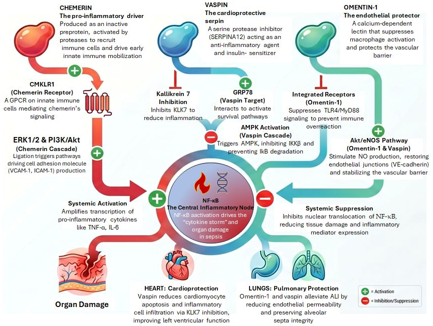
35]. Following removal of a 20-amino acid N-terminal signal peptide, the mature form undergoes C-terminal proteolytic processing by inflammatory and coagulation serine proteases, including plasmin, neutrophil elastase, and cathepsins, generating a spectrum of isoforms with varying receptor affinities. The most biologically potent isoform, chemerin 21–157, serves as the primary endogenous ligand for the chemokine-like receptor 1 (CMKLR1), also known as chemerin receptor 23 (ChemR23) or chemerin receptor 1, a G protein-coupled receptor (GPCR) highly expressed on innate immune cells including plasmacytoid dendritic cells (pDCs), macrophages, and natural killer (NK) cells [
36]. This activation of prochemerin by coagulation serine proteases is particularly relevant in sepsis, where simultaneous coagulation cascade activation may drive autocrine and paracrine chemerin signaling at sites of infection [
37,
38].
Chemerin is produced not only by adipocytes but also by hepatocytes, fibroblasts, and epithelial cells of the lung, kidney, adrenal gland, pancreas, and skin [
21]. Its cationic regions can disrupt bacterial membranes, conferring direct antimicrobial properties. Additionally, chemerin regulates adipogenesis, glucose homeostasis, and insulin signaling in skeletal muscle and contributes to the development of insulin resistance, a common and clinically significant complication in sepsis [
39].
3.2. Immunological Functions: Pro- and Anti-Inflammatory Duality
Chemerin signals through CMKLR1 (ChemR23), a Gαi-coupled GPCR highly expressed on macrophages, neutrophils, DCs, and NK cells [
35]. Receptor signaling proceeds through Gαi1/i2/i3 and Gαo subtypes, followed by recruitment of β-arrestin 1 and 2. Downstream, MAPK/ERK1/2 and PI3K/Akt activation requires both Gαi/o and β-arrestin 2 [
40,
41].
ERK1/2 and PI3K/Akt converge on NF-κB activation, driving the expression of vascular cell adhesion molecule-1 (VCAM-1), intercellular adhesion molecule-1 (ICAM-1), E-selectin, and monocyte chemoattractant protein-1 (MCP-1) in endothelial cells, enhancing monocyte–endothelial adhesion [
42]. IL-1β acts synergistically with chemerin to amplify NF-κB-mediated inflammation [
42].
A critical nuance is that CMKLR1 signaling is context-dependent. Chemerin acts as a potent chemoattractant for pDCs and macrophages to sites of inflammation, facilitating early innate immune activation, promoting pro-inflammatory macrophage/DC recruitment. Conversely, CMKLR1 activation can also induce pro-resolving pathways. The nanopeptide chemerin9, derived from the C-terminus of active chemerin, has been shown to induce pro-resolving macrophage phenotype changes via Gi signaling, reducing inflammatory mediator production [
35]. Resolvin E1 (RvE1), a specialized anti-inflammatory lipid mediator that inhibits leukocyte infiltration and pro-inflammatory gene expression, has also been proposed to signal through CMKLR1, further linking chemerin biology to inflammation resolution, a process critically dysregulated in sepsis [
36,
43]. Additionally, CMKLR1-deficient mice paradoxically show enhanced inflammation in some models, possibly because CMKLR1 also recruits tolerogenic pDCs [
43]. This dual nature, pro-inflammatory via chemerin and pro-resolving via RvE1, establish CMKLR1 as a multifunctional receptor whose net effect depends on the ligand milieu [
35,
36,
43].
Receptor internalization and desensitization are regulated by G protein-coupled receptor kinase 6 (GRK6) and β-arrestin 2. GRK6-deficient macrophages show increased migration toward chemerin and altered Akt/ERK signaling, suggesting that impaired receptor desensitization could amplify chemerin-driven inflammation in disease states [
41]. Conversely, the nanopeptide chemerin9 and RvE1 signal through CMKLR1 to promote pro-resolving macrophage phenotypes and suppress inflammation [
35,
36].
Hence, chemerin occupies a paradoxical position in immunology; it can exert both pro- and anti-inflammatory effects depending on the physiological context, isoform generated, and receptor subtype engaged [
36].
3.3. In Vitro Evidence
In vitro data on the immunological functions of chemerin have yielded complex and sometimes contradictory results, reflecting the isoform- and context-dependent nature of its biology.
In human endothelial cells, chemerin exhibits opposing effects depending on the experimental conditions. Chemerin has been shown to induce NF-κB activation in HUVECs via MAPK/ERK1/2 and PI3K/Akt pathways, driving expression of the adhesion molecules E-selectin, VCAM-1, and ICAM-1 [
42]. It has also been shown to enhance monocyte–endothelial adhesion in functional assays, a critical early step in atherosclerosis and vascular inflammation [
42]. Moreover, IL-1β acts synergistically with chemerin to amplify NF-κB-mediated inflammation, suggesting chemerin potentiates cytokine-driven endothelial activation [
42]. Conversely, chemerin has been shown to inhibit TNF-α-induced VCAM-1 expression in HUVECs by activating the Akt/endothelial nitric oxide synthase (eNOS) pathway and increasing nitric oxide (NO) production [
44]. Furthermore, the same study showed that chemerin rapidly phosphorylated Akt and eNOS, increasing intracellular cGMP. This, in turn, suppressed TNF-α-induced phosphorylation of NF-κB p65 and p38 MAPK, reducing VCAM-1 expression and monocyte adhesion. The protective effect was NO-dependent, since NOS inhibitors could reverse chemerin’s anti-inflammatory actions, while NO donors mimicked them. Similar anti-inflammatory effects were observed in rat isolated aorta ex vivo [
44]. These opposing effects likely reflect timing and context. Acute chemerin exposure (minutes) activates Akt/eNOS/NO signaling with anti-inflammatory consequences, while prolonged exposure or co-stimulation with IL-1β activates MAPK/NF-κB pathways with pro-inflammatory outcomes. Hence, chemerin concentration, endothelial cell activation state, and the presence of other inflammatory mediators determine the net effect.
In HMVECs and VSMCs, chemerin promotes pro-inflammatory and proliferative responses by inducing NADPH oxidase (NOX)-dependent reactive oxygen species (ROS) production [
45]. In this study, chemerin increased ROS production and phosphorylation of MAPK (ERK1/2, p38, JNK), effects blocked by NOX inhibitors and the ROS scavenger N-acetylcysteine. In VSMCs, chemerin stimulated proliferation via redox-sensitive MAPK signaling, decreased PI3K/Akt activation, and increased TUNEL-positive VSMCs, indicating pro-apoptotic effects. In HMVECs, chemerin reduced eNOS activity and NO production, impairing endothelial function. Moreover, chemerin increased the mRNA expression of pro-inflammatory mediators (IL-6, IL-8, MCP-1) and enhanced monocyte-to-endothelial cell attachment. Finally, adipocyte-conditioned medium from obese/diabetic mice, which have elevated chemerin, increased ROS generation in VSMCs, while medium from control mice had no effect. These effects were blocked by CCX 832, a ChemR23 antagonist, confirming CMKLR1 dependence [
45].
In macrophage models, no direct effect has been demonstrated on LPS-induced cytokine production. Specifically, mouse peritoneal macrophages and human monocyte-derived macrophages have been shown to express functional ChemR23 [
46]. However, using peritoneal macrophages generated from wild-type or
CMKLR1-/- knockout (KO) mice, the authors demonstrated that bioactive chemerin did not modulate cytokine responses despite functional ChemR23 expression, with identical null results in human blood monocyte-derived macrophages [
46]. Hence, despite functional
CMKLR1 expression, chemerin did not seem to modulate LPS-induced cytokine production in macrophages. This contradicts earlier reports of direct anti-inflammatory effects of chemerin on macrophages and suggests that chemerin’s anti-inflammatory actions in vivo occur through indirect mechanisms, likely recruitment of tolerogenic pDCs or modulation of the tissue microenvironment rather than direct suppression of macrophage cytokine production [
47].
While chemerin does not seem to affect macrophage cytokine production, it potently promotes macrophage adhesion to extracellular matrix and endothelium. Chemerin could stimulate adhesion of mouse peritoneal exudate cells to fibronectin and VCAM-1, via ChemR23 and Gαi signaling. Moreover, 89% of adhesion to fibronectin was mediated by increased avidity of integrin VLA-5 (α5β1), while 88% of adhesion to VCAM-1 was mediated by VLA-4 (α4β1). Chemerin did not increase integrin affinity but instead promoted integrin clustering, as visualized by confocal microscopy. Key signaling mediators included PI3K, Akt, and p38 MAPK. Pertussis toxin and
CMKLR1-/--KO macrophages confirmed Gαi-coupled receptor dependence. This rapid adhesion response, combined with chemotactic activity, suggests chemerin promotes both recruitment and retention of macrophages at inflammatory sites [
48].
The atypical chemerin receptor, C-C motif chemokine receptor-like 2 (CCRL2), which binds chemerin but does not signal, is expressed on endothelial cells and regulates chemerin bioavailability [
49]. CCRL2 and VCAM-1 were found co-upregulated in human and mouse vascular endothelial cells by pro-inflammatory stimuli (TNF-α, IL-1β, LPS) via NF-κB and JAK/STAT signaling. CCRL2 was constitutively expressed at high levels by pulmonary endothelial cells and at lower levels by liver endothelium. Liver, but not pulmonary, endothelial cells further upregulated CCRL2 in response to systemic LPS. It was demonstrated that CCRL2 bound chemerin and presented it to CMKLR1 on nearby leukocytes, enhancing local chemerin bioactivity. Moreover, plasma chemerin levels were elevated in
CCRL2-KO mice and increased further after LPS treatment, confirming that CCRL2 regulates circulating chemerin levels. Chemerin binding to endothelial CCRL2 triggered robust adhesion of CMKLR1+ lymphoid cells (NK cells) through an α4β1 integrin/VCAM-1-dependent mechanism. Lastly, in LPS-induced acute lung inflammation, CMKLR1+ NK cell recruitment to airways was significantly impaired in
CCRL2-KO mice, demonstrating that endothelial CCRL2 is required for efficient chemerin-mediated leukocyte recruitment in vivo [
49].
In inflammatory macrophages, CMKLR1 signaling and function were tightly regulated by GRK6 and β-arrestin 2, which modulate receptor desensitization and internalization. Chemerin stimulation led to GRK6-mediated phosphorylation of CMKLR1 intracellular domains, recruitment of β-arrestin 2, and signaling termination. β-arrestin recruitment to CMKLR1 was enhanced by co-expression of GRK6. CMKLR1 internalization following chemerin stimulation was decreased in GRK6- and β-arrestin 2-deficient macrophages. These deficient macrophages displayed increased migration toward chemerin and altered Akt/ERK signaling, suggesting impaired receptor desensitization amplifying chemerin-driven responses. This regulatory mechanism may be therapeutically relevant, as defective GRK6/β-arrestin 2 function could lead to exaggerated chemerin-mediated inflammation in rheumatic diseases [
41].
Finally, in peritoneal macrophages,
CMKLR1 expression was demonstrated to be dynamically regulated by inflammatory stimuli. Pro-inflammatory cytokines and Toll-like receptor (TLR) ligands suppressed macrophage CMKLR1 expression, while TGF-β upregulated the receptor. This stimulus-specific regulation might suggest that
CMKLR1 expression is downregulated during acute but upregulated during resolution [
50].
In
Table 1, the findings from the in vitro septic/inflammatory models studying chemerin are listed.
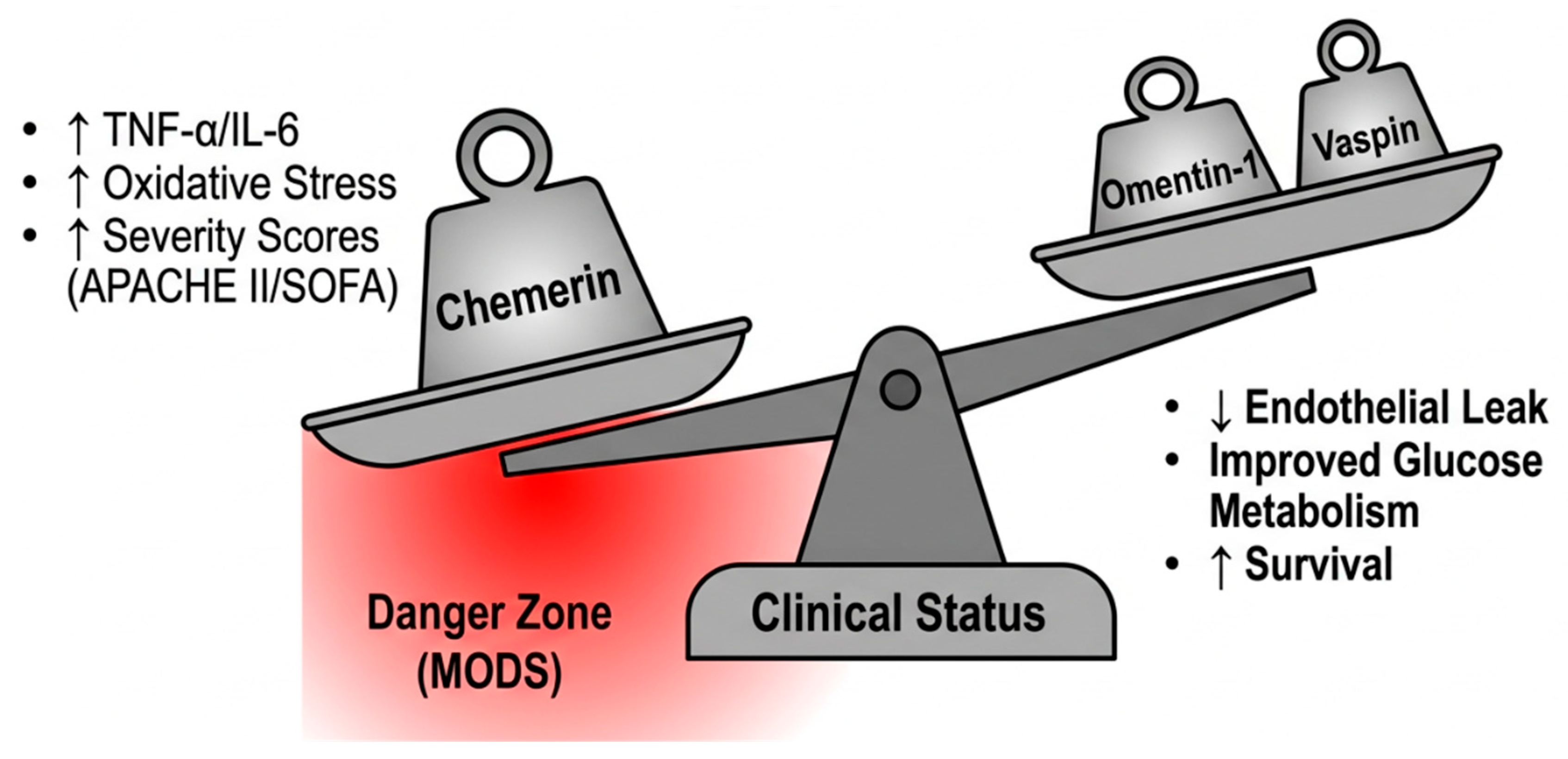
The in vitro data reveal that chemerin’s role in sepsis cannot be reduced to simply pro-inflammatory or anti-inflammatory. Instead, chemerin acts as a context-dependent immunomodulator whose effects depend on the cell type: pro-inflammatory in endothelial cells and VSMCs (via NOX/ROS/MAPK), anti-inflammatory in whole lung tissue (via pDC recruitment), neutral in macrophage cytokine production. Timing is important, as acute exposure activates protective Akt/eNOS/NO pathways, while chronic exposure or co-stimulation with IL-1β/TNF-α activates MAPK/NF-κB inflammation. Receptor regulation is another important component. CMKLR1 is downregulated by TLR ligands during acute inflammation but upregulated by TGF-β during resolution, suggesting chemerin’s role shifts across sepsis phases. Receptor type comprises another aspect of chemerin’s role. The signaling receptor CMKLR1 mediates direct cellular effects, while the non-signaling CCRL2 on endothelium concentrates chemerin to enhance local bioactivity. This complexity may explain why circulating chemerin is elevated in sepsis and predicts mortality. Yet, ChemR23-KO mice show worse outcomes in LPS models, so chemerin likely exerts both harmful (endothelial activation, ROS generation) and beneficial (tolerogenic DC recruitment) effects simultaneously, with the net outcome determined by disease stage and tissue context.
3.4. In Vivo Experimental Evidence
Animal models have provided important insights into the role of the chemerin/CMKLR1 axis in inflammatory lung disease and infection, conditions that are highly relevant to the pathophysiology of sepsis-associated organ injury.
In contrast to the mixed in vitro findings, chemerin exhibits potent anti-inflammatory effects in mouse LPS-induced lung inflammation. In a murine model of LPS-induced acute lung injury, the administration of exogenous chemerin acted as a protective agent by significantly reducing neutrophil infiltration and the release of inflammatory cytokines. This anti-inflammatory activity is strictly dependent on the ChemR23 (CMKLR1) receptor, as
CMKLR1-KO mice failed to respond to chemerin treatment and instead demonstrated increased neutrophil accumulation following an LPS challenge. The primary mechanism for this protection likely involved the recruitment of tolerogenic pDCs, which express high levels of the ChemR23 receptor. While expression is highest in immature pDCs, the receptor is also present at lower levels on myeloid DCs, macrophages, and NK cells. This functional study indicated that chemerin promotes essential immune responses such as calcium mobilization and chemotaxis in these cells, both of which are entirely abrogated in
CMKLR1-deficient models [
47].
In a model of acute viral pneumonia using the pneumonia virus of mice (PVM),
CMKLR1-KO mice exhibited higher mortality and morbidity, altered lung function, delayed viral clearance, reduced pDC recruitment, and diminished type I interferon production compared to wild-type controls, establishing the chemerin/CMKLR1 axis as an important mediator of anti-viral innate immunity [
51]. In LPS-induced lung inflammation,
CMKLR1-KO mice showed exacerbated pulmonary inflammatory responses [
51].
Recombinant chemerin at picomolar concentrations has been reported to exert anti-inflammatory effects on zymosan-induced murine peritonitis in a proteolysis-dependent manner, by reducing pro-inflammatory mediator expression [
52]. More specifically, chemerin15 (C15) was shown to inhibit macrophage activation to a similar extent as proteolyzed chemerin. Intraperitoneal administration of C15 to mice before zymosan challenge conferred significant protection against zymosan-induced peritonitis, suppressing neutrophil and monocyte recruitment with a concomitant reduction in pro-inflammatory mediator expression. Importantly, C15 was unable to ameliorate zymosan-induced peritonitis in
CMKLR1-KO mice, demonstrating that C15’s anti-inflammatory effects are entirely ChemR23-dependent [
52]. The same group demonstrated that during peritoneal inflammation, C15 administration enhanced microbial particle clearance and apoptotic neutrophil ingestion (efferocytosis) by macrophages in wild-type but not
CMKLR1-KO mice, profoundly reducing levels of apoptotic and necrotic cells at the inflammatory site [
53].
CCRL2-KO mice displayed exaggerated local and systemic inflammatory responses in both zymosan- and thioglycollate-induced peritonitis, characterized by increased myeloid cell recruitment. This amplified inflammation was associated with increased circulating and local chemerin levels. Antibody neutralization of chemerin in
CCRL2-KO mice abrogated the amplified inflammatory responses, confirming that the phenotype is chemerin-dependent [
54].
In another study,
CCRL2-KO mice exhibited impaired NK cell recruitment in LPS-induced lung inflammation. Plasma chemerin levels were elevated in
CCRL2-KO mice and further enhanced after systemic LPS, confirming CCRL2’s role in regulating circulating chemerin levels. This demonstrates that endothelial CCRL2 is required for efficient local concentration of chemerin at inflammatory sites to recruit CMKLR1
+ immune cells [
49].
Moreover, cathepsin K- and L-truncated chemerin displayed direct antibacterial activity against Enterobacteriaceae in addition to triggering robust migration of human pDCs ex vivo [
55]. Another study using single-cell (sc)RNA sequencing in an ALI model identified that reverse-migrated neutrophils (those migrating away from the inflammatory site back into the vasculature) exhibited increased
CCRL2 expression. Circulating chemerin concentrations increased in the late stage of inflammation, and neutralizing chemerin decreased the reverse-migrated neutrophil ratio in blood, suggesting chemerin/CCRL2 interaction promotes neutrophil reverse migration, a mechanism potentially involved in dissemination of inflammation [
56].
RvE1 serves as a ChemR23 ligand and acts as an endogenous pro-resolving lipid mediator. Administration of RvE1 6 h post-LPS in rats improved survival, increased alveolar fluid clearance, reduced lung wet–dry weight ratio, and mitigated lung injury scores [
57]. In bacterial pneumonia models, RvE1 selectively decreased lung neutrophil accumulation, enhanced
E. coli clearance, and markedly improved survival. Mechanistically, RvE1 seemed to limit collateral lung damage by independently downregulating pro-inflammatory cytokines such as IL-1β, IL-6, and high-mobility group box 1 (HMGB1) without impairing pathogen killing [
58].
In a pulmonary inflammation model, RvE1 promoted phagocytosis-induced neutrophil apoptosis via the leukotriene B4 receptor 1 (BLT1), enhancing NADPH oxidase-derived ROS and caspase-8/3 activation, while attenuating anti-apoptosis signals from myeloperoxidase (MPO) and serum amyloid A (SAA) [
59].
These discordant findings likely reflect differences in experimental design, genetic background, and the specific chemerin isoforms tested, and underscore that the direct anti-inflammatory effect of chemerin on macrophages cannot be generalized across all in vitro settings. Notwithstanding these complexities, chemerin has been consistently shown to drive chemotaxis of pDCs, NK cells, and immature DCs through CMKLR1 in transwell migration assays, establishing its role as a potent chemoattractant in inflammatory conditions [
36]. Structural studies using cryo-electron microscopy (cryo-EM) have elucidated the molecular basis of CMKLR1 signaling by chemerin9, revealing agonist-induced conformational changes in the receptor that activate Gi signaling pathways, and providing a structural framework for the development of small-molecule CMKLR1 agonists that could promote resolution of inflammation [
35].
Table 2 lists the chemerin studies in in vivo models of sepsis and organ injury.
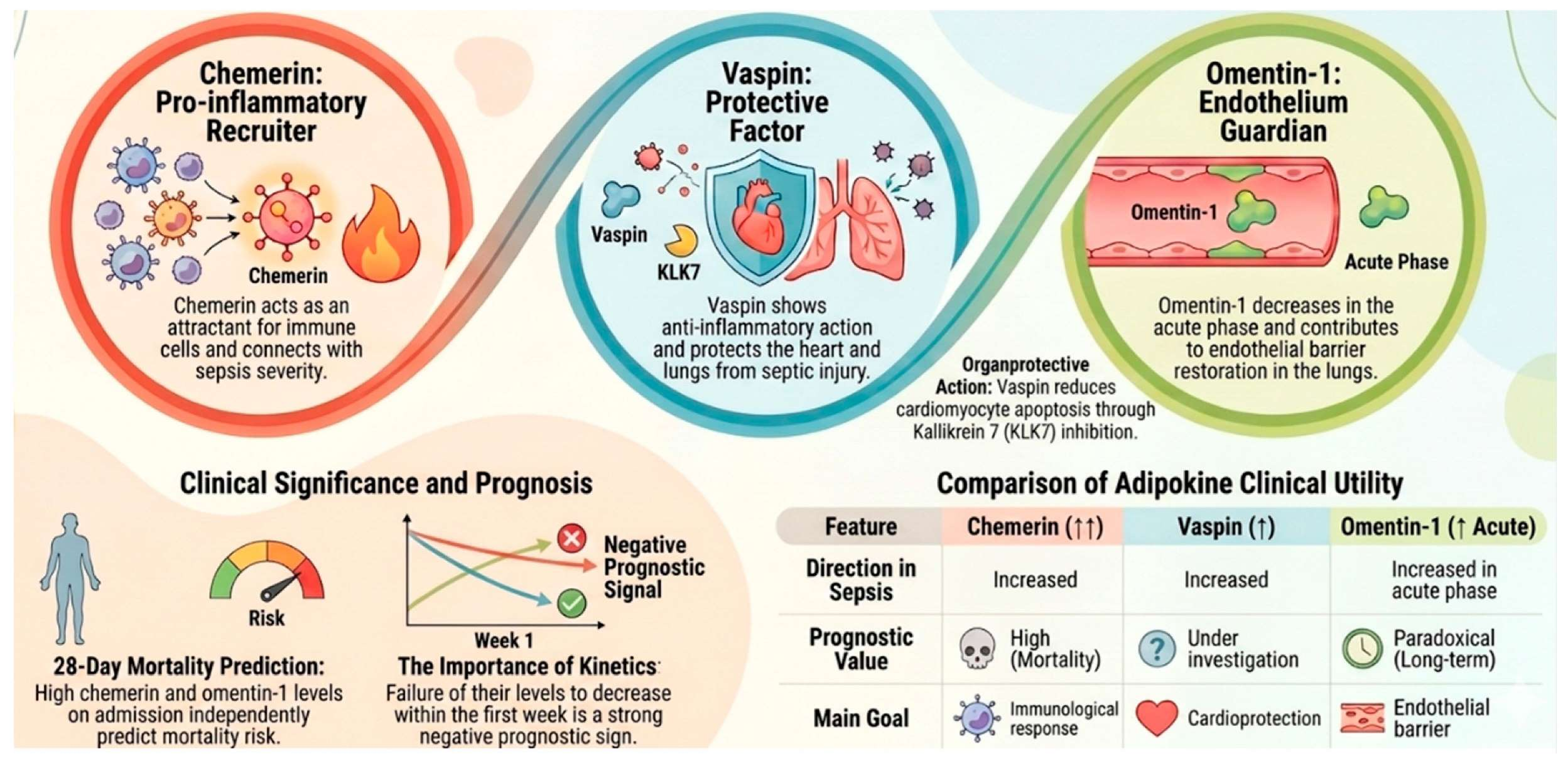
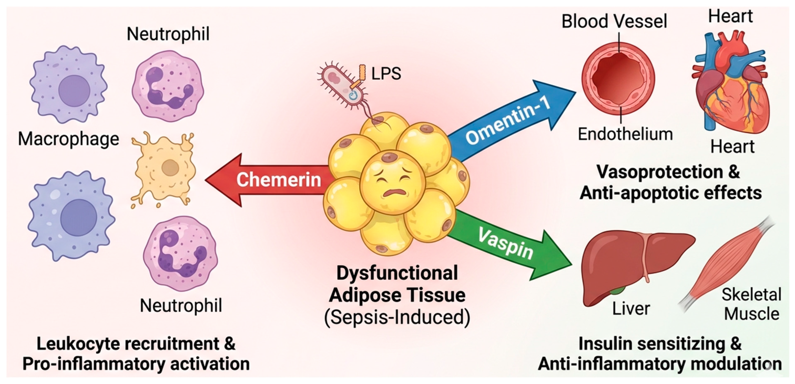
These in vivo findings collectively support a context-dependent role for chemerin in infection and inflammation: pro-inflammatory and chemoattractive in early innate immune mobilization, but capable of promoting resolution when appropriate receptor signaling is engaged.
3.5. Clinical Evidence in Sepsis and Critical Illness
The most comprehensive prospective clinical data on chemerin in sepsis come from Karampela et al., who measured serum chemerin in 102 critically ill patients with sepsis within 48 h of onset and again one week later, compared to 102 age- and sex-matched healthy controls [
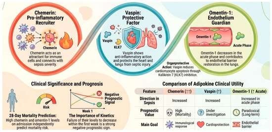
21]. Serum chemerin was markedly elevated at sepsis onset and, while it declined significantly over the first week, it remained above control levels throughout follow-up. Levels were substantially higher in patients with septic shock than in those with sepsis alone, and in non-survivors compared to survivors at both timepoints. Crucially, Cox proportional hazards regression analysis revealed that elevated chemerin at admission was an independent predictor of 28-day mortality, yielding a Hazard Ratio (HR) of 3.58 (95% CI: 1.48–8.65,
p = 0.005). When evaluated dynamically at one-week post-onset, the prognostic power of sustained chemerin elevation strengthened dramatically, escalating to an HR of 10.01 (95% CI: 4.32–23.20,
p < 0.001), underlining its utility in tracking ongoing, unresolved systemic inflammation. The diagnostic performance for severity discrimination (AUC 0.78) was comparable to CRP. Chemerin correlated significantly with the acute physiology and chronic health evaluation (APACHE) II and the sequential organ function assessment (SOFA) scores, white blood cell (WBC) count, lactate, CRP, and procalcitonin [
21].
A critical nuance was established by Horn et al. in peritoneal sepsis. In this study, chemerin correlated with intraoperative glucose, positioning it as a metabolic biomarker [
60]. Crucially, the prognostic relationship was context-dependent; among patients with stress hyperglycemia (SHG), non-survivors had paradoxically lower chemerin, while non-survivors without SHG trended toward higher chemerin. Despite elevated circulating levels, visceral adipose
RARRES2 mRNA was decreased in sepsis, suggesting extra-adipose sources or altered clearance [
60]. This paradoxical finding suggests that chemerin may serve different functions depending on metabolic context. In SHG, higher chemerin may reflect a compensatory insulin-sensitizing response that is protective, while in non-SHG patients, elevated chemerin may reflect greater inflammatory burden.
Amend et al. further showed that Gram-positive infection was associated with significantly higher plasma chemerin than Gram-negative infection or COVID-19, raising the possibility that chemerin could serve as an early biomarker to distinguish infecting organism class, a distinction with direct therapeutic implications for empiric antibiotic selection [
61]. Importantly, patients with liver cirrhosis had markedly lower chemerin, highlighting the need to adjust for hepatic function when interpreting circulating levels in heterogeneous ICU populations [
61].
Multiple studies have evaluated chemerin in COVID-19, providing the closest clinical analog to sepsis-associated ARDS. In 88 COVID-19 patients (40 ICU), plasma chemerin was significantly higher in ICU patients than healthy controls at all time points and higher in non-survivors than survivors. Moreover, the multivariate analysis showed that chemerin at day 14 was an independent risk factor for death. Immunohistochemistry of autopsied COVID-19 lungs revealed strong ChemR23 expression on smooth muscle cells and chemerin expression on myofibroblasts in advanced ARDS, suggesting active chemerin/ChemR23 signaling in the fibroproliferative phase [
62]. A separate study confirmed sustained chemerin elevation in hospitalized COVID-19 patients with a trend toward further increase over 7 days [
63]. However, one study reported decreased chemerin in COVID-19 patients, highlighting inconsistency across cohorts, likely reflecting differences in disease severity, timing of sampling, and assay methodology [
19]. Thus, in SARS-CoV-2 infection, anti-inflammatory adipokines including chemerin are altered relative to controls, though direction seems to vary by disease severity.
A recent study provided novel insights into the resolution pathway in critically ill COVID-19 patients. Among a panel of cytokines and resolvins, RvE1 was the single best discriminator of COVID-19 severity, outperforming all cytokines, including IL-6. RvE1 was paradoxically elevated in the most severe patients, mechanically ventilated patients, and non-survivors, suggesting failed resolution rather than insufficient resolvin production.
CMKLR1 mRNA exhibited an opposite profile; higher expression correlated with lower inflammation, better respiratory function, and shorter hospital stay. This RvE1–ChemR23 axis dysregulation suggests that in severe ARDS, the resolution machinery is activated but functionally impaired, possibly due to receptor downregulation or desensitization [
64].
Ebihara et al. performed a comprehensive adipocytokine profiling study on 37 septic patients with serial measurements over 15 days. Hierarchical clustering analysis revealed that chemerin does not cluster with the core inflammatory network (IL-6, IL-8, MCP-1, IL-10) dominated by resistin, suggesting it reflects a parallel immunometabolic pathway offering complementary prognostic information [
17].





 ,
,