


Abstract
Background/Objectives: Electroconvulsive therapy (ECT) is among the most effective treatments for severe major depression, treatment-resistant depression, psychotic and bipolar depression, catatonia, and selected psychotic disorders. Yet response, remission, seizure adequacy, and cognitive tolerability remain difficult to predict for an individual patient. This review examines whether oral and gut microbial signatures can inform precision ECT as contextual biological markers, rather than as standalone explanations of ECT efficacy. Methods: A structured narrative PubMed/MEDLINE search was conducted on 1 May 2026 and supplemented by targeted manual searches of Crossref, Google Scholar, journal websites, and reference lists updated through 17 May 2026. Evidence was grouped as direct human ECT–microbiome studies, indirect human ECT biomarker studies, preclinical electroconvulsive shock (ECS) studies, and mechanistic microbiome–gut–brain literature. Results: Direct human ECT–microbiome evidence remains very limited and currently consists of two small prospective cohorts with sufficient microbiome data, totaling approximately 25 patients across studies, plus one single-patient case report. In severe or treatment-resistant depression, a pilot oral microbiome study with sufficient microbiological data from 14 patients reported higher pre-treatment oral alpha diversity in responders than in non-responders, without a consistent global oral microbiome shift after ECT. In schizophrenia, a small stool microbiome cohort of 11 patients suggested that baseline Bifidobacterium and Lactobacillus proportions may relate to symptom improvement, although sample size and confounding preclude firm inference. Conclusions: Microbiome-informed precision ECT remains a biologically plausible research direction, but current human evidence supports only cautious evaluation of baseline microbial context as a candidate predictor, not clinical microbiome-guided ECT, mediation, or microbiome-modifying intervention. The strongest current biological bridge comes from inflammatory markers, particularly baseline CRP and IL-6. Preclinical ECS studies support gut inflammatory, motility and vagal mechanisms, but they cannot substitute for human validation.
1. Introduction
Electroconvulsive therapy (ECT) occupies a singular position in contemporary psychiatry. It is simultaneously one of the most effective treatments for severe depressive illness and one of the most clinically misunderstood interventions in medicine [1,2]. In carefully selected patients with severe major depressive disorder, treatment-resistant depression, psychotic depression, bipolar depression, catatonia or selected psychotic disorders, ECT can produce a speed and magnitude of response that is rarely matched by pharmacotherapy alone [1,3]. Its underuse reflects not a lack of efficacy but the combined weight of stigma, limited service access, uncertainty about cognitive adverse effects and the persistent difficulty of predicting which patient will respond, how quickly, and at what tolerability cost [1,2].
The clinical challenge is familiar at the bedside. One patient with psychotic depression and psychomotor retardation may improve dramatically after several treatments; another, apparently similar in diagnosis and rating-scale severity, may require a prolonged course, experience short EEG seizures under propofol and benzodiazepine exposure, and show only partial improvement [1,4]. A catatonic patient may arrive dehydrated, immobile and constipated after weeks of reduced intake [1,3]. A patient with schizophrenia receiving clozapine may be referred for ECT with severe constipation, metabolic syndrome and anticholinergic burden [1,5]. These are not marginal details. They are the biological and clinical context in which ECT is delivered.
Conventional predictors of ECT response, including older age, psychotic features, psychomotor disturbance, shorter current episode duration, lower degree of antidepressant resistance and selected cognitive or illness-course variables, are useful at group level but remain insufficient for individual decision making [1,6,7]. Contemporary precision-ECT efforts have therefore moved toward integrative models combining clinical features with neuroimaging, electrophysiology, immune-inflammatory markers, epigenetic signals, molecular biomarkers and seizure parameters [8,9,10]. However, most models still omit an increasingly relevant biological system: the oral and gut microbiome.
The microbiome is not proposed here as an alternative explanation of how ECT works [11,12]. Such a claim would be premature and clinically unhelpful [13]. A more defensible question is whether pre-treatment oral and gut microbial states, microbial metabolites, gut barrier biology, inflammatory tone, autonomic regulation and gastrointestinal phenotype contribute to the biological context in which an induced therapeutic seizure is converted into neuroplastic and symptomatic responses [11,14,15,16,17]. In this formulation, microbiome-informed ECT is not about whether ECT merely changes bacteria; it is about whether microbial and immune–metabolic states help predict response, remission, seizure quality, cognitive tolerability, gastrointestinal symptoms and relapse.
This reframing is important because psychiatric microbiome research has often been centered on diagnosis. The literature has compared depression, bipolar disorder or schizophrenia with healthy controls, producing valuable but heterogeneous findings shaped by diet, smoking, medications, body mass index, constipation, hospitalization, sampling methods and analytic pipelines [18,19]. ECT provides a different clinical model: a time-limited biological intervention with measurable session-level physiology, standardized outcome definitions and repeated sampling opportunities [20]. The field is therefore well suited to ask whether microbial signatures add incremental value to precision psychiatry [21,22,23].
The direct human ECT–microbiome literature is small and should be interpreted accordingly. At present, it consists of two small prospective cohorts with sufficient microbiome data, totaling approximately 25 patients across studies, together with one single-patient case report. A prospective oral microbiome pilot study in severe or treatment-resistant depression reported that ECT responders had higher pre-treatment oral alpha diversity than non-responders, based on sufficient microbiological data from 14 patients, with no clear global oral microbiome shift after the ECT course [24]. A small prospective schizophrenia stool microbiome cohort including 11 patients found that baseline stool Bifidobacterium and Lactobacillus proportions were associated with symptomatic improvement after ECT, again without major pre–post microbial changes [25,26,27,28,29]. A single case report described a Clostridium change after ECT in schizophrenia, but this remains hypothesis-generating. These early observations suggest that baseline microbial ecology may be more relevant as a candidate predictor or contextual marker than as a simple downstream outcome [11,30].
This review synthesizes the evidence for microbiome-informed precision ECT. It deliberately separates three roles that are often conflated: microbiome as predictor, microbiome as mediator and microbiome as modifier. It also integrates seizure biology, inflammation, kynurenine metabolism, short-chain fatty acids, hypothalamic–pituitary–adrenal (HPA)-axis regulation, neuroplasticity, anesthetic exposure, cognitive tolerability, pharmacomicrobiomics, and gastrointestinal phenotype [11,31,32]. The aim is to provide a clinically disciplined framework for future studies rather than to overstate the readiness of microbiome testing for ECT practice.
2. Methods
This manuscript is a narrative review with a structured literature search and mechanistic synthesis. It was not designed as a systematic review or scoping review and therefore did not include protocol registration, duplicate independent screening, formal risk-of-bias assessment, quantitative synthesis, or PRISMA-ScR flow-diagram reporting. The structured search was used to improve transparency and reproducibility of the narrative evidence mapping, while the final synthesis remained interpretive and clinically oriented. Reporting was informed in spirit by PRISMA-ScR, a general transparency framework; no formal protocol, duplicate-screening workflow, quantitative synthesis, or PRISMA-ScR flow diagram was produced [33]. STORMS was not used as a reporting framework for the present review but was used as a reference point when formulating recommendations for future primary ECT–microbiome studies, particularly regarding sampling metadata, sequencing and bioinformatic workflow, contamination control, medication exposure, diet, gastrointestinal phenotype, oral-health metadata, and other clinically relevant confounders [34].
The core structured PubMed/MEDLINE search was conducted on 1 May 2026. Supplementary searches in Crossref, Google Scholar, journal websites, and reference lists were updated manually through 17 May 2026 to capture relevant articles published or indexed after the original draft, including 2025–2026 work on ECT biomarkers, microbiome pilot data, anesthetic effects, gastrointestinal motility, and inflammatory predictors. This manual update was narrative and targeted rather than a formal systematic-search update.
The core PubMed search string was: (“electroconvulsive therapy” OR “ECT” OR “electroconvulsive shock” OR “ECS”) AND (“microbiome” OR “microbiota” OR “dysbiosis” OR “gut microbiome” OR “gut microbiota” OR “oral microbiome” OR “oral microbiota” OR “salivary microbiome” OR “short-chain fatty acids” OR “SCFA” OR “butyrate” OR “propionate” OR “acetate” OR “kynurenine” OR “tryptophan” OR “lipopolysaccharide” OR “LPS” OR “zonulin” OR “intestinal permeability” OR “gut-brain axis” OR “microbiota-gut-brain axis” OR “adenosine” OR “purine” OR “bile acids” OR “indole”) AND (“response” OR “remission” OR “predictor” OR “biomarker” OR “treatment outcome” OR “seizure duration” OR “seizure quality” OR “cognition” OR “memory” OR “constipation” OR “gastrointestinal symptoms”).
Eligible evidence included human ECT studies with oral microbiome, gut microbiome, stool microbiota, salivary microbiome or microbial metabolite measures; human ECT studies with inflammatory, kynurenine, metabolomic or gut–brain-relevant biomarkers; human ECT studies on seizure duration, seizure quality, seizure threshold, medication effects, anesthetics or cognitive tolerability; studies on gastrointestinal symptoms, constipation, autonomic changes or gut motility during ECT; and ECS animal studies examining microbiome composition, gut inflammation, gut motility, vagal signaling, microbial metabolites or depressive-like behavior. Broader microbiome–gut–brain literature was included only when it directly supported an ECT-specific mechanism. The evidence domains, search focus, interpretive role and methodological safeguards used for evidence classification are summarized in Table 1.
Table 1.
Search framework and evidence classification used for the structured narrative review.
Exclusion criteria were general depression–microbiome or schizophrenia–microbiome studies without ECT relevance, probiotic trials without ECT relevance, neuromodulation studies involving rTMS, tDCS, DBS, VNS or electroacupuncture unless used only as contextual evidence, opinion pieces without primary data, and non-peer-reviewed preprints unless explicitly labeled as hypothesis-generating. The final narrative corpus discussed in detail comprised direct human ECT–microbiome studies, ECT biomarker and seizure-parameter studies, ECS translational studies, microbiome methodology papers, and mechanistic microbiome–gut–brain studies relevant to inflammation, HPA-axis tone, kynurenine biology, SCFAs, gut barrier function, oral microbiome ecology and pharmacomicrobiomics. Key human and translational studies informing the synthesis are summarized in Supplementary Table S1.
Evidence was interpreted with five safeguards. First, oral and gut microbiomes were treated as distinct compartments rather than interchangeable measures. Second, predictor, mediator and modifier hypotheses were separated. Third, direct human ECT evidence was distinguished from indirect biomarker evidence and from animal ECS data. Fourth, microbial taxa were not overinterpreted without considering function, metabolites and compositional data constraints. Fifth, medication, diet, body mass index, smoking, antibiotics, proton-pump inhibitors, laxatives, constipation, oral hygiene, periodontal disease, hospitalization and session-level ECT protocol variables were treated as central confounders. The rationale for this conservative approach is reinforced by the large inter-individual variation in the healthy human microbiome and population-level microbiome structure [35,36].
3. Current Predictors of ECT Response: Why New Biomarkers Are Needed
ECT response is clinically patterned but individually uncertain. Meta-analytic evidence supports older age, psychotic features, psychomotor disturbance, shorter episode duration and lower treatment resistance as predictors of better response or remission in major depression [6,37,38]. These variables remain important because they represent readily available clinical knowledge. A severely melancholic or psychotic depression with prominent psychomotor slowing is often a different ECT proposition from a long-standing nonpsychotic depression complicated by personality pathology, benzodiazepine dependence, chronic pain, metabolic disease and polypharmacy [1,39].
The problem is that these predictors do not provide enough precision at the individual level. The Dutch ECT Consortium prediction model illustrates both progress and limitation: readily available clinical variables can reduce uncertainty, but substantial variability remains unexplained [40]. Recent narrative and systematic work on ECT biomarkers similarly emphasize that immune-inflammatory markers, neuroimaging, epigenetic markers, electrophysiology and clinical symptoms converge only partially and remain difficult to implement at the bedside [7,20,41]. Precision ECT therefore requires additive, biologically coherent variables, not isolated new biomarkers [42].
Seizure physiology is fundamental to this problem. ECT is not simply a procedure that delivers electricity; it is a treatment that induces a controlled generalized seizure under anesthesia. Clinical outcome depends on electrode placement, pulse width, stimulus dosing relative to seizure threshold, EEG seizure duration, postictal suppression, number of treatments, anesthetic agent and concomitant medications [1,43,44]. A large Swedish registry study of 6998 patients with major depressive disorder treated with unilateral ECT found that EEG seizure duration was associated with remission and that anticonvulsants and benzodiazepines were associated with shorter seizures and less favorable outcomes [42]. This does not make seizure duration a perfect surrogate for treatment quality, but it confirms that any biological predictor must be interpreted in relation to session-level ECT physiology [42,45].
Inflammation is currently a more mature bridge than the microbiome itself. A 2025 meta-analysis of 14 studies including 556 patients with depression reported that higher baseline CRP and IL-6 were associated with greater depressive symptom reduction after ECT [46]. The same literature also suggested a role for kynurenine metabolites and IL-8 dynamics [46,47]. These findings fit a broader model in which inflammatory depression may be more responsive to ECT-induced neuroimmune and neuroplastic modulation, while also providing a rational entry point for microbiome biology [20,41,48].
Microbiome-informed ECT should therefore be evaluated not as a competing predictor but as a potential layer in a multimodal model. The clinically meaningful question is whether oral or gut microbial data improve prediction beyond age, psychosis, episode duration, treatment resistance, baseline cognition, CRP/IL-6, medication exposure and seizure quality [41,49,50,51]. This incremental-value criterion should define the field.
4. Oral Microbiome and ECT Response in Severe Depression
Within this very small direct evidence base, the most directly relevant human depression evidence comes from the prospective oral microbiome pilot study by Ammer-Herrmenau and colleagues [24]. The investigators enrolled patients with severe or treatment-resistant depression undergoing ECT and collected oral swabs before and after treatment. Sufficient microbiological data were available for 14 patients, divided into seven responders and seven non-responders using the conventional criterion of at least 50% reduction in Montgomery–Åsberg Depression Rating Scale score [24,49]. The small sample size precludes definitive inference, but the study is important because it places the oral microbiome inside the ECT prediction problem rather than inside a generic depression–microbiome framework [11,24,51]. The study is summarized in Supplementary Table S1, Ammer-Herrmenau et al. [24] row.
The central observation was that responders had higher pre-treatment oral alpha diversity than non-responders [24,52]. Importantly, the study did not show a robust global oral microbiome shift after the ECT course. Responders and non-responders reportedly did not differ substantially in baseline depression severity or in key ECT treatment variables, although age and atypical antipsychotic exposure remained clinically relevant factors [53]. This pattern favors a baseline-predictor interpretation: oral microbial ecology may reflect a biological state that makes response more likely, rather than a microbial change caused by ECT [11,16,52].
The oral compartment deserves attention for pragmatic and biological reasons. It is accessible, acceptable to patients, feasible for repeated sampling during an ECT course and potentially easier to integrate into clinical workflows than stool collection [54,55]. It may also reflect systemic inflammatory state, salivary immune activity, medication exposure, xerostomia, smoking, diet, dental disease and oral hygiene [56]. Recent population-based and review work increasingly supports the oral–brain axis as an underexplored component of mental health research [17,51]. However, the oral microbiome should not be treated as a proxy for stool microbiota. It is its own ecological niche, with distinct local determinants and distinct sources of confounding [57,58,59].
Taxonomic signals from the pilot study should be handled with restraint. Streptococcus-dominant patterns were described more often among non-responders, while Gemella and Aggregatibacter signals were noted among responders. These observations are not clinical biomarkers. Streptococcus is a dominant oral genus and may reflect plaque biofilm ecology, salivary flow, diet, xerostomia, dental disease, antibiotic history, smoking, hospitalization or local mucosal inflammation rather than an ECT-specific biological state [22,23]. Gemella is also a common oral and upper aerodigestive commensal, and its clinical meaning depends strongly on species-level identity, co-occurring taxa and host context. Aggregatibacter includes species linked to periodontal inflammation, particularly A. actinomycetemcomitans, but genus-level detection alone cannot determine whether the signal reflects periodontal disease, oral dysbiosis or systemic immune relevance [59,60]. Therefore, these taxa should be interpreted as markers of oral ecological context, not as functional predictors of ECT response. For a top-tier ECT–microbiome study, species names are less important than reproducible functional signatures, inflammatory markers and metabolite profiles [16,52].
A clinically experienced interpretation is that oral microbiome sampling may become useful only if embedded within oral-health, medication, inflammatory and ECT-procedural metadata, rather than interpreted as an isolated microbial readout [16,55,61].
5. Gut Microbiome and ECT Response in Schizophrenia and Psychotic Disorders
Direct stool microbiome evidence in ECT-treated psychotic disorders is very limited. Kanayama and colleagues studied 11 patients with schizophrenia receiving ECT and examined stool bacterial proportions before and after treatment [62]. In the primary linear regression model for change in BPRS score, baseline Bifidobacterium abundance showed a positive association with symptom improvement (β = 2.383, p = 0.023), whereas baseline Lactobacillus abundance showed an inverse association (β = −1.903, p = 0.020). For catatonia severity, the corresponding BFCRS associations did not reach statistical significance (Bifidobacterium: β = 2.314, p = 0.093; Lactobacillus: β = −1.416, p = 0.132). Confidence intervals were not reported, and the sample size was only 11 patients; these coefficients should therefore be interpreted as exploratory regression signals rather than stable effect estimates. No bacterial group showed a significant pre–post change in proportion. Like the depression oral microbiome pilot, this finding supports a baseline-predictor hypothesis more than a simple post-ECT-change hypothesis. This cohort is summarized in Supplementary Table S1, Kanayama et al. [63] row.
The quantitative interpretability of this schizophrenia cohort is limited. The study reported directional associations between baseline bacterial proportions and symptomatic improvement, but the sample size was too small to derive clinically meaningful thresholds or stable multivariable estimates. Effect sizes and confidence intervals, where unavailable or insufficiently precise, should therefore not be inferred from the reported associations. The Bifidobacterium and Lactobacillus findings should be presented as exploratory signals requiring replication rather than as validated predictors of ECT response.
The clinical context of schizophrenia makes this signal interesting but fragile. Patients referred for ECT in schizophrenia are often complex: treatment-resistant psychosis, catatonia, severe agitation, clozapine augmentation, polypharmacy, metabolic syndrome, smoking, low physical activity, constipation and variable inpatient diet are all common [63,64,65]. Antipsychotic exposure is not merely a medication variable; it can change weight, insulin resistance, appetite, bowel transit and microbial ecology. Clozapine and olanzapine are particularly important because they are associated with metabolic changes and gastrointestinal hypomotility that can reshape stool microbiota and microbial metabolites [63,66,67,68,69].
The 2019 case report describing decreased Clostridium abundance after ECT in schizophrenia should be cited only as hypothesis-generating [62]. This case report is summarized in Supplementary Table S1, Kanayama et al. [70] row. Single-case microbial trajectories are vulnerable to diet, antibiotics, laxatives, sampling timing, bowel transit and analytic noise [71,72]. Their value is not to establish causality but to show why repeated sampling and careful metadata are needed. In ECT-treated psychosis cohorts, constipation and clozapine exposure should be treated as core biological variables, not as footnotes [64].
Broader schizophrenia microbiome studies provide mechanistic plausibility but not ECT evidence. Fecal microbiota transfer studies have linked schizophrenia-associated microbiota to behavioral changes, glutamate–glutamine–GABA cycle disturbances and kynurenine pathway abnormalities in mice [68,69]. These findings align with ECT-relevant pathways, but they do not show that gut microbiota predicts ECT response. For this reason, they should be used as mechanistic background, not as evidence for clinical microbial stratification [29,53,71].
In psychotic disorders, the most useful future ECT–microbiome cohorts will likely be diagnosis-specific or stratified by clinical indication. Catatonia, clozapine-resistant schizophrenia and acute psychotic agitation are biologically and clinically different reasons for ECT referral [1,73,74]. Combining them without phenotypic clarity would obscure signals. A pragmatic strategy would be to treat schizophrenia and catatonia as exploratory cohorts while focusing primary statistical power on severe depressive illness, where ECT outcome definitions and comparative data are stronger.
6. Preclinical ECS Evidence: Microbiota, Gut Inflammation, Vagal Signaling and Depressive-like Behavior
Electroconvulsive shock (ECS) animal models are not clinical ECT, but they allow experimental interrogation of pathways that are difficult to study in acutely ill patients. Their value is mechanistic: investigators can control stress exposure, collect gut tissue, measure local cytokines, test motility, manipulate the vagus nerve and examine depressive-like behaviors [75,76,77]. Their limitation is equally clear: mouse taxa, stress models and ECS protocols do not translate directly into human ECT biomarkers.
In a chronic unpredictable mild stress model, Ji and colleagues reported that ECS/ECT-like treatment improved depressive-like behavior, reduced gut inflammatory cytokines including TNF-alpha, IL-6 and IL-1beta, and altered microbial diversity and taxa [11]. The finding supports a gut inflammatory pathway but does not justify species-level extrapolation. In translational terms, the important message is that seizure-based neuromodulation may interact with intestinal inflammation and microbial ecology in a bidirectional manner [11,78,79]. This preclinical evidence is summarized in Supplementary Table S1, Ji et al. [80] row.
Dai and colleagues provided a complementary gut physiology signal [81]. In a clinical component, ECT was associated with improvement in gastrointestinal symptoms, particularly constipation, among patients with major depressive disorder [81]. In a depressive-like mouse model, ECS improved distal colonic motility, and subdiaphragmatic vagotomy attenuated this effect. This supports involvement of a hypothalamic paraventricular nucleus–vagus–gut pathway [79,81]. For microbiome-informed ECT, this is highly relevant because gut transit time is a major determinant of stool microbial composition and metabolite concentration [82,83,84]. The clinical and translational findings are summarized in Supplementary Table S1, Dai et al. [81] row.
Because ECS studies are heterogeneous and cannot be read as direct human ECT evidence, Table 2 summarizes the main preclinical signals and their translational boundaries. The purpose of the table is not to upgrade ECS findings to clinical biomarkers but to specify which findings are mechanistically useful and which should not be directly extrapolated to patient-level ECT prediction or treatment selection.
These studies sharpen a methodological point. If ECT changes stool microbiota after treatment, the change could reflect improved mood, improved appetite, hospital diet, increased mobility, altered laxative use, decreased constipation, medication changes, autonomic shifts or a direct gut–brain effect of the induced seizure. A pre–post microbial change is therefore not automatically a mechanism of antidepressant response. It may be a secondary marker of altered gut physiology [62,83,85,86].
Table 2.
Preclinical ECS findings and limits of clinical translation.
Preclinical reports involving microbiota-derived purines or adenosine and ECS efficacy should be treated carefully unless they are peer-reviewed and independently replicated. Purinergic signaling is biologically relevant to seizure threshold, adenosinergic inhibition and neuroplasticity, but the clinical evidence that microbiota-derived adenosine determines ECT response remains insufficient [86,87]. Such work is useful for hypothesis generation and metabolomic panel design, not for clinical decision making.
7. Inflammation, Kynurenines and the Immune–Metabolic Bridge
The strongest current bridge between microbiome biology and ECT response is not a bacterial taxon. It is an immune-inflammatory phenotype. Higher baseline CRP and IL-6 have been associated with greater depressive symptom reduction after ECT in meta-analytic evidence [46,88]. This is a crucial observation because CRP and IL-6 are clinically accessible, mechanistically interpretable and biologically upstream or downstream of multiple microbiome-related processes [89,90].
Inflammation may influence ECT response through neuroplasticity, glial activation, neurovascular function, monoamine metabolism, HPA-axis tone and glutamatergic signaling [14,20,91]. ECT itself can induce acute immune and endocrine responses and may produce longer-term changes in inflammatory and neurotrophic systems [92]. The correct interpretation is not that inflammation is uniformly bad or that ECT simply reduces inflammation. A more nuanced model is that a subgroup of depression with elevated inflammatory tone may be especially sensitive to ECT-induced resetting of neuroimmune and neuroplastic networks [20,89].
Kynurenine biology is particularly important because it links inflammation, microbial metabolism and neural excitability. Pro-inflammatory signaling can shunt tryptophan metabolism toward kynurenine pathway metabolites [72,93]. Quinolinic acid may influence glutamatergic neurotoxicity, whereas kynurenic acid can modulate NMDA receptor signaling. ECT studies have reported changes in kynurenine pathway metabolites in treatment-resistant depression, and some data suggest that kynurenine metabolite dynamics may relate to symptom change [32,94,95]. Microbes can influence tryptophan availability, indole production and immune regulation, making kynurenines a biologically coherent component of a microbiome-informed ECT panel [93,96].
From a clinical standpoint, kynurenines have a better future as part of a panel than as a single biomarker. A patient with high CRP, high IL-6, altered KYN/TRP ratio, low butyrate-producing capacity, constipation and short EEG seizures under benzodiazepine exposure represents a different biological context from a patient with low inflammatory markers, normal bowel transit, no anticonvulsant exposure and robust seizures [46,97]. Precision ECT should capture these patterns rather than seek one universal marker.
The same logic applies to IL-8, TNF-alpha and IL-1beta. These markers may be relevant, but current clinical ECT evidence is less mature than for CRP and IL-6 [46,98]. Future studies should use prespecified immune panels, fasting blood draws where feasible, batch correction, symptom timing relative to treatment, and repeated measures that can distinguish baseline prediction from acute postictal immune response [20,72].
8. Short-Chain Fatty Acids, Gut Barrier Biology and Functional Microbiome Readouts
Functional readouts are central to microbiome-informed ECT research because microbial metabolites are more clinically interpretable than genus-level taxonomic patterns alone [99,100]. Short-chain fatty acids (SCFAs), particularly butyrate, acetate and propionate, are central candidates because they influence epithelial barrier function, immune tone, microglial maturation, energy metabolism, HPA-axis signaling and neuroplasticity [97,101,102].
Butyrate is especially relevant because of its role in intestinal barrier integrity and histone deacetylase inhibition [103,104]. In theory, reduced butyrate-producing capacity could promote low-grade inflammation, gut permeability and altered immune signaling, creating a biological context in which ECT response and cognitive tolerability differ. However, this remains a hypothesis in ECT. It should be tested with measured fecal and plasma metabolites, not inferred from the presence or absence of a few butyrate-associated taxa [11,97].
Gut barrier biology is another plausible but methodologically difficult pathway. Lipopolysaccharide-related signaling, lipopolysaccharide-binding protein, soluble CD14, intestinal fatty-acid binding protein and zonulin-like markers are often invoked as indicators of microbial translocation or barrier dysfunction [9,10]. These markers can support a gut–immune model, but they are not interchangeable and some, particularly commercial zonulin assays, require cautious interpretation [105,106]. In ECT cohorts, barrier markers should be analyzed alongside constipation, gut transit, medications, diet and systemic inflammation [107].
Indoles and bile acids are also relevant. Microbial indole derivatives can influence epithelial barrier function, aryl hydrocarbon receptor signaling and immune regulation [108,109]. Bile acid metabolism links gut microbiota to metabolic syndrome, inflammation and central signaling [110,111,112]. In patients receiving ECT, these pathways may be confounded by obesity, diabetes, metformin, diet, antipsychotic exposure and hospitalization. This is precisely why metabolomics should accompany metagenomics in future studies. Microbial tryptophan catabolites and SCFA-related signaling provide particularly plausible functional readouts for this type of integrated model [99,113].
The methodological implication is clear. Shotgun metagenomics is preferable to 16S rRNA sequencing when the research question concerns functional capacity, strain-level variation or microbial metabolic pathways [53,114]. If 16S is used, it should be treated as a screening tool, not a definitive functional assay. Relative abundance data must also be handled as compositional data, with appropriate statistical methods and clear reporting of normalization, batch correction and multiple-comparison control [115,116,117].
9. HPA Axis, Autonomic Regulation and the Vagus Nerve
The HPA axis offers a second bridge between microbiome biology and ECT physiology. Foundational microbiome work showed that microbial colonization can shape stress-reactivity systems in early life [118,119,120]. Subsequent gut–brain research has linked microbial states with stress responsiveness, autonomic regulation and immune–endocrine signaling [99,121]. ECT, in turn, produces acute autonomic and endocrine responses, including hypothalamic and cardiovascular activation [122]. These systems converge on the same clinical variables: seizure threshold, postictal recovery, delirium vulnerability, appetite, sleep, gut motility and treatment tolerability [123,124,125].
In severe depression, HPA-axis dysregulation is common but heterogeneous. Some patients show hypercortisolemia, sleep–wake disruption, weight loss and psychomotor slowing; others have metabolic syndrome, chronic inflammation and atypical features [123,126]. The microbiome may shape this endocrine context through microbial metabolites, vagal afferents, immune signaling and barrier function [99,121,127,128]. However, HPA biomarkers are rarely integrated into ECT–microbiome studies. This is a missed opportunity.
The vagus nerve is especially relevant because it connects gut physiology with central autonomic and affective circuits [129]. Dai and colleagues’ ECS–vagotomy findings provide a concrete translational signal that seizure-based neuromodulation can affect colonic motility through vagal pathways [81]. In future human studies, heart-rate variability, bowel motility, constipation measures and autonomic medication burden could help connect microbial findings to ECT physiology [122,130,131]. This would be more informative than taxonomy alone.
Clinically, autonomic state is visible. A patient with severe psychotic depression may present with dehydration, tachycardia, poor intake, constipation and insomnia. A catatonic patient may have immobility, reduced bowel sounds, and a high risk of medical complications. A clozapine-treated patient may have profound gastrointestinal hypomotility [132,133]. These states alter both the conduct of ECT and the validity of stool microbiome data [134]. Future studies should not sanitize them away; they should measure them [135,136].
10. Neuroplasticity, Brain Networks and the Biological Context of Therapeutic Seizures
ECT’s therapeutic effect is commonly conceptualized through neuroplasticity. Human neuroimaging studies consistently show structural and functional brain changes during ECT, including hippocampal and amygdala volume changes, network-level alterations and associations between brain changes and clinical outcome [15,45,76]. These findings do not reduce ECT to a single mechanism; rather, they indicate that induced seizures produce broad plasticity-related effects across mood and cognitive circuits [1,137].
Microbiome-informed ECT adds a contextual hypothesis: microbial, immune and metabolic states may influence the brain’s capacity to translate seizure-induced perturbation into adaptive plasticity. Inflammation can prime microglia, alter neurotrophin signaling, affect glutamatergic balance and modify cortical–striatal or limbic network function [12,14,91]. SCFA can influence microglial maturation and immune tone [138,139]. Kynurenine metabolites can affect NMDA receptor-related signaling [31,140]. HPA-axis state can influence neurogenesis, sleep and cognitive recovery [1,123,141]. These pathways are individually plausible; their ECT-specific integration remains untested.
A useful clinical analogy is wound healing. The same procedural stimulus can produce different outcomes depending on the host inflammatory, metabolic and endocrine state. ECT-induced seizure activity may similarly occur within a biological host context that shapes the durability and direction of neuroplastic response [12,92]. This analogy should not be pushed too far, but it clarifies why microbiome markers should be considered as contextual variables rather than causal replacements for established ECT mechanisms.
Future studies should therefore combine microbial and immune–metabolic measures with ECT-relevant brain outcomes where feasible: baseline cognition, postictal reorientation, autobiographical memory, mood-network imaging, hippocampal change, resting-state connectivity or electrophysiological metrics [15,45,137]. The field will not advance if microbiome data are collected in isolation from the neurobiology that ECT already measures.
11. Seizure Biology, ECT Parameters and Anesthetic Confounding
Any microbiome-informed ECT model that ignores seizure biology will fail clinically. The induced seizure is the proximate physiological event. Its quality is shaped by seizure threshold, stimulus dose, pulse width, electrode placement, anesthetic agent, medication exposure, ventilation, physiological state and number of treatments [4,142]. The microbiome may eventually prove relevant to this physiology, but only if studies record ECT parameters with sufficient precision [1,42,44].
The registry study by Gillving and colleagues is particularly useful because it places seizure duration back into the prediction conversation at scale [42]. The study found that EEG seizure duration was associated with remission in major depressive disorder and that anticonvulsant and benzodiazepine exposure was associated with shorter seizures and less favorable outcomes [42,142]. The implication for microbiome studies is direct: if non-responders have shorter seizures because of benzodiazepines, anticonvulsants or anesthetic depth, a microbial non-responder signal may be spurious unless these variables are modeled [143,144,145].
Anesthetic choice is not a technical afterthought. Propofol, methohexital, thiopental, etomidate, ketamine and esketamine differ in anticonvulsant properties, hemodynamic profile, recovery characteristics and potential neurobiological effects [146,147,148]. Propofol may shorten seizures; etomidate may prolong them but can have recovery and side-effect trade-offs; ketamine and esketamine have independent antidepressant and glutamatergic effects [142,148,149]. The anesthetic–ECT time interval and depth of anesthesia can also influence seizure dynamics [4]. In microbiome-informed research, anesthetic agent and dose should be recorded at the session level [4,150].
Ketamine deserves special attention because it intersects with depression, ECT, inflammation and cognition. Ketamine-assisted ECT has been evaluated in systematic reviews and network meta-analyses, but benefits are not uniformly established and cognitive effects require caution [148,149,151,152,153]. If a future ECT–microbiome study includes ketamine or esketamine anesthesia, it must prespecify whether ketamine is considered an anesthetic exposure, antidepressant co-intervention or mechanistic confounder. Otherwise, microbial or inflammatory signals could be incorrectly attributed to ECT [148].
Medication effects remain relevant to seizure physiology. Benzodiazepines and anticonvulsants may reduce seizure adequacy or shorten seizure duration [42,143,144,145]. Lithium may influence delirium risk or cognitive tolerability [154,155]. Antipsychotic and anticholinergic exposure may affect gastrointestinal physiology, constipation risk, metabolic status and microbial ecology [62,156,157,158]. These exposures are discussed in greater detail in the pharmacomicrobiomics section.
12. Cognitive Tolerability and the Microbiome: An Underdeveloped Endpoint
Response and remission are necessary but insufficient outcomes for precision ECT. Cognitive tolerability is central to patient-centered decision making. ECT can produce transient cognitive impairment, acute postictal disorientation and, in some patients, autobiographical memory disturbance [1,159,160]. Risk is influenced by electrode placement, pulse width, stimulus dose, treatment frequency, baseline cognition, age, concomitant medication and illness severity [1,160]. A precision model that predicts symptom response but ignores cognitive tolerability is incomplete [161,162,163].
Microbiome-informed ECT has not yet adequately addressed cognition. This is a major gap. Microbial and inflammatory states may plausibly influence cognitive vulnerability through systemic inflammation, HPA-axis dysregulation, sleep disruption, delirium susceptibility, metabolic syndrome, gut barrier signaling and neurovascular function [164,165,166,167]. These pathways are speculative in ECT but biologically coherent. They justify measurement, not clinical claims.
The practical endpoint should not be a vague statement that cognition improved or worsened. Studies should include orientation time, delirium or prolonged confusion, MoCA or comparable global cognitive measure, verbal memory where feasible, and a structured measure of autobiographical memory [159,168]. Baseline cognitive function should be recorded before treatment because it may predict both ECT response and vulnerability to adverse cognitive effects [169,170,171].
From a clinician’s perspective, the relevant question is not whether the microbiome causes memory problems. It is whether a combined phenotype—older age, high inflammatory markers, metabolic syndrome, constipation, sedating medications, lithium exposure, poor sleep, low baseline cognition and bilateral electrode placement—predicts a higher risk of cognitive adverse effects [172,173]. Microbial data may eventually add to this risk model, but they must be tested against variables already known to matter [172,174,175].
13. Psycho-Pharmacomicrobiomics and Medication Exposure
Pharmacomicrobiomics is central to this review because ECT is rarely delivered in medication-free patients. In severe depression and psychotic disorders, patients often receive antidepressants, antipsychotics, mood stabilizers, benzodiazepines, anticonvulsants, anticholinergic agents, proton-pump inhibitors, laxatives and sometimes antibiotics. Each can affect either the microbiome, ECT physiology or both [62,143].
A systematic review and meta-analysis of psycho-pharmacomicrobiomics concluded that psychotropic medications are associated with altered gut microbiome composition and that the gut microbiome may, in turn, influence psychotropic efficacy and tolerability [62]. This is highly relevant to ECT cohorts because medication exposure may differ between responders and non-responders for reasons unrelated to microbial biology. For example, a patient with severe anxiety and insomnia may receive higher benzodiazepine doses, leading to shorter seizures and lower remission probability, while also differing in diet, sleep and gut physiology [145,176].
Antipsychotics are particularly important. Olanzapine has been linked in animal and clinical literature to microbiome changes and metabolic dysfunction; clozapine is strongly associated with constipation and gastrointestinal hypomotility [64,67,158]. In a clozapine-treated patient referred for ECT, stool microbiota may reflect slow transit and laxative exposure as much as psychiatric pathophysiology [177,178]. Reporting clozapine dose without documenting bowel habit is inadequate for microbiome interpretation [131].
Antidepressants and mood stabilizers also matter. SSRIs, SNRIs, tricyclics, lithium and valproate may influence gut function, inflammation, appetite, weight or seizure threshold [179,180,181]. Proton-pump inhibitors can alter oral and gut ecology [182]. Antibiotics can cause large and prolonged microbial shifts [176,183]. Metformin, often used in antipsychotic-associated metabolic syndrome, has well-described microbiome effects [183]. Future ECT–microbiome studies must therefore treat medication exposure as a biological dataset, not merely as a demographic descriptor [85,184].
Microbiome signals in ECT studies cannot be interpreted without medication stratification. At minimum, investigators should record current and recent antibiotics, PPIs, probiotics, laxatives, benzodiazepines, anticonvulsants, lithium, antipsychotics, antidepressants, anticholinergics, metformin and anesthetic agents, with dose, duration and timing relative to sampling and treatment [53,176].
14. Gastrointestinal Phenotype: Constipation, Transit Time and Hospital Ecology
Gastrointestinal phenotype is not peripheral to microbiome-informed ECT. It is one of the determinants of microbiome validity. Constipation, slow transit, dehydration, reduced mobility, low food intake, anticholinergic burden, opioid exposure, clozapine treatment and hospital diet can substantially alter stool microbiota and microbial metabolites [185,186,187]. In severe psychiatric illness, these factors are common rather than exceptional [64,67].
The Dai study provides a rare ECT-specific gastrointestinal signal [81]. The clinical observation of improved gastrointestinal symptoms, particularly constipation, and the ECS–vagotomy component in mice suggest that ECT may modulate gut motility via autonomic pathways [81]. If so, stool microbiome changes after ECT may partially reflect altered transit time. This is not a problem; it is a biological observation. But it means that transit must be measured [82].
Future studies should include a minimum gastrointestinal panel: bowel movement frequency, Bristol Stool Scale, Rome-style constipation criteria where feasible, laxative exposure, anticholinergic burden, diet and fiber intake, clozapine/olanzapine exposure, physical activity or mobility status, inpatient versus outpatient setting, and timing of stool sampling relative to bowel movement and ECT session [132,135,178,188,189]. Without these data, stool microbiome findings are uninterpretable.
Hospital ecology should also be acknowledged as a care-setting confounder rather than as an ECT-specific microbiome mechanism [33,135,189]. Inpatient ECT may standardize medication administration and meal timing, but it may also introduce changes in mobility, sleep, institutional diet, infection exposure, bowel regimen, hospitalization duration before sampling, and acute medication exposure [85,135,176,182,189]. Each of these variables can influence stool consistency, gut transit, microbial composition, or microbial metabolite profiles [82,83,84,135,185,186,187]. Outpatient ECT has a different confounding structure, including more variable diet, oral-health routines, home bowel regimen, medication adherence, physical activity, and timing of sampling relative to meals, bowel movement, and ECT sessions [54,55,135,189]. Therefore, inpatient/outpatient status and hospitalization duration before sampling should be recorded and modeled together with medication exposure, diet, stool form, gut transit, constipation measures, laxative use, and oral-health metadata [33,54,55,82,83,84,85,134,135,176,182,189].
15. Mechanistic Synthesis: From Microbial Ecology to Seizure-Induced Neuroplasticity
A coherent model requires two directional pathways. Pathway A is microbiome-to-ECT response. In this model, baseline oral and gut microbial ecology influence microbial metabolites, gut barrier function, immune activation, HPA-axis tone, vagal afferent signaling, kynurenine metabolism, glutamate/GABA balance, microglial priming, BDNF-related plasticity and cortical–limbic network responsiveness [11,97,99]. These factors may then shape seizure threshold, EEG seizure duration, postictal suppression, cognitive tolerability and clinical response [41,82,83].
Pathway B is ECT-to-gut physiology. The induced seizure triggers autonomic, hypothalamic, endocrine and neuroimmune responses [14]. These may alter appetite, gut motility, constipation, vagal signaling and inflammatory tone [20,81]. Subsequent microbial changes may therefore be secondary to changes in host physiology rather than primary mechanisms of psychiatric improvement. This distinction is essential because it determines study design. A conceptual framework linking baseline oral and gut microbial ecology with immune–metabolic signaling, gastrointestinal physiology, ECT-induced seizure biology and clinical outcomes is shown in Figure 1.
Figure 1.
Microbiome-informed precision ECT model. The figure summarizes two directional pathways relevant to microbiome-informed ECT research: baseline oral and gut microbial ecology as a potential contextual predictor of ECT response and ECT-induced seizure physiology as a potential driver of secondary gastrointestinal and microbial changes. The model links microbial metabolites, gut barrier function, immune-inflammatory signaling, kynurenine biology, HPA/autonomic regulation, seizure physiology and clinical outcomes, while emphasizing major confounders including ECT parameters, anesthesia, medication exposure, diet, oral health and gastrointestinal phenotype. The clinical-phenotype endpoint panel is intended as an illustrative transdiagnostic endpoint space across severe depression, psychotic depression, catatonia and schizophrenia-spectrum presentations, rather than as a diagnosis-specific outcome model. In the trial framework proposed in this review, severe unipolar or bipolar depression remains the primary target population, with schizophrenia/catatonia considered exploratory cohorts. Created in BioRender. Szarpak, L. (2026). https://BioRender.com/xreuwxy (accessed on 22 June 2026).
Session-level ECT variables should be captured in detail: electrode placement, pulse width, stimulus charge, seizure threshold, EEG seizure duration, postictal suppression, anesthetic agent and dose, number of sessions, treatment frequency and continuation or maintenance ECT [41]. Medication and clinical confounders should be prespecified: diet, BMI, smoking, alcohol use, antibiotics, probiotics, proton-pump inhibitors, laxatives, benzodiazepines, anticonvulsants, lithium, antipsychotics, clozapine/olanzapine exposure, anticholinergic burden, periodontal disease, oral hygiene and constipation. The minimum confounder domains and reporting requirements for microbiome-informed ECT research are summarized in Table 3.
Table 3.
Minimum confounder framework for microbiome-informed ECT research.
Applied to this study design, Figure 1 should be read as a temporal and covariate map rather than as a causal claim. The microbiome-to-ECT pathway requires baseline oral and stool sampling before the first treatment, prespecified clinical and tolerability endpoints, blood inflammatory and metabolomic measures, and session-level ECT variables. The ECT-to-gut pathway requires post-treatment sampling aligned with ECT sessions, bowel movement timing, appetite, diet, mobility, laxative exposure, anesthesia, medication changes and gastrointestinal phenotype. In this framework, every microbiome measurement should be linked to sampling compartment, timing, clinical outcome, seizure quality, medication/anesthesia exposure and gastrointestinal context.
A predictor hypothesis asks whether baseline microbiome or metabolomic features identify patients more likely to respond to or tolerate ECT. This requires sampling before the first treatment, prespecified responder definitions and adjustment for clinical predictors, CRP/IL-6, medication and ECT parameters [22,190]. A mediator hypothesis asks whether ECT-induced microbial or metabolic changes contribute causally to improvement. This requires repeated measures, temporal modeling, mediation analysis and ideally experimental support [191]. A modifier hypothesis asks whether changing the microbial context—through diet, bowel regimen, prebiotics, probiotics, antibiotic stewardship or medication review—improves efficacy or tolerability. This requires interventional trials [192]. The distinction between predictor, mediator and modifier hypotheses is illustrated in Figure 2.
Figure 2.
Predictor–mediator–modifier framework for microbiome-informed ECT research. The figure distinguishes three non-equivalent roles of microbiome findings in ECT studies: baseline microbial profiles as candidate predictors, treatment-associated microbial or metabolic changes as candidate mediators, and externally modified microbial or gastrointestinal context as a candidate modifier of ECT efficacy or tolerability. These roles require different study designs and should not be interpreted interchangeably. Created in BioRender. Szarpak, L. (2026). https://BioRender.com/a7mw7kn (accessed on 22 June 2026).
Figure 2 provides a practical classification rule for future studies. If oral or gut microbiome measures are obtained before ECT and tested against response, remission, seizure quality or cognitive tolerability, the study addresses a predictor hypothesis. If serial microbial, metabolic or immune changes are tested as mechanisms linking ECT exposure with clinical improvement, the study addresses a mediator hypothesis and requires repeated sampling, temporal precedence and mediation analysis. If diet, bowel regimen, oral health, probiotics, prebiotics, antibiotic stewardship or medication review are manipulated to improve ECT efficacy or tolerability, the study addresses a modifier hypothesis and requires a controlled interventional design. These roles should be prespecified before microbiome associations are interpreted.
Most existing evidence is compatible with the predictor hypothesis but insufficient for mediation or modification claims. The oral microbiome pilot and schizophrenia stool study both suggest baseline differences without robust pre–post shifts [24,63]. The ECS studies support gut inflammatory and motility mechanisms [11,81]. The inflammatory meta-analysis indicates that CRP and IL-6 are more mature candidates than microbiome measures [46,89]. Together, these findings argue for multimodal prediction rather than microbiome determinism.
The most plausible future biomarker will probably not be a single bacterium. It will be a composite: clinical phenotype, CRP/IL-6, kynurenines, SCFAs or metabolomic signatures, oral and stool microbiome features, constipation/transit data, medication exposures and seizure-quality metrics [41,50,192,193]. The incremental value of each layer must be tested explicitly.
16. Toward a Microbiome-Informed Precision ECT Trial
The next step should not be another very small pre–post microbiome study. The decisive question is incremental prediction: does adding oral/gut microbiome and metabolomic information improve prediction beyond clinical phenotype, inflammatory markers and ECT seizure parameters [41,49]? If it does not, microbiome testing will remain biologically interesting but clinically redundant. A minimum design framework for a future microbiome-informed precision ECT cohort is proposed in Table 4.
Table 4.
Minimum design framework for a future microbiome-informed precision ECT cohort.
A credible prospective study would enroll 150–250 patients with severe unipolar or bipolar depression referred for ECT, with an exploratory schizophrenia or catatonia cohort [8,48]. Severe depression should be the primary population because outcome definitions are better standardized, ECT indications are common and comparison with existing predictors is feasible [7,41]. Psychosis/catatonia cohorts should be phenotyped separately rather than collapsed into a single severe-mental-disorder category.
Sampling should include stool, oral swab or saliva and blood at baseline, after two ECT sessions, after six sessions, at the end of the acute course, at one month and at three months [47,49]. Baseline sampling is essential for prediction. Early sampling can detect biological change before clinical remission [194]. End-of-course sampling can distinguish treatment response from exposure effects. Follow-up sampling can address relapse, maintenance treatment and persistent cognitive or gastrointestinal outcomes [1]. A proposed longitudinal sampling and assessment schedule for a microbiome-informed precision ECT cohort is shown in Figure 3.
Figure 3.
Sampling timeline for a future microbiome-informed ECT cohort. The figure illustrates a longitudinal assessment framework with baseline sampling at T0 (1–7 days before the first ECT treatment), followed by repeated evaluations after two sessions (T1), after six sessions (T2), at the end of the acute course (T3), at 1 month (T4), and at 3 months (T5). Stool, oral/salivary samples, blood biomarkers, clinical scales, cognitive measures, gastrointestinal phenotype and session-level ECT parameters are aligned temporally across the study course. The design is intended to distinguish baseline predictors from early biological change, end-of-course response and later relapse or tolerability signals. Created in BioRender. Szarpak, L. (2026). https://BioRender.com/5z6g3qi (accessed on 22 June 2026).
The primary endpoint should be response, defined as at least a 50% reduction in MADRS or Hamilton Depression Rating Scale score, and remission using accepted cutoffs [40,195]. Secondary endpoints should include speed of response, number of sessions to remission, relapse after ECT, cognitive tolerability, autobiographical memory, postictal reorientation, gastrointestinal symptoms, constipation and adverse events [1,160]. This outcome set reflects actual clinical decisions: not simply will the patient improve? but how quickly, how completely, with what cognitive cost, and with what maintenance plan?
Microbiome analysis should preferably use shotgun metagenomics, with 16S rRNA sequencing as a minimum if resources are constrained [196]. Oral and gut compartments should be analyzed separately. Metabolomics should include SCFAs, bile acids, indoles, kynurenines and purines/adenosine-related metabolites [47,197]. Immune measures should include CRP, IL-6, TNF-alpha, IL-1beta, LBP and soluble CD14 [20,194]. Gut barrier markers such as zonulin and intestinal fatty-acid binding protein may be included but must be interpreted cautiously.
For future primary ECT–microbiome studies, reporting should include STORMS-aligned microbiome metadata rather than only taxonomic results. At minimum, investigators should report the sampling compartment, collection device, time from collection to freezing, storage temperature, extraction kit, sequencing platform, negative and positive controls, contamination-control strategy, bioinformatic pipeline, normalization approach, batch handling, and missing-data handling. Clinical metadata should include recent antibiotics, probiotics/prebiotics, proton-pump inhibitors, laxatives, bowel frequency, Bristol Stool Scale, diet or fiber estimate, oral-health status, hospitalization duration before sampling, and session-level ECT/anesthesia variables [33].
Session-level ECT variables should be captured in detail: electrode placement, pulse width, stimulus charge, seizure threshold, EEG seizure duration, postictal suppression, anesthetic agent and dose, number of sessions, treatment frequency and continuation or maintenance ECT [41,136]. Medication and clinical confounders should be prespecified: diet, BMI, smoking, alcohol use, antibiotics, probiotics, proton-pump inhibitors, laxatives, benzodiazepines, anticonvulsants, lithium, antipsychotics, clozapine/olanzapine exposure, anticholinergic burden, periodontal disease, oral hygiene and constipation [198]. The minimum confounder domains and reporting requirements for microbiome-informed ECT research are summarized in Table 5.
Table 5.
Mechanistic axes linking microbiome biology with ECT response and tolerability.
Statistical modeling should compare clinical-only prediction with clinical plus inflammatory biomarkers and then with clinical plus inflammatory plus microbiome/metabolome plus ECT seizure-parameter models [199,200]. Nested cross-validation, calibration assessment, overfitting control and external validation should be used where possible [201]. Decision-curve analysis would help determine whether microbial testing changes clinical decisions enough to justify cost and complexity.
17. Clinical Implications
The immediate clinical implication is restraint. Microbiome testing should not be used to decide whether a patient is referred for ECT, which electrode placement is chosen, how stimulus dosing is set or whether continuation of ECT is prescribed [41,202]. There is no validated oral or stool microbial biomarker for ECT decision making [41,46]. Given the very small size of the direct human ECT–microbiome literature, the term “microbiome-informed precision ECT” should be understood as a research framework rather than a clinically deployable strategy; current data support hypothesis generation and baseline risk stratification research but not microbiome-guided ECT decision making.
A second implication is that routine ECT research assessments should become more microbiome-aware even before microbial testing enters clinical practice. Clinicians already document diagnosis, medications, anesthesia and seizure duration [1,41]. Future research protocols should similarly document constipation, laxative use, PPI exposure, recent antibiotics, smoking, hospital diet, oral health, clozapine/olanzapine exposure and anticholinergic burden [18]. These variables improve interpretation even if no microbiome assay is performed.
A third implication is that inflammatory markers are the most realistic near-term bridge. CRP and IL-6 are inexpensive, clinically accessible and supported by more direct ECT outcome evidence than microbial taxa [46,89,90]. Microbiome-informed ECT should therefore be developed as a multimodal prediction framework, not as a replacement for clinical assessment or inflammatory phenotyping [41,203].
Finally, the clinician’s language matters. Patients should not be told that their microbiome predicts ECT response. A scientifically accurate formulation is that researchers are studying whether gut and oral microbial patterns, inflammation and gastrointestinal physiology may one day help personalize ECT [202]. This distinction protects patients from overclaiming and protects the field from premature translation.
18. Clinical Readiness and Limitations
18.1. Why This Is Not Clinically Actionable Yet
Microbiome-informed precision ECT is not currently ready for clinical decision making. The direct human evidence is limited to very small studies, including a 14-patient oral microbiome pilot, an 11-patient schizophrenia stool microbiome cohort, and a single case report. These observations have not been independently replicated, have not been validated in adequately powered prospective cohorts, and do not establish microbiome mediation of ECT response. No study has yet shown that oral or gut microbiome measures improve prediction beyond established clinical variables, inflammatory markers, medication exposure, anesthesia, and seizure-quality parameters. Similarly, there are no prospective modifier trials showing that changing diet, bowel regimen, oral health, probiotics, prebiotics, or microbial ecology improves ECT efficacy or tolerability. For these reasons, microbiome findings should currently be interpreted only as hypothesis-generating candidate contextual markers, not as tools for ECT referral, protocol selection, or treatment modification.
18.2. Methodological Limitations of the Current Evidence
Beyond the lack of current clinical actionability, the evidence base remains methodologically immature. Available studies are small, diagnostically heterogeneous and difficult to compare because of varied ECT protocols, different sampling compartments, inconsistent microbiome methods, limited functional resolution and incomplete confounder control. Baseline microbial differences may reflect age, diet, smoking, medication exposure, oral health, constipation, hospitalization or inflammatory comorbidity, while post-treatment microbial changes may reflect altered appetite, mobility, bowel transit, hospital diet or medication changes rather than ECT-specific biology [18,29]. The current translational readiness of candidate microbiome-related markers and multimodal precision-ECT models is summarized in Table 6.
Table 6.
Translational readiness of candidate biomarkers for microbiome-informed precision ECT.
The oral microbiome depression pilot and the gut microbiome–schizophrenia cohort are important because they open the field, not because they settle it [24,63]. Neither provides a clinical biomarker. The Clostridium case report is a clinical observation, not causal evidence. ECS animal studies support plausibility but cannot establish human mechanisms or treatment recommendations [11].
Causality is unresolved. Baseline microbial differences may reflect age, diet, smoking, medication, oral health, constipation, hospitalization, inflammatory comorbidity or illness severity [18,29,53]. Pre–post microbial changes may reflect altered appetite, mobility, bowel transit, hospital diet or medication changes rather than ECT-specific biology. Taxonomic changes should not be overinterpreted without metabolomics, immune markers and clinical context [53,204,205].
There is also a risk of publication bias. Positive pilot findings are more likely to appear than null studies, especially in a young field [18,53]. Future studies should report negative results, share analytic pipelines when possible and avoid taxon-by-taxon storytelling unsupported by correction for multiple testing or functional validation [29,205].
Another limitation is that microbiome studies can create an illusion of precision. Sequencing produces thousands of features, but high-dimensional data in small samples are vulnerable to overfitting [18,53]. A microbial classifier built on 14 or 11 patients is not a biomarker; it is a hypothesis. Top-tier research will require adequate sample size, independent validation, prespecified analysis and integration with clinical variables already known to influence ECT outcome [41,49].
19. Future Directions
Future research should move from descriptive microbiome comparisons to precision-ECT modeling. The key question is not whether ECT changes bacteria; it is whether microbial, immune and metabolic phenotypes improve prediction or mechanistic understanding of ECT response beyond standard clinical and seizure-related variables [41,203].
The highest priorities are replication of baseline oral alpha diversity findings in larger depression cohorts; stool and oral sampling in the same patients; integration of CRP, IL-6, kynurenines, SCFAs and gut barrier markers; rigorous control of medications and constipation; standardized seizure-quality metrics; cognitive follow-up; and external validation of prediction models [41,46,49]. Studies should also test whether bowel-regimen optimization, dietary stabilization or medication review improves interpretability before microbial interventions are attempted. Recent mechanism reviews, national outcome data and network/prediction analyses reinforce the need for integrative ECT models that combine biology with clinical and session-level data rather than relying on single-marker stratification [20,206].
Interventional microbiome modulation—probiotics, prebiotics, dietary fiber, synbiotics or targeted metabolic strategies—should not be combined with ECT as an efficacy-enhancing intervention until observational groundwork is stronger [207]. If tested, such interventions should be adjunctive, controlled, mechanistically measured and designed with attention to safety, timing, seizure threshold, antibiotic exposure and psychiatric acuity [208,209].
The field should also consider the oral microbiome as a practical clinical signal. Oral sampling may be easier to implement than stool sampling in ECT services [210]. However, its interpretation requires dental and periodontal metadata [210]. A future oral–microbiome classifier without oral-health measures would be clinically weak regardless of statistical significance [16,211].
Finally, future reports should make the predictor–mediator–modifier distinction explicit. A baseline-predictor study, a mechanistic mediator study and a microbiome-modification trial are different scientific projects [202,212]. Confusing them leads to overinterpretation. Separating them will make the field more credible [203,213].
20. Conclusions
Microbiome-informed precision ECT is biologically credible and clinically interesting, but it is not clinically actionable. Direct human evidence is limited to small pilot and early prospective studies, while the strongest current biological bridge comes from inflammatory markers, particularly baseline CRP and IL-6. Preclinical ECS studies support gut inflammatory, motility and vagal mechanisms, but they cannot substitute for human validation.
The most defensible conclusion is that oral and gut microbiome measures should be evaluated as part of the biological context in which ECT-induced seizure activity produces neuroplastic and clinical change. The future is unlikely to be a standalone microbiome test. A more realistic and clinically useful approach is multimodal stratification integrating clinical phenotype, inflammatory biomarkers, microbial and metabolic profiles, gastrointestinal phenotype, medication exposure and session-level ECT seizure parameters. The central task for the field is therefore not clinical microbiome-guided ECT, but rigorous testing of whether microbial and metabolomic data add incremental value to multimodal prediction models anchored in clinical phenotype, inflammation and seizure physiology.
For clinicians and researchers, the conceptual shift is simple but important: the microbiome should not be framed as an alternative mechanism of ECT, nor as a fashionable adjunct to a mature procedure. It should be studied as a possible contextual layer in precision ECT—one that may help explain why the same therapeutic seizure produces rapid remission in one patient, partial response in another and unacceptable tolerability in a third.
Supplementary Materials
The following supporting information can be downloaded at: https://www.mdpi.com/article/10.3390/biomedicines14071467/s1, Supplementary Table S1: Key human and translational evidence informing microbiome-informed precision ECT.
Author Contributions
Conceptualization, B.R.; methodology, B.R. and L.S.; validation, M.M., M.P., M.J., I.N. and L.S.; investigation, B.R., M.M. and M.P.; resources, B.R. and L.S.; data curation, B.R. and M.P.; writing—original draft preparation, B.R.; writing—review and editing, B.R., M.M., M.P., M.J., I.N. and L.S.; visualization, B.R. and L.S.; supervision, L.S.; project administration, B.R. All authors have read and agreed to the published version of the manuscript.
Funding
This research received no external funding.
Institutional Review Board Statement
Not applicable.
Informed Consent Statement
Not applicable.
Data Availability Statement
No new data were created or analyzed in this study. Data sharing is not applicable to this article. The evidence discussed in this review is available in the published articles cited in the Reference list.
Acknowledgments
During preparation of this manuscript, QuillBot v. 3.0.1 was used solely for English-language editing and grammar/style improvement. It was not used to generate scientific content, analyze data, create references or formulate conclusions. The authors reviewed and approved all AI-assisted edits, and they take full responsibility for the content of this manuscript.
Conflicts of Interest
The authors declare no conflicts of interest.
Abbreviations
The following abbreviations are used in this manuscript:
| 16S | 16S ribosomal RNA gene sequencing |
| ACTH | Adrenocorticotropic hormone |
| BDNF | Brain-derived neurotrophic factor |
| BMI | Body mass index |
| BPRS | Brief Psychiatric Rating Scale |
| CGI | Clinical Global Impression |
| CRP | C-reactive protein |
| DBS | Deep brain stimulation |
| ECS | Electroconvulsive shock |
| ECT | Electroconvulsive therapy |
| EEG | Electroencephalography |
| GABA | Gamma-aminobutyric acid |
| GI | Gastrointestinal |
| HAM-D | Hamilton Depression Rating Scale |
| HPA | Hypothalamic–pituitary–adrenal |
| HRV | Heart-rate variability |
| I-FABP | Intestinal fatty acid-binding protein |
| IL | Interleukin |
| KYN/TRP | Kynurenine-to-tryptophan ratio |
| KYNA | Kynurenic acid |
| LBP | Lipopolysaccharide-binding protein |
| LPS | Lipopolysaccharide |
| MADRS | Montgomery–Åsberg Depression Rating Scale |
| MEDLINE | Medical Literature Analysis and Retrieval System Online |
| MoCA | Montreal Cognitive Assessment |
| MRI | Magnetic resonance imaging |
| NMDA | N-methyl-D-aspartate |
| PPI | Proton-pump inhibitor |
| PRISMA-ScR | Preferred Reporting Items for Systematic Reviews and Meta-Analyses extension for Scoping Reviews |
| QUIN | Quinolinic acid |
| rTMS | Repetitive transcranial magnetic stimulation |
| sCD14 | Soluble CD14 |
| SCFA | Short-chain fatty acid |
| SCFAs | Short-chain fatty acids |
| SNRI | Serotonin–norepinephrine reuptake inhibitor |
| SSRI | Selective serotonin reuptake inhibitor |
| STORMS | Strengthening The Organization and Reporting of Microbiome Studies |
| tDCS | Transcranial direct current stimulation |
| TNF-alpha | Tumor necrosis factor-alpha |
| VNS | Vagus nerve stimulation |
References
- Espinoza, R.T.; Kellner, C.H. Electroconvulsive Therapy. N. Engl. J. Med. 2022, 386, 667–672. [Google Scholar] [CrossRef] [Scilit] [PubMed]
- Kellner, C.H.; Obbels, J.; Sienaert, P. When to consider electroconvulsive therapy (ECT). Acta Psychiatr. Scand. 2020, 141, 304–315. [Google Scholar] [PubMed]
- American Psychiatric Association. Resource Document on Catatonia; American Psychiatric Association: Washington, DC, USA, 2025; Available online: https://www.psychiatry.org/getattachment/bd1b5149-2c2e-472c-8fa9-1e5e79aeb4d5/Resource-Document-on-Catatonia.pdf (accessed on 17 May 2026).
- Ninke, T.; Groene, P. Electroconvulsive therapy: Recent advances and anesthetic considerations. Curr. Opin. Anesthesiol. 2023, 36, 441–446. [Google Scholar] [CrossRef] [Scilit]
- Sanghani, S.N.; Petrides, G.; Kellner, C.H. Electroconvulsive therapy (ECT) in schizophrenia: A review of recent literature. Curr. Opin. Psychiatry 2018, 31, 213–222. [Google Scholar] [PubMed]
- van Diermen, L.; van den Ameele, S.; Kamperman, A.M.; Sabbe, B.C.G.; Vermeulen, T.; Schrijvers, D.; Birkenhäger, T.K. Prediction of electroconvulsive therapy response and remission in major depression: Meta-analysis. Br. J. Psychiatry 2018, 212, 71–80, Erratum in Br. J. Psychiatry 2018, 212, 322. [Google Scholar] [CrossRef] [Scilit] [PubMed]
- Pinna, M.; Manchia, M.; Oppo, R.; Scano, F.; Pillai, G.; Loche, A.P.; Salis, P.; Minnai, G.P. Clinical and biological predictors of response to electroconvulsive therapy (ECT): A review. Neurosci. Lett. 2018, 669, 32–42. [Google Scholar] [CrossRef] [Scilit] [PubMed]
- Bruin, W.B.; Oltedal, L.; Bartsch, H.; Abbott, C.; Argyelan, M.; Barbour, T.; Camprodon, J.; Chowdhury, S.; Espinoza, R.; Mulders, P.; et al. Development and validation of a multimodal neuroimaging biomarker for electroconvulsive therapy outcome in depression: A multicenter machine learning analysis. Psychol. Med. 2024, 54, 495–506. [Google Scholar] [PubMed]
- Sartorius, A.; Zilles-Wegner, D. A unified voice for evidence-based psychiatry: A global response to the WHO guidance on ECT. Eur. Arch. Psychiatry Clin. Neurosci. 2026, 276, 1351–1353. [Google Scholar] [CrossRef] [Scilit] [PubMed]
- Lundin, R.M.; Falcao, V.P.; Kannangara, S.; Eakin, C.W.; Abdar, M.; O’Neill, J.; Khosravi, A.; Eyre, H.; Nahavandi, S.; Loo, C.; et al. Machine Learning in Electroconvulsive Therapy: A Systematic Review. J. ECT 2024, 40, 245–253. [Google Scholar] [CrossRef] [Scilit] [PubMed]
- Ji, J.; Guo, J.; Huang, Y.; Chen, K.; Xu, Y.; Liang, W.; Lin, Z.; Xiong, C.; Han, X.; Liu, J.; et al. Electroconvulsive therapy modulates brain plasticity in male depression: Links to gut microbial metabolites and diet-derived regulation of Wnt/BDNF signaling. J. Nutr. Biochem. 2026, 150, 110240. [Google Scholar] [PubMed]
- João, R.B.; Ragazzo, P.C. Delayed gastric emptying and antiseizure medication absorption: A potential pharmacokinetic consideration for GLP-1/GIP receptor agonists. Eur. J. Clin. Pharmacol. 2026, 82, 115. [Google Scholar] [CrossRef] [Scilit] [PubMed]
- Al Noman, A.; Alhudhaibi, A.M.; Afroza, M.; Tonni, S.D.; Shehab, H.M.; Jahan Iba, N.; Taha, T.H.; Abdallah, E.M. Neuroplasticity and the microbiome: How microorganisms influence brain change. Front. Microbiol. 2025, 16, 1629349. [Google Scholar] [CrossRef] [Scilit] [PubMed]
- Belge, J.B.; Mulders, P.; Van Diermen, L.; Sienaert, P.; Sabbe, B.; Abbott, C.C.; Tendolkar, I.; Schrijvers, D.; van Eijndhoven, P. Reviewing the neurobiology of electroconvulsive therapy on a micro- meso- and macro-level. Prog. Neuropsychopharmacol. Biol. Psychiatry 2023, 127, 110809. [Google Scholar] [CrossRef] [Scilit] [PubMed]
- Laroy, M.; Emsell, L.; Vandenbulcke, M.; Bouckaert, F. Mapping electroconvulsive therapy induced neuroplasticity: Towards a multilevel understanding of the available clinical literature—A scoping review. Neurosci. Biobehav. Rev. 2025, 173, 106143. [Google Scholar] [PubMed]
- Malan-Müller, S.; Vidal, R.; O’Shea, E.; Montero, E.; Figuero, E.; Zorrilla, I.; de Diego-Adeliño, J.; Cano, M.; García-Portilla, M.P.; González-Pinto, A.; et al. Probing the oral-brain connection: Oral microbiome patterns in a large community cohort with anxiety, depression, and trauma symptoms, and periodontal outcomes. Transl. Psychiatry 2024, 14, 419. [Google Scholar] [CrossRef] [Scilit] [PubMed]
- Tao, K.; Yuan, Y.; Xie, Q.; Dong, Z. Relationship between human oral microbiome dysbiosis and neuropsychiatric diseases: An updated overview. Behav. Brain Res. 2024, 471, 115111. [Google Scholar] [CrossRef] [Scilit] [PubMed]
- McGuinness, A.J.; Davis, J.A.; Dawson, S.L.; Loughman, A.; Collier, F.; O’Hely, M.; Simpson, C.A.; Green, J.; Marx, W.; Hair, C.; et al. A systematic review of gut microbiota composition in observational studies of major depressive disorder, bipolar disorder and schizophrenia. Mol. Psychiatry 2022, 27, 1920–1935. [Google Scholar] [CrossRef] [Scilit] [PubMed]
- Espinosa, P.; Hinojosa-Figueroa, M.S.; Vallejo, P.; Pérez, F.; Burneo, G.; Villarreal, C.; Rodas, J.A.; Leon-Rojas, J.E. Microbial dysbiosis as a diagnostic marker in psychiatric disorders: A systematic review of gut-brain axis disruptions. Front. Neurosci. 2026, 20, 1728473. [Google Scholar] [PubMed]
- João, R.B.; Toiansk de Azevedo, J.P.R.P.; Pereira, D.A.; Ragazzo, P.C.; de Oliveira, P.M. Immune-inflammatory, neuroplastic, and epigenetic effects of electroconvulsive therapy in mood disorders: An overview of recent studies. Front. Psychiatry 2025, 16, 1577530. [Google Scholar] [CrossRef] [Scilit] [PubMed]
- McGuinness, A.J.; Loughman, A.; Foster, J.A.; Jacka, F. Mood Disorders: The Gut Bacteriome and Beyond. Biol. Psychiatry 2024, 95, 319–328. [Google Scholar] [CrossRef] [Scilit] [PubMed]
- Xie, Y.; Zhu, H.; Yuan, Y.; Guan, X.; Xie, Q.; Dong, Z. Baseline gut microbiota profiles affect treatment response in patients with depression. Front. Microbiol. 2024, 15, 1429116. [Google Scholar] [CrossRef] [Scilit] [PubMed]
- Wang, Y.; Zhou, J.; Ye, J.; Sun, Z.; He, Y.; Zhao, Y.; Ren, S.; Zhang, G.; Liu, M.; Zheng, P.; et al. Multi-omics reveal microbial determinants impacting the treatment outcome of antidepressants in major depressive disorder. Microbiome 2023, 11, 195. [Google Scholar] [CrossRef] [Scilit] [PubMed]
- Ammer-Herrmenau, C.; Hamm, J.; Neesse, A.; Günther, K.; Besse, M.; Zilles-Wegner, D. Response to electroconvulsive therapy is associated with a more diverse oral microbiome—A prospective longitudinal cohort pilot study. Eur. Arch. Psychiatry Clin. Neurosci. 2025, 275, 1851–1858. [Google Scholar] [CrossRef] [Scilit] [PubMed]
- Liu, F.; Yang, Y.; Zhang, N.; Wang, S.; Wang, C.X. The association of depression and oral microbiota. Sci. Rep. 2025, 15, 42434. [Google Scholar] [CrossRef] [Scilit] [PubMed]
- Wingfield, B.; Lapsley, C.; McDowell, A.; Miliotis, G.; McLafferty, M.; O’Neill, S.M.; Coleman, S.; McGinnity, T.M.; Bjourson, A.J.; Murray, E.K. Variations in the oral microbiome are associated with depression in young adults. Sci. Rep. 2021, 11, 15009. [Google Scholar] [CrossRef] [Scilit] [PubMed]
- Murray, N.; Al Khalaf, S.; Bastiaanssen, T.F.S.; Kaulmann, D.; Lonergan, E.; Cryan, J.F.; Clarke, G.; Khashan, A.S.; O’Connor, K. Compositional and Functional Alterations in Intestinal Microbiota in Patients with Psychosis or Schizophrenia: A Systematic Review and Meta-analysis. Schizophr. Bull. 2023, 49, 1239–1255. [Google Scholar] [CrossRef] [Scilit] [PubMed]
- Kong, L.; Wang, X.; Chen, G.; Zhu, Y.; Wang, L.; Yan, M.; Zeng, J.; Zhou, X.; Lui, S.S.Y.; Chan, R.C.K. Gut microbiome characteristics in individuals across different stages of schizophrenia spectrum disorders: A systematic review and meta-analysis. Neurosci. Biobehav. Rev. 2025, 173, 106167. [Google Scholar] [CrossRef] [Scilit] [PubMed]
- Šimunović Filipčić, I.; Kolčić, I.; Grošić, V.; Filipčić, I. The role of gut microbiota in psychiatric disorders: Current findings. Curr. Opin. Psychiatry 2025, 38, 327–333. [Google Scholar] [PubMed]
- Gao, M.; Tu, H.; Liu, P.; Zhang, Y.; Zhang, R.; Jing, L.; Zhang, K. Association analysis of gut microbiota and efficacy of SSRIs antidepressants in patients with major depressive disorder. J. Affect. Disord. 2023, 330, 40–47. [Google Scholar] [CrossRef] [Scilit] [PubMed]
- Savitz, J. The kynurenine pathway: A finger in every pie. Mol. Psychiatry 2020, 25, 131–147. [Google Scholar] [PubMed]
- Ryan, K.M.; Allers, K.A.; McLoughlin, D.M.; Harkin, A. Tryptophan metabolite concentrations in depressed patients before and after electroconvulsive therapy. Brain Behav. Immun. 2020, 83, 153–162. [Google Scholar] [CrossRef] [Scilit] [PubMed]
- Tricco, A.C.; Lillie, E.; Zarin, W.; O’Brien, K.K.; Colquhoun, H.; Levac, D.; Moher, D.; Peters, M.D.J.; Horsley, T.; Weeks, L.; et al. PRISMA Extension for Scoping Reviews (PRISMA-ScR): Checklist and Explanation. Ann. Intern. Med. 2018, 169, 467–473. [Google Scholar] [CrossRef] [Scilit] [PubMed]
- Mirzayi, C.; Renson, A.; Genomic Standards Consortium; Massive Analysis and Quality Control Society; Zohra, F.; Elsafoury, S.; Geistlinger, L.; Kasselman, L.J.; Eckenrode, K.; van de Wijgert, J.; et al. Reporting guidelines for human microbiome research: The STORMS checklist. Nat. Med. 2021, 27, 1885–1892. [Google Scholar]
- Human Microbiome Project Consortium. Structure, function and diversity of the healthy human microbiome. Nature 2012, 486, 207–214. [Google Scholar] [CrossRef] [Scilit] [PubMed]
- Falony, G.; Joossens, M.; Vieira-Silva, S.; Wang, J.; Darzi, Y.; Faust, K.; Kurilshikov, A.; Bonder, M.J.; Valles-Colomer, M.; Vandeputte, D.; et al. Population-level analysis of gut microbiome variation. Science 2016, 352, 560–564. [Google Scholar] [CrossRef] [Scilit] [PubMed]
- Jelovac, A.; Landau, S.; Beeley, P.; McCaffrey, C.; Finnegan, M.; Gusciute, G.; Whooley, E.; McDonogh, S.; Thompson, S.; Igoe, A.; et al. Why is electroconvulsive therapy for depression more effective in older age? A causal mediation analysis. Psychol. Med. 2025, 55, e110. [Google Scholar] [CrossRef] [Scilit] [PubMed]
- Heijnen, W.T.C.J.; Kamperman, A.M.; Tjokrodipo, L.D.; Hoogendijk, W.J.G.; van den Broek, W.W.; Birkenhager, T.K. Influence of age on ECT efficacy in depression and the mediating role of psychomotor retardation and psychotic features. J. Psychiatr. Res. 2019, 109, 41–47. [Google Scholar] [CrossRef] [Scilit] [PubMed]
- van Diermen, L.; Poljac, E.; Van der Mast, R.; Plasmans, K.; Van den Ameele, S.; Heijnen, W.; Birkenhäger, T.; Schrijvers, D.; Kamperman, A. Toward Targeted ECT: The Interdependence of Predictors of Treatment Response in Depression Further Explained. J. Clin. Psychiatry 2020, 82, 20m13287. [Google Scholar] [PubMed]
- van der Does, Y.; Turner, R.J.; Bartels, M.J.H.; Hagoort, K.; Metselaar, A.; Scheepers, F.; Grünwald, P.D.; Somers, M.; van Dellen, E. Outcome prediction of electroconvulsive therapy for depression. Psychiatry Res. 2023, 326, 115328. [Google Scholar] [CrossRef] [Scilit] [PubMed]
- Zilles-Wegner, D.; von Mücke-Heim, I.A.; Yrondi, A.; Takamiya, A. Clinical and biological markers of electroconvulsive therapy effectiveness: A narrative review. Transl. Psychiatry 2026, 16, 87. [Google Scholar] [CrossRef] [Scilit] [PubMed]
- Gillving, C.; Ekman, C.J.; Hammar, Å.; Landén, M.; Lundberg, J.; Movahed Rad, P.; Nordanskog, P.; von Knorring, L.; Nordenskjöld, A. Seizure Duration and Electroconvulsive Therapy in Major Depressive Disorder. JAMA Netw. Open 2024, 7, e2422738. [Google Scholar] [CrossRef] [Scilit] [PubMed]
- Sjödin, J.; Zimmer, K.; Bell, M.; Tiger, M. Role of Anesthesia in ECT for Major Depressive Disorder. J. ECT 2026. Online ahead of print. [Google Scholar] [CrossRef] [Scilit]
- Gill, S.; Hussain, S.; Purushothaman, S.; Sarma, S.; Weiss, A.; Chamoli, S.; Fasnacht, M.; Gandhi, A.; Fitzgerald, P.B.; Simpson, B.; et al. Prescribing electroconvulsive therapy for depression: Not as simple as it used to be. Aust. N. Z. J. Psychiatry 2023, 57, 1202–1207. [Google Scholar] [CrossRef] [Scilit] [PubMed]
- Leaver, A.M.; Espinoza, R.; Wade, B.; Narr, K.L. Parsing the Network Mechanisms of Electroconvulsive Therapy. Biol. Psychiatry 2022, 92, 193–203. [Google Scholar] [CrossRef] [Scilit] [PubMed]
- Dellink, A.; Vanderhaegen, G.; Coppens, V.; Ryan, K.M.; McLoughlin, D.M.; Kruse, J.; van Exel, E.; van Diermen, L.; Belge, J.B.; Aarsland, T.I.M.; et al. Inflammatory markers associated with electroconvulsive therapy response in patients with depression: A meta-analysis. Neurosci. Biobehav. Rev. 2025, 170, 106060. [Google Scholar] [CrossRef] [Scilit] [PubMed]
- Uchida, T.; Sugiura, Y.; Sugiyama, E.; Maeda, R.; Tanaka, K.F.; Suematsu, M.; Mimura, M.; Uchida, H. Metabolites for monitoring symptoms and predicting remission in patients with depression who received electroconvulsive therapy: A pilot study. Sci. Rep. 2023, 13, 13218. [Google Scholar] [CrossRef] [Scilit] [PubMed]
- Desfossés, C.Y.; Peredo, R.; Chabot, A.; Carmel, J.P.; Tremblay, P.M.; Mérette, C.; Picher, G.; Lachance, I.; Patry, S.; Lemasson, M. The Pattern of Change in Depressive Symptoms and Inflammatory Markers After Electroconvulsive Therapy: A Systematic Review. J. ECT 2021, 37, 291–297. [Google Scholar] [CrossRef] [Scilit] [PubMed]
- von Mücke-Heim, I.A.; Pape, J.C.; Grandi, N.C.; Erhardt, A.; Deussing, J.M.; Binder, E.B. Multiomics and blood-based biomarkers of electroconvulsive therapy in severe and treatment-resistant depression: Study protocol of the DetECT study. Eur. Arch. Psychiatry Clin. Neurosci. 2024, 274, 673–684. [Google Scholar] [PubMed]
- Ha, S.; Kang, H.J.; Lee, T.; Kang, K.; Kim, J.M.; Lee, H. Multimodal machine learning models for predicting remission in major depressive disorder using clinical data, blood biomarkers, and DNA methylation. J. Affect. Disord. 2026, 402, 121259. [Google Scholar] [CrossRef] [Scilit] [PubMed]
- Rafique, Q.T.; Gogoi, V.; Barah, P. Beyond the Gut: Integrating Oral Microbiota into the Microbiota-Brain Axis in Depression. Mol. Neurobiol. 2025, 63, 207. [Google Scholar] [PubMed]
- Zeng, Y.; Jia, X.; Li, H.; Zhou, N.; Liang, X.; Liu, K.; Yang, B.Z.; Xiang, B. Oral microbiota among treatment-naïve adolescents with depression: A case-control study. J. Affect. Disord. 2025, 375, 93–102. [Google Scholar] [CrossRef] [Scilit] [PubMed]
- Nikolova, V.L.; Smith, M.R.B.; Hall, L.J.; Cleare, A.J.; Stone, J.M.; Young, A.H. Perturbations in Gut Microbiota Composition in Psychiatric Disorders: A Review and Meta-analysis. JAMA Psychiatry 2021, 78, 1343–1354. [Google Scholar] [CrossRef] [Scilit] [PubMed]
- Brzychczy-Sroka, B.; Talaga-Ćwiertnia, K.; Sroka-Oleksiak, A.; Gurgul, A.; Zarzecka-Francica, E.; Ostrowski, W.; Kąkol, J.; Drożdż, K.; Brzychczy-Włoch, M.; Zarzecka, J. Standardization of the protocol for oral cavity examination and collecting of the biological samples for microbiome research using the next-generation sequencing (NGS): Own experience with the COVID-19 patients. Sci. Rep. 2024, 14, 3717. [Google Scholar] [CrossRef] [Scilit] [PubMed]
- Varoni, E.M.; Bavarian, R.; Robledo-Sierra, J.; Porat Ben-Amy, D.; Wade, W.G.; Paster, B.; Kerr, R.; Peterson, D.E.; Frandsen Lau, E. World Workshop on Oral Medicine VII: Targeting the microbiome for oral medicine specialists-Part 1. A methodological guide. Oral Dis. 2019, 25, 12–27. [Google Scholar] [CrossRef] [Scilit] [PubMed]
- Kerff, F.; Pasco, J.A.; Williams, L.J.; Jacka, F.N.; Loughman, A.; Dawson, S.L. Associations Between Oral Microbiota Pathogens and Elevated Depressive and Anxiety Symptoms in Men. Depress. Anxiety 2025, 2025, 9961595. [Google Scholar] [CrossRef] [Scilit] [PubMed]
- Kunath, B.J.; De Rudder, C.; Laczny, C.C.; Letellier, E.; Wilmes, P. The oral-gut microbiome axis in health and disease. Nat. Rev. Microbiol. 2024, 22, 791–805. [Google Scholar] [PubMed]
- Colombo, A.P.V.; Lourenço, T.G.B.; de Oliveira, A.M.; da Costa, A.L.A. Link Between Oral and Gut Microbiomes: The Oral-Gut Axis. Adv. Exp. Med. Biol. 2025, 1472, 71–87. [Google Scholar] [PubMed]
- Rashidi, A.; Gem, H.; McLean, J.S.; Kerns, K.; Dean, D.R.; Dey, N.; Minot, S. Multi-cohort shotgun metagenomic analysis of oral and gut microbiota overlap in healthy adults. Sci. Data 2024, 11, 75. [Google Scholar] [CrossRef] [Scilit] [PubMed]
- Tran, A.H.; Zaidi, A.H.; Bolger, A.F.; Del Brutto, O.H.; Hegde, R.; Patton, L.L.; Rausch, J.; Zachariah, J.P.; American Heart Association Cardiovascular Disease Prevention Committee of the Council on Lifelong Congenital Heart Disease and Heart Health in the Young; Council on Clinical Cardiology; et al. Periodontal Disease and Atherosclerotic Cardiovascular Disease: A Scientific Statement from the American Heart Association. Circulation 2026, 153, e73–e88. [Google Scholar] [PubMed]
- Jin, W.; Tang, L.; Yang, J.; Hu, X.; Guo, W.; Ai, H.; Zuo, Y.; Jin, Z. The oral—X axis: From periodontal dysbiosis to systemic disease. Front. Immunol. 2026, 17, 1806445. [Google Scholar] [PubMed]
- Minichino, A.; Preston, T.; Fanshawe, J.B.; Fusar-Poli, P.; McGuire, P.; Burnet, P.W.J.; Lennox, B.R. Psycho-Pharmacomicrobiomics: A Systematic Review and Meta-Analysis. Biol. Psychiatry 2024, 95, 611–628. [Google Scholar] [PubMed]
- Kanayama, M.; Nagahama, M.; Otsuki, K.; Miyaoka, T.; Horiguchi, J.; Inagaki, M. The gut microbiota response to electroconvulsive therapy for schizophrenia: A prospective cohort study. Shimane J. Med. Sci. 2024, 41, 83–91. [Google Scholar]
- Vasileva, S.S.; Yang, Y.; Baker, A.; Siskind, D.; Gratten, J.; Eyles, D. Associations of the Gut Microbiome with Treatment Resistance in Schizophrenia. JAMA Psychiatry 2024, 81, 292–302. [Google Scholar] [CrossRef] [Scilit] [PubMed]
- Li, Z.; Qing, Y.; Cui, G.; Li, M.; Liu, T.; Zeng, Y.; Zhou, C.; Hu, X.; Jiang, J.; Wang, D.; et al. Shotgun metagenomics reveals abnormal short-chain fatty acid-producing bacteria and glucose and lipid metabolism of the gut microbiota in patients with schizophrenia. Schizophr. Res. 2023, 255, 59–66. [Google Scholar] [PubMed]
- Borkent, J.; Ioannou, M.; Laman, J.D.; Haarman, B.C.M.; Sommer, I.E.C. Role of the gut microbiome in three major psychiatric disorders. Psychol. Med. 2022, 52, 1222–1242. [Google Scholar] [CrossRef] [Scilit] [PubMed]
- Cai, Y.; Eguchi, A.; Murayama, R.; Ding, X.; Yue, Y.; Niitsu, T.; Oda, Y.; Futamura, T.; Yang, J.J.; Nakamura, H.; et al. Clozapine disrupts the gut-lung microbiota axis, linking gastrointestinal hypomotility to increased respiratory vulnerability. Transl. Psychiatry 2026. [Google Scholar] [CrossRef] [Scilit] [PubMed]
- Zheng, P.; Zeng, B.; Liu, M.; Chen, J.; Pan, J.; Han, Y.; Liu, Y.; Cheng, K.; Zhou, C.; Wang, H.; et al. The gut microbiome from patients with schizophrenia modulates the glutamate-glutamine-GABA cycle and schizophrenia-relevant behaviors in mice. Sci. Adv. 2019, 5, eaau8317, Erratum in Sci. Adv. 2019, 5, eaay2759. [Google Scholar] [PubMed]
- Wang, Z.; Yuan, X.; Zhu, Z.; Pang, L.; Ding, S.; Li, X.; Kang, Y.; Hei, G.; Zhang, L.; Zhang, X.; et al. Multiomics Analyses Reveal Microbiome-Gut-Brain Crosstalk Centered on Aberrant Gamma-Aminobutyric Acid and Tryptophan Metabolism in Drug-Naïve Patients with First-Episode Schizophrenia. Schizophr. Bull. 2024, 50, 187–198. [Google Scholar] [PubMed]
- Kanayama, M.; Hayashida, M.; Hashioka, S.; Miyaoka, T.; Inagaki, M. Decreased Clostridium abundance after electroconvulsive therapy in the gut microbiota of a patient with schizophrenia. Case Rep. Psychiatry 2019, 2019, 4576842. [Google Scholar] [CrossRef] [Scilit] [PubMed]
- Su, B.W.; Li, Y.; Yang, L.Y.; Yang, H.X.; Wang, W.H.; Ren, H.W.; Bao, Y.N.; Lao, J.Y.; Luan, Z.L. The role of the microbiota-gut-brain axis in schizophrenia: An immunological perspective. Front. Immunol. 2025, 16, 1711756. [Google Scholar] [PubMed]
- Więdłocha, M.; Marcinowicz, P.; Janoska-Jaździk, M.; Szulc, A. Gut microbiota, kynurenine pathway and mental disorders—Review. Prog. Neuropsychopharmacol. Biol. Psychiatry 2021, 106, 110145. [Google Scholar] [PubMed]
- Heckers, S.; Walther, S. Catatonia. N. Engl. J. Med. 2023, 389, 1797–1802. [Google Scholar] [CrossRef] [Scilit] [PubMed]
- Sinclair, D.J.; Zhao, S.; Qi, F.; Nyakyoma, K.; Kwong, J.S.; Adams, C.E. Electroconvulsive therapy for treatment-resistant schizophrenia. Cochrane Database Syst. Rev. 2019, 3, CD011847. [Google Scholar] [CrossRef] [Scilit] [PubMed]
- Kokolakis, E.; Gottschalk, M.G.; Kläffgen, S.; Deussing, J.M.; Erhardt, A.; Pape, J.C.; von Mücke-Heim, I.A. Contemporary in vivo rodent electroconvulsive therapy (ECT) models in translational depression research: A systematic review. Transl. Psychiatry 2025, 15, 515. [Google Scholar] [CrossRef] [Scilit] [PubMed]
- Abe, Y.; Erchinger, V.J.; Ousdal, O.T.; Oltedal, L.; Tanaka, K.F.; Takamiya, A. Neurobiological mechanisms of electroconvulsive therapy for depression: Insights into hippocampal volumetric increases from clinical and preclinical studies. J. Neurochem. 2024, 168, 1738–1750. [Google Scholar] [CrossRef] [Scilit] [PubMed]
- Rosenthal, Z.P.; Majeski, J.B.; Somarowthu, A.; Quinn, D.K.; Lindquist, B.E.; Putt, M.E.; Karaj, A.; Favilla, C.G.; Baker, W.B.; Hosseini, G.; et al. Electroconvulsive therapy generates a postictal wave of spreading depolarization in mice and humans. Nat. Commun. 2025, 16, 4619. [Google Scholar] [CrossRef] [Scilit] [PubMed]
- Wang, H.; Wu, J.; Wang, P.; Wang, W.; Gao, L.; Liu, D.; Ding, X.; Su, T. The relationship between “microbiota-gut-brain” axis and depression: Chronic stress-induced inflammation. Physiol. Behav. 2025, 294, 114881. [Google Scholar] [CrossRef] [Scilit] [PubMed]
- Tan, C.; Yan, Q.; Ma, Y.; Fang, J.; Yang, Y. Recognizing the role of the vagus nerve in depression from microbiota-gut brain axis. Front. Neurol. 2022, 13, 1015175. [Google Scholar] [CrossRef] [Scilit] [PubMed]
- Ji, J.; Guo, J.; Yang, J.; Zeng, S.; Han, X.; Hei, Z.; Yao, W.; Chen, C. Mechanisms of microbial-gut-brain axis modulation by electroconvulsive therapy in the treatment of depression. Anesthesiol. Perioper. Sci. 2025, 3, 22. [Google Scholar] [CrossRef] [Scilit]
- Dai, M.; Li, Y.; Tian, Y. Electroconvulsive therapy improves distal colonic motility via the subdiaphragmatic vagus nerve in depressive-like mice. Sci. Rep. 2025, 15, 19597. [Google Scholar] [CrossRef] [Scilit] [PubMed]
- Minnebo, Y.; Delbaere, K.; Goethals, V.; Raes, J.; Van de Wiele, T.; De Paepe, K. Gut microbiota response to in vitro transit time variation is mediated by microbial growth rates, nutrient use efficiency and adaptation to in vivo transit time. Microbiome 2023, 11, 240. [Google Scholar] [CrossRef] [Scilit] [PubMed]
- Procházková, N.; Falony, G.; Dragsted, L.O.; Licht, T.R.; Raes, J.; Roager, H.M. Advancing human gut microbiota research by considering gut transit time. Gut 2023, 72, 180–191. [Google Scholar] [PubMed]
- Asnicar, F.; Leeming, E.R.; Dimidi, E.; Mazidi, M.; Franks, P.W.; Al Khatib, H.; Valdes, A.M.; Davies, R.; Bakker, E.; Francis, L.; et al. Blue poo: Impact of gut transit time on the gut microbiome using a novel marker. Gut 2021, 70, 1665–1674. [Google Scholar] [CrossRef] [Scilit] [PubMed]
- Michaelis, L.; Berg, L.; Maier, L. Confounder or Confederate? The Interactions Between Drugs and the Gut Microbiome in Psychiatric and Neurological Diseases. Biol. Psychiatry 2024, 95, 361–369. [Google Scholar] [PubMed]
- Müller, M.; Hermes, G.D.A.; Canfora, E.E.; Smidt, H.; Masclee, A.A.M.; Zoetendal, E.G.; Blaak, E.E. Distal colonic transit is linked to gut microbiota diversity and microbial fermentation in humans with slow colonic transit. Am. J. Physiol. Gastrointest. Liver Physiol. 2020, 318, G361–G369. [Google Scholar] [CrossRef] [Scilit] [PubMed]
- Beamer, E.; Kuchukulla, M.; Boison, D.; Engel, T. ATP and adenosine-Two players in the control of seizures and epilepsy development. Prog. Neurobiol. 2021, 204, 102105. [Google Scholar] [CrossRef] [Scilit] [PubMed]
- Nikolic, L.; Nobili, P.; Shen, W.; Audinat, E. Role of astrocyte purinergic signaling in epilepsy. Glia 2020, 68, 1677–1691. [Google Scholar] [PubMed]
- Carlier, A.; Berkhof, J.G.; Rozing, M.; Bouckaert, F.; Sienaert, P.; Eikelenboom, P.; Veerhuis, R.; Vandenbulcke, M.; Berkhof, J.; Stek, M.L.; et al. Inflammation and remission in older patients with depression treated with electroconvulsive therapy; findings from the MODECT study. J. Affect. Disord. 2019, 256, 509–516. [Google Scholar] [CrossRef] [Scilit] [PubMed]
- Kruse, J.L.; Congdon, E.; Olmstead, R.; Njau, S.; Breen, E.C.; Narr, K.L.; Espinoza, R.; Irwin, M.R. Inflammation and Improvement of Depression Following Electroconvulsive Therapy in Treatment-Resistant Depression. J. Clin. Psychiatry 2018, 79, 17m11597. [Google Scholar] [CrossRef] [Scilit] [PubMed]
- Giacobbe, J.; Pariante, C.M.; Borsini, A. The innate immune system and neurogenesis as modulating mechanisms of electroconvulsive therapy in pre-clinical studies. J. Psychopharmacol. 2020, 34, 1086–1097. [Google Scholar] [CrossRef] [Scilit] [PubMed]
- Falhani, N.; Brunner, L.M.; Melchner, D.; Schwarzbach, J.V.; Rupprecht, R.; Nothdurfter, C. Electroconvulsive Therapy Changes Peripheral Blood Neurotrophic and Inflammatory Markers in Depressed Patients. J. ECT 2025, 42, 96–105. [Google Scholar] [CrossRef] [Scilit] [PubMed]
- Kennedy, P.J.; Cryan, J.F.; Dinan, T.G.; Clarke, G. Kynurenine pathway metabolism and the microbiota-gut-brain axis. Neuropharmacology 2017, 112, 399–412. [Google Scholar] [CrossRef] [Scilit] [PubMed]
- Guloksuz, S.; Arts, B.; Walter, S.; Drukker, M.; Rodriguez, L.; Myint, A.M.; Schwarz, M.J.; Ponds, R.; van Os, J.; Kenis, G.; et al. The impact of electroconvulsive therapy on the tryptophan-kynurenine metabolic pathway. Brain Behav. Immun. 2015, 48, 48–52. [Google Scholar] [CrossRef] [Scilit] [PubMed]
- Schwieler, L.; Samuelsson, M.; Frye, M.A.; Bhat, M.; Schuppe-Koistinen, I.; Jungholm, O.; Johansson, A.G.; Landén, M.; Sellgren, C.M.; Erhardt, S. Electroconvulsive therapy suppresses the neurotoxic branch of the kynurenine pathway in treatment-resistant depressed patients. J. Neuroinflammation 2016, 13, 51. [Google Scholar] [CrossRef] [Scilit] [PubMed]
- Lukić, I.; Ivković, S.; Mitić, M.; Adžić, M. Tryptophan metabolites in depression: Modulation by gut microbiota. Front. Behav. Neurosci. 2022, 16, 987697. [Google Scholar] [CrossRef] [Scilit] [PubMed]
- Korenblik, V.; Schilder, N.K.M.; de Lange, I.G.S.; Daams, J.G.; Bockting, C.L.H.; Brul, S.; Nieuwdorp, M.; Lok, A.; Korosi, A. From gut to glee: Is butyrate a promising antidepressant? A systematic review and mechanistic insights. Brain Behav. Immun. 2026, 132, 106237. [Google Scholar] [PubMed]
- Ryan, K.M.; McLoughlin, D.M. Peripheral blood inflammatory markers in depression: Response to electroconvulsive therapy and relationship with cognitive performance. Psychiatry Res. 2022, 315, 114725. [Google Scholar] [CrossRef] [Scilit] [PubMed]
- Li, Y.; Li, T.; Zhang, Y.; Wang, Y.; Wu, G.; Tan, Y. Microbial metabolites in the gut-brain axis: Their impact on depression pathophysiology and treatment. Neuroscience 2026, 593, 1–7. [Google Scholar] [PubMed]
- O’Riordan, K.J.; Collins, M.K.; Moloney, G.M.; Knox, E.G.; Aburto, M.R.; Fülling, C.; Morley, S.J.; Clarke, G.; Schellekens, H.; Cryan, J.F. Short chain fatty acids: Microbial metabolites for gut-brain axis signalling. Mol. Cell. Endocrinol. 2022, 546, 111572. [Google Scholar] [CrossRef] [Scilit] [PubMed]
- Sajdel-Sulkowska, E.M. Neuropsychiatric Ramifications of COVID-19: Short-Chain Fatty Acid Deficiency and Disturbance of Microbiota-Gut-Brain Axis Signaling. Biomed. Res. Int. 2021, 2021, 7880448. [Google Scholar] [PubMed]
- van de Wouw, M.; Boehme, M.; Lyte, J.M.; Wiley, N.; Strain, C.; O’Sullivan, O.; Clarke, G.; Stanton, C.; Dinan, T.G.; Cryan, J.F. Short-chain fatty acids: Microbial metabolites that alleviate stress-induced brain-gut axis alterations. J. Physiol. 2018, 596, 4923–4944. [Google Scholar] [CrossRef] [Scilit] [PubMed]
- Alpino, G.C.Á.; Pereira-Sol, G.A.; Dias, M.M.E.; Aguiar, A.S.; Peluzio, M.D.C.G. Beneficial effects of butyrate on brain functions: A view of epigenetic. Crit. Rev. Food Sci. Nutr. 2024, 64, 3961–3970. [Google Scholar] [PubMed]
- Rahmani, D.; Chodari, L.; Kakallahpour, M.; Niknam, Z. Therapeutic Potential of Sodium Butyrate in Neurological and Psychiatric Disorders. Mol. Neurobiol. 2025, 63, 90. [Google Scholar] [CrossRef] [Scilit] [PubMed]
- Ioannou, M.; Borkent, J.; Severance, E.G.; Yolken, R.H.; Fasano, A.; Sommer, I.E.C.; Haarman, B.C.M. Biomarkers of intestinal permeability in major psychiatric disorders: Distinct biological roles call for a more nuanced application. Prog. Neuropsychopharmacol. Biol. Psychiatry 2025, 139, 111405. [Google Scholar] [CrossRef] [Scilit] [PubMed]
- Safadi, J.M.; Quinton, A.M.G.; Lennox, B.R.; Burnet, P.W.J.; Minichino, A. Gut dysbiosis in severe mental illness and chronic fatigue: A novel trans-diagnostic construct? A systematic review and meta-analysis. Mol. Psychiatry 2022, 27, 141–153. [Google Scholar] [PubMed]
- Wasiak, J.; Gawlik-Kotelnicka, O. Intestinal permeability and its significance in psychiatric disorders—A narrative review and future perspectives. Behav. Brain Res. 2023, 448, 114459. [Google Scholar] [PubMed]
- Zhou, Y.; Chen, Y.; He, H.; Peng, M.; Zeng, M.; Sun, H. The role of the indoles in microbiota-gut-brain axis and potential therapeutic targets: A focus on human neurological and neuropsychiatric diseases. Neuropharmacology 2023, 239, 109690. [Google Scholar] [PubMed]
- Peng, M.; Sun, H. The indole-brain connection: Neuroimmune mechanisms and therapy. Curr. Opin. Immunol. 2026, 98, 102708. [Google Scholar] [PubMed]
- Darmanto, A.G.; Yen, T.L.; Jan, J.S.; Linh, T.T.D.; Taliyan, R.; Yang, C.H.; Sheu, J.R. Beyond metabolic messengers: Bile acids and TGR5 as pharmacotherapeutic intervention for psychiatric disorders. Pharmacol. Res. 2025, 211, 107564. [Google Scholar] [CrossRef] [Scilit] [PubMed]
- Wang, J.; Zhang, Y.; Wu, Q.; Zhong, Y.; Xu, Z.; Yang, J. Interactions of bile acids and gut microbiota modulate neurological health: A comprehensive review on mechanisms and therapeutic potential of dietary phytochemicals. Front. Microbiol. 2026, 17, 1757551. [Google Scholar] [CrossRef] [Scilit] [PubMed]
- Lirong, W.; Mingliang, Z.; Mengci, L.; Qihao, G.; Zhenxing, R.; Xiaojiao, Z.; Tianlu, C. The clinical and mechanistic roles of bile acids in depression, Alzheimer’s disease, and stroke. Proteomics 2022, 22, e2100324. [Google Scholar] [PubMed]
- Wang, X.; Liu, R.; Liu, J.; Lin, Y.; Zhou, M. Gut microbiota-derived indole metabolites in depression: Mechanisms and therapeutic potential. Eur. J. Pharmacol. 2026, 1019, 178720. [Google Scholar] [CrossRef] [Scilit] [PubMed]
- Bars-Cortina, D.; Ramon, E.; Rius-Sansalvador, B.; Guinó, E.; Garcia-Serrano, A.; Mach, N.; Khannous-Lleiffe, O.; Saus, E.; Gabaldón, T.; Ibáñez-Sanz, G.; et al. Comparison between 16S rRNA and shotgun sequencing in colorectal cancer, advanced colorectal lesions, and healthy human gut microbiota. BMC Genom. 2024, 25, 730. [Google Scholar] [CrossRef] [Scilit]
- Wang, B.; Sun, F.; Luan, Y. Comparison of the effectiveness of different normalization methods for metagenomic cross-study phenotype prediction under heterogeneity. Sci. Rep. 2024, 14, 7024. [Google Scholar] [CrossRef] [Scilit] [PubMed]
- Abdelkader, A.; Ferdous, N.A.; El-Hadidi, M.; Burzykowski, T.; Mysara, M. metaGEENOME: An integrated framework for differential abundance analysis of microbiome data in cross-sectional and longitudinal studies. BMC Bioinform. 2025, 26, 189. [Google Scholar]
- Greenacre, M.; Martínez-Álvaro, M.; Blasco, A. Compositional Data Analysis of Microbiome and Any-Omics Datasets: A Validation of the Additive Logratio Transformation. Front. Microbiol. 2021, 12, 727398. [Google Scholar] [CrossRef] [Scilit] [PubMed]
- Vagnerová, K.; Vodička, M.; Hermanová, P.; Ergang, P.; Šrůtková, D.; Klusoňová, P.; Balounová, K.; Hudcovic, T.; Pácha, J. Interactions Between Gut Microbiota and Acute Restraint Stress in Peripheral Structures of the Hypothalamic-Pituitary-Adrenal Axis and the Intestine of Male Mice. Front. Immunol. 2019, 10, 2655. [Google Scholar] [PubMed]
- Ortega, V.A.; Mercer, E.M.; Giesbrecht, G.F.; Arrieta, M.C. Evolutionary Significance of the Neuroendocrine Stress Axis on Vertebrate Immunity and the Influence of the Microbiome on Early-Life Stress Regulation and Health Outcomes. Front. Microbiol. 2021, 12, 634539. [Google Scholar] [CrossRef] [Scilit] [PubMed]
- Wu, W.L.; Adame, M.D.; Liou, C.W.; Barlow, J.T.; Lai, T.T.; Sharon, G.; Schretter, C.E.; Needham, B.D.; Wang, M.I.; Tang, W.; et al. Microbiota regulate social behaviour via stress response neurons in the brain. Nature 2021, 595, 409–414. [Google Scholar] [CrossRef] [Scilit] [PubMed]
- Bautista, J.; Hidalgo-Tinoco, C.; Di Capua Delgado, M.; Viteri-Recalde, J.; Guerra-Guerrero, A.; López-Cortés, A. The gut-brain-circadian axis in anxiety and depression: A critical review. Front. Psychiatry 2025, 16, 1697200. [Google Scholar] [PubMed]
- Chen, X.; He, C.; Zhang, H.; Yang, H.; Li, J. The acute effect of bitemporal electroconvulsive therapy on synchronous changes in heart rate variability and heart rate in patients with depression. Physiol. Meas. 2025, 46, 015005. [Google Scholar] [CrossRef] [Scilit] [PubMed]
- Mickey, B.J.; Ginsburg, Y.; Sitzmann, A.F.; Grayhack, C.; Sen, S.; Kirschbaum, C.; Maixner, D.F.; Abelson, J.L. Cortisol trajectory, melancholia, and response to electroconvulsive therapy. J. Psychiatr. Res. 2018, 103, 46–53. [Google Scholar] [CrossRef] [Scilit] [PubMed]
- Hermida, A.P.; Mohsin, M.; Marques Pinheiro, A.P.; McCord, E.; Lisko, J.C.; Head, L.W. The Cardiovascular Side Effects of Electroconvulsive Therapy and Their Management. J. ECT 2022, 38, 2–9. [Google Scholar] [CrossRef] [Scilit] [PubMed]
- Watanabe, T.; Miyajima, M.; Ohta, K.; Yoshida, N.; Omoya, R.; Fujiwara, M.; Suzuki, Y.; Murata, I.; Ozaki, S.; Nakamura, M.; et al. Predicting postictal suppression in electroconvulsive therapy using analysis of heart rate variability. J. Affect. Disord. 2019, 246, 355–360. [Google Scholar] [CrossRef] [Scilit] [PubMed]
- Haskett, R.F. Electroconvulsive therapy’s mechanism of action: Neuroendocrine hypotheses. J. ECT 2014, 30, 107–110. [Google Scholar] [PubMed]
- Zhang, H.; Wang, Z.; Wang, G.; Song, X.; Qian, Y.; Liao, Z.; Sui, L.; Ai, L.; Xia, Y. Understanding the Connection between Gut Homeostasis and Psychological Stress. J. Nutr. 2023, 153, 924–939. [Google Scholar] [CrossRef] [Scilit] [PubMed]
- Nazir, M.M.; Ghaffar, W.; Mustafa, G.; Saeed, S.; Ijaz, M.U.; Ashraf, A. Modulating depression through the gut-brain axis: The role of gut microbiota in therapeutic interventions. Naunyn Schmiedeb. Arch. Pharmacol. 2025, 398, 16893–16911. [Google Scholar]
- Xing, T.; Ozkaya, K.S.; Nassrallah, Z.; Travagli, R.A. The vagus connection: Exploring the neurobiology of brain-gut communication. J. Neurophysiol. 2026, 135, 261–272. [Google Scholar] [CrossRef] [Scilit] [PubMed]
- McCall, W.V.; Thomas, A.; Miller, B.J.; Rosenquist, P.B. The Role of the Autonomic Nervous System in the Mediation of the Resolution of Suicidal Ideation with Electroconvulsive Therapy: A Hypothesis and Review of Heart Rate Variability over a Course of Electroconvulsive Therapy. J. ECT 2023, 39, 214–219. [Google Scholar] [PubMed]
- Ebert, A.; Jochum, T.; Ritter, J.; Boettger, M.K.; Schulz, S.; Voss, A.; Bär, K.J. Does parasympathetic modulation prior to ECT treatment influence therapeutic outcome? Prog. Neuropsychopharmacol. Biol. Psychiatry 2010, 34, 1174–1180. [Google Scholar] [CrossRef] [Scilit] [PubMed]
- Gurrera, R.J.; Gearin, P.F.; Love, J.; Li, K.J.; Xu, A.; Donaghey, F.H.; Gerace, M.R. Recognition and management of clozapine adverse effects: A systematic review and qualitative synthesis. Acta Psychiatr. Scand. 2022, 145, 423–441. [Google Scholar] [CrossRef] [Scilit] [PubMed]
- Handley, S.A.; Every-Palmer, S.; Ismail, A.; Flanagan, R.J. Clozapine-induced gastrointestinal hypomotility: Presenting features and outcomes, UK pharmacovigilance reports, 1992–2017. Br. J. Psychiatry 2022, 220, 355–363. [Google Scholar] [CrossRef] [Scilit] [PubMed]
- Vandeputte, D.; Falony, G.; Vieira-Silva, S.; Tito, R.Y.; Joossens, M.; Raes, J. Stool consistency is strongly associated with gut microbiota richness and composition, enterotypes and bacterial growth rates. Gut 2016, 65, 57–62. [Google Scholar] [PubMed]
- Porcari, S.; Mullish, B.H.; Asnicar, F.; Ng, S.C.; Zhao, L.; Hansen, R.; O’Toole, P.W.; Raes, J.; Hold, G.; Putignani, L.; et al. International consensus statement on microbiome testing in clinical practice. Lancet Gastroenterol. Hepatol. 2025, 10, 154–167. [Google Scholar] [PubMed]
- Van Ameringen, M.; Turna, J.; Patterson, B.; Pipe, A.; Mao, R.Q.; Anglin, R.; Surette, M.G. The gut microbiome in psychiatry: A primer for clinicians. Depress. Anxiety 2019, 36, 1004–1025. [Google Scholar] [CrossRef] [Scilit] [PubMed]
- Deng, Z.D.; Argyelan, M.; Miller, J.; Quinn, D.K.; Lloyd, M.; Jones, T.R.; Upston, J.; Erhardt, E.; McClintock, S.M.; Abbott, C.C. Electroconvulsive therapy, electric field, neuroplasticity, and clinical outcomes. Mol. Psychiatry 2022, 27, 1676–1682. [Google Scholar] [PubMed]
- Ortiz-Samur, N.S.; Vijaya, A.K.; Burokas, A.; Mela, V. Exploring the Role of Microglial Cells in the Gut-Brain Axis Communication: A Systematic Review. J. Neurochem. 2025, 169, e70154. [Google Scholar] [PubMed]
- Erny, D.; Dokalis, N.; Mezö, C.; Castoldi, A.; Mossad, O.; Staszewski, O.; Frosch, M.; Villa, M.; Fuchs, V.; Mayer, A.; et al. Microbiota-derived acetate enables the metabolic fitness of the brain innate immune system during health and disease. Cell Metab. 2021, 33, 2260–2276.e7. [Google Scholar] [CrossRef] [Scilit] [PubMed]
- O’Regan, G.; Plummer, Z.; Christie, B.R.; Shultz, S.R.; Allen, J. Kynurenine pathway dysregulation in major depressive disorder: The convergence of excitotoxicity, neuroinflammation, and oxidative stress. J. Neuroinflammation 2026, 23, 179. [Google Scholar] [CrossRef] [Scilit] [PubMed]
- Rotheneichner, P.; Lange, S.; O’Sullivan, A.; Marschallinger, J.; Zaunmair, P.; Geretsegger, C.; Aigner, L.; Couillard-Despres, S. Hippocampal neurogenesis and antidepressive therapy: Shocking relations. Neural Plast. 2014, 2014, 723915. [Google Scholar] [CrossRef] [Scilit] [PubMed]
- Akhtar, S.M.M.; Saleem, S.Z.; Rizvi, S.H.A.; Raja, S.; Asghar, M.S. Beyond the surface: Analyzing etomidate and propofol as anesthetic agents in electroconvulsive therapy-A systematic review and meta-analysis of seizure duration outcomes. Front. Neurol. 2023, 14, 1251882. [Google Scholar] [PubMed]
- Swierkosz-Lenart, K.; Ranjbar, S.; Compagne, C.; Mall, J.F.; von Gunten, A.; Vandel, P.; Pozuelo Moyano, B. Differential Factor-Level MADRS Response Associated with Seizure Duration in Electroconvulsive Therapy: Exploratory Findings. J. ECT 2026. Online ahead of print. [Google Scholar] [CrossRef] [Scilit]
- Pariwatcharakul, P.; Homhual, P.; Prachgosin, P.; Sa-Nguanpanich, N.; Thongchot, L.; Liao, P.; Suansanae, T. Dose-Dependent Effects of Benzodiazepines and Other Psychotropic Drugs on Seizure Duration in Electroconvulsive Therapy. J. ECT 2025. Online ahead of print. [Google Scholar] [CrossRef] [Scilit]
- McDonnell, K.; Jelovac, A.; Mohan, C.; Whooley, E.; Igoe, A.; McCaffrey, C.; Thompson, S.; McLoughlin, D.M. Association between concomitant anticonvulsants and clinical and cognitive outcomes of electroconvulsive therapy for depression. Brain Stimul. 2025, 18, 1600–1607. [Google Scholar] [CrossRef] [Scilit] [PubMed]
- Tang, V.M.; Pasricha, A.N.; Blumberger, D.M.; Voineskos, D.; Pasricha, S.; Mulsant, B.H.; Daskalakis, Z.J. Should Benzodiazepines and Anticonvulsants Be Used During Electroconvulsive Therapy?: A Case Study and Literature Review. J. ECT 2017, 33, 237–242. [Google Scholar] [PubMed]
- Hoyer, C.; Kranaster, L.; Janke, C.; Sartorius, A. Impact of the anesthetic agents ketamine, etomidate, thiopental, and propofol on seizure parameters and seizure quality in electroconvulsive therapy: A retrospective study. Eur. Arch. Psychiatry Clin. Neurosci. 2014, 264, 255–261. [Google Scholar] [PubMed]
- Rhee, T.G.; Shim, S.R.; Popp, J.H.; Trikalinos, T.A.; Rosenheck, R.A.; Kellner, C.H.; Seiner, S.J.; Espinoza, R.T.; Forester, B.P.; McIntyre, R.S. Efficacy and safety of ketamine-assisted electroconvulsive therapy in major depressive episode: A systematic review and network meta-analysis. Mol. Psychiatry 2024, 29, 750–759. [Google Scholar] [PubMed]
- Ren, L.; Yu, J.; Zeng, J.; Wei, K.; Li, P.; Luo, J.; Shen, Y.; Lv, F.; Min, S. Comparative efficacy and tolerability of different anesthetics in electroconvulsive therapy for major depressive disorder: A systematic review and network meta-analysis. J. Psychiatr. Res. 2024, 171, 116–125. [Google Scholar] [CrossRef] [Scilit] [PubMed]
- Lihua, P.; Su, M.; Ke, W.; Ziemann-Gimmel, P. Different regimens of intravenous sedatives or hypnotics for electroconvulsive therapy (ECT) in adult patients with depression. Cochrane Database Syst. Rev. 2014, 2014, CD009763. [Google Scholar] [CrossRef] [Scilit] [PubMed]
- Sicignano, D.J.; Kantesaria, R.; Mastropietro, M.; Sedensky, A.; Kohlbrecher, R.; Hernandez, A.V.; White, C.M. The Impact of Ketamine-Based Versus Non-Ketamine-Based ECT Anesthesia Regimens on the Severity of Patients’ Depression and Occurrence of Adverse Events: A Systematic Review with Meta-Analysis. Ann. Pharmacother. 2025, 59, 250–261. [Google Scholar] [PubMed]
- Kumpf, K.T.; Wilkinson, S.T.; Hu, B.; Chen, R.; Krishnan, K.; Chakrabarti, S.; Rhee, T.G.; Grezmak, T.; Mathew, S.J.; Sanacora, G.; et al. Comparing the Cognitive Effects of Repeated Intravenous Ketamine and Electroconvulsive Therapy in Patients with Treatment-Resistant Depression: A Secondary Analysis of the ELEKT-D Trial. J. Clin. Psychiatry 2025, 86, 25m15781. [Google Scholar] [PubMed]
- Zheng, W.; Li, X.H.; Zhu, X.M.; Cai, D.B.; Yang, X.H.; Ungvari, G.S.; Ng, C.H.; Ning, Y.P.; Hu, Y.D.; He, S.H.; et al. Adjunctive ketamine and electroconvulsive therapy for major depressive disorder: A meta-analysis of randomized controlled trials. J. Affect. Disord. 2019, 250, 123–131. [Google Scholar] [CrossRef] [Scilit] [PubMed]
- Van den Eynde, L.; Takamiya, A.; Vansteelandt, K.; Obbels, J.; Denayer, N.; Verspecht, S.; Hebbrecht, K.; Sienaert, P. Lithium, Electroconvulsive Therapy and Cognition: A Systematic Review and Meta-Analysis. Acta Psychiatr. Scand. 2026, 1–15. [Google Scholar] [CrossRef] [Scilit] [PubMed]
- Youssef, N.A.; Madangarli, N.; Bachu, A.; Patel, R.S. Electroconvulsive therapy plus lithium is associated with less cognitive impairment and drug-induced delirium in bipolar depression compared to unipolar depression. Ann. Clin. Psychiatry 2023, 35, 103–108. [Google Scholar] [CrossRef] [Scilit] [PubMed]
- Chiang, C.Y.; Lo, S.C.; Beckstead, J.W.; Yang, C.Y. Associations between constipation risk and lifestyle, medication use, and affective symptoms in patients with schizophrenia: A multicenter cross-sectional study. Soc. Psychiatry Psychiatr. Epidemiol. 2025, 60, 427–440, Erratum in Soc. Psychiatry Psychiatr. Epidemiol. 2025, 60, 441. [Google Scholar] [PubMed]
- Every-Palmer, S.; Newton-Howes, G.; Clarke, M.J. Pharmacological treatment for antipsychotic-related constipation. Cochrane Database Syst. Rev. 2017, 1, CD011128. [Google Scholar] [CrossRef] [Scilit] [PubMed]
- Dias, M.F.; Nogueira, Y.J.A.; Romano-Silva, M.A.; Marques de Miranda, D. Effects of antipsychotics on the gastrointestinal microbiota: A systematic review. Psychiatry Res. 2024, 336, 115914. [Google Scholar] [CrossRef] [Scilit] [PubMed]
- Mathiassen, A.B.; Semkovska, M.; Lundsgaard, C.C.; Gbyl, K.; Videbech, P. Autobiographical memory after electroconvulsive therapy: Systematic review and meta-analysis. Br. J. Psychiatry 2026, 228, 263–273. [Google Scholar] [PubMed]
- Landry, M.; Moreno, A.; Patry, S.; Potvin, S.; Lemasson, M. Current Practices of Electroconvulsive Therapy in Mental Disorders: A Systematic Review and Meta-Analysis of Short and Long-Term Cognitive Effects. J. ECT 2021, 37, 119–127. [Google Scholar] [PubMed]
- Guo, Q.; Wang, Y.; Guo, L.; Li, X.; Ma, X.; He, X.; Li, J.; Zhang, X.; Shang, S. Long-term cognitive effects of electroconvulsive therapy in major depressive disorder: A systematic review and meta-analysis. Psychiatry Res. 2024, 331, 115611. [Google Scholar] [PubMed]
- Hebbrecht, K.; Giltay, E.J.; Birkenhäger, T.K.; Sabbe, B.; Verwijk, E.; Obbels, J.; Roelant, E.; Schrijvers, D.; Van Diermen, L. Cognitive change after electroconvulsive therapy in mood disorders measured with the Montreal Cognitive Assessment. Acta Psychiatr. Scand. 2020, 142, 413–422. [Google Scholar] [CrossRef] [Scilit] [PubMed]
- Verdijk, J.P.A.J.; van Kessel, M.A.; Oud, M.; Kellner, C.H.; Hofmeijer, J.; Verwijk, E.; van Waarde, J.A. Pharmacological interventions to diminish cognitive side effects of electroconvulsive therapy: A systematic review and meta-analysis. Acta Psychiatr. Scand. 2022, 145, 343–356. [Google Scholar] [PubMed]
- Zhou, L.; Wu, Q.; Jiang, L.; Rao, J.; Gao, J.; Zhao, F.; Wang, X. Role of the microbiota in inflammation-related related psychiatric disorders. Front. Immunol. 2025, 16, 1613027. [Google Scholar] [CrossRef] [Scilit] [PubMed]
- Frileux, S.; Boltri, M.; Doré, J.; Leboyer, M.; Roux, P. Cognition and gut microbiota in schizophrenia spectrum and mood disorders: A systematic review. Neurosci. Biobehav. Rev. 2024, 162, 105722. [Google Scholar] [CrossRef] [Scilit] [PubMed]
- Nayak, A.; Bera, S.; Purohit, S.; Jain, C.K. Gut microbiota-mediated neuroinflammation in psychiatric disorders: Current perspectives and challenges. Behav. Brain Res. 2026, 501, 116019. [Google Scholar] [PubMed]
- Wu, W.; Li, S.; Ye, Z. Targeting the gut microbiota-inflammation-brain axis as a potential therapeutic strategy for psychiatric disorders: A Mendelian randomization analysis. J. Affect. Disord. 2025, 374, 150–159. [Google Scholar] [PubMed]
- Deng, Z.D.; Luber, B.; McClintock, S.M.; Weiner, R.D.; Husain, M.M.; Lisanby, S.H. Clinical Outcomes of Magnetic Seizure Therapy vs Electroconvulsive Therapy for Major Depressive Episode: A Randomized Clinical Trial. JAMA Psychiatry 2024, 81, 240–249. [Google Scholar] [PubMed]
- Luccarelli, J.; Forester, B.P.; Dooley, M.; Patrick, R.E.; Harper, D.G.; Seiner, S.J.; Petrides, G.; Mueller, M.; Henry, M.E. The Effects of Baseline Impaired Global Cognitive Function on the Efficacy and Cognitive Effects of Electroconvulsive Therapy in Geriatric Patients: A Retrospective Cohort Study. Am. J. Geriatr. Psychiatry 2022, 30, 790–798. [Google Scholar] [PubMed]
- Wenfeng, Z.; Han, W.; Li, R.; Dandi, Z.; Jing, Y.; Xiao, W.; Qing, E.Z. Cognitive function in late-life depression after modified electroconvulsive therapy: A stratified analysis. J. Affect. Disord. 2026, 407, 121754. [Google Scholar] [CrossRef] [Scilit] [PubMed]
- Obbels, J.; Vansteelandt, K.; Verwijk, E.; Dols, A.; Bouckaert, F.; Oudega, M.L.; Vandenbulcke, M.; Stek, M.; Sienaert, P. MMSE Changes During and After ECT in Late-Life Depression: A Prospective Study. Am. J. Geriatr. Psychiatry 2019, 27, 934–944. [Google Scholar] [CrossRef] [Scilit] [PubMed]
- Karimi, H.; Najafi, A.; Farahmand, K.; Mosaddeghi-Heris, R.; Shamabadi, A.; Naseri, A.; Shahabifard, H.; Cattarinussi, G.; Sambataro, F.; Bressi, C.; et al. Inflammatory markers and electroconvulsive therapy-related cognitive outcomes in Depressive Disorders: A systematic review. J. Affect. Disord. 2026, 406, 121639. [Google Scholar] [CrossRef] [Scilit] [PubMed]
- Jo, Y.T.; Joo, S.W.; Lee, J.; Joo, Y.H. Factors associated with post-electroconvulsive therapy delirium: A retrospective chart review study. Medicine 2021, 100, e24508. [Google Scholar] [PubMed]
- Thisayakorn, P.; Thipakorn, Y.; Tantavisut, S.; Sirivichayakul, S.; Vojdani, A.; Maes, M. Increased IgA-mediated responses to the gut paracellular pathway and blood-brain barrier proteins predict delirium due to hip fracture in older adults. Front. Neurol. 2024, 15, 1294689. [Google Scholar] [PubMed]
- Carlier, A.; Rhebergen, D.; Veerhuis, R.; Schouws, S.; Oudega, M.L.; Eikelenboom, P.; Bouckaert, F.; Sienaert, P.; Obbels, J.; Stek, M.L.; et al. Inflammation and Cognitive Functioning in Depressed Older Adults Treated with Electroconvulsive Therapy: A Prospective Cohort Study. J. Clin. Psychiatry 2021, 82, 20m13631. [Google Scholar] [PubMed]
- Aasmets, O.; Taba, N.; Krigul, K.L.; Andreson, R.; Estonian Biobank Research Team; Org, E. A hidden confounder for microbiome studies: Medications used years before sample collection. MSystems 2025, 10, e0054125. [Google Scholar] [CrossRef] [Scilit] [PubMed]
- Igudesman, D.; Abbaspour, A.; Reed, K.K.; Flatt, R.E.; Becken, B.; Thornton, L.M.; Bulik, C.M.; Carroll, I.M. Laxative Abuse Is Associated with a Depleted Gut Microbial Community Structure Among Women and Men with Binge-Eating Disorder or Bulimia Nervosa: The Binge Eating Genetics Initiative. Psychosom. Med. 2023, 85, 727–735. [Google Scholar] [CrossRef] [Scilit] [PubMed]
- Xu, Y.; Shao, M.; Fang, X.; Tang, W.; Zhou, C.; Hu, X.; Zhang, X.; Su, K.P. Antipsychotic-induced gastrointestinal hypomotility and the alteration in gut microbiota in patients with schizophrenia. Brain Behav. Immun. 2022, 99, 119–129. [Google Scholar] [CrossRef] [Scilit] [PubMed]
- Bui, T.A.; O’Croinin, B.R.; Dennett, L.; Winship, I.R.; Greenshaw, A.J. Pharmaco-psychiatry and gut microbiome: A systematic review of effects of psychotropic drugs for bipolar disorder. Microbiology 2025, 171, 001568. [Google Scholar] [CrossRef] [Scilit] [PubMed]
- Gamboa, J.; Le, G.H.; Wong, S.; Alteza, E.A.I.; Zachos, K.A.; Teopiz, K.M.; McIntyre, R.S. Impact of antidepressants on the composition of the gut microbiome: A systematic review and meta-analysis of in vivo studies. J. Affect. Disord. 2025, 369, 819–833. [Google Scholar] [PubMed]
- Lin, S.K.; Chen, H.C.; Chen, C.H.; Chen, I.M.; Lu, M.L.; Hsu, C.D.; Chiu, Y.H.; Wang, T.Y.; Chen, H.M.; Chung, Y.E.; et al. Exploring the human gut microbiota targets in relation to the use of contemporary antidepressants. J. Affect. Disord. 2024, 344, 473–484. [Google Scholar] [PubMed]
- Vich Vila, A.; Collij, V.; Sanna, S.; Sinha, T.; Imhann, F.; Bourgonje, A.R.; Mujagic, Z.; Jonkers, D.M.A.E.; Masclee, A.A.M.; Fu, J.; et al. Impact of commonly used drugs on the composition and metabolic function of the gut microbiota. Nat. Commun. 2020, 11, 362. [Google Scholar] [CrossRef] [Scilit] [PubMed]
- Luo, C.; Wang, X.; Huang, H.; Mao, X.; Zhou, H.; Liu, Z. Effect of Metformin on Antipsychotic-Induced Metabolic Dysfunction: The Potential Role of Gut-Brain Axis. Front. Pharmacol. 2019, 10, 371. [Google Scholar] [CrossRef] [Scilit] [PubMed]
- Dilmore, A.H.; Kuplicki, R.; McDonald, D.; Kumar, M.; Estaki, M.; Youngblut, N.; Tyakht, A.; Ackermann, G.; Blach, C.; MahmoudianDehkordi, S.; et al. Medication use is associated with distinct microbial features in anxiety and depression. Mol. Psychiatry 2025, 30, 2545–2557. [Google Scholar] [CrossRef] [Scilit] [PubMed]
- Hu, L.; Liu, Q.; Ke, X.; Zhao, P.; Fang, W.; Ren, Y. Correlation of the intestinal flora and its metabolites with the colonic transport function in functional constipation. Front. Microbiol. 2025, 16, 1591697. [Google Scholar] [CrossRef] [Scilit] [PubMed]
- Parthasarathy, G.; Chen, J.; Chen, X.; Chia, N.; O’Connor, H.M.; Wolf, P.G.; Gaskins, H.R.; Bharucha, A.E. Relationship Between Microbiota of the Colonic Mucosa vs Feces and Symptoms, Colonic Transit, and Methane Production in Female Patients with Chronic Constipation. Gastroenterology 2016, 150, 367–379.e1. [Google Scholar] [CrossRef] [Scilit] [PubMed]
- Yang, L.; Wang, Y.; Zhang, Y.; Li, W.; Jiang, S.; Qian, D.; Duan, J. Gut microbiota: A new avenue to reveal pathological mechanisms of constipation. Appl. Microbiol. Biotechnol. 2022, 106, 6899–6913. [Google Scholar] [CrossRef] [Scilit] [PubMed]
- Tess, A.V.; Smetana, G.W. Medical evaluation of patients undergoing electroconvulsive therapy. N. Engl. J. Med. 2009, 360, 1437–1444, Erratum in N. Engl. J. Med. 2011, 364, 1582. [Google Scholar] [CrossRef] [Scilit] [PubMed]
- Diacova, T.; Cifelli, C.J.; Davis, C.D.; Holscher, H.D.; Kable, M.E.; Lampe, J.W.; Latulippe, M.E.; Swanson, K.S.; Karl, J.P. Best Practices and Considerations for Conducting Research on Diet-Gut Microbiome Interactions and Their Impact on Health in Adult Populations: An Umbrella Review. Adv. Nutr. 2025, 16, 100419. [Google Scholar] [CrossRef] [Scilit] [PubMed]
- Madan, A.; Thompson, D.; Fowler, J.C.; Ajami, N.J.; Salas, R.; Frueh, B.C.; Bradshaw, M.R.; Weinstein, B.L.; Oldham, J.M.; Petrosino, J.F. The gut microbiota is associated with psychiatric symptom severity and treatment outcome among individuals with serious mental illness. J. Affect. Disord. 2020, 264, 98–106. [Google Scholar] [CrossRef] [Scilit] [PubMed]
- Landau, S.; Emsley, R.; Dunn, G. Beyond total treatment effects in randomised controlled trials: Baseline measurement of intermediate outcomes needed to reduce confounding in mediation investigations. Clin. Trials 2018, 15, 247–256. [Google Scholar] [CrossRef] [Scilit] [PubMed]
- Borkent, J.; Ioannou, M.; Neijzen, D.; Haarman, B.C.M.; Sommer, I.E.C. Probiotic Formulation for Patients with Bipolar or Schizophrenia Spectrum Disorder: A Double-Blind, Randomized Placebo-Controlled Trial. Schizophr. Bull. 2025, 51, 1568–1580. [Google Scholar] [PubMed]
- Brydges, C.R.; Bhattacharyya, S.; Dehkordi, S.M.; Milaneschi, Y.; Penninx, B.; Jansen, R.; Kristal, B.S.; Han, X.; Arnold, M.; Kastenmüller, G.; et al. Metabolomic and inflammatory signatures of symptom dimensions in major depression. Brain Behav. Immun. 2022, 102, 42–52. [Google Scholar] [CrossRef] [Scilit] [PubMed]
- Belschner, H.; Frank, J.; Zillich, E.; Sirignano, L.; Kachel, K.; Engelmann, J.; Schneider, E.; Karl, S.; Pedraz-Petrozzi, B.; Ziegler, N.; et al. Longitudinal characterization of inflammatory plasma protein signatures in ECT response. J. Affect. Disord. 2026, 394, 120494. [Google Scholar] [PubMed]
- Blumberger, D.M.; McClintock, S.M.; Thorpe, K.E.; Tamminga, C.A.; Foley, K.; Husain, M.M.; Kaster, T.S.; Knyahnytska, Y.; Voineskos, D.; Bailey, K.J.; et al. Confirmatory efficacy and safety trial of magnetic seizure therapy versus right unilateral ultra-brief electroconvulsive therapy in depression (CREST-MST): A randomised, double-blind, non-inferiority trial in Canada and the USA. Lancet Psychiatry 2026, 13, 376–386, Erratum in Lancet Psychiatry 2026, 13, 19. [Google Scholar] [PubMed]
- Liu, J.; Wang, X.; Xie, H.; Zhong, Q.; Xia, Y. Analysis and evaluation of different sequencing depths from 5 to 20 million reads in shotgun metagenomic sequencing, with optimal minimum depth being recommended. Genome 2022, 65, 491–504. [Google Scholar] [CrossRef] [Scilit] [PubMed]
- Cavaleri, D.; Bassetti, C.; Cucchi, G.; De Fazio, P.; de Filippis, R.; Albert, U.; Pellegrini, L.; Carrà, G.; Bartoli, F. Metabolomics biomarkers for precision psychiatry. Front. Psychiatry 2026, 17, 1736206. [Google Scholar] [CrossRef] [Scilit] [PubMed]
- Ferdous, T.; Jiang, L.; Dinu, I.; Groizeleau, J.; Kozyrskyj, A.L.; Greenwood, C.M.T.; Arrieta, M.C. The rise to power of the microbiome: Power and sample size calculation for microbiome studies. Mucosal Immunol. 2022, 15, 1060–1070. [Google Scholar] [CrossRef] [Scilit] [PubMed]
- Sathyanarayanan, A.; Mueller, T.T.; Ali Moni, M.; Schueler, K.; ECNP TWG Network Members; Baune, B.T.; Lio, P.; Mehta, D.; Baune, B.T.; Dierssen, M.; et al. Multi-omics data integration methods and their applications in psychiatric disorders. Eur. Neuropsychopharmacol. 2023, 69, 26–46. [Google Scholar] [CrossRef] [Scilit] [PubMed]
- Khatami, S.H.; Anoosheh, S.; Khodaparast, M.; Maghsoudloonejad, A.; Dadgostar, E.; Asadi, A.; Kaveh, M.; Haghighi, M.M. Multi-omics biomarkers in psychiatric disorders diagnosis and stratification. Clin. Chim. Acta 2026, 585, 120887. [Google Scholar] [CrossRef] [Scilit] [PubMed]
- Poldrack, R.A.; Huckins, G.; Varoquaux, G. Establishment of Best Practices for Evidence for Prediction: A Review. JAMA Psychiatry 2020, 77, 534–540. [Google Scholar] [CrossRef] [Scilit] [PubMed]
- Cevoli, F.; Manji, H.K.; Miller, A.H.; Penninx, B.W.J.H.; Kas, M.; Pariante, C.; De Picker, L.; Swieboda, P.; Leboyer, M. Implementing Precision Medicine in Psychiatry. JAMA Psychiatry 2026, 83, 207–211. [Google Scholar] [CrossRef] [Scilit] [PubMed]
- Kas, M.J.H.; Do, K.Q.; Sand, M.S.; Kozak, R.; Tunbridge, E.M.; Oquendo, M.A.; Tamminga, C.; Koutsouleris, N.; Knudsen, G.M.; Penninx, B.W.J.H.; et al. Biomarker innovations in precision psychiatry diagnostics and treatment strategies. Eur. Neuropsychopharmacol. 2026, 105, 112762. [Google Scholar] [CrossRef] [Scilit] [PubMed]
- Lee, J.J.; Piras, E.; Tamburini, S.; Bu, K.; Wallach, D.S.; Remsen, B.; Cantor, A.; Kong, J.; Goetz, D.; Hoffman, K.W.; et al. Gut and oral microbiome modulate molecular and clinical markers of schizophrenia-related symptoms: A transdiagnostic, multilevel pilot study. Psychiatry Res. 2023, 326, 115279. [Google Scholar] [CrossRef] [Scilit] [PubMed]
- Chen, L.L.; Abbaspour, A.; Mkoma, G.F.; Bulik, C.M.; Rück, C.; Djurfeldt, D. Gut Microbiota in Psychiatric Disorders: A Systematic Review. Psychosom. Med. 2021, 83, 679–692. [Google Scholar] [CrossRef] [Scilit] [PubMed]
- Kaster, T.S.; Rhee, T.G.; Adler, E.; Kirov, G. Electroconvulsive therapy: Improved understanding of long-term risks and benefits from advances in administrative health data. Br. J. Psychiatry 2026, 1–6. [Google Scholar] [CrossRef] [Scilit] [PubMed]
- Ribera, C.; Sánchez-Ortí, J.V.; Clarke, G.; Marx, W.; Mörkl, S.; Balanzá-Martínez, V. Probiotic, prebiotic, synbiotic and fermented food supplementation in psychiatric disorders: A systematic review of clinical trials. Neurosci. Biobehav. Rev. 2024, 158, 105561. [Google Scholar] [CrossRef] [Scilit] [PubMed]
- Forth, E.; Buehner, B.; Storer, A.; Sgarbossa, C.; Milev, R.; Chinna Meyyappan, A. Systematic review of probiotics as an adjuvant treatment for psychiatric disorders. Front. Behav. Neurosci. 2023, 17, 1111349. [Google Scholar] [CrossRef] [Scilit] [PubMed]
- Sgarbossa, C.; Forth, E.; Squires, S.; Groth, A.; Farid, M.; Gallant, K.; Desai, D.; Redfearn, W.; Milev, R. Neurobiological effects of microbial treatments within psychiatry: A systematic review. Front. Psychiatry 2026, 17, 1745964. [Google Scholar] [CrossRef] [Scilit] [PubMed]
- Martínez, M.; Postolache, T.T.; García-Bueno, B.; Leza, J.C.; Figuero, E.; Lowry, C.A.; Malan-Müller, S. The Role of the Oral Microbiota Related to Periodontal Diseases in Anxiety, Mood and Trauma- and Stress-Related Disorders. Front. Psychiatry 2022, 12, 814177. [Google Scholar] [CrossRef] [Scilit] [PubMed]
- Li, Z.; Xie, C.; Pan, C. The oral-gut-brain axis: How periodontitis influence depression. Front. Microbiol. 2026, 17, 1778744. [Google Scholar] [PubMed]
- Comai, S.; Manchia, M.; Bosia, M.; Miola, A.; Poletti, S.; Benedetti, F.; Nasini, S.; Ferri, R.; Rujescu, D.; Leboyer, M.; et al. Moving toward precision and personalized treatment strategies in psychiatry. Int. J. Neuropsychopharmacol. 2025, 28, pyaf025. [Google Scholar] [CrossRef] [Scilit] [PubMed]
- Jones, C.; Nemeroff, C.B. Precision Psychiatry: Biomarker-Guided Tailored Therapy for Effective Treatment and Prevention in Major Depression. Adv. Exp. Med. Biol. 2021, 1305, 535–563. [Google Scholar] [CrossRef] [Scilit] [PubMed]
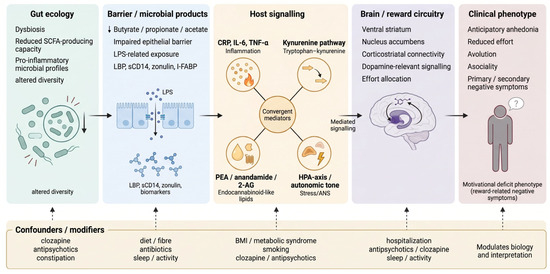
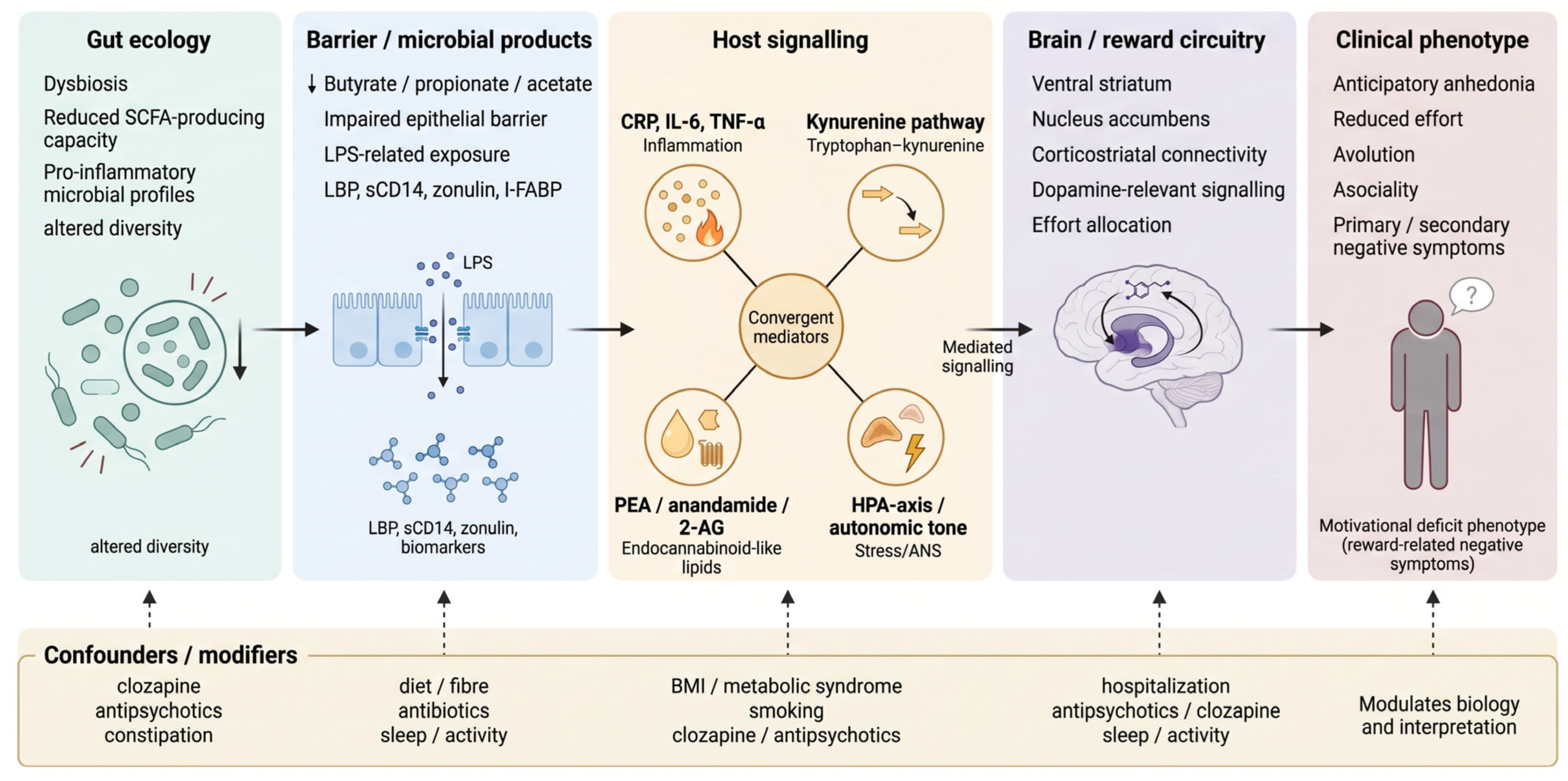
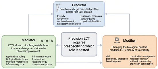
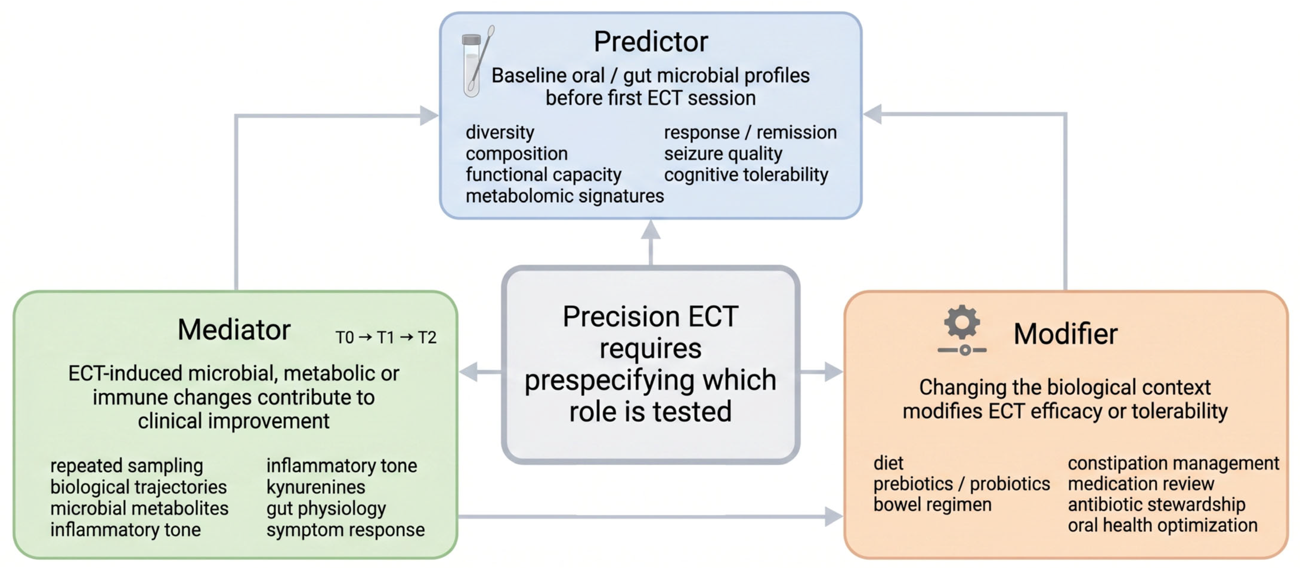
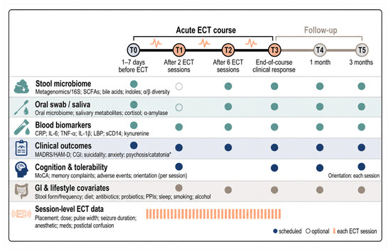
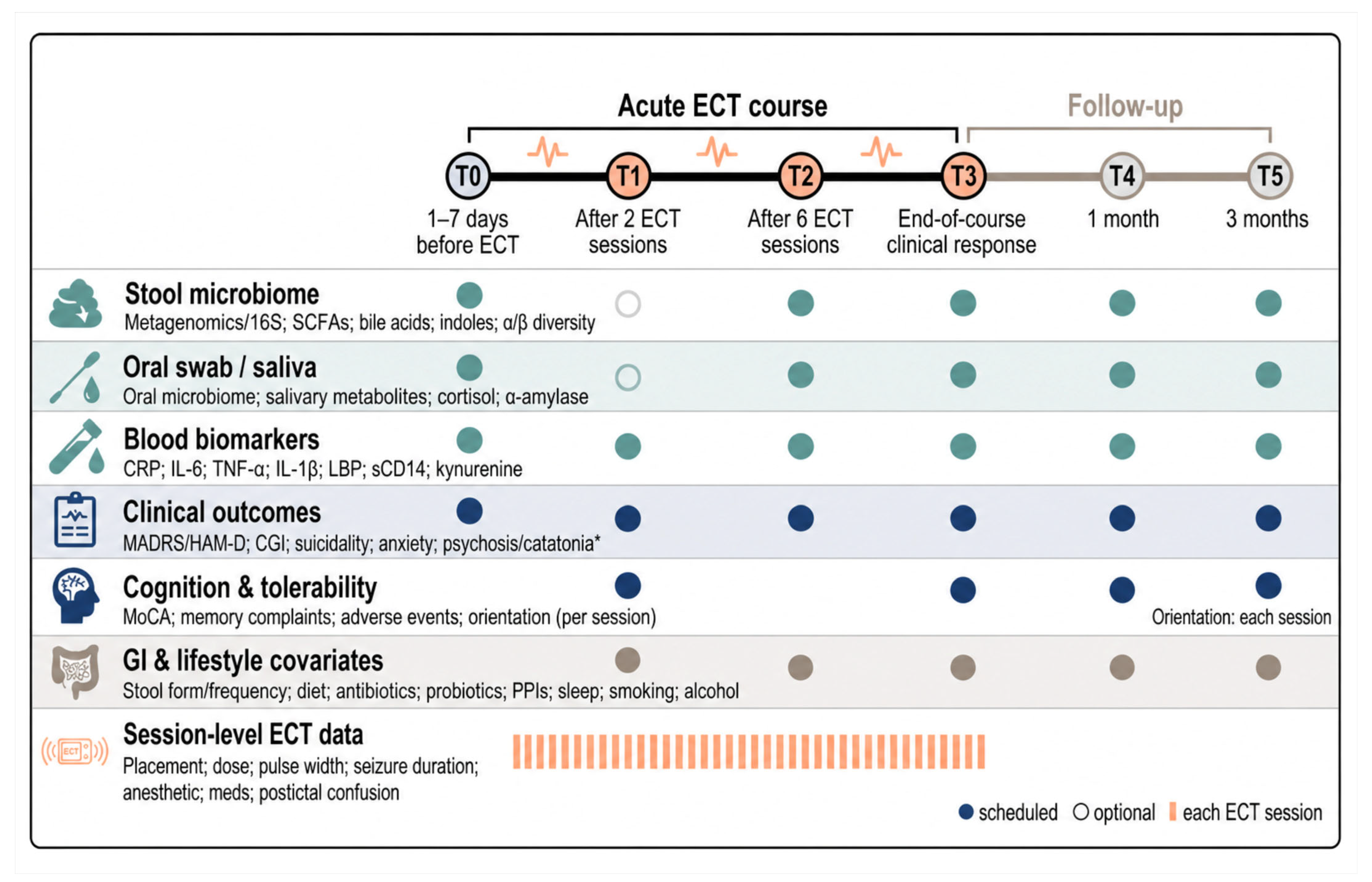

Disclaimer/Publisher’s Note: The statements, opinions and data contained in all publications are solely those of the individual author(s) and contributor(s) and not of MDPI and/or the editor(s). MDPI and/or the editor(s) disclaim responsibility for any injury to people or property resulting from any ideas, methods, instructions or products referred to in the content. |
© 2026 by the authors. Licensee MDPI, Basel, Switzerland. This article is an open access article distributed under the terms and conditions of the Creative Commons Attribution (CC BY) license.