Incomplete Concordance Between Nominal Eosinophilic Labels and Molecular Burden in Chronic Rhinosinusitis with Nasal Polyps

Abstract
1. Introduction
2. Materials and Methods
2.1. Study Design
2.2. Public Datasets and Overall Analytical Framework
2.3. Construction of the Molecular Burden Framework
2.4. Consistency and Sensitivity Analyses
2.5. Discovery Epithelial Single-Cell Analysis
2.6. Independent Validation scRNA-Seq Analysis
2.7. GeoMx Spatial Profiling Analysis
2.8. Bulk Transcriptomic Analysis
2.9. Statistical Analysis
3. Results
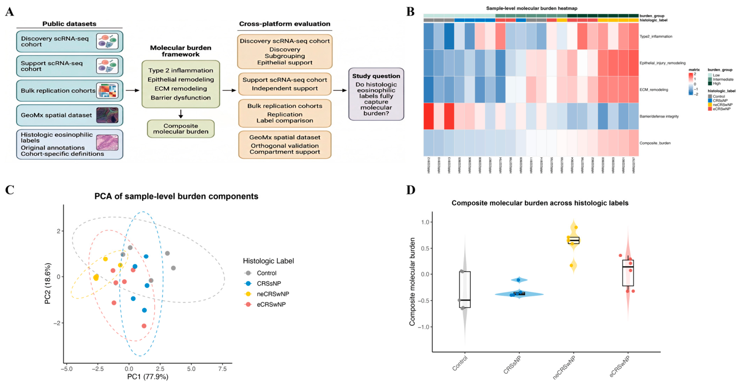
3.1. Public Transcriptomic Data Reveal a Graded and Partially Overlapping Molecular Burden Landscape Across Histologic Labels in the Discovery scRNA-Seq Cohort
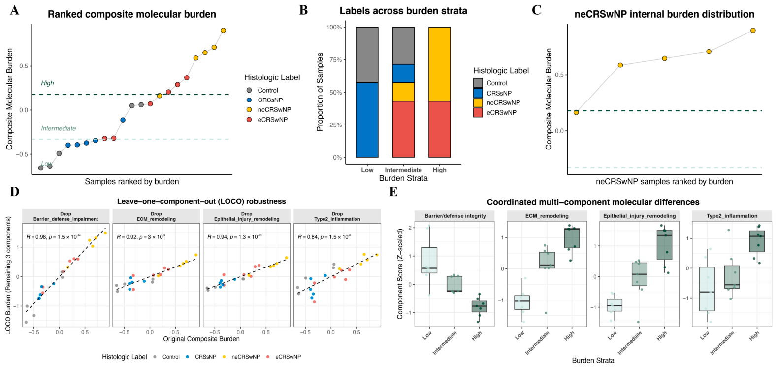
3.2. Nominally Non-Eosinophilic CRSwNP Is Represented Across the Intermediate-to-High Molecular Burden Range in the Discovery Cohort
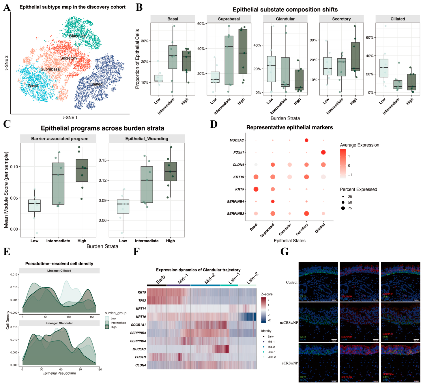
3.3. Single-Cell Analysis Links Higher Molecular Burden to Epithelial State Reorganization, Injury-Associated Programs, and Trajectory-Associated Remodeling
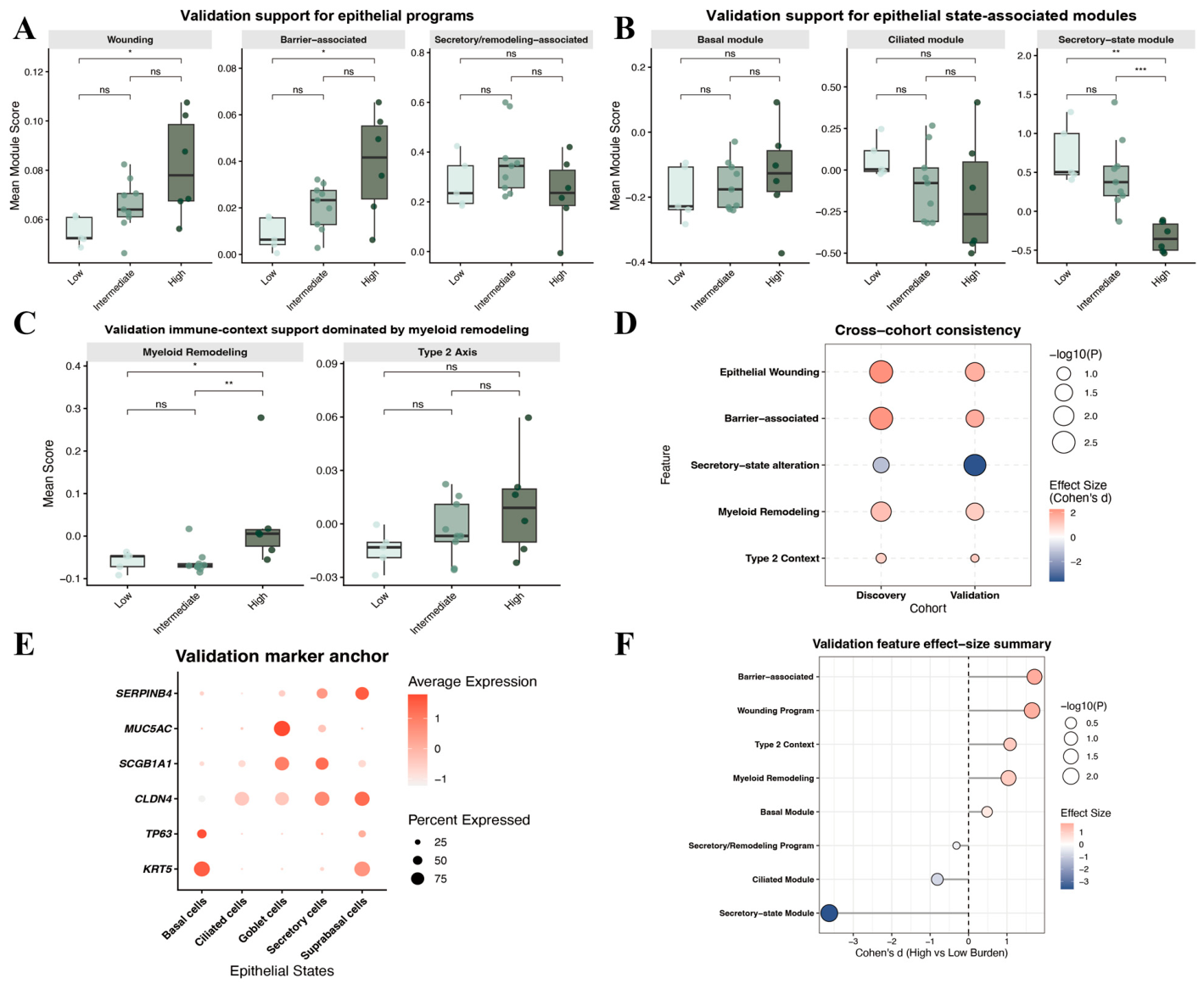
3.4. Independent Validation Provides Partial and Uneven External Support for Framework-Related Features
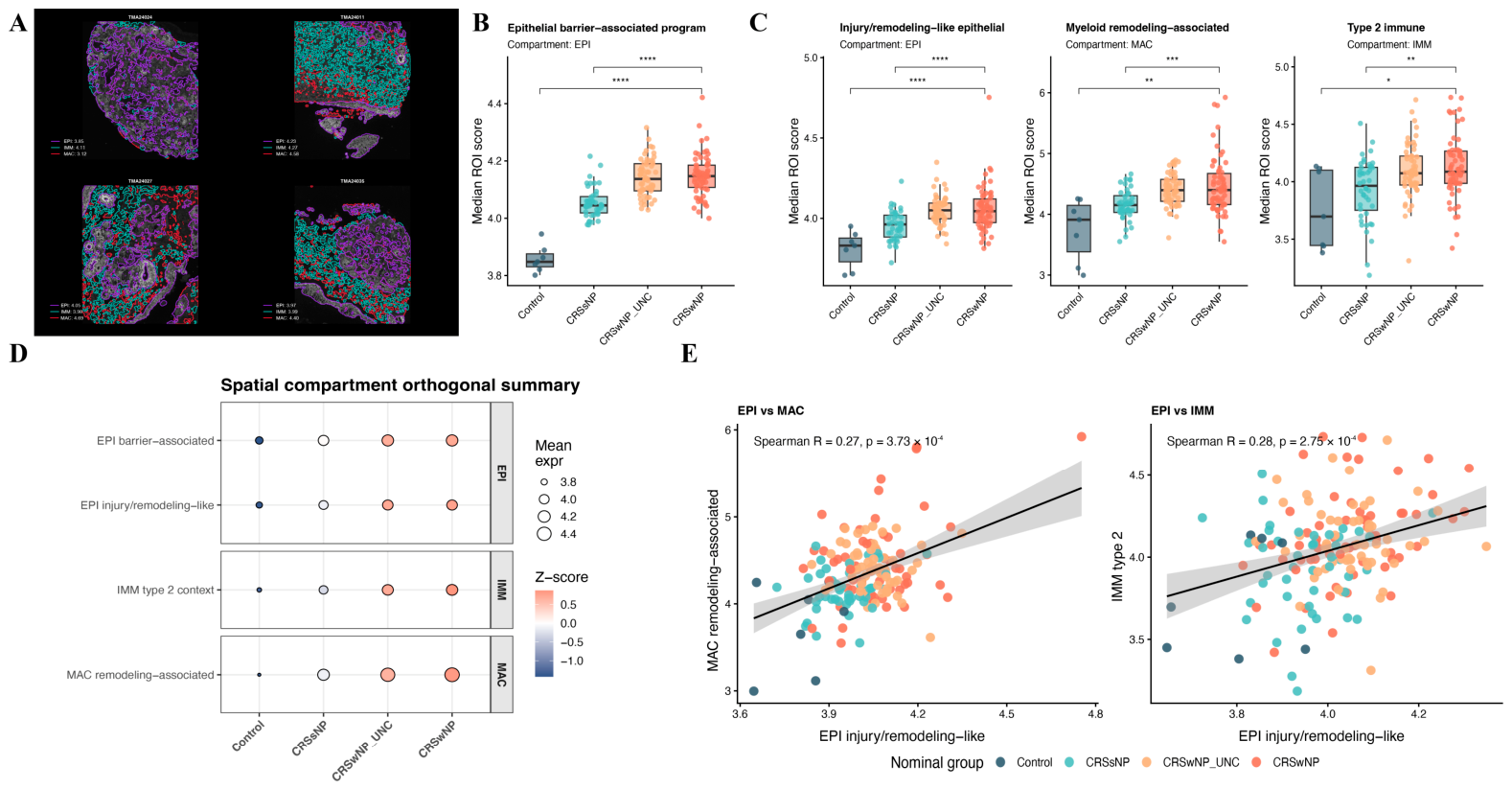
3.5. GeoMx Spatial Profiling Provides Compartment-Resolved Support While Illustrating Heterogeneous Molecular Organization Across Nominal Disease Groups
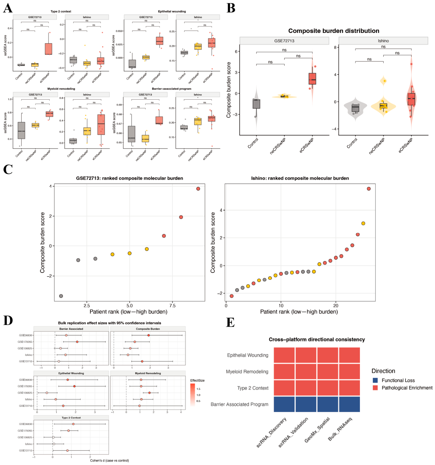
3.6. Independent Bulk Transcriptomic Cohorts Support the Burden Framework and Further Illustrate Incomplete Concordance Between Nominal Eosinophilic Labels and Molecular Burden
4. Discussion
5. Conclusions
Supplementary Materials
Author Contributions
Funding
Institutional Review Board Statement
Informed Consent Statement
Data Availability Statement
Acknowledgments
Conflicts of Interest
Abbreviations
| CRS | chronic rhinosinusitis |
| CRSwNP | chronic rhinosinusitis with nasal polyps |
| CRSsNP | chronic rhinosinusitis without nasal polyps |
| HRQoL | health-related quality of life |
| JESREC | Japanese Epidemiological Survey of Refractory Eosinophilic Chronic Rhinosinusitis |
| scRNA-seq | single-cell RNA sequencing |
| GeoMx | GeoMx digital spatial profiling |
| ECM | extracellular matrix |
| TMM | trimmed mean of M values |
| logCPM | log counts per million |
| GSVA | gene set variation analysis |
| ssGSEA | single-sample gene set enrichment analysis |
| t-SNE | t-distributed stochastic neighbor embedding |
| ROI | region of interest |
| FOV | field of view |
| HPF | high power field |
| ECRS | eosinophilic chronic rhinosinusitis |
| T/NK | T cell/natural killer cell |
| CT | control tissue |
| NP | nasal polyp |
References
- Fokkens, W.J.; Lund, V.J.; Hopkins, C. European Position Paper on Rhinosinusitis and Nasal Polyps. Rhinology 2020, 58, 1–464. [Google Scholar] [CrossRef] [Scilit]
- Orlandi, R.R.; Kingdom, T.T.; Smith, T.L.; Bleier, B.; DeConde, A.; Luong, A.U.; Poetker, D.M.; Soler, Z.; Welch, K.C.; Wise, S.K.; et al. International consensus statement on allergy and rhinology: Rhinosinusitis 2021. Int. Forum Allergy Rhinol. 2021, 11, 213–739. [Google Scholar] [CrossRef] [Scilit]
- Mullol, J.; Azar, A.; Buchheit, K.M.; Hopkins, C.; Bernstein, J.A. Chronic Rhinosinusitis with Nasal Polyps: Quality of Life in the Biologics Era. J. Allergy Clin. Immunol. Pract. 2022, 10, 1434–1453.e9. [Google Scholar] [CrossRef] [Scilit] [PubMed]
- Fokkens, W.J.; Lund, V.; Bachert, C.; Mullol, J.; Bjermer, L.; Bousquet, J.; Canonica, G.W.; Deneyer, L.; Desrosiers, M.; Diamant, Z.; et al. EUFOREA consensus on biologics for CRSwNP with or without asthma. Allergy 2019, 74, 2312–2319. [Google Scholar] [CrossRef] [Scilit]
- Bachert, C.; Han, J.K.; Desrosiers, M.; Hellings, P.W.; Amin, N.; Lee, S.E.; Mullol, J.; Greos, L.S.; Bosso, J.V.; Laidlaw, T.M.; et al. Efficacy and safety of dupilumab in patients with severe chronic rhinosinusitis with nasal polyps (LIBERTY NP SINUS-24 and LIBERTY NP SINUS-52): Results from two multicentre, randomised, double-blind, placebo-controlled, parallel-group phase 3 trials. Lancet 2019, 394, 1638–1650. [Google Scholar] [CrossRef] [Scilit] [PubMed]
- DeConde, A.S.; Mace, J.C.; Levy, J.M.; Rudmik, L.; Alt, J.A.; Smith, T.L. Prevalence of polyp recurrence after endoscopic sinus surgery for chronic rhinosinusitis with nasal polyposis. Laryngoscope 2016, 127, 550–555. [Google Scholar] [CrossRef] [Scilit] [PubMed]
- Kato, A.; Schleimer, R.P.; Bleier, B.S. Mechanisms and pathogenesis of chronic rhinosinusitis. J. Allergy Clin. Immunol. 2022, 149, 1491–1503. [Google Scholar] [CrossRef] [Scilit]
- Wang, X.; Sima, Y.; Zhao, Y.; Zhang, N.; Zheng, M.; Du, K.; Wang, M.; Wang, Y.; Hao, Y.; Li, Y.; et al. Endotypes of chronic rhinosinusitis based on inflammatory and remodeling factors. J. Allergy Clin. Immunol. 2023, 151, 458–468. [Google Scholar] [CrossRef] [Scilit]
- Subspecialty Group of Rhinology, Editorial Board of Chinese Journal of Otorhinolaryngology Head and Neck Surgery; Subspecialty Group of Rhinology, Society of Otorhinolaryngology Head and Neck Surgery, Chinese Medical Association. Chinese Guidelines for Diagnosis and Treatment of Chronic Rhinosinusitis (2024). World J. Otorhinolaryngol.—Head Neck Surg. 2025, 12, 25–55. [Google Scholar]
- Tokunaga, T.; Sakashita, M.; Haruna, T.; Asaka, D.; Takeno, S.; Ikeda, H.; Nakayama, T.; Seki, N.; Ito, S.; Murata, J.; et al. Novel scoring system and algorithm for classifying chronic rhinosinusitis: The JESREC Study. Allergy 2015, 70, 995–1003. [Google Scholar] [CrossRef] [Scilit]
- Lou, H.; Meng, Y.; Piao, Y.; Wang, C.; Zhang, L.; Bachert, C. Predictive Significance of Tissue Eosinophilia for Nasal Polyp Recurrence in the Chinese Population. Am. J. Rhinol. Allergy 2015, 29, 350–356. [Google Scholar] [CrossRef] [Scilit]
- Lou, H.; Zhang, N.; Bachert, C.; Zhang, L. Highlights of eosinophilic chronic rhinosinusitis with nasal polyps in definition, prognosis, and advancement. Int. Forum Allergy Rhinol. 2018, 8, 1218–1225. [Google Scholar] [CrossRef] [Scilit]
- Yang, S.K.; Kim, J.-W.; Won, T.-B.; Rhee, C.-S.; Han, Y.B.; Cho, S.-W. Differences in Clinical and Immunological Characteristics According to the Various Criteria for Tissue Eosinophilia in Chronic Rhinosinusitis with Nasal Polyps. Clin. Exp. Otorhinolaryngol. 2023, 16, 359–368. [Google Scholar] [CrossRef] [Scilit]
- Huang, Z.Q.; Liu, J.; Sun, L.Y.; Ong, H.H.; Ye, J.; Xu, Y.; Wang, D.Y. Updated epithelial barrier dysfunction in chronic rhinosinusitis: Targeting pathophysiology and treatment response of tight junctions. Allergy 2024, 79, 1146–1165. [Google Scholar] [CrossRef] [Scilit]
- Xie, X.; Wang, P.; Jin, M.; Wang, Y.; Qi, L.; Wu, C.; Guo, S.; Li, C.; Zhang, X.; Yuan, Y.; et al. IL-1β-induced epithelial cell and fibroblast transdifferentiation promotes neutrophil recruitment in chronic rhinosinusitis with nasal polyps. Nat. Commun. 2024, 15, 9101. [Google Scholar] [CrossRef] [Scilit]
- Liao, G.; Nakayama, T.; Zhu, B.; Lee, I.T.; Yeung, J.; Yeo, Y.Y.; Chang, Y.; Wang, C.; Liao, S.C.-K.; Nkosi, D.; et al. Multi-scaled transcriptomics of chronically inflamed nasal epithelium reveals immune-epithelial dynamics and tissue remodeling in nasal polyp formation. Immunity 2025, 58, 2593–2608.e6. [Google Scholar] [CrossRef] [Scilit] [PubMed]
- Zhang, Y.; Gevaert, E.; Lou, H.; Wang, X.; Zhang, L.; Bachert, C.; Zhang, N. Chronic rhinosinusitis in Asia. J. Allergy Clin. Immunol. 2017, 140, 1230–1239. [Google Scholar] [CrossRef] [Scilit] [PubMed]
- Wang, W.; Xu, Y.; Wang, L.; Zhu, Z.; Aodeng, S.; Chen, H.; Cai, M.; Huang, Z.; Han, J.; Wang, L.; et al. Single-cell profiling identifies mechanisms of inflammatory heterogeneity in chronic rhinosinusitis. Nat. Immunol. 2022, 23, 1484–1494. [Google Scholar] [CrossRef] [Scilit] [PubMed]
- Wang, W.; Gao, Z.; Wang, H.; Li, T.; He, W.; Lv, W.; Zhang, J. Transcriptome Analysis Reveals Distinct Gene Expression Profiles in Eosinophilic and Noneosinophilic Chronic Rhinosinusitis with Nasal Polyps. Sci. Rep. 2016, 6, 26604. [Google Scholar] [CrossRef] [Scilit]
- Ishino, T.; Takeno, S.; Takemoto, K.; Yamato, K.; Oda, T.; Nishida, M.; Horibe, Y.; Chikuie, N.; Kono, T.; Taruya, T.; et al. Distinct Gene Set Enrichment Profiles in Eosinophilic and Non-Eosinophilic Chronic Rhinosinusitis with Nasal Polyps by Bulk RNA Barcoding and Sequencing. Int. J. Mol. Sci. 2022, 23, 5653. [Google Scholar] [CrossRef] [Scilit]
- Ordovas-Montanes, J.; Dwyer, D.F.; Nyquist, S.K.; Buchheit, K.M.; Vukovic, M.; Deb, C.; Wadsworth, M.H.; 2nd Hughes, T.K.; Kazer, S.W.; Yoshimoto, E.; et al. Allergic inflammatory memory in human respiratory epithelial progenitor cells. Nature 2018, 560, 649–654. [Google Scholar] [CrossRef] [Scilit]
- Stuart, T.; Butler, A.; Hoffman, P.; Hafemeister, C.; Papalexi, E.; Mauck, W.M.; Hao, Y., 3rd; Stoeckius, M.; Smibert, P.; Satija, R. Comprehensive Integration of Single-Cell Data. Cell 2019, 177, 1888–1902.e21. [Google Scholar] [CrossRef] [Scilit]
- Street, K.; Risso, D.; Fletcher, R.B.; Das, D.; Ngai, J.; Yosef, N.; Purdom, E.; Dudoit, S. Slingshot: Cell lineage and pseudotime inference for single-cell transcriptomics. BMC Genom. 2018, 19, 477. [Google Scholar] [CrossRef] [Scilit] [PubMed]
- Merritt, C.R.; Ong, G.T.; Church, S.E.; Barker, K.; Danaher, P.; Geiss, G.; Hoang, M.; Jung, J.; Liang, Y.; McKay-Fleisch, J.; et al. Multiplex digital spatial profiling of proteins and RNA in fixed tissue. Nat. Biotechnol. 2020, 38, 586–599. [Google Scholar] [CrossRef] [Scilit]
- Bachert, C.; Marple, B.; Schlosser, R.J.; Hopkins, C.; Schleimer, R.P.; Lambrecht, B.N.; Bröker, B.M.; Laidlaw, T.; Song, W.-J. Adult chronic rhinosinusitis. Nat. Rev. Dis. Prim. 2020, 6, 86. [Google Scholar] [CrossRef] [Scilit] [PubMed]
- Schleimer, R.P. Immunopathogenesis of Chronic Rhinosinusitis and Nasal Polyposis. Annu. Rev. Pathol. Mech. Dis. 2017, 12, 331–357. [Google Scholar] [CrossRef] [Scilit]
- Choi, T.; Ryu, S.; Bae, J.-S.; Yoo, S.H.; Mo, J.-H. Epithelial-Mesenchymal Transition in Chronic Rhinosinusitis. J. Rhinol. 2024, 31, 67–77. [Google Scholar] [CrossRef] [Scilit]
- Takabayashi, T.; Kato, A.; Peters, A.T.; Hulse, K.E.; Suh, L.A.; Carter, R.; Norton, J.; Grammer, L.C.; Cho, S.H.; Tan, B.K.; et al. Excessive Fibrin Deposition in Nasal Polyps Caused by Fibrinolytic Impairment through Reduction of Tissue Plasminogen Activator Expression. Am. J. Respir. Crit. Care Med. 2013, 187, 49–57. [Google Scholar] [CrossRef] [Scilit]
- Bachert, C.; Han, J.K.; Desrosiers, M.Y.; Gevaert, P.; Heffler, E.; Hopkins, C.; Tversky, J.R.; Barker, P.; Cohen, D.; Emson, C.; et al. Efficacy and safety of benralizumab in chronic rhinosinusitis with nasal polyps: A randomized, placebo-controlled trial. J. Allergy Clin. Immunol. 2022, 149, 1309–1317.e12. [Google Scholar] [CrossRef] [Scilit]
- Gevaert, P.; Omachi, T.A.; Corren, J.; Mullol, J.; Han, J.; Lee, S.E.; Kaufman, D.; Ligueros-Saylan, M.; Howard, M.; Zhu, R.; et al. Efficacy and safety of omalizumab in nasal polyposis: 2 randomized phase 3 trials. J. Allergy Clin. Immunol. 2020, 146, 595–605. [Google Scholar] [CrossRef] [Scilit] [PubMed]
- Zimmerman, K.D.; Espeland, M.A.; Langefeld, C.D. A practical solution to pseudoreplication bias in single-cell studies. Nat. Commun. 2021, 12, 738. [Google Scholar] [CrossRef] [Scilit]
- Delemarre, T.; Bochner, B.S.; Simon, H.-U.; Bachert, C. Rethinking neutrophils and eosinophils in chronic rhinosinusitis. J. Allergy Clin. Immunol. 2021, 148, 327–335. [Google Scholar] [CrossRef] [Scilit] [PubMed]
- Jeican, I.I.; Gheban, D.; Barbu-Tudoran, L.; Inisca, P.; Albu, C.; Ilies, M.; Albu, S.; Vica, M.L.; Matei, H.V.; Tripon, S.; et al. Respiratory Nasal Mucosa in Chronic Rhinosinusitis with Nasal Polyps versus COVID-19: Histopathology, Electron Microscopy Analysis and Assessing of Tissue Interleukin-33. J. Clin. Med. 2021, 10, 4110. [Google Scholar] [CrossRef] [Scilit] [PubMed]


Disclaimer/Publisher’s Note: The statements, opinions and data contained in all publications are solely those of the individual author(s) and contributor(s) and not of MDPI and/or the editor(s). MDPI and/or the editor(s) disclaim responsibility for any injury to people or property resulting from any ideas, methods, instructions or products referred to in the content. |
© 2026 by the authors. Licensee MDPI, Basel, Switzerland. This article is an open access article distributed under the terms and conditions of the Creative Commons Attribution (CC BY) license.
Share and Cite
Tan, S.; Lai, J.; Zhi, H.; Tang, W.; Jin, L.; Yu, S. Incomplete Concordance Between Nominal Eosinophilic Labels and Molecular Burden in Chronic Rhinosinusitis with Nasal Polyps. Biomedicines 2026, 14, 1189. https://doi.org/10.3390/biomedicines14061189
Tan S, Lai J, Zhi H, Tang W, Jin L, Yu S. Incomplete Concordance Between Nominal Eosinophilic Labels and Molecular Burden in Chronic Rhinosinusitis with Nasal Polyps. Biomedicines. 2026; 14(6):1189. https://doi.org/10.3390/biomedicines14061189
Chicago/Turabian StyleTan, Shiwang, Ju Lai, Heng Zhi, Wei Tang, Ling Jin, and Shaoqing Yu. 2026. "Incomplete Concordance Between Nominal Eosinophilic Labels and Molecular Burden in Chronic Rhinosinusitis with Nasal Polyps" Biomedicines 14, no. 6: 1189. https://doi.org/10.3390/biomedicines14061189
APA StyleTan, S., Lai, J., Zhi, H., Tang, W., Jin, L., & Yu, S. (2026). Incomplete Concordance Between Nominal Eosinophilic Labels and Molecular Burden in Chronic Rhinosinusitis with Nasal Polyps. Biomedicines, 14(6), 1189. https://doi.org/10.3390/biomedicines14061189