Evolution of Concepts Regarding the Diagnostic and Prognostic Significance of Glial Fibrillary Acidic Protein (GFAP)-Positive Extracellular Vesicles

 , , , , , and
Abstract
1. Introduction and Review Methodology
2. Structure and Function of GFAP
3. Expression, Secretion, and Study of Serum/Plasma GFAP as Diagnostic and Prognostic Marker of CNS Pathology
4. GFAP-Positive Vesicles as Marker of CNS Pathology
5. GFAP-Positive Vesicles as Marker of PNS Pathology
6. Methodological Aspects of the Study of GFAP-Positive Extracellular Vesicles
7. Conclusions
Author Contributions
Funding
Institutional Review Board Statement
Informed Consent Statement
Data Availability Statement
Conflicts of Interest
Abbreviations
| PC | Protein corona |
| EVs | Extracellular vesicles |
| GFAP | Glial fibrillary acidic protein |
| ADEVs | Astrocyte-derived EVs |
| CD9, CD63, TSG101, ALIX | Canonical EV markers |
| SCs | Schwann cells |
| SGCs | Satellite glial cells |
| SC-EVs | EVs from SCs |
| SGC-EVs | EVs from SGCs |
| DNP | Diabetic neuropathy |
| DINP | Drug-induced neuropathy |
| PTEN | A dual-substrate specificity phosphatase, a product of the PTEN gene, a negative regulator of the PI3K/AKT/mTOR signaling pathway |
| PNS | Peripheral nervous system |
| CNS | Central nervous system |
| BBB | Blood–brain barrier |
| DRG | Dorsal root ganglia of spinal nerves |
| NTA | Nanoparticle tracking analysis |
| MS | Mass spectrometry |
| NfL | Neurofilament light chain |
| STAT3, AP-1 | Transcription factors, regulating GFAP expression |
| CT | Computer tomography |
| AD | Alzheimer’s disease |
| MMD | Major depressive disorder |
| GLAST | Glutamine aspartate transporter |
| SED | Stress-induced exhaustion disorder |
| AQP4 | Aquaporin 4 |
| MMPs | Matrix metalloproteinases |
| ELISA | Enzyme-linked immunosorbent assay |
References
- Eng, L.F.; Ghirnikar, R.S.; Lee, Y.L. Glial fibrillary acidic protein: GFAP-thirty-one years (1969–2000). Neurochem. Res. 2000, 25, 1439–1451. [Google Scholar] [CrossRef] [PubMed]
- Abdelhak, A.; Foschi, M.; Abu-Rumeileh, S.; Yue, J.K.; D’Anna, L.; Huss, A.; Oeckl, P.; Ludolph, A.C.; Kuhle, J.; Petzold, A.; et al. Blood GFAP as an emerging biomarker in brain and spinal cord disorders. Nat. Rev. Neurol. 2022, 18, 158–172. [Google Scholar] [CrossRef]
- Zheng, X.; Yang, J.; Hou, Y.; Shi, X.; Liu, K. Prediction of clinical progression in nervous system diseases: Plasma glial fibrillary acidic protein (GFAP). Eur. J. Med. Res. 2024, 29, 51. [Google Scholar] [CrossRef]
- Welsh, J.A.; Goberdhan, D.C.I.; O’Driscoll, L.; Buzas, E.I.; Blenkiron, C.; Bussolati, B.; Cai, H.; Di Vizio, D.; Driedonks, T.A.P.; Erdbrügger, U.; et al. Minimal information for studies of extracellular vesicles (MISEV2023): From basic to advanced approaches. J. Extracell. Vesicles 2024, 13, e12404. [Google Scholar] [CrossRef] [PubMed]
- Ralton, J.E.; Lu, X.; Hutcheson, A.M.; Quinlan, R.A. Identification of two N-terminal non-alpha-helical domain motifs important in the assembly of glial fibrillary acidic protein. Cell Sci. 1994, 107, 1935–1948. [Google Scholar] [CrossRef]
- Hol, E.M.; Pekny, M. Glial fibrillary acidic protein (GFAP) and the astrocyte intermediate filament system in diseases of the central nervous system. Curr. Opin. Cell Biol. 2015, 32, 121–130. [Google Scholar] [CrossRef] [PubMed]
- Lim, M.C.; Maubach, G.; Zhuo, L. Glial fibrillary acidic protein splice variants in hepatic stellate cells--expression and regulation. Mol. Cells 2008, 25, 376–384. [Google Scholar] [CrossRef]
- Messing, A.; Brenner, M. GFAP at 50. ASN Neuro 2020, 12, 1759091420949680. [Google Scholar] [CrossRef]
- Galea, E.; Dupouey, P.; Feinstein, D.L. Glial fibrillary acidic protein mRNA isotypes: Expression in vitro and in vivo. J. Neurosci. Res. 1995, 41, 452–461. [Google Scholar] [CrossRef]
- Yang, Z.; Wang, K.K. Glial fibrillary acidic protein: From intermediate filament assembly and gliosis to neurobiomarker. Trends Neurosci. 2015, 38, 364–374. [Google Scholar] [CrossRef]
- Kamphuis, W.; Mamber, C.; Moeton, M.; Kooijman, L.; Sluijs, J.A.; Jansen, A.H.; Verveer, M.; de Groot, L.R.; Smith, V.D.; Rangarajan, S.; et al. GFAP isoforms in adult mouse brain with a focus on neurogenic astrocytes and reactive astrogliosis in mouse models of Alzheimer disease. PLoS ONE 2012, 7, e42823. [Google Scholar] [CrossRef]
- Kamphuis, W.; Middeldorp, J.; Kooijman, L.; Sluijs, J.A.; Kooi, E.J.; Moeton, M.; Freriks, M.; Mizee, M.R.; Hol, E.M. Glial fibrillary acidic protein isoform expression in plaque related astrogliosis in Alzheimer’s disease. Neurobiol. Aging 2014, 35, 492–510. [Google Scholar] [CrossRef]
- Boer, K.; Middeldorp, J.; Spliet, W.G.; Razavi, F.; van Rijen, P.C.; Baayen, J.C.; Hol, E.M.; Aronica, E. Immunohistochemical characterization of the out-of frame splice variants GFAP Delta164/Deltaexon 6 in focal lesions associated with chronic epilepsy. Epilepsy Res. 2010, 90, 99–109. [Google Scholar] [CrossRef] [PubMed]
- Zelenika, D.; Grima, B.; Brenner, M.; Pessac, B. A novel glial fibrillary acidic protein mRNA lacking exon 1. Brain Res. Mol. Brain Res. 1995, 30, 251–258. [Google Scholar] [CrossRef]
- Potokar, M.; Stenovec, M.; Gabrijel, M.; Li, L.; Kreft, M.; Grilc, S.; Pekny, M.; Zorec, R. Intermediate filaments attenuate stimulation-dependent mobility of endosomes/lysosomes in astrocytes. Glia 2010, 58, 1208–1219. [Google Scholar] [CrossRef]
- Tang, Y.; Han, L.; Li, S.; Hu, T.; Xu, Z.; Fan, Y.; Liang, X.; Yu, H.; Wu, J.; Wang, J. Plasma GFAP in Parkinson’s disease with cognitive impairment and its potential to predict conversion to dementia. npj Park. Dis. 2023, 9, 23. [Google Scholar] [CrossRef] [PubMed]
- Bandyopadhyay, U.; Sridhar, S.; Kaushik, S.; Kiffin, R.; Cuervo, A.M. Identification of regulators of chaperone-mediated autophagy. Mol. Cell. 2010, 39, 535–547. [Google Scholar] [CrossRef]
- Arias, E.; Koga, H.; Diaz, A.; Mocholi, E.; Patel, B.; Cuervo, A.M. Lysosomal mTORC2/PHLPP1/Akt Regulate Chaperone-Mediated Autophagy. Mol. Cell 2015, 59, 270–284. [Google Scholar] [CrossRef]
- Kalra, L.P.; Khatter, H.; Ramanathan, S.; Sapehia, S.; Devi, K.; Kaliyaperumal, A.; Bal, D.; Sebastian, I.; Kakarla, R.; Singhania, A.; et al. Serum GFAP for stroke diagnosis in regions with limited access to brain imaging (BE FAST India). Eur. Stroke J. 2021, 6, 176–184. [Google Scholar] [CrossRef]
- Triolo, D.; Dina, G.; Lorenzetti, I.; Malaguti, M.; Morana, P.; Del Carro, U.; Comi, G.; Messing, A.; Quattrini, A.; Previtali, S.C. Loss of glial fibrillary acidic protein (GFAP) impairs Schwann cell proliferation and delays nerve regeneration after damage. J. Cell Sci. 2006, 119, 3981–3993. [Google Scholar] [CrossRef] [PubMed]
- Stenzel, W.; Soltek, S.; Schlüter, D.; Deckert, M. The intermediate filament GFAP is important for the control of experimental murine Staphylococcus aureus-induced brain abscess and Toxoplasma encephalitis. J. Neuropathol. Exp. Neurol. 2004, 63, 631–640. [Google Scholar] [CrossRef]
- Middeldorp, J.; Hol, E.M. GFAP in health and disease. Prog. Neurobiol. 2011, 93, 421–443. [Google Scholar] [CrossRef]
- Laranjeira, C.; Sandgren, K.; Kessaris, N.; Richardson, W.; Potocnik, A.; Vanden Berghe, P.; Pachnis, V. Glial cells in the mouse enteric nervous system can undergo neurogenesis in response to injury. J. Clin. Investig. 2011, 121, 3412–3424. [Google Scholar] [CrossRef]
- Gulbransen, B.D.; Sharkey, K.A. Novel functional roles for enteric glia in the gastrointestinal tract. Nat. Rev. Gastroenterol. Hepatol. 2012, 9, 625–632. [Google Scholar] [CrossRef]
- Davidoff, M.S.; Middendorff, R.; Köfüncü, E.; Müller, D.; Jezek, D.; Holstein, A.F. Leydig cells of the human testis possess astrocyte and oligodendrocyte marker molecules. Acta Histochem. 2002, 104, 39–49. [Google Scholar] [CrossRef]
- Yeo, S.; Bandyopadhyay, S.; Messing, A.; Brenner, M. Transgenic analysis of GFAP promoter elements. Glia 2013, 61, 1488–1499. [Google Scholar] [CrossRef] [PubMed]
- Brenner, M.; Messing, A.; Olsen, M.L. AP-1 and the injury response of the GFAP gene. J. Neurosci. Res. 2019, 97, 149–161. [Google Scholar] [CrossRef] [PubMed]
- Liddelow, S.A.; Barres, B.A. Reactive Astrocytes: Production, Function, and Therapeutic Potential. Immunity 2017, 46, 957–967. [Google Scholar] [CrossRef] [PubMed]
- Escartin, C.; Guillemaud, O.; Carrillo-de Sauvage, M.A. Questions and (some) answers on reactive astrocytes. Glia 2019, 67, 2221–2247. [Google Scholar] [CrossRef]
- Aibaidula, A.; Gharibi Loron, A.; Bouchal, S.M.; Bauman, M.M.J.; You, H.B.; Lucien, F.; Parney, I.F. Plasma Extracellular Vesicles as Liquid Biopsies for Glioblastoma: Biomarkers, Subpopulation Enrichment, and Clinical Translation. Int. J. Mol. Sci. 2025, 26, 11686. [Google Scholar] [CrossRef]
- Tumani, H.; Huss, A.; Bachhuber, F. The cerebrospinal fluid and barriers—anatomic and physiologic considerations. Handb. Clin. Neurol. 2017, 146, 21–32. [Google Scholar] [PubMed]
- Plog, B.A.; Dashnaw, M.L.; Hitomi, E.; Peng, W.; Liao, Y.; Lou, N.; Deane, R.; Nedergaard, M. Biomarkers of traumatic injury are transported from brain to blood via the glymphatic system. J. Neurosci. 2015, 35, 518–526. [Google Scholar] [CrossRef]
- Anderson, T.N.; Hwang, J.; Munar, M.; Papa, L.; Hinson, H.E.; Vaughan, A.; Rowell, S.E. Blood-based biomarkers for prediction of intracranial hemorrhage and outcome in patients with moderate or severe traumatic brain injury. J. Trauma Acute Care Surg. 2020, 89, 80–86. [Google Scholar] [CrossRef]
- Krämer-Albers, E.M.; Bretz, N.; Tenzer, S.; Winterstein, C.; Möbius, W.; Berger, H.; Nave, K.A.; Schild, H.; Trotter, J. Oligodendrocytes secrete exosomes containing major myelin and stress-protective proteins: Trophic support for axons? Proteom. Clin. Appl. 2007, 1, 1446–1461. [Google Scholar] [CrossRef] [PubMed]
- Potolicchio, I.; Carven, G.J.; Xu, X.; Stipp, C.; Riese, R.J.; Stern, L.J.; Santambrogio, L. Proteomic analysis of microglia-derived exosomes: Metabolic role of the aminopeptidase CD13 in neuropeptide catabolism. J. Immunol. 2005, 175, 2237–2243. [Google Scholar] [CrossRef]
- Willis, C.M.; Ménoret, A.; Jellison, E.R.; Nicaise, A.M.; Vella, A.T.; Crocker, S.J. A Refined Bead-Free Method to Identify Astrocytic Exosomes in Primary Glial Cultures and Blood Plasma. Front. Neurosci. 2017, 11, 335. [Google Scholar] [CrossRef]
- Goetzl, E.J.; Mustapic, M.; Kapogiannis, D.; Eitan, E.; Lobach, I.V.; Goetzl, L.; Schwartz, J.B.; Miller, B.L. Cargo proteins of plasma astrocyte-derived exosomes in Alzheimer’s disease. FASEB J. 2016, 30, 3853–3859. [Google Scholar] [CrossRef] [PubMed]
- Ranganathan, M.; Rahman, M.; Ganesh, S.; D’Souza, D.C.; Skosnik, P.D.; Radhakrishnan, R.; Pathania, S.; Mohanakumar, T. Analysis of circulating exosomes reveals a peripheral signature of astrocytic pathology in schizophrenia. World J. Biol. Psychiatry 2022, 23, 33–45. [Google Scholar] [CrossRef]
- Wallensten, J.; Nager, A.; Åsberg, M.; Borg, K.; Beser, A.; Wilczek, A.; Mobarrez, F. Leakage of astrocyte-derived extracellular vesicles in stress-induced exhaustion disorder: A cross-sectional study. Sci. Rep. 2021, 11, 2009. [Google Scholar] [CrossRef]
- Forró, T.; Manu, D.R.; Băjenaru, O.L.; Bălașa, R. GFAP as Astrocyte-Derived Extracellular Vesicle Cargo in Acute Ischemic Stroke Patients-A Pilot Study. Int. J. Mol. Sci. 2024, 25, 5726. [Google Scholar] [CrossRef]
- Babaee, A.; Wichmann, T.O.; Rasmussen, M.M.; Brink, O.; Olsen, D.A.; Borris, L.C.; Lesbo, M.; Rasmussen, R.W.; Salomon, C.; Handberg, A.; et al. Extracellular Vesicle Glial Fibrillary Acidic Protein as a Circulating Biomarker of Traumatic Brain Injury Severity. J. Mol. Neurosci. 2025, 75, 69. [Google Scholar] [CrossRef]
- Flynn, S.; Leete, J.; Shahim, P.; Pattinson, C.; Guedes, V.A.; Lai, C.; Devoto, C.; Qu, B.X.; Greer, K.; Moore, B.; et al. Extracellular vesicle concentrations of glial fibrillary acidic protein and neurofilament light measured 1 year after traumatic brain injury. Sci. Rep. 2021, 11, 3896. [Google Scholar] [CrossRef]
- Yunusova, N.; Tulendinov, E.; Svarovsky, D.; Ryabova, A.; Kondakova, I.; Ponomaryova, A.; Vtorushin, S.; Tabakaev, S.; Korshunov, D.; Shtam, T.; et al. Levels of Proangiogenic Molecules and Terminal Complement Complex C5b-9 in the Crown of Circulating sEVs in Patients with Recurrent Glioblastomas: Relationship with Tumor Molecular Characteristics. Curr. Issues Mol. Biol. 2025, 47, 132. [Google Scholar] [CrossRef] [PubMed]
- Galbo, P.M., Jr.; Ciesielski, M.J.; Figel, S.; Maguire, O.; Qiu, J.; Wiltsie, L.; Minderman, H.; Fenstermaker, R.A. Circulating CD9+/GFAP+/survivin+ exosomes in malignant glioma patients following survivin vaccination. Oncotarget 2017, 8, 114722–114735. [Google Scholar] [CrossRef] [PubMed]
- Ryabova, A.I.; Novikov, V.A.; Choynzonov, E.L.; Spirina, L.V.; Yunusova, N.V.; Ponomareva, A.A.; Tamkovich, S.N.; Gribova, O.V. The role of liquid biopsy in the diagnosis of glioblastoma progression. Sib. J. Oncol. 2022, 21, 104–116. [Google Scholar] [CrossRef]
- Wu, M.; Shi, Y.; Liu, Y.; Huang, H.; Che, J.; Shi, J.; Xu, C. Exosome-transmitted podoplanin promotes tumor-associated macrophage-mediated immune tolerance in glioblastoma. CNS Neurosci. Ther. 2024, 30, e14643. [Google Scholar] [CrossRef]
- Sartori, M.T.; Della Puppa, A.; Ballin, A.; Campello, E.; Radu, C.M.; Saggiorato, G.; d’Avella, D.; Scienza, R.; Cella, G.; Simioni, P. Circulating microparticles of glial origin and tissue factor bearing in high-grade glioma: A potential prothrombotic role. Thromb. Haemost. 2013, 110, 378–385. [Google Scholar] [CrossRef] [PubMed]
- Li, K.; Du, S.; Li, H.; Li, Z.; Zhu, Q.; Peng, Q.; Liao, B.; Qi, L. A novel three-dimensional co-culture model for studying exosome-mediated cell interactions in glioblastoma. Biochim. Biophys. Acta Gen. Subj. 2025, 1869, 130752. [Google Scholar] [CrossRef]
- Gudbergsson, J.M.; Kostrikov, S.; Johnsen, K.B.; Fliedner, F.P.; Stolberg, C.B.; Humle, N.; Hansen, A.E.; Kristensen, B.W.; Christiansen, G.; Kjær, A.; et al. A tumorsphere model of glioblastoma multiforme with intratumoral heterogeneity for quantitative analysis of cellular migration and drug response. Exp. Cell Res. 2019, 379, 73–82. [Google Scholar] [CrossRef]
- Tóth, E.Á.; Turiák, L.; Visnovitz, T.; Cserép, C.; Mázló, A.; Sódar, B.W.; Försönits, A.I.; Petővári, G.; Sebestyén, A.; Komlósi, Z.; et al. Formation of a protein corona on the surface of extracellular vesicles in blood plasma. J. Extracell. Vesicles 2021, 10, e12140. [Google Scholar] [CrossRef]
- Petrova, E.S. Current Views on Schwann Cells: Development, Plasticity, Functions. J. Evol. Biochem. Phys. 2019, 55, 433–447. [Google Scholar] [CrossRef]
- Griffin, J.W.; Thompson, W.J. Biology and pathology of nonmyelinating Schwann cells. Glia 2008, 56, 1518–1531. [Google Scholar] [CrossRef]
- Sugimoto, K.; Yasujima, M.; Yagihashi, S. Role of advanced glycation end products in diabetic neuropathy. Curr. Pharm. Des. 2008, 14, 953–961. [Google Scholar] [CrossRef]
- Gomes, M.B.; Negrato, C.A. Alpha-lipoic acid as a pleiotropic compound with potential therapeutic use in diabetes and other chronic diseases. Diabetol. Metab. Syndr. 2014, 6, 80. [Google Scholar] [CrossRef]
- Yunusova, N.V.; Dandarova, E.E.; Svarovsky, D.A. Production and Internalization of Extracellular Vesicles in Norm and under Conditions of Hyperglycemia and Insulin Resistance. Biochem. (Mosc.) Suppl. Ser. Biomed. Chem. 2022, 16, 104–112. [Google Scholar] [CrossRef]
- Yunusova, N.; Svarovsky, D.; Kaigorodova, E.; Dobrodeev, A.; Sisakian, V.; Tamkovich, S. Comparative Analysis of Methodological Aspects of the Study of Extracellular Vesicles and Extracellular Mitochondria: From Isolation to Internalization. Curr. Issues Mol. Biol. 2026, 48, 217. [Google Scholar] [CrossRef] [PubMed]
- Yunusova, N.V.; Kaigorodova, E.V.; Panfilova, P.A.; Popova, N.O.; Udintseva, I.N.; Kondakova, I.V.; Svarovsky, D.A.; Goldberg, V.E. Internalization of extracellular vesicles of cancer patients by peripheral blood mononuclear cells during polychemotherapy: Connection with neurotoxicity. Biomeditsinskaya Khimiya 2024, 70, 240–247. [Google Scholar] [CrossRef]
- Li, J.; Wu, G.; Li, W.; Zhou, X.; Li, W.; Xu, X.; Xu, K.; Cao, R.; Cui, S. Plasma exosomes improve peripheral neuropathy via miR-20b-3p/Stat3 in type I diabetic rats. J. Nanobiotechnol. 2023, 21, 447. [Google Scholar] [CrossRef]
- Wang, L.; Chopp, M.; Szalad, A.; Lu, X.; Zhang, Y.; Wang, X.; Cepparulo, P.; Lu, M.; Li, C.; Zhang, Z.G. Exosomes Derived From Schwann Cells Ameliorate Peripheral Neuropathy in Type 2 Diabetic Mice. Diabetes 2020, 69, 749–759. [Google Scholar] [CrossRef] [PubMed]
- You, M.; Xing, H.; Yan, M.; Zhang, J.; Chen, J.; Chen, Y.; Liu, X.; Zhu, J. Schwann Cell-Derived Exosomes Ameliorate Paclitaxel-Induced Peripheral Neuropathy Through the miR-21-Mediated PTEN Signaling Pathway. Mol. Neurobiol. 2023, 60, 6840–6851. [Google Scholar] [CrossRef]
- Zhao, L.; Liu, S.; Zhang, X.; Yang, J.; Mao, M.; Zhang, S.; Xu, S.; Feng, S.; Wang, X. Satellite glial cell-secreted exosomes after in-vitro oxaliplatin treatment presents a pro-nociceptive effect for dorsal root ganglion neurons and induce mechanical hypersensitivity in naïve mice. Mol. Cell Neurosci. 2023, 126, 103881. [Google Scholar] [CrossRef]
- Keerthikumar, S.; Gangoda, L.; Liem, M.; Fonseka, P.; Atukorala, I.; Ozcitti, C.; Mechler, A.; Adda, C.G.; Ang, C.S.; Mathivanan, S. Proteogenomic analysis reveals exosomes are more oncogenic than ectosomes. Oncotarget 2015, 6, 15375–15396. [Google Scholar] [CrossRef]
- Ferreira, J.V.; da Rosa Soares, A.; Ramalho, J.; Máximo Carvalho, C.; Cardoso, M.H.; Pintado, P.; Carvalho, A.S.; Beck, H.C.; Matthiesen, R.; Zuzarte, M.; et al. LAMP2A regulates the loading of proteins into exosomes. Sci. Adv. 2022, 8, eabm1140. [Google Scholar] [CrossRef] [PubMed]
- Park, J.E.; Tan, H.S.; Datta, A.; Lai, R.C.; Zhang, H.; Meng, W.; Lim, S.K.; Sze, S.K. Hypoxic tumor cell modulates its microenvironment to enhance angiogenic and metastatic potential by secretion of proteins and exosomes. Mol. Cell. Proteom. 2010, 9, 1085–1099. [Google Scholar] [CrossRef] [PubMed] [PubMed Central]
- Lee, J.E.; Moon, P.G.; Lee, I.K.; Baek, M.C. Proteomic Analysis of Extracellular Vesicles Released by Adipocytes of Otsuka Long-Evans Tokushima Fatty (OLETF) Rats. Protein J. 2015, 34, 220–235. [Google Scholar] [CrossRef]
- Kalra, H.; Adda, C.G.; Liem, M.; Ang, C.S.; Mechler, A.; Simpson, R.J.; Hulett, M.D.; Mathivanan, S. Comparative proteomics evaluation of plasma exosome isolation techniques and assessment of the stability of exosomes in normal human blood plasma. Proteomics 2013, 13, 3354–3364. [Google Scholar] [CrossRef] [PubMed]
- Hurwitz, S.N.; Rider, M.A.; Bundy, J.L.; Liu, X.; Singh, R.K.; Meckes, D.G., Jr. Proteomic profiling of NCI-60 extracellular vesicles uncovers common protein cargo and cancer type-specific biomarkers. Oncotarget 2016, 7, 86999–87015. [Google Scholar] [CrossRef]



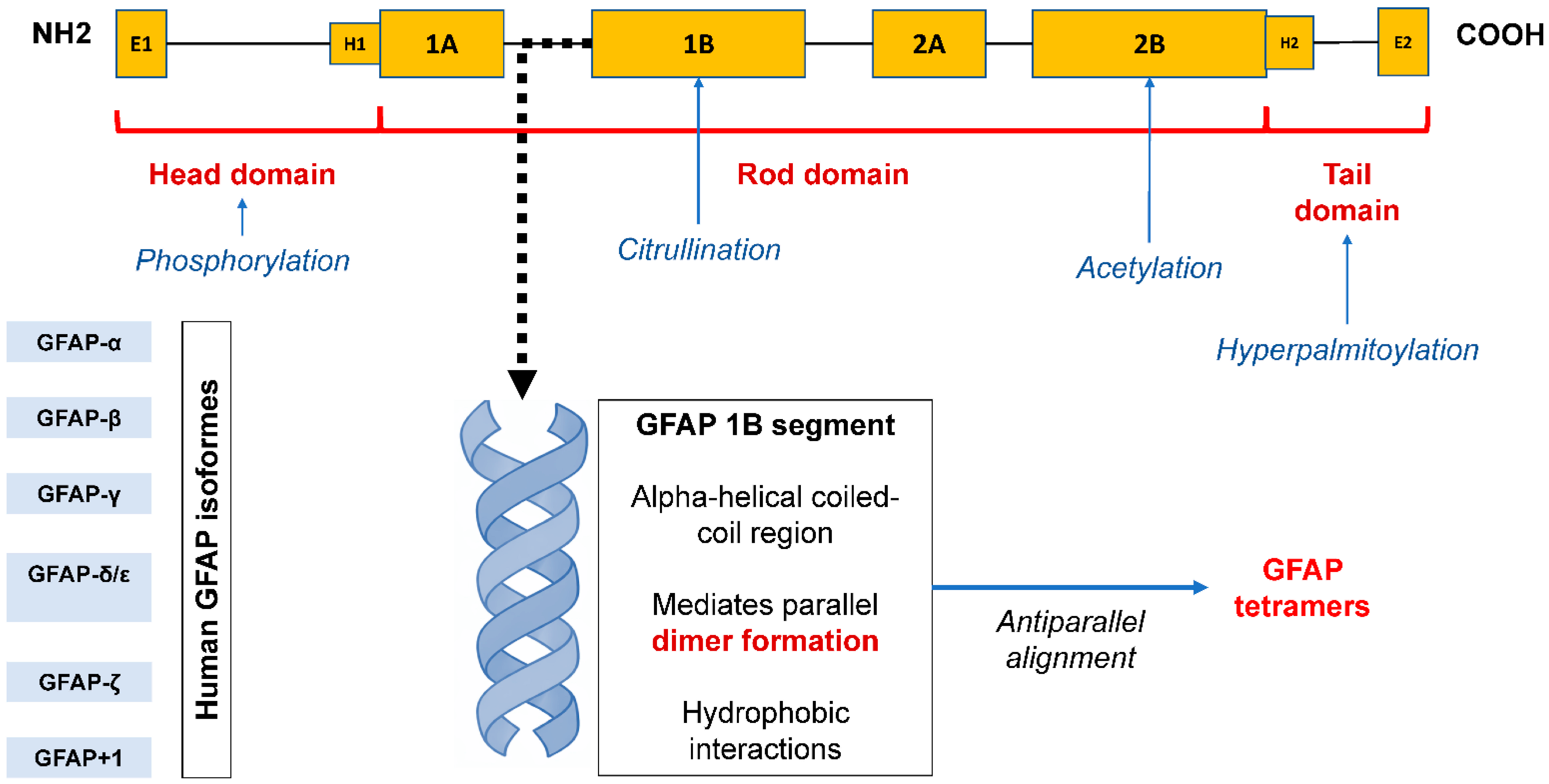
| GFAP Isoforms | Number of Amino Acids | Localization | References | |
|---|---|---|---|---|
| GFAP-α | 432 |
| [5,8,9] | |
| GFAP-β | More than 432 |
| [7,9] | |
| GFAP-γ | Less than 432 |
| [8,10,11] | |
| GFAP-δ/GFAP-ε | 431 |
| [11,15] | |
| GFAP-κ | 438 |
| [7,8,11] | |
| GFAP-ζ | More than 432 |
| [11,12] | |
| GFAP+1 | GFAPΔEx6 | 347 |
| [11,12] |
| GFAPΔ164 | 366 | [11,12] | ||
| GFAPΔ135 | 374 | [13] | ||
| GFAPΔEx7 | 418 | [9] | ||
| Type of Biofluidic | Isolation Method | GFAP Detection Platform | Enrichment Strategy | Cohort Size | Clinical Association | Ref. |
|---|---|---|---|---|---|---|
| Blood plasma | ExoQuick exosome kit | ELISA | Sorption on streptavidine-agarose ultralink resin with GLAST biotinylated antibodies | 12 pts. with early stage of AD vs. 10 matched cognitive normal controls | GFAP level in plasma ADEVs was significantly less in AD pts. than controls pts. | [37] |
| Blood plasma | ExoQuick exosome kit | Western blotting | No enrichment | 12 pts. with schizophrenia vs. 12 controls | The significantly higher concentration of exosomal GFAP in the schizophrenia smpl. is suggestive of selective enrichment of exosome protein astrocytic origin only in the pts. samples. Exosomal samples from both groups were similar in the level of synaptophysin, suggestive of the presence of neuronal-derived exosomes irrespective of disease status. | [38] |
| Blood plasma | Differential centrifugation | High-sensitivity flow cytometry (individual detection) | No enrichment | Patients with SED (n = 31), MDD (n = 31), and healthy matched controls (n = 61) | Patients with SED had significantly higher concentrations of AQP4-positive and GFAP-positive EVs and EVs co-expressing AQP4/GFAP than patients with MDD and healthy controls. | [39] |
| Blood plasma | Differential centrifugation with ultracentrifugation and ultrafiltration | High-sensitivity flow cytometry (beads- based method) | Sorption of EVs on latex beads coated with antibodies to GFAP | Glioblastoma multiforme pts. with no tumor recurrence for over one year (n = 6) and after first relapse (n = 14) | In both groups, C5b-9 was predominantly detected on tumor-specific circulating EVs (GFAP+ EVs) with high VEGF-A expression, while C5b-9 was significantly less frequent on EVs with low VEGF-A expression. GFAP+VEGF+dimMMP2-C5b-9+ EVs were rarely detected in pts. without relapse, suggesting their potential utility as biomarkers for a favorable relapse-free prognosis. In recurrent pts., a positive correlation was observed between GFAP+VEGF+bright MMP2+C5b-9+ EVs and MGMT gene promoter methylation levels (r = 0.543; p < 0.05). | [43] |
| Blood serum | Differential centrifugation with ultracentrifugation | High-sensitivity flow cytometry (individual detection) | No enrichment strategy | 8 pts with glioblastoma multiforme progressed early, late and without progression vs. 3 controls (non-cancers) | Pts. with glioblastoma have CD9+/GFAP+/Survivin+ and CD9+/Survivin+ exosomes that are released into the circulation and that early reductions in their numbers following anti-survivin immunotherapy might be associated with longer progression-free survival. | [44] |
| Blood plasma | Size-exclusion chromatography | Ultrasensitive single-molecule array | No enrichment | 93 trauma patients (75 with TBI and 18 without TBI) were analyzed | EV-GFAP levels were significantly elevated in TBI patients compared with non-TBI trauma patients at admission and 15 h. A positive head CT was associated with 2.85 (95% CI: 1.18–6.91)-fold increased EV-GFAP, whereas EV-NfL and EV-T-Tau levels were not affected. None of the tested EV biomarkers were associated with 1-year mortality or 6–12 months’ functional outcome. | [41] |
| Blood serum | ExoQuick exosome kit | Ultrasensitive single-molecule array | No enrichment | 72 TBI patients and 20 controls | EV GFAP concentrations were elevated in moderate and severe TBI compared with controls (p < 0.001) and could distinguish controls from moderate (AUC = 0.86) or severe TBI (AUC = 0.88). Increased EV GFAP and EV NfL levels were associated with lower 1-year Glasgow Outcome Scale–Extended scores (p < 0.05). | [42] |
| Blood plasma | ExoQuick ultra-EV kit | Western blotting | Exo-flow beads coated with GLAST biotinylated antibody | Plasma samples from 18 acute ischemic stroke pts. at 24 h (D1), 7 days (D7), and 30 days (D30) post-symptoms onset, and 9 healthy controls | Post-stroke ADEV GFAP levels were elevated at D1 and D7 but not D30 compared with controls (p = 0.007, p = 0.019, and p = 0.344, respectively). A positive correlation was observed between the modified Rankin scale at D7 and ADEV GFAP at D1 (r = 0.58; p = 0.010) and D7 (r = 0.57; p = 0.013), respectively. | [40] |
| Database | ID | Ref. | Sample Type/Source (Biological Origin) | Method | Localization of GFAP in EVs (Established/Not Established) | Summary of Localization Evidence |
|---|---|---|---|---|---|---|
| ExoCarta | GFAP (gene_id=2670; ExoCarta_2670), Experiment ID 224 | [62] | Homo sapiens; neuroblastoma cells (SH-SY5Y) | Mass spectrometry; Western blotting | Not established | GFAP is reported as a “protein identified” within EVs based on MS; the study provides general physical/molecular EV characterization (electron microscopy and enrichment markers), but lacks GFAP-specific evidence (e.g., immunogold labeling and protease-based treatment). |
| ExoCarta | GFAP (gene_id=2670; ExoCarta_2670), Experiment ID 834–835 | [63] | Homo sapiens; retinal pigment epithelial cells (ARPE-19) | Western blotting; mass spectrometry | Not established | The record confirms the presence of GFAP by MS and general EV validation (microscopy, NTA, and EV markers), but does not indicate its specific localization (intraluminal, surface-associated, or co-precipitated); GFAP is not investigated in localization-specific experiments. |
| ExoCarta | GFAP (gene_id=2670; ExoCarta_2670), Experiment ID 191 | [64] | Homo sapiens; squamous-cell carcinoma cells (A431) | Mass spectrometry | Not established | The study emphasizes protein/exosome isolation and proteomic analysis of the secretome/100,000 g pellet; GFAP appears in the proteomic list, but no data are provided to support its vesicular or sub-vesicular topology. |
| ExoCarta | Gfap (gene_id=24387; ExoCarta_24387) | [65] | Rattus norvegicus; adipocytes/adipose tissue (OLETF rats) | Unspecified | Not established | The ExoCarta entry indicates the presence of GFAP in adipose tissue-derived EVs; however, the extracted record fragment does not specify the identification method for GFAP. Based on the PubMed annotation, this is EV proteomics using MS, which does not establish the intravesicular localization of GFAP. |
| Vesiclepedia | exp_id=354 | [66] | Homo sapiens; plasma | Mass spectrometry [Orbitrap Velos]; Western blotting | Not established | Presence of GFAP is based on MS in the exosome fraction; the study compares proteomics across different isolation/stability conditions but does not provide GFAP-focused localization evidence (e.g., immunogold labeling or protease-based treatment). |
| Vesiclepedia | exp_id=590 | [67] | Homo sapiens; colorectal cancer cells (e.g., HCT-15) | Mass spectrometry [LTQ] | Not established | High-throughput EV proteomics: GFAP is reported as an identified protein, corresponding to the level of “presence in an EV-enriched fraction,” without direct evidence of sub-vesicular localization. |
Disclaimer/Publisher’s Note: The statements, opinions and data contained in all publications are solely those of the individual author(s) and contributor(s) and not of MDPI and/or the editor(s). MDPI and/or the editor(s) disclaim responsibility for any injury to people or property resulting from any ideas, methods, instructions or products referred to in the content. |
© 2026 by the authors. Licensee MDPI, Basel, Switzerland. This article is an open access article distributed under the terms and conditions of the Creative Commons Attribution (CC BY) license.
Share and Cite
Yunusova, N.; Svarovsky, D.; Panfilova, P.; Ryabova, A.; Kaigorodova, E.; Sidenko, E.; Gervas, P.; Molokov, A.; Kondakova, I. Evolution of Concepts Regarding the Diagnostic and Prognostic Significance of Glial Fibrillary Acidic Protein (GFAP)-Positive Extracellular Vesicles. Biomedicines 2026, 14, 1116. https://doi.org/10.3390/biomedicines14051116
Yunusova N, Svarovsky D, Panfilova P, Ryabova A, Kaigorodova E, Sidenko E, Gervas P, Molokov A, Kondakova I. Evolution of Concepts Regarding the Diagnostic and Prognostic Significance of Glial Fibrillary Acidic Protein (GFAP)-Positive Extracellular Vesicles. Biomedicines. 2026; 14(5):1116. https://doi.org/10.3390/biomedicines14051116
Chicago/Turabian StyleYunusova, Natalia, Dmitry Svarovsky, Polina Panfilova, Anastasia Ryabova, Evgeniya Kaigorodova, Evgeniya Sidenko, Polina Gervas, Aleksey Molokov, and Irina Kondakova. 2026. "Evolution of Concepts Regarding the Diagnostic and Prognostic Significance of Glial Fibrillary Acidic Protein (GFAP)-Positive Extracellular Vesicles" Biomedicines 14, no. 5: 1116. https://doi.org/10.3390/biomedicines14051116
APA StyleYunusova, N., Svarovsky, D., Panfilova, P., Ryabova, A., Kaigorodova, E., Sidenko, E., Gervas, P., Molokov, A., & Kondakova, I. (2026). Evolution of Concepts Regarding the Diagnostic and Prognostic Significance of Glial Fibrillary Acidic Protein (GFAP)-Positive Extracellular Vesicles. Biomedicines, 14(5), 1116. https://doi.org/10.3390/biomedicines14051116