Natural Polymers in Tissue Engineering and Regeneration: Material–Cell Mechanotransduction, Biofabrication Strategies, and Clinical Translation

 , , ,
, , ,  and
and
Abstract
1. Introduction
2. Natural Polymers and Composites
2.1. Natural Polymers in the Biological Environment (ECM-Mimicking, Enzymatic Degradation)
2.2. Natural Polymers and Composites in Tissue Engineering

| Polymer | Source | Tensile/ Compressive Strength | Degradation Rate | Porosity (%) | Cell Viability (%) | Benefits | Limitations | Reference |
|---|---|---|---|---|---|---|---|---|
| Collagen | Mammalian connective tissue | 50–500 MPa (tensile) | 4–6 weeks (non-crosslinked) | 70–95 | >90 | High bioactivity, supports osteogenesis | Immunogenicity risk, fast degradation | [131,132,133] |
| Gelatin | Denatured collagen | 10–100 kPa (hydrogel modulus) | 2–3 weeks | 80–98 | >85 | Easy processing, bioactive | Low mechanical strength | [134,135] |
| Chitosan | Crustacean shells | 2–12 MPa (blends) | Weeks–months | 65–85 | >90 | Antibacterial, osteoconductive | Poor solubility at neutral pH | [136,137,138,139] |
| Alginate | Brown algae | <1 MPa | Weeks–months | 80–95 | 70–90 | Mild gelation, printable | Lacks adhesion motifs | [140,141,142] |
| Silk fibroin | Silkworm cocoons | Up to 740 MPa | Months–years | 60–85 | >90 | High strength, tunable degradation | Hydrophobicity | [143,144] |
| Hyaluronic acid | ECM component | Low modulus | <24 h (native) | 85–98 | >95 | ECM mimicry, promotes angiogenesis | Rapid degradation | [145,146,147] |
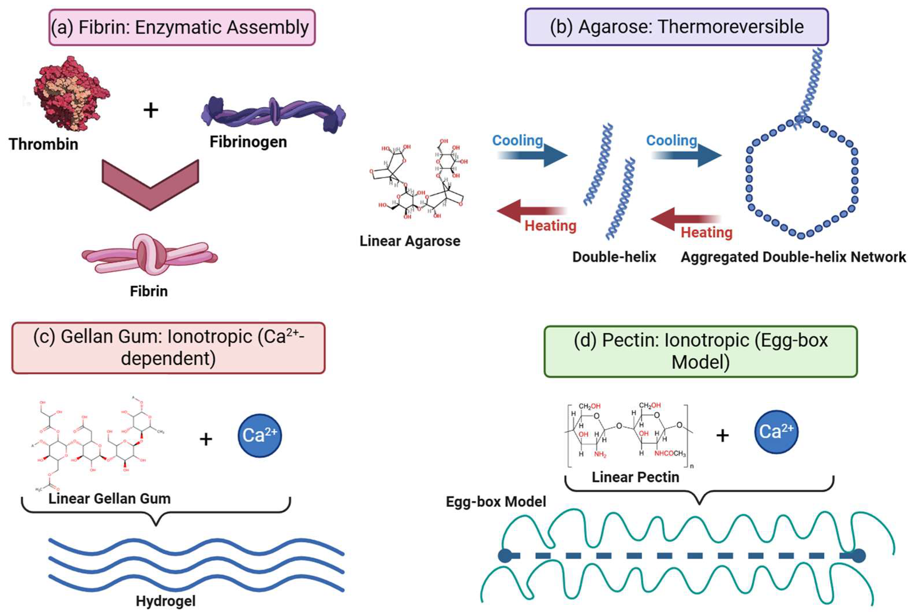
| Fibrin | Blood plasma (fibrinogen + thrombin) | Low; strain-stiffening | Rapid (days–weeks); proteolytic (plasmin/MMP); highly cell-responsive | High (>90%) | >95% | Native RGD motifs; intrinsic bioactivity; chemotactic; remodelable | Mechanically weak (<30 kPa); degrades too fast for long-term support | [120,148] |
| Agarose | Red algae (Rhodophyceae such as Gelidium) | ~25 kPa (2%) | Very slow/non-degradable in mammals (lack agarases); stable for months | Nanoporous (decreases with conc., e.g., <100 nm at 3%) | Good (50–75%) | High stiffness (cartilage mimic); shape retention; thermoreversible | Inert (no adhesion); non-degradable (foreign body risk); nanoporosity limits integration | [149] |
| Pectin | Plant cell walls (citrus/apple peel) | 6–100 kPa (Ca-crosslinked) | Moderate/tunable (weeks); enzymatic (pectinase/lysozyme); Mass loss ~30% in 3 days (blends) | ~50% | >90% | Versatile gelation (pH/ions); low cost; high tensile strength in blends | Ionic instability (Ca2+ loss); brittle if over-crosslinked; variable source properties | [150] |
| Carrageenan | Red algae (Rhodophyceae) | Compressive modulus: 30–60 kPa (hybrid); tensile: ~1.3 MPa (double network) | Tunable (weeks); Enzymatic (carrageenase) or hydrolytic | 70–90% | >90% | Thermoreversible gelation, mimics sulfated GAGs, antiviral/anti-inflammatory properties | Brittleness in pure form; potential inflammation from low-MW byproducts | [151,152,153] |
| Decellularized ECM (dECM) | Porcine/bovine tissues (heart, skin, bone) | Flexural: 90–130 MPa (bone composites) | Weeks to months | 80–95% | >95% | Perfect biochemical mimicry, retains cryptic peptides/growth factors, tissue-specific cues | Batch-to-batch variability; risk of residual immunogens (α-Gal); complex processing | [154,155,156] |
| Keratin | Wool, human hair | Compressive: 0.2–1.2 MPa (reinforced with HA) | Proteolytic degradation (weeks); tunable via crosslinking | 75–85% | >90% | Intrinsic cell binding motifs (LDV, EDS); osteoinductive; high cysteine content | Fragile in pure form; extensive extraction processing required | [157,158] |
| Ulvan | Green algae (Ulva species) | Modulus: ~4 MPa (PCL blends); weak pure hydrogels | Moderate (weeks); depends on degree of crosslinking | 80–90% | >85% | Structurally similar to heparin; promotes osteogenic differentiation; renewable resource | Seasonal chemical variability; difficult extraction/purification; low gel strength | [159,160] |
| Xanthan gum | Xanthomonas campestris fermentation | Compressive modulus: 3–200 kPa (ionic crosslinking) | Slow in vivo degradation; highly stable | >90% | >95% | Excellent rheological modifier (shear-thinning); bio-inert backbone; tunable stiffness | Lack of intrinsic cell adhesion sites; susceptible to microbial contamination if not sterile | [161,162,163] |
2.3. 3D and 4D Printing, Implant Coatings, and Advanced Fabrication Approaches
| Technique | Typical Materials | Advantages | Restraints | References |
|---|---|---|---|---|
| Electrospinning | Collagen, chitosan, silk blends | ECM-like nanofibers; large surface area | Limited cell infiltration unless porous | [180,181,182] |
| 3D bioprinting | GelMA, alginate, GelMA–alginate hybrids | Spatial control of cells and growth factors | Bioink rheology and crosslinking constraints | [183,184] |
| Injectable in situ gels | Alginate, thermogelling chitosan/gelatin | Minimal invasiveness; defect conformation | Controlling gelation and retention in vivo | [185,186] |
| Freeze-drying/porogen leaching | Collagen, gelatin | High porosity; simple | Poor mechanical strength; batch variability | [187,188,189] |
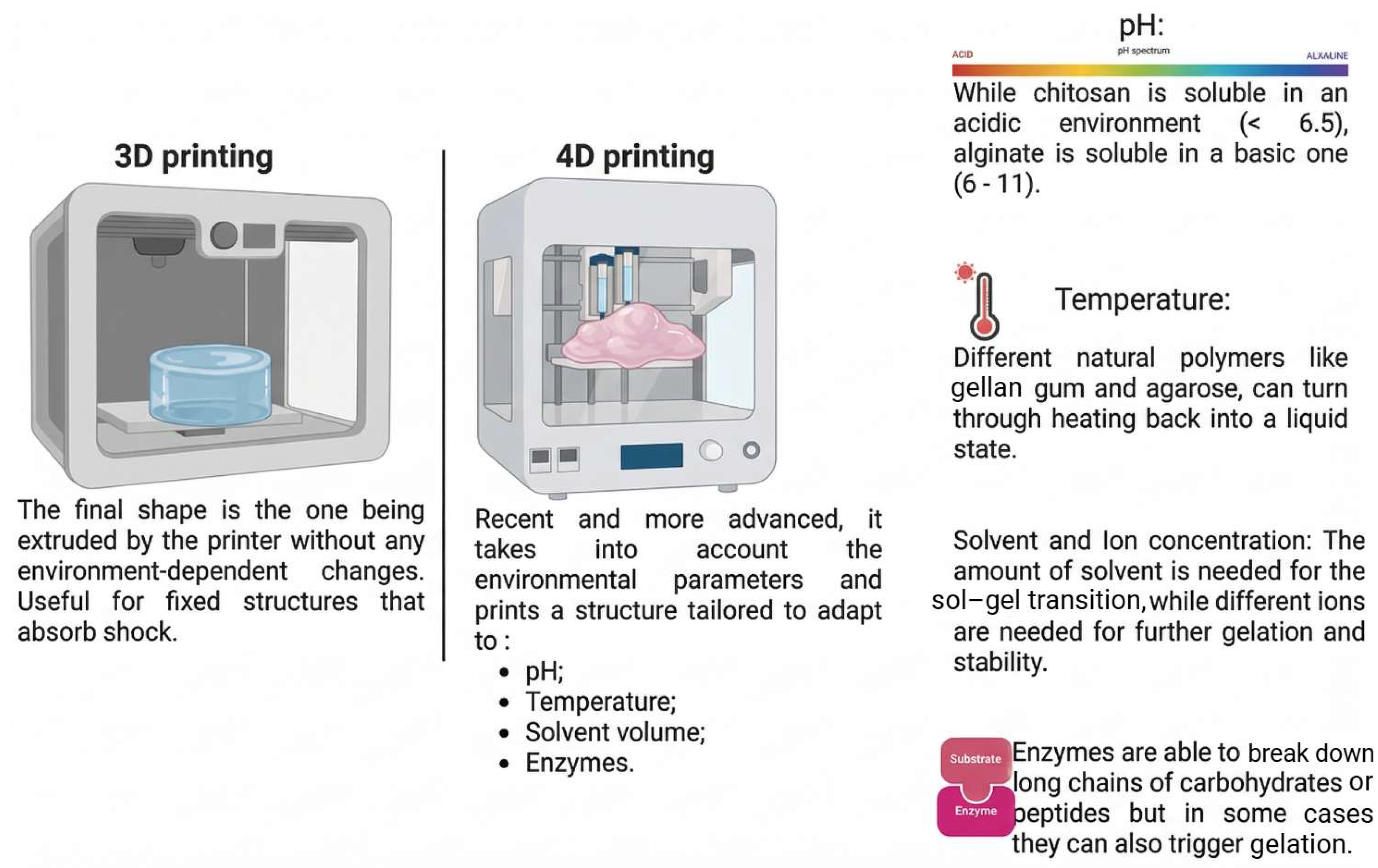
| 4D printing/stimuli-responsive | Smart hydrogels, modified natural polymers | Dynamic remodeling; shape change after implantation | Early technology; limited biological validation | [190,191,192] |
| Melt electrowriting (MEW) | PCL/chitosan blends, PCL/gelatin | Solvent-free fabrication; highly ordered microfibrous architecture (2–50 µm); combines porosity with mechanical resilience | Limited to thermally stable polymers; requires specialized equipment; slower vertical build rates than extrusion | [193,194] |
| Digital Light Processing (DLP) | GelMA, methacrylated silk (SilMA), PEGDA/alginate | Ultra-high resolution (~25 µm); rapid layer-by-layer curing; prints complex geometries without support structures | Requires photo-curable moieties (methacrylation); potential cytotoxicity of photo-initiators; limited material viscosity range | [195,196,197] |
| Microfluidics | Alginate, gelatin, silk fibroin | Monodisperse microgel production; high control over internal architecture (core–shell); protects encapsulated cells from shear | Low throughput for macroscale scaffold production; complex device fabrication; potential channel clogging | [198,199,200] |
| Gas foaming | Alginate, gelatin, chitosan | Solvent-free and low-temperature process; creates highly interconnected pores; preserves bioactive agents | Lack of precise control over pore architecture; formation of non-porous skin layer; requires high-pressure equipment | [201,202,203] |
| Coaxial bioprinting | Alginate/pluronic, GelMA/alginate | Fabrication of vessel-like (tubular) structures; enhanced cell viability via core–shell shielding; multi-material deposition | Complex rheological matching required between core and shell; lower resolution than DLP; limited to continuous filaments | [204,205,206] |
| Stimulus | Responsive Material | Mechanism of Action | Biomedical Application | Reference |
|---|---|---|---|---|
| Water/Ion | Alginate/Cellulose Bilayers | Differential swelling/anisotropic expansion | Self-folding tracheal stents, vascular grafts | [215,216] |
| Temperature | Gelatin–PNIPAAm, Silk–PNIPAAm | Coil-to-globule transition around LCST (~32–37 °C) | Injectable bone scaffolds, smart wound dressings | [217,218,219] |
| pH | Chitosan, Alginate | Protonation/deprotonation causing electrostatic repulsion/attraction | Smart wound dressings sensitive to infection | [220,221] |
| Magnetic | Alginate + Fe3O4 NPs | Magneto-mechanical alignment or induction heating | Remote actuation, mechanotransduction stimulation | [222] |
| Enzymatic | Collagen, HA (MMP-Sensitive) | Cleavage of crosslinks by specific enzymes (e.g., MMPs) | Cell-mediated remodeling, degrading scaffolds | [223,224] |
| Light (UV/IR) | Methacrylated Silk, GelMA–Gold Nanorods | Photo-cleavage of crosslinks or photothermal heating inducing phase transition/shrinkage | On-demand drug release, remote actuation of soft grippers, spatiotemporal control of cell adhesion | [225,226] |
| Electric Field | Chitosan–Graphene, Alginate–CNT | Electro-osmotic ion migration causing asymmetric swelling/bending | Artificial muscles, electro-stimulated nerve regeneration, smart drug-release devices | [227,228] |
| Multi-Stimuli (pH + Temp) | Chitosan–PNIPAAm, Alginate–Pluronic | Dual-response logic (e.g., sol–gel at body temp. + swelling at acidic pH) | Tumor-targeted delivery (acidic/warm microenvironment), complex shape-morphing scaffolds | [229,230,231] |
| Glucose | Concanavalin A-Modified Alginate/Chitosan | Competitive binding of glucose disrupts polymer crosslinks, increasing porosity | Self-regulated insulin delivery systems (artificial pancreas), diabetes management | [232,233] |
| Reactive Oxygen Species (ROS) | Thioketal-Modified Keratin or Hyaluronic Acid | Cleavage of ROS-sensitive linkers triggers degradation or drug release | Targeting inflamed tissues (e.g., chronic wounds, cardiac infarction) with antioxidant release | [234,235] |
3. The Use of Injectable Gels and Scaffolds in Experimental Therapy
3.1. Bone Regeneration
3.2. Cartilage Repair
3.3. Nerve Regeneration
3.4. Cardiac Applications
3.5. Soft Tissue and Wound Healing
| Application | System/Composition | Gelation/ Crosslinking | Mechanical Properties | Biological/Clinical Outcomes | References |
|---|---|---|---|---|---|
| Bone regeneration | GelMA + HA microspheres | Photocrosslinking | Compressive stress: ~138 ± 5 kPa; modulus: ↑ from 4.2 to 50 kPa (5–15% GelMA) | Enhanced MSC osteogenesis | [299] |
| Alginate–HA hydrogel | Ionic (Ca2+) | 50–70 kPa | 65% new bone vs. 40% pristine alginate (rat calvarial defect) | [236] | |
| PVA + bioactive glass | Dual-network | Compressive strength: ~34 MPa; modulus: ~0.8 MPa | Improved bone-like mechanical fidelity | [257] | |
| Osteochondral regeneration | Bilayered chitosan/xanthan gum + biphasic calcium phosphate | Freeze-drying/ionic crosslinking | Compressive strength; ~1–5 MPa (gradient stiffness) | Integrated repair of bone-cartilage interface; support of subchondral bone formation | [300] |
| Cartilage repair | HA–tyramine | Enzymatic (HRP/H2O2) | Compressive modulus: 15–30 kPa | IKDC ↑ 25–30% at 6 months (clinical) | [301,302] |
| Nano-composite hydrogels (PCL–HA, GelMA–IL-4, etc.) | Photocrosslinked/ionic | 0.4–70 MPa depending on filler | Promoted BMSC chondrogenesis, improved load-bearing | [303,304,305] | |
| Cellulose–PAM composite | Dual-network | 3–10 MPa under cyclic load (90–120 days) | Maintained function in rabbit osteochondral repair | [264,306,307] | |
| Intervertebral disc repair | Chitosan–hyaluronic acid/silk–PU blends | Thermosensitive (sol–gel at 37 °C)/chemical | Compressive modulus: 10–50 kPa (matches nucleus pulposus); fatigue-resistant | Restoration of Disc Height Index (DHI) and MRI signal intensity in rabbit models; prevents further degeneration | [308] |
| Nerve regeneration | Electrospun gelatin–chitosan | Injectable aligned scaffold | Not reported | 1.5–2× longer neurites in vitro; motor recovery in 8 weeks (rat sciatic) | [266] |
| HA granular hydrogel | Injectable granular packing | Not reported | Restored CMAP amplitude; ↑ conduction velocity after 12 weeks | [267] | |
| Silk fibroin–HA hybrid | Self-assembling | Not reported | Enhanced Schwann cell proliferation; improved remyelination | [267] | |
| Advanced nerve repair | Conductive chitosan/polypyrrole or aligned fibrin | In situ polymerization/magnetic alignment | Conductivity: ~10−3 S/cm; modulus: ~1 kPa (soft tissue match) | Restored nerve conduction velocity and CMAP to autograft levels at 12 weeks; enhanced myelination | [309] |
| Cardiac repair | Alginate (VEGF-loaded) | Ionic | Not reported | LVEF ↑ 9% in porcine MI model | [249] |
| Chitosan–gelatin + IGF-1 | Thermosensitive | Not reported | ↓ infarct size by 22%; ↑ angiogenesis in rats | [271] | |
| Hydrogel–cell/growth factor composites | Various | Not reported | Meta-analysis: LVEF ↑ 8.9% (rats), ↑ 16.5% (mice); FS ↑ ~6% | [272,274,310] | |
| Theranostic hydrogel | Injectable/responsive | Not reported | Radial strain ↑ 52.7%; circumferential strain ↑ 44.1% (porcine MI) | [311,312,313,314] | |
| Soft tissue/wound healing | Collagen–elastin blend | Self-assembly | Strain-to-failure: >80% | 35% faster re-epithelialization vs. collagen | [251] |
| HA injectable hydrogel | Crosslinked | Not reported | 25–40% faster ulcer healing vs. controls | [251] | |
| Natural gum-based gels | Thermosensitive/ionic | Not reported | Sustained release up to 14 days; improved angiogenesis | [294] | |
| Hemostasis | Carboxymethyl chitosan + oxidized dextran/gelatin | Schiff base reaction (rapid self-healing) | Storage modulus: >1 kPa; adhesive strength: >10 kPa | Stopped bleeding in rat liver trauma in <40 s (vs. 167 s for gauze); reduced total blood loss by >80% | [315] |
4. Translational and Regulatory Considerations
5. Conclusions
6. Future Directions
Author Contributions
Funding
Institutional Review Board Statement
Informed Consent Statement
Data Availability Statement
Conflicts of Interest
References
- Borgström, F.; Karlsson, L.; Ortsäter, G.; Norton, N.; Halbout, P.; Cooper, C.; Lorentzon, M.; McCloskey, E.V.; Harvey, N.C.; Javaid, M.K.; et al. Fragility Fractures in Europe: Burden, Management and Opportunities. Arch. Osteoporos. 2020, 15, 59. [Google Scholar] [CrossRef] [Scilit]
- Mitchell, P.J.; Chan, D.-C.; Lee, J.-K.; Tabu, I.; Alpuerto, B.B. The Global Burden of Fragility Fractures—What Are the Differences, and Where Are the Gaps. Best Pract. Res. Clin. Rheumatol. 2022, 36, 101777. [Google Scholar] [CrossRef] [Scilit]
- Mitchell, P.J.; Cooper, C.; Fujita, M.; Halbout, P.; Åkesson, K.; Costa, M.; Dreinhöfer, K.E.; Marsh, D.R.; Lee, J.-K.; Chan, D.-C.; et al. Quality Improvement Initiatives in Fragility Fracture Care and Prevention. Curr. Osteoporos. Rep. 2019, 17, 510–520. [Google Scholar] [CrossRef] [Scilit]
- Cauley, J.A. The Global Burden of Fractures. Lancet Healthy Longev. 2021, 2, e535–e536. [Google Scholar] [CrossRef] [Scilit]
- Wu, A.-M.; Bisignano, C.; James, S.L.; Abady, G.G.; Abedi, A.; Abu-Gharbieh, E.; Alhassan, R.K.; Alipour, V.; Arabloo, J.; Asaad, M.; et al. Global, Regional, and National Burden of Bone Fractures in 204 Countries and Territories, 1990–2019: A Systematic Analysis from the Global Burden of Disease Study 2019. Lancet Healthy Longev. 2021, 2, e580–e592. [Google Scholar] [CrossRef] [Scilit]
- Zhang, S.; Lee, Y.; Liu, Y.; Yu, Y.; Han, I. Stem Cell and Regenerative Therapies for the Treatment of Osteoporotic Vertebral Compression Fractures. Int. J. Mol. Sci. 2024, 25, 4979. [Google Scholar] [CrossRef] [Scilit] [PubMed]
- Perez, J.R.; Kouroupis, D.; Li, D.J.; Best, T.M.; Kaplan, L.; Correa, D. Tissue Engineering and Cell-Based Therapies for Fractures and Bone Defects. Front. Bioeng. Biotechnol. 2018, 6, 105. [Google Scholar] [CrossRef] [Scilit] [PubMed]
- Pearlin; Nayak, S.; Manivasagam, G.; Sen, D. Progress of Regenerative Therapy in Orthopedics. Curr. Osteoporos. Rep. 2018, 16, 169–181. [Google Scholar] [CrossRef] [Scilit]
- Gómez-Barrena, E.; Padilla-Eguiluz, N.G.; Avendaño-Solá, C.; Payares-Herrera, C.; Velasco-Iglesias, A.; Torres, F.; Rosset, P.; Gebhard, F.; Baldini, N.; Rubio-Suarez, J.C.; et al. A Multicentric, Open-Label, Randomized, Comparative Clinical Trial of Two Different Doses of Expanded hBM-MSCs Plus Biomaterial versus Iliac Crest Autograft, for Bone Healing in Nonunions after Long Bone Fractures: Study Protocol. Stem Cells Int. 2018, 2018, 6025918. [Google Scholar] [CrossRef] [Scilit] [PubMed]
- Guo, L.; Liang, Z.; Yang, L.; Du, W.; Yu, T.; Tang, H.; Li, C.; Qiu, H. The Role of Natural Polymers in Bone Tissue Engineering. J. Control. Release 2021, 338, 571–582. [Google Scholar] [CrossRef] [Scilit]
- Gomez-Florit, M.; Pardo, A.; Domingues, R.M.A.; Graça, A.L.; Babo, P.S.; Reis, R.L.; Gomes, M.E. Natural-Based Hydrogels for Tissue Engineering Applications. Molecules 2020, 25, 5858. [Google Scholar] [CrossRef] [Scilit]
- Ebhodaghe, S.O. Natural Polymeric Scaffolds for Tissue Engineering Applications. J. Biomater. Sci. Polym. Ed. 2021, 32, 2144–2194. [Google Scholar] [CrossRef] [Scilit]
- Bose, S.; Koski, C.; Vu, A.A. Additive Manufacturing of Natural Biopolymers and Composites for Bone Tissue Engineering. Mater. Horiz. 2020, 7, 2011–2027. [Google Scholar] [CrossRef] [Scilit]
- Puertas-Bartolomé, M.; Mora-Boza, A.; García-Fernández, L. Emerging Biofabrication Techniques: A Review on Natural Polymers for Biomedical Applications. Polymers 2021, 13, 1209. [Google Scholar] [CrossRef] [Scilit] [PubMed]
- Alberti, K.A.; Xu, Q. Biocompatibility and Degradation of Tendon-Derived Scaffolds. Regen. Biomater. 2016, 3, 1–11. [Google Scholar] [CrossRef] [Scilit] [PubMed]
- Park, S.-H.; Gil, E.S.; Shi, H.; Kim, H.J.; Lee, K.; Kaplan, D.L. Relationships Between Degradability of Silk Scaffolds and Osteogenesis. Biomaterials 2010, 31, 6162–6172. [Google Scholar] [CrossRef] [Scilit]
- Joshi, S.R.; Pendyala, G.S.; Shah, P.; Mopagar, V.P.; Padmawar, N.; Padubidri, M. Scaffolds—The Ground for Regeneration: A Narrative Review. J. Int. Soc. Prev. Community Dent. 2020, 10, 692–699. [Google Scholar] [CrossRef] [Scilit] [PubMed]
- Segneanu, A.-E.; Bejenaru, L.E.; Bejenaru, C.; Blendea, A.; Mogoşanu, G.D.; Biţă, A.; Boia, E.R. Advancements in Hydrogels: A Comprehensive Review of Natural and Synthetic Innovations for Biomedical Applications. Polymers 2025, 17, 2026. [Google Scholar] [CrossRef] [Scilit]
- Liu, T.; Yu, S.; Zhang, L.; Ji, W.; Wang, G.; Wang, N.; Li, M.; Hu, T.; Shi, Z. Yes-Associated Protein 1 in Cancer: Bridging Mechanical Transduction and Epigenetic Regulation. Cancer Biol. Ther. 2025, 26, 2562726. [Google Scholar] [CrossRef] [Scilit]
- Zhang, W.; Li, Q.; Gao, H.; Wang, Y.; Cheng, M.; Wang, Y.-X. The Regulation of Yes-Associated Protein/Transcriptional Coactivator with PDZ-Binding Motif and Their Roles in Vascular Endothelium. Front. Cardiovasc. Med. 2022, 9, 925254. [Google Scholar] [CrossRef] [Scilit]
- Filippi, M.; Born, G.; Chaaban, M.; Scherberich, A. Natural Polymeric Scaffolds in Bone Regeneration. Front. Bioeng. Biotechnol. 2020, 8, 474. [Google Scholar] [CrossRef] [Scilit]
- Donnaloja, F.; Jacchetti, E.; Soncini, M.; Raimondi, M.T. Natural and Synthetic Polymers for Bone Scaffolds Optimization. Polymers 2020, 12, 905. [Google Scholar] [CrossRef] [Scilit]
- Keshvardoostchokami, M.; Majidi, S.S.; Huo, P.; Ramachandran, R.; Chen, M.; Liu, B. Electrospun Nanofibers of Natural and Synthetic Polymers as Artificial Extracellular Matrix for Tissue Engineering. Nanomaterials 2021, 11, 21. [Google Scholar] [CrossRef] [Scilit]
- Grigore, M.; Vasilache, I.-A.; Cianga, P.; Constantinescu, D.; Duma, O.; Matasariu, R.D.; Scripcariu, I.-S. Acceptability of Human Papilloma Virus Self-Sampling for Cervical Cancer Screening in a Cohort of Patients from Romania (Stage 2). J. Clin. Med. 2022, 11, 2503. [Google Scholar] [CrossRef] [Scilit]
- Kužma, J.; Suchý, T.; Horný, L.; Šupová, M.; Sucharda, Z.; Kužma, J.; Suchý, T.; Horný, L.; Šupová, M.; Sucharda, Z. Comparative Study of the Dehydrothermal Crosslinking of Electrospun Collagen Nanofibers: The Effects of Vacuum Conditions and Subsequent Chemical Crosslinking. Polymers 2024, 16, 2453. [Google Scholar] [CrossRef] [Scilit]
- Tousignant, M.N.; Lin, Z.S.; Brusso, J.; Lessard, B.H. Interfacial Ultraviolet Cross-Linking of Green Bilayer Dielectrics. ACS Appl. Mater. Interfaces 2023, 15, 3680–3688. [Google Scholar] [CrossRef] [Scilit]
- Chen, J.; Garcia, E.S.; Zimmerman, S.C. Intramolecularly Cross-Linked Polymers: From Structure to Function with Applications as Artificial Antibodies and Artificial Enzymes. Acc. Chem. Res. 2020, 53, 1244–1256. [Google Scholar] [CrossRef] [Scilit] [PubMed]
- Li, Z.; Lu, F.; Liu, Y. A Review of the Mechanism, Properties, and Applications of Hydrogels Prepared by Enzymatic Cross-Linking. J. Agric. Food Chem. 2023, 71, 10238–10249. [Google Scholar] [CrossRef] [Scilit] [PubMed]
- Brovold, M.; Almeida, J.I.; Pla-Palacín, I.; Sainz-Arnal, P.; Sánchez-Romero, N.; Rivas, J.J.; Almeida, H.; Royo Dachary, P.; Serrano-Aulló, T.; Soker, S.; et al. Naturally-Derived Biomaterials for Tissue Engineering Applications. Adv. Exp. Med. Biol. 2018, 1077, 421–449. [Google Scholar] [CrossRef] [Scilit] [PubMed]
- Sell, S.A.; Wolfe, P.S.; Garg, K.; McCool, J.M.; Rodriguez, I.A.; Bowlin, G.L. The Use of Natural Polymers in Tissue Engineering: A Focus on Electrospun Extracellular Matrix Analogues. Polymers 2010, 2, 522–553. [Google Scholar] [CrossRef] [Scilit]
- Sionkowska, A. Natural Polymers as Components of Blends for Biomedical Applications. In Polymeric Biomaterials; CRC Press: Boca Raton, FL, USA, 2013. [Google Scholar]
- Hadad, E.; Rudnick-Glick, S.; Grinberg, I.; Kolitz-Domb, M.; Chill, J.H.; Margel, S. Synthesis and Characterization of Poly(RGD) Proteinoid Polymers and NIR Fluorescent Nanoparticles of Optimal d,l-Configuration for Drug-Delivery Applications—In Vitro Study. ACS Omega 2020, 5, 23568–23577. [Google Scholar] [CrossRef] [Scilit]
- Yang, M.; Zhang, Z.-C.; Liu, Y.; Chen, Y.-R.; Deng, R.-H.; Zhang, Z.-N.; Yu, J.-K.; Yuan, F.-Z. Function and Mechanism of RGD in Bone and Cartilage Tissue Engineering. Front. Bioeng. Biotechnol. 2021, 9, 773636. [Google Scholar] [CrossRef] [Scilit] [PubMed]
- Psarra, E.; König, U.; Müller, M.; Bittrich, E.; Eichhorn, K.-J.; Welzel, P.B.; Stamm, M.; Uhlmann, P. In Situ Monitoring of Linear RGD-Peptide Bioconjugation with Nanoscale Polymer Brushes. ACS Omega 2017, 2, 946–958. [Google Scholar] [CrossRef] [Scilit]
- Kang, Z.; Wang, Y.; Xu, J.; Song, G.; Ding, M.; Zhao, H.; Wang, J. An RGD-Containing Peptide Derived from Wild Silkworm Silk Fibroin Promotes Cell Adhesion and Spreading. Polymers 2018, 10, 1193. [Google Scholar] [CrossRef] [Scilit]
- Zhou, F.; Zhang, X.; Cai, D.; Li, J.; Mu, Q.; Zhang, W.; Zhu, S.; Jiang, Y.; Shen, W.; Zhang, S.; et al. Silk Fibroin-Chondroitin Sulfate Scaffold with Immuno-Inhibition Property for Articular Cartilage Repair. Acta Biomater. 2017, 63, 64–75. [Google Scholar] [CrossRef] [Scilit]
- Farokhi, M.; Solouk, A.; Mirzadeh, H.; Herbert Teuschl, A.; Redl, H. An Injectable Enzymatically Crosslinked and Mechanically Tunable Silk Fibroin/Chondroitin Sulfate Chondro-Inductive Hydrogel. Macromol. Mater. Eng. 2023, 308, 2200503. [Google Scholar] [CrossRef] [Scilit]
- Pereira, R.V.S.; EzEldeen, M.; Ugarte-Berzal, E.; Martens, E.; Malengier-Devlies, B.; Vandooren, J.; Vranckx, J.J.; Matthys, P.; Opdenakker, G. Physiological Fibrin Hydrogel Modulates Immune Cells and Molecules and Accelerates Mouse Skin Wound Healing. Front. Immunol. 2023, 14, 1170153. [Google Scholar] [CrossRef] [Scilit] [PubMed]
- Eichinger, J.F.; Haeusel, L.J.; Paukner, D.; Aydin, R.C.; Humphrey, J.D.; Cyron, C.J. Mechanical Homeostasis in Tissue Equivalents: A Review. Biomech. Model. Mechanobiol. 2021, 20, 833–850. [Google Scholar] [CrossRef] [Scilit] [PubMed]
- Kearney, K.J.; Ariëns, R.A.S.; Macrae, F.L. The Role of Fibrin(Ogen) in Wound Healing and Infection Control. Semin. Thromb. Hemost. 2022, 48, 174–187. [Google Scholar] [CrossRef] [Scilit]
- Zhao, X.; Chen, J.; Sun, H.; Zhang, Y.; Zou, D. New Insights into Fibrosis from the ECM Degradation Perspective: The Macrophage-MMP-ECM Interaction. Cell Biosci. 2022, 12, 117. [Google Scholar] [CrossRef] [Scilit]
- Hernandez-Guillamon, M.; Mawhirt, S.; Blais, S.; Montaner, J.; Neubert, T.A.; Rostagno, A.; Ghiso, J. Sequential Amyloid-β Degradation by the Matrix Metalloproteases MMP-2 and MMP-9*. J. Biol. Chem. 2015, 290, 15078–15091. [Google Scholar] [CrossRef] [Scilit]
- Jabłońska-Trypuć, A.; Matejczyk, M.; Rosochacki, S. Matrix Metalloproteinases (MMPs), the Main Extracellular Matrix (ECM) Enzymes in Collagen Degradation, as a Target for Anticancer Drugs. J. Enzyme Inhib. Med. Chem. 2016, 31, 177–183. [Google Scholar] [CrossRef] [Scilit]
- Sikorski, D.; Gzyra-Jagieła, K.; Draczyński, Z. The Kinetics of Chitosan Degradation in Organic Acid Solutions. Mar. Drugs 2021, 19, 236. [Google Scholar] [CrossRef] [Scilit]
- Lončarević, A.; Ivanković, M.; Rogina, A. Lysozyme-Induced Degradation of Chitosan: The Characterisation of Degraded Chitosan Scaffolds. J. Tissue Repair Regen. 2017, 1, 12–22. [Google Scholar] [CrossRef] [Scilit]
- Poshina, D.N.; Raik, S.V.; Poshin, A.N.; Skorik, Y.A. Accessibility of Chitin and Chitosan in Enzymatic Hydrolysis: A Review. Polym. Degrad. Stab. 2018, 156, 269–278. [Google Scholar] [CrossRef] [Scilit]
- Singh, R.; Gautam, P.; Sharma, C.; Osmolovskiy, A. Fibrin and Fibrinolytic Enzyme Cascade in Thrombosis: Unravelling the Role. Life 2023, 13, 2196. [Google Scholar] [CrossRef] [Scilit] [PubMed]
- Weisel, J.W.; Litvinov, R.I. Fibrin Formation, Structure and Properties. In Fibrous Proteins: Structures and Mechanisms; Parry, D.A.D., Squire, J.M., Eds.; Springer International Publishing: Cham, Switzerland, 2017; pp. 405–456. ISBN 978-3-319-49674-0. [Google Scholar]
- Wolbank, S.; Pichler, V.; Ferguson, J.C.; Meinl, A.; van Griensven, M.; Goppelt, A.; Redl, H. Non-Invasive In Vivo Tracking of Fibrin Degradation by Fluorescence Imaging. J. Tissue Eng. Regen. Med. 2015, 9, 973–976. [Google Scholar] [CrossRef] [Scilit] [PubMed]
- Nencini, F.; Bettiol, A.; Argento, F.R.; Borghi, S.; Giurranna, E.; Emmi, G.; Prisco, D.; Taddei, N.; Fiorillo, C.; Becatti, M. Post-Translational Modifications of Fibrinogen: Implications for Clotting, Fibrin Structure and Degradation. Mol. Biomed. 2024, 5, 45. [Google Scholar] [CrossRef] [Scilit]
- Sillmann, Y.M.; Baggio, A.M.P.; Eber, P.; Freedman, B.R.; Liu, C.; Jounaidi, Y.; Schramm, A.; Wilde, F.; Guastaldi, F.P.S. Advancing Scaffold Architecture for Bone Tissue Engineering: A Comparative Study of 3D-Printed β-TCP Constructs in Dynamic Culture with pBMSC. J. Funct. Biomater. 2025, 16, 327. [Google Scholar] [CrossRef] [Scilit]
- Brusatin, G.; Panciera, T.; Gandin, A.; Citron, A.; Piccolo, S. Biomaterials and Engineered Microenvironments to Control YAP/TAZ-Dependent Cell Behaviour. Nat. Mater. 2018, 17, 1063–1075. [Google Scholar] [CrossRef] [Scilit] [PubMed]
- Virdi, J.K.; Pethe, P. Biomaterials Regulate Mechanosensors YAP/TAZ in Stem Cell Growth and Differentiation. Tissue Eng. Regen. Med. 2021, 18, 199–215. [Google Scholar] [CrossRef] [Scilit] [PubMed]
- Totaro, A.; Castellan, M.; Battilana, G.; Zanconato, F.; Azzolin, L.; Giulitti, S.; Cordenonsi, M.; Piccolo, S. YAP/TAZ Link Cell Mechanics to Notch Signalling to Control Epidermal Stem Cell Fate. Nat. Commun. 2017, 8, 15206. [Google Scholar] [CrossRef] [Scilit] [PubMed]
- Williams, S.; Neumann, A.; Bremer, I.; Su, Y.; Dräger, G.; Kasper, C.; Behrens, P. Nanoporous Silica Nanoparticles as Biomaterials: Evaluation of Different Strategies for the Functionalization with Polysialic Acid by Step-by-Step Cytocompatibility Testing. J. Mater. Sci. Mater. Med. 2015, 26, 125. [Google Scholar] [CrossRef] [Scilit] [PubMed]
- Wu, J.; Zhan, X.; Liu, L.; Xia, X. Bioproduction, Purification, and Application of Polysialic Acid. Appl. Microbiol. Biotechnol. 2018, 102, 9403–9409. [Google Scholar] [CrossRef] [Scilit]
- Zhang, Q.; Li, S.; He, L.; Feng, X. A Brief Review of Polysialic Acid-Based Drug Delivery Systems. Int. J. Biol. Macromol. 2023, 230, 123151. [Google Scholar] [CrossRef] [Scilit]
- Cao, Y.; Song, W.; Chen, X. Multivalent Sialic Acid Materials for Biomedical Applications. Biomater. Sci. 2023, 11, 2620–2638. [Google Scholar] [CrossRef] [Scilit]
- Skog, M.S.; Nystedt, J.; Korhonen, M.; Anderson, H.; Lehti, T.A.; Pajunen, M.I.; Finne, J. Expression of Neural Cell Adhesion Molecule and Polysialic Acid in Human Bone Marrow-Derived Mesenchymal Stromal Cells. Stem Cell Res. Ther. 2016, 7, 113. [Google Scholar] [CrossRef] [Scilit]
- Werneburg, S.; Buettner, F.F.R.; Mühlenhoff, M.; Hildebrandt, H. Polysialic Acid Modification of the Synaptic Cell Adhesion Molecule SynCAM 1 in Human Embryonic Stem Cell-Derived Oligodendrocyte Precursor Cells. Stem Cell Res. 2015, 14, 339–346. [Google Scholar] [CrossRef] [Scilit]
- Gretenkort, L.; Thiesler, H.; Hildebrandt, H. Neuroimmunomodulatory Properties of Polysialic Acid. Glycoconj. J. 2023, 40, 277–294. [Google Scholar] [CrossRef] [Scilit]
- Wang, X.-J.; Peng, C.-H.; Zhang, S.; Xu, X.-L.; Shu, G.-F.; Qi, J.; Zhu, Y.-F.; Xu, D.-M.; Kang, X.-Q.; Lu, K.-J.; et al. Polysialic-Acid-Based Micelles Promote Neural Regeneration in Spinal Cord Injury Therapy. Nano Lett. 2019, 19, 829–838. [Google Scholar] [CrossRef] [Scilit]
- Liu, F.; Liu, S.; Patterson, T.A.; Fogle, C.; Hanig, J.P.; Slikker, W.; Wang, C. Effects of Xenon-Based Anesthetic Exposure on the Expression Levels of Polysialic Acid Neural Cell Adhesion Molecule (PSA-NCAM) on Human Neural Stem Cell–Derived Neurons. Mol. Neurobiol. 2020, 57, 217–225. [Google Scholar] [CrossRef] [Scilit] [PubMed]
- Sankar, S.; O’Neill, K.; Bagot D’Arc, M.; Rebeca, F.; Buffier, M.; Aleksi, E.; Fan, M.; Matsuda, N.; Gil, E.S.; Spirio, L. Clinical Use of the Self-Assembling Peptide RADA16: A Review of Current and Future Trends in Biomedicine. Front. Bioeng. Biotechnol. 2021, 9, 679525. [Google Scholar] [CrossRef] [Scilit]
- Yao, X.; Hu, Y.; Lin, M.; Peng, K.; Wang, P.; Gao, Y.; Gao, X.; Guo, T.; Zhang, X.; Zhou, H. Self-Assembling Peptide RADA16: A Promising Scaffold for Tissue Engineering and Regenerative Medicine. Nanomedicine 2023, 18, 1305–1326. [Google Scholar] [CrossRef] [Scilit] [PubMed]
- Gelain, F.; Luo, Z.; Zhang, S. Self-Assembling Peptide EAK16 and RADA16 Nanofiber Scaffold Hydrogel. Chem. Rev. 2020, 120, 13434–13460. [Google Scholar] [CrossRef] [Scilit] [PubMed]
- Wang, R.; Wang, Z.; Guo, Y.; Li, H.; Chen, Z. Design of a RADA16-Based Self-Assembling Peptide Nanofiber Scaffold for Biomedical Applications. J. Biomater. Sci. Polym. Ed. 2019, 30, 713–736. [Google Scholar] [CrossRef] [Scilit]
- Sionkowska, A. Collagen Blended with Natural Polymers: Recent Advances and Trends. Prog. Polym. Sci. 2021, 122, 101452. [Google Scholar] [CrossRef] [Scilit]
- Rico-Llanos, G.A.; Borrego-González, S.; Moncayo-Donoso, M.; Becerra, J.; Visser, R. Collagen Type I Biomaterials as Scaffolds for Bone Tissue Engineering. Polymers 2021, 13, 599. [Google Scholar] [CrossRef] [Scilit]
- Salvatore, L.; Gallo, N.; Natali, M.L.; Terzi, A.; Sannino, A.; Madaghiele, M. Mimicking the Hierarchical Organization of Natural Collagen: Toward the Development of Ideal Scaffolding Material for Tissue Regeneration. Front. Bioeng. Biotechnol. 2021, 9, 644595. [Google Scholar] [CrossRef] [Scilit]
- Stefanache, A.; Lungu, I.I.; Cernei, R.E.; Diana, O. Zinc-based dental cements: Properties, applications, and advancements. Rom. J. Oral Rehabil. 2023, 15, 354–364. [Google Scholar]
- Bozso, S.J.; EL-Andari, R.; Al-Adra, D.; Moon, M.C.; Freed, D.H.; Nagendran, J.; Nagendran, J. A Review of the Immune Response Stimulated by Xenogenic Tissue Heart Valves. Scand. J. Immunol. 2021, 93, e13018. [Google Scholar] [CrossRef] [Scilit]
- Hwang, J.W.; Lee, N.K.; Yang, J.H.; Son, H.J.; Bang, S.I.; Chang, J.W.; Na, D.L. A Comparison of Immune Responses Exerted Following Syngeneic, Allogeneic, and Xenogeneic Transplantation of Mesenchymal Stem Cells into the Mouse Brain. Int. J. Mol. Sci. 2020, 21, 3052. [Google Scholar] [CrossRef] [Scilit] [PubMed]
- Bielenstein, J.; Radenković, M.; Najman, S.; Liu, L.; Ren, Y.; Cai, B.; Beuer, F.; Rimashevskiy, D.; Schnettler, R.; Alkildani, S.; et al. In Vivo Analysis of the Regeneration Capacity and Immune Response to Xenogeneic and Synthetic Bone Substitute Materials. Int. J. Mol. Sci. 2022, 23, 10636. [Google Scholar] [CrossRef] [Scilit] [PubMed]
- Lungu, A.; Sarbu, G.-E.; Cotlet, A.S.; Savin, I.-A.; Damian, I.-R.; Juncu, S.; Muzica, C.; Girleanu, I.; Sîngeap, A.-M.; Stanciu, C.; et al. Acute Kidney Injury in Patients with Liver Cirrhosis: From Past to Present Definition and Diagnosis. Life 2025, 15, 1249. [Google Scholar] [CrossRef] [Scilit] [PubMed]
- Grierosu, C.; Mihai, C.; Lungu, I.I.; Calin, G.; Stefanache, A.; Papusa Vasiliu, M.; Haliţchi, G.L.; Tomita, D.I.; Gheban, D.M. Stress-induced variability in local anesthesia response: Exploring implications for dental anesthetic management. Rom. J. Oral Rehabil. 2024, 16, 608–617. [Google Scholar] [CrossRef] [Scilit]
- Pavoni, J.M.F.; dos Santos, N.Z.; May, I.C.; Pollo, L.D.; Tessaro, I.C. Impact of Acid Type and Glutaraldehyde Crosslinking in the Physicochemical and Mechanical Properties and Biodegradability of Chitosan Films. Polym. Bull. 2021, 78, 981–1000. [Google Scholar] [CrossRef] [Scilit]
- Mugnaini, G.; Gelli, R.; Mori, L.; Bonini, M. How to Cross-Link Gelatin: The Effect of Glutaraldehyde and Glyceraldehyde on the Hydrogel Properties. ACS Appl. Polym. Mater. 2023, 5, 9192–9202. [Google Scholar] [CrossRef] [Scilit]
- Taghipour, Y.D.; Hokmabad, V.R.; Del Bakhshayesh, A.R.; Asadi, N.; Salehi, R.; Nasrabadi, H.T. The Application of Hydrogels Based on Natural Polymers for Tissue Engineering. Curr. Med. Chem. 2020, 27, 2658–2680. [Google Scholar] [CrossRef] [Scilit]
- Ehrmann, A. Non-Toxic Crosslinking of Electrospun Gelatin Nanofibers for Tissue Engineering and Biomedicine—A Review. Polymers 2021, 13, 1973. [Google Scholar] [CrossRef] [Scilit] [PubMed]
- Lukin, I.; Erezuma, I.; Maeso, L.; Zarate, J.; Desimone, M.F.; Al-Tel, T.H.; Dolatshahi-Pirouz, A.; Orive, G. Progress in Gelatin as Biomaterial for Tissue Engineering. Pharmaceutics 2022, 14, 1177. [Google Scholar] [CrossRef] [Scilit]
- Sharifi, S.; Islam, M.M.; Sharifi, H.; Islam, R.; Koza, D.; Reyes-Ortega, F.; Alba-Molina, D.; Nilsson, P.H.; Dohlman, C.H.; Mollnes, T.E.; et al. Tuning Gelatin-Based Hydrogel towards Bioadhesive Ocular Tissue Engineering Applications. Bioact. Mater. 2021, 6, 3947–3961. [Google Scholar] [CrossRef] [Scilit]
- Bartmański, M.; Rościszewska, M.; Wekwejt, M.; Ronowska, A.; Nadolska-Dawidowska, M.; Mielewczyk-Gryń, A. Properties of New Composite Materials Based on Hydroxyapatite Ceramic and Cross-Linked Gelatin for Biomedical Applications. Int. J. Mol. Sci. 2022, 23, 9083. [Google Scholar] [CrossRef] [Scilit]
- Seidenstuecker, M.; Schmeichel, T.; Ritschl, L.; Vinke, J.; Schilling, P.; Schmal, H.; Bernstein, A. Mechanical Properties of the Composite Material Consisting of β-TCP and Alginate-Di-Aldehyde-Gelatin Hydrogel and Its Degradation Behavior. Materials 2021, 14, 1303. [Google Scholar] [CrossRef] [Scilit]
- Sri Ramakrishnan, L.; Ps, U.; Sabu, C.K.; Krishnan, A.G.; Nair, M.B. Effect of Wheat Gluten on Improved Thermal Cross-Linking and Osteogenesis of Hydroxyapatite-Gelatin Composite Scaffolds. Int. J. Biol. Macromol. 2021, 183, 1200–1209. [Google Scholar] [CrossRef] [Scilit]
- Motta, S.D.; Passos Leite, P.H.; Nascimento, A.D.; Sinisterra, R.D.; Cortés, M.E. Development of Biomimetic Gelatin-Hydroxyapatite Composites Containing Doxycycline with Osteogenic Potential. ACS Appl. Bio Mater. 2025, 8, 6784–6798. [Google Scholar] [CrossRef] [Scilit]
- Shahrebabaki, K.E.; Labbaf, S.; Karimzadeh, F.; Goli, M.; Mirhaj, M. Alginate-Gelatin Based Nanocomposite Hydrogel Scaffold Incorporated with Bioactive Glass Nanoparticles and Fragmented Nanofibers Promote Osteogenesis: From Design to In Vitro Studies. Int. J. Biol. Macromol. 2024, 282, 137104. [Google Scholar] [CrossRef] [Scilit]
- Ching, K.Y.; Andriotis, O.; Sengers, B.; Stolz, M. Genipin Crosslinked Chitosan/PEO Nanofibrous Scaffolds Exhibiting an Improved Microenvironment for the Regeneration of Articular Cartilage. J. Biomater. Appl. 2021, 36, 503–516. [Google Scholar] [CrossRef] [Scilit]
- Wang, Z.; Liu, H.; Luo, W.; Cai, T.; Li, Z.; Liu, Y.; Gao, W.; Wan, Q.; Wang, X.; Wang, J.; et al. Regeneration of Skeletal System with Genipin Crosslinked Biomaterials. J. Tissue Eng. 2020, 11, 2041731420974861. [Google Scholar] [CrossRef] [Scilit] [PubMed]
- Ishihara, S.; Kurosawa, H.; Haga, H.; Ishihara, S.; Kurosawa, H.; Haga, H. Stiffness-Modulation of Collagen Gels by Genipin-Crosslinking for Cell Culture. Gels 2023, 9, 148. [Google Scholar] [CrossRef] [Scilit] [PubMed]
- Yan, M.; An, X.; Duan, S.; Jiang, Z.; Liu, X.; Zhao, X.; Li, Y. A Comparative Study on Cross-Linking of Fibrillar Gel Prepared by Tilapia Collagen and Hyaluronic Acid with EDC/NHS and Genipin. Int. J. Biol. Macromol. 2022, 213, 639–650. [Google Scholar] [CrossRef] [Scilit]
- Figueiredo, L.; Fonseca, R.; Pinto, L.F.V.; Ferreira, F.C.; Almeida, A.; Rodrigues, A. Strategy to Improve the Mechanical Properties of Bioabsorbable Materials Based on Chitosan for Orthopedic Fixation Applications. J. Mech. Behav. Biomed. Mater. 2020, 103, 103572. [Google Scholar] [CrossRef] [Scilit] [PubMed]
- Wahba, M.I. Enhancement of the Mechanical Properties of Chitosan. J. Biomater. Sci. Polym. Ed. 2020, 31, 350–375. [Google Scholar] [CrossRef] [Scilit]
- Ma, W.; Zhang, S.; Xie, C.; Wan, X.; Li, X.; Chen, K.; Zhao, G. Preparation of High Mechanical Strength Chitosan Nanofiber/NanoSiO2/PVA Composite Scaffolds for Bone Tissue Engineering Using Sol–Gel Method. Polymers 2022, 14, 2083. [Google Scholar] [CrossRef] [Scilit]
- Shelly, M.; Raghavendra, M.; Prabhu, A.; Ravikumar, H.B.; Mathew, M.; Francis, T. Improved Mechanical and Microstructural Performance of High-Density Polyethylene–Chitosan–Hydroxyapatite Composites as Potential Bone Implant Materials. Mater. Today Sustain. 2022, 19, 100186. [Google Scholar] [CrossRef] [Scilit]
- Koyyada, A.; Orsu, P. Natural Gum Polysaccharides as Efficient Tissue Engineering and Drug Delivery Biopolymers. J. Drug Deliv. Sci. Technol. 2021, 63, 102431. [Google Scholar] [CrossRef] [Scilit]
- Pandit, A.; Indurkar, A.; Deshpande, C.; Jain, R.; Dandekar, P. A Systematic Review of Physical Techniques for Chitosan Degradation. Carbohydr. Polym. Technol. Appl. 2021, 2, 100033. [Google Scholar] [CrossRef] [Scilit]
- Kapadnis, G.; Dey, A.; Dandekar, P.; Jain, R. Effect of Degree of Deacetylation on Solubility of Low-Molecular-Weight Chitosan Produced via Enzymatic Breakdown of Chitosan. Polym. Int. 2019, 68, 1054–1063. [Google Scholar] [CrossRef] [Scilit]
- Ali, G.; Sharma, M.; Salama, E.-S.; Ling, Z.; Li, X. Applications of Chitin and Chitosan as Natural Biopolymer: Potential Sources, Pretreatments, and Degradation Pathways. Biomass Convers. Biorefinery 2024, 14, 4567–4581. [Google Scholar] [CrossRef] [Scilit]
- Zhou, Q.; Lyu, S.; Bertrand, A.A.; Hu, A.C.; Chan, C.H.; Ren, X.; Dewey, M.J.; Tiffany, A.S.; Harley, B.A.C.; Lee, J.C. Stiffness of Nanoparticulate Mineralized Collagen Scaffolds Triggers Osteogenesis via Mechanotransduction and Canonical Wnt Signaling. Macromol. Biosci. 2021, 21, e2000370. [Google Scholar] [CrossRef] [Scilit]
- Dong, D.-L.; Jin, G.-Z. YAP and ECM Stiffness: Key Drivers of Adipocyte Differentiation and Lipid Accumulation. Cells 2024, 13, 1905. [Google Scholar] [CrossRef] [Scilit] [PubMed]
- Abasalizadeh, F.; Moghaddam, S.V.; Alizadeh, E.; Akbari, E.; Kashani, E.; Fazljou, S.M.B.; Torbati, M.; Akbarzadeh, A. Alginate-Based Hydrogels as Drug Delivery Vehicles in Cancer Treatment and Their Applications in Wound Dressing and 3D Bioprinting. J. Biol. Eng. 2020, 14, 8. [Google Scholar] [CrossRef] [Scilit]
- Gonzalez-Fernandez, T.; Tenorio, A.J.; Campbell, K.T.; Silva, E.A.; Leach, J.K. Alginate-Based Bioinks for 3D Bioprinting and Fabrication of Anatomically Accurate Bone Grafts. Tissue Eng. Part A 2021, 27, 1168–1181. [Google Scholar] [CrossRef] [Scilit]
- Lungu, I.I.; Huzum, B.; Humulescu, I.A.; Cioancă, O.; Morariu, D.; Șerban, I.-L.; Hăncianu, M. Flavonoids as Promising Therapeutic and Dietary Agents. Med.-Surg. J. 2020, 124, 151–156. [Google Scholar]
- Yao, B.; Hu, T.; Cui, X.; Song, W.; Fu, X.; Huang, S. Enzymatically Degradable Alginate/Gelatin Bioink Promotes Cellular Behavior and Degradation In Vitro and In Vivo. Biofabrication 2019, 11, 045020. [Google Scholar] [CrossRef] [Scilit]
- Lueckgen, A.; Garske, D.S.; Ellinghaus, A.; Mooney, D.J.; Duda, G.N.; Cipitria, A. Enzymatically-Degradable Alginate Hydrogels Promote Cell Spreading and In Vivo Tissue Infiltration. Biomaterials 2019, 217, 119294. [Google Scholar] [CrossRef] [Scilit]
- Lujerdean, C.; Baci, G.-M.; Cucu, A.-A.; Dezmirean, D.S. The Contribution of Silk Fibroin in Biomedical Engineering. Insects 2022, 13, 286. [Google Scholar] [CrossRef] [Scilit]
- Sun, W.; Gregory, D.A.; Tomeh, M.A.; Zhao, X. Silk Fibroin as a Functional Biomaterial for Tissue Engineering. Int. J. Mol. Sci. 2021, 22, 1499. [Google Scholar] [CrossRef] [Scilit]
- Wang, L.; Chen, Z.; Yan, Y.; He, C.; Li, X. Fabrication of Injectable Hydrogels from Silk Fibroin and Angiogenic Peptides for Vascular Growth and Tissue Regeneration. Chem. Eng. J. 2021, 418, 129308. [Google Scholar] [CrossRef] [Scilit]
- Lu, K.; Li, K.; Zhang, M.; Fang, Z.; Wu, P.; Feng, L.; Deng, K.; Yu, C.; Deng, Y.; Xiao, Y.; et al. Adipose-Derived Stem Cells (ADSCs) and Platelet-Rich Plasma (PRP) Loaded Gelatin/Silk Fibroin Hydrogels for Improving Healing in a Murine Pressure Ulcer Model. Chem. Eng. J. 2021, 424, 130429. [Google Scholar] [CrossRef] [Scilit]
- Lee, H.R.; Lee, H.-Y.; Heo, J.; Jang, J.Y.; Shin, Y.S.; Kim, C.-H. Liquid-Type Nonthermal Atmospheric Plasma Enhanced Regenerative Potential of Silk–Fibrin Composite Gel in Radiation-Induced Wound Failure. Mater. Sci. Eng. C 2021, 128, 112304. [Google Scholar] [CrossRef] [Scilit] [PubMed]
- Stojic, M.; Ródenas-Rochina, J.; López-Donaire, M.L.; González de Torre, I.; González Pérez, M.; Rodríguez-Cabello, J.C.; Vojtová, L.; Jorcano, J.L.; Velasco, D. Elastin-Plasma Hybrid Hydrogels for Skin Tissue Engineering. Polymers 2021, 13, 2114. [Google Scholar] [CrossRef] [Scilit] [PubMed]
- Varghese, S.A.; Rangappa, S.M.; Siengchin, S.; Parameswaranpillai, J. Chapter 2—Natural Polymers and the Hydrogels Prepared from Them. In Hydrogels Based on Natural Polymers; Chen, Y., Ed.; Elsevier: Amsterdam, The Netherlands, 2020; pp. 17–47. ISBN 978-0-12-816421-1. [Google Scholar]
- Hwang, H.S.; Lee, C.-S. Recent Progress in Hyaluronic-Acid-Based Hydrogels for Bone Tissue Engineering. Gels 2023, 9, 588. [Google Scholar] [CrossRef] [Scilit]
- Sekar, M.P.; Suresh, S.; Zennifer, A.; Sethuraman, S.; Sundaramurthi, D. Hyaluronic Acid as Bioink and Hydrogel Scaffolds for Tissue Engineering Applications. ACS Biomater. Sci. Eng. 2023, 9, 3134–3159. [Google Scholar] [CrossRef] [Scilit]
- Schuurmans, C.C.L.; Brouwer, A.J.; Jong, J.A.W.; Boons, G.-J.P.H.; Hennink, W.E.; Vermonden, T. Hydrolytic (In)Stability of Methacrylate Esters in Covalently Cross-Linked Hydrogels Based on Chondroitin Sulfate and Hyaluronic Acid Methacrylate. ACS Omega 2021, 6, 26302–26310. [Google Scholar] [CrossRef] [Scilit] [PubMed]
- Chandika, P.; Khan, F.; Heo, S.-Y.; Kim, T.-H.; Kim, Y.-M.; Yi, M.; Jung, W.-K. Multifunctional Dual Cross-Linked Poly (Vinyl Alcohol)/Methacrylate Hyaluronic Acid/Chitooligosaccharide-Sinapic Acid Wound Dressing Hydrogel. Int. J. Biol. Macromol. 2022, 222, 1137–1150. [Google Scholar] [CrossRef] [Scilit]
- Wang, S.; Ren, K.; Zhang, M.; Shen, L.; Zhou, G.; Ding, Y.; Xin, Q.; Luo, J.; Xie, J.; Li, J. Self-Adhesive, Strong Antifouling, and Mechanically Reinforced Methacrylate Hyaluronic Acid Cross-Linked Carboxybetaine Zwitterionic Hydrogels. Biomacromolecules 2024, 25, 474–485. [Google Scholar] [CrossRef] [Scilit]
- Zhou, G.; Zhu, J.; Inverarity, C.; Fang, Y.; Zhang, Z.; Ye, H.; Cui, Z.; Nguyen, L.; Wan, H.; Dye, J.F. Fabrication of Fibrin/Polyvinyl Alcohol Scaffolds for Skin Tissue Engineering via Emulsion Templating. Polymers 2023, 15, 1151. [Google Scholar] [CrossRef] [Scilit]
- Rowe, S.L.; Lee, S.; Stegemann, J.P. Influence of Thrombin Concentration on the Mechanical and Morphological Properties of Cell-Seeded Fibrin Hydrogels. Acta Biomater. 2007, 3, 59–67. [Google Scholar] [CrossRef] [Scilit] [PubMed]
- Ahearne, M.; Kelly, D. A Comparison of Fibrin, Agarose and Gellan Gum Hydrogels as Carriers of Stem Cells and Growth Factor Delivery Microspheres for Cartilage Regeneration. Biomed. Mater. 2013, 8, 035004. [Google Scholar] [CrossRef] [Scilit] [PubMed]
- Ahmann, K.A.; Weinbaum, J.S.; Johnson, S.L.; Tranquillo, R.T. Fibrin Degradation Enhances Vascular Smooth Muscle Cell Proliferation and Matrix Deposition in Fibrin-Based Tissue Constructs Fabricated In Vitro. Tissue Eng. Part A 2010, 16, 3261–3270. [Google Scholar] [CrossRef] [Scilit]
- Jiang, F.; Xu, X.-W.; Chen, F.-Q.; Weng, H.-F.; Chen, J.; Ru, Y.; Xiao, Q.; Xiao, A.-F. Extraction, Modification and Biomedical Application of Agarose Hydrogels: A Review. Mar. Drugs 2023, 21, 299. [Google Scholar] [CrossRef] [Scilit]
- Mukundan, L.; Rajasekaran, R.; Das, S.; Seesala, V.; Ganguly, D.; Kumar, N.; Dhara, S.; Chattopadhyay, S. Tailoring of Agarose Hydrogel to Modulate Its 3D Bioprintability and Mechanical Properties for Stem Cell Mediated Bone Tissue Engineering. Int. J. Biol. Macromol. 2025, 309, 142795. [Google Scholar] [CrossRef] [Scilit]
- Zigan, C.; Benito Alston, C.; Chatterjee, A.; Solorio, L.; Chan, D.D. Characterization of Composite Agarose–Collagen Hydrogels for Chondrocyte Culture. Ann. Biomed. Eng. 2025, 53, 120–132. [Google Scholar] [CrossRef] [Scilit] [PubMed]
- Huang, Y.; Peng, S.; Chen, Y.; Chu, B. Agarose Hydrogels for Bone Tissue Engineering, from Injectables to Bioprinting. Gels 2025, 11, 255. [Google Scholar] [CrossRef] [Scilit]
- Ng, K.W.; Kugler, L.E.; Doty, S.B.; Ateshian, G.A.; Hung, C.T. Scaffold Degradation Elevates the Collagen Content and Dynamic Compressive Modulus in Engineered Articular Cartilage. Osteoarthr. Cartil. OARS Osteoarthr. Res. Soc. 2009, 17, 220–227. [Google Scholar] [CrossRef] [Scilit]
- López-Marcial, G.R.; Zeng, A.Y.; Osuna, C.; Dennis, J.; García, J.M.; O’Connell, G.D. Agarose-Based Hydrogels as Suitable Bioprinting Materials for Tissue Engineering. ACS Biomater. Sci. Eng. 2018, 4, 3610–3616. [Google Scholar] [CrossRef] [Scilit]
- Rubio-Martin del Campo, K.N.; Rivas-Gastelum, M.F.; Garcia-Amezquita, L.E.; Sepulveda-Villegas, M.; López-Mena, E.R.; Mejía-Méndez, J.L.; Sánchez-López, A.L. From Nature to Science: A Review of the Applications of Pectin-Based Hydrogels. Macromol 2025, 5, 58. [Google Scholar] [CrossRef] [Scilit]
- Fares, M.M.; Sani, E.S.; Lara, R.P.; Oliveira, R.B.; Khademhosseini, A.; Annabi, N. Interpenetrating Network Gelatin Methacryloyl (GelMA) and Pectin-g-PCL Hydrogels with Tunable Properties for Tissue Engineering. Biomater. Sci. 2018, 6, 2938–2950. [Google Scholar] [CrossRef] [Scilit]
- Wang, H. A Review of the Effects of Collagen Treatment in Clinical Studies. Polymers 2021, 13, 3868. [Google Scholar] [CrossRef] [Scilit]
- Mathew-Steiner, S.S.; Roy, S.; Sen, C.K. Collagen in Wound Healing. Bioengineering 2021, 8, 63. [Google Scholar] [CrossRef] [Scilit] [PubMed]
- Singh, D.; Rai, V.; Agrawal, D.K. Regulation of Collagen I and Collagen III in Tissue Injury and Regeneration. Cardiol. Cardiovasc. Med. 2023, 7, 5–16. [Google Scholar] [CrossRef] [Scilit] [PubMed]
- Asim, S.; Tabish, T.A.; Liaqat, U.; Ozbolat, I.T.; Rizwan, M. Advances in Gelatin Bioinks to Optimize Bioprinted Cell Functions. Adv. Healthc. Mater. 2023, 12, 2203148. [Google Scholar] [CrossRef] [Scilit] [PubMed]
- Milano, F.; Masi, A.; Madaghiele, M.; Sannino, A.; Salvatore, L.; Gallo, N. Current Trends in Gelatin-Based Drug Delivery Systems. Pharmaceutics 2023, 15, 1499. [Google Scholar] [CrossRef] [Scilit]
- Aderibigbe, B.A.; Naki, T. Chitosan-Based Nanocarriers for Nose to Brain Delivery. Appl. Sci. 2019, 9, 2219. [Google Scholar] [CrossRef] [Scilit]
- Bahmanpour, A.; Ghaffari, M.; Milan, P.B.; Moztarzadeh, F.; Mozafari, M. Synthesis and Characterization of Thermosensitive Hydrogel Based on Quaternized Chitosan for Intranasal Delivery of Insulin. Biotechnol. Appl. Biochem. 2021, 68, 247–256. [Google Scholar] [CrossRef] [Scilit]
- Dabija, M.G.; Olaru, I.; Ciuhodaru, T.; Stefanache, A.; Mihai, C.; Lungu, I.I.; Calin, G.; Stadoleanu, C.; Damir, D.L. Chitosan as a Plurivalent Biopolymer in Nanodelivery Systems. Polymers 2025, 17, 558. [Google Scholar] [CrossRef] [Scilit]
- Lungu, R.; Anisiei, A.; Rosca, I.; Sandu, A.-I.; Ailincai, D.; Marin, L. Double Functionalization of Chitosan Based Nanofibers towards Biomaterials for Wound Healing. React. Funct. Polym. 2021, 167, 105028. [Google Scholar] [CrossRef] [Scilit]
- Ahmad Raus, R.; Wan Nawawi, W.M.F.; Nasaruddin, R.R. Alginate and Alginate Composites for Biomedical Applications. Asian J. Pharm. Sci. 2021, 16, 280–306. [Google Scholar] [CrossRef] [Scilit] [PubMed]
- Sahoo, D.R.; Biswal, T. Alginate and Its Application to Tissue Engineering. SN Appl. Sci. 2021, 3, 30. [Google Scholar] [CrossRef] [Scilit]
- Hurtado, A.; Aljabali, A.A.A.; Mishra, V.; Tambuwala, M.M.; Serrano-Aroca, Á. Alginate: Enhancement Strategies for Advanced Applications. Int. J. Mol. Sci. 2022, 23, 4486. [Google Scholar] [CrossRef] [Scilit]
- DeBari, M.K.; King, C.I.I.; Altgold, T.A.; Abbott, R.D. Silk Fibroin as a Green Material. ACS Biomater. Sci. Eng. 2021, 7, 3530–3544. [Google Scholar] [CrossRef] [Scilit] [PubMed]
- Li, G.; Sun, S. Silk Fibroin-Based Biomaterials for Tissue Engineering Applications. Molecules 2022, 27, 2757. [Google Scholar] [CrossRef] [Scilit] [PubMed]
- Abatangelo, G.; Vindigni, V.; Avruscio, G.; Pandis, L.; Brun, P. Hyaluronic Acid: Redefining Its Role. Cells 2020, 9, 1743. [Google Scholar] [CrossRef] [Scilit]
- Marinho, A.; Nunes, C.; Reis, S. Hyaluronic Acid: A Key Ingredient in the Therapy of Inflammation. Biomolecules 2021, 11, 1518. [Google Scholar] [CrossRef] [Scilit] [PubMed]
- Walker, K.; Basehore, B.M.; Goyal, A.; Zito, P.M. Hyaluronic Acid. In StatPearls [Internet]; StatPearls Publishing: Treasure Island, FL, USA, 2023. [Google Scholar]
- Jimenez, J.; Tuttle, T.; Guo, Y.; Miles, D.; Buganza Tepole, A.; Calve, S. Multiscale Mechanical Characterization and Computational Modeling of Fibrin Gels. Acta Biomater. 2023, 162, 292–303. [Google Scholar] [CrossRef] [Scilit] [PubMed]
- Gasperini, L.; Mano, J.F.; Reis, R.L. Natural Polymers for the Microencapsulation of Cells. J. R. Soc. Interface 2014, 11, 20140817. [Google Scholar] [CrossRef] [Scilit]
- Said, N.S.; Olawuyi, I.F.; Lee, W.Y. Pectin Hydrogels: Gel-Forming Behaviors, Mechanisms, and Food Applications. Gels 2023, 9, 732. [Google Scholar] [CrossRef] [Scilit]
- Mahardika, M.; Jatul Fitri, M.V.; Kusumastuti, Y.; Suryanegara, L.; Fitriani; Amelia, D.; Holilah; Sapuan, S.M.; Siddiqui, V.U.; Mohamad Haafiz, M.K. Effect of Plasticizer on Biodegradability, Physical, and Morphological Properties of κ-Carrageenan Biopolymer Films. Biomass Bioenergy 2025, 201, 108096. [Google Scholar] [CrossRef] [Scilit]
- Asadzadeh, N.; Ghorbanpour, M.; Sayyah, A. Effects of Filler Type and Content on Mechanical, Thermal, and Physical Properties of Carrageenan Biocomposite Films. Int. J. Biol. Macromol. 2023, 253, 127551. [Google Scholar] [CrossRef] [Scilit]
- Qamar, S.A.; Junaid, M.; Riasat, A.; Jahangeer, M.; Bilal, M.; Mu, B.-Z. Carrageenan-Based Hybrids with Biopolymers and Nano-Structured Materials for Biomimetic Applications. Starch-Stärke 2024, 76, 2200018. [Google Scholar] [CrossRef] [Scilit]
- Ergun, C.; Parmaksiz, M.; Vurat, M.T.; Elçin, A.E.; Elçin, Y.M. Decellularized Liver ECM-Based 3D Scaffolds: Compositional, Physical, Chemical, Rheological, Thermal, Mechanical, and In Vitro Biological Evaluations. Int. J. Biol. Macromol. 2022, 200, 110–123. [Google Scholar] [CrossRef] [Scilit]
- Parmaksiz, M. Decellularized Tendon-Based Heparinized Nanocomposite Scaffolds for Prospective Regenerative Applications: Chemical, Physical, Thermal, Mechanical and In Vitro Biological Evaluations. J. Mech. Behav. Biomed. Mater. 2022, 134, 105387. [Google Scholar] [CrossRef] [Scilit]
- Kuşoğlu, A.; Yangın, K.; Özkan, S.N.; Sarıca, S.; Örnek, D.; Solcan, N.; Karaoğlu, İ.C.; Kızılel, S.; Bulutay, P.; Fırat, P.; et al. Different Decellularization Methods in Bovine Lung Tissue Reveals Distinct Biochemical Composition, Stiffness, and Viscoelasticity in Reconstituted Hydrogels. ACS Appl. Bio Mater. 2023, 6, 793–805. [Google Scholar] [CrossRef] [Scilit]
- Sasikanth, V.; Meganathan, B.; Rathinavel, T.; Seshachalam, S.; Nallappa, H.; Gopi, B. General Overview of Biopolymers: Structure and Properties. Phys. Sci. Rev. 2024, 9, 1857–1883. [Google Scholar] [CrossRef] [Scilit]
- Gunawan, G.; Heryanto, H.; Tahir, D. Keratin-Based Bioplastics Extracted from Chicken Feathers: Effect of Chitosan Concentration on the Structural, Chemical Bonding, and Mechanical Properties of Bioplastics. Int. J. Biol. Macromol. 2024, 265, 130722. [Google Scholar] [CrossRef] [Scilit] [PubMed]
- Foroughi, J.; Ruhparwar, A.; Aloko, S.; Wang, C.H. Manufacturing Ulvan Biopolymer for Wound Dressings. Macromol. Mater. Eng. 2024, 309, 2300268. [Google Scholar] [CrossRef] [Scilit]
- Manikandan, N.A.; Lens, P.N.L. Sustainable Biorefining and Bioprocessing of Green Seaweed (Ulva Spp.) for the Production of Edible (Ulvan) and Non-Edible (Polyhydroxyalkanoate) Biopolymeric Films. Microb. Cell Factories 2023, 22, 140. [Google Scholar] [CrossRef] [Scilit]
- Sulaiman, H.; Taha, M.R.; Abd Rahman, N.; Mohd Taib, A. Performance of Soil Stabilized with Biopolymer Materials—Xanthan Gum and Guar Gum. Phys. Chem. Earth Parts ABC 2022, 128, 103276. [Google Scholar] [CrossRef] [Scilit]
- Wan, J.; Ouyang, F.; Xiao, H.; Wang, L.; Tao, G. Experimental Study on the Physical and Mechanical Properties of Modified Clay Using Xanthan Gum and Guar Gum Composite Materials. Sustainability 2024, 16, 5432. [Google Scholar] [CrossRef] [Scilit]
- Mohamed, A.-M.O.; O’Kelly, B.C.; Soltani, A. Xanthan Gum Production and Structure. In Sustainability in Ground Improvement: The Case of Xanthan Gum Biopolymer; Mohamed, A.-M.O., O’Kelly, B.C., Soltani, A., Eds.; Springer Nature Switzerland: Cham, Switzerland, 2024; pp. 25–55. ISBN 978-3-031-75313-8. [Google Scholar]
- Fayyazbakhsh, F.; Khayat, M.; Leu, M.-C. 3d-Printed Gelatin-Alginate Hydrogel Dressings for Burn Wound Healing: A Comprehensive Study. Int. J. Bioprinting 2022, 8, 274–291. [Google Scholar] [CrossRef] [Scilit]
- Tytgat, L.; Van Damme, L.; Ortega Arevalo, M.d.P.; Declercq, H.; Thienpont, H.; Otteveare, H.; Blondeel, P.; Dubruel, P.; Van Vlierberghe, S. Extrusion-Based 3D Printing of Photo-Crosslinkable Gelatin and κ-Carrageenan Hydrogel Blends for Adipose Tissue Regeneration. Int. J. Biol. Macromol. 2019, 140, 929–938. [Google Scholar] [CrossRef] [Scilit]
- Boretti, G.; Baldursson, H.E.; Buonarrivo, L.; Simonsson, S.; Brynjólfsson, S.; Gargiulo, P.; Sigurjónsson, Ó.E. Mechanical and Biological Characterization of Ionic and Photo-Crosslinking Effects on Gelatin-Based Hydrogel for Cartilage Tissue Engineering Applications. Polymers 2024, 16, 2741. [Google Scholar] [CrossRef] [Scilit]
- Reynolds, J.P.; Rau, D.A.; Williams, C.B.; Bortner, M.J. A Rheological Method to Predict Printability of High Solids Content Inks via Ultraviolet-Assisted Material Extrusion. Addit. Manuf. 2023, 75, 103753. [Google Scholar] [CrossRef] [Scilit]
- Jalalvandi, E.; Shavandi, A. Shear Thinning/Self-Healing Hydrogel Based on Natural Polymers with Secondary Photocrosslinking for Biomedical Applications. J. Mech. Behav. Biomed. Mater. 2019, 90, 191–201. [Google Scholar] [CrossRef] [Scilit]
- Chang, F.-L.; Hu, B.; Huang, W.-T.; Chen, L.; Yin, X.-C.; Cao, X.-W.; He, G.-J. Improvement of Rheology and Mechanical Properties of PLA/PBS Blends by in-Situ UV-Induced Reactive Extrusion. Polymer 2022, 259, 125336. [Google Scholar] [CrossRef] [Scilit]
- Bonino, C.A.; Samorezov, J.E.; Jeon, O.; Alsberg, E.; Khan, S.A. Real-Time in Siturheology of Alginate Hydrogel Photocrosslinking. Soft Matter 2011, 7, 11510–11517. [Google Scholar] [CrossRef] [Scilit]
- Maji, K.; Dasgupta, S. Comparative Study on Mechanical Strength of Macroporous Hydroxyapatite-Biopolymer Based Composite Scaffold. Ratio 2014, 1, 80. [Google Scholar]
- Martin, C.; Radhakrishnan, S.; Martin, J.; EAK, N.; GM, S.; Dhayanithy, G.; Frank-Kamenetskaya, O.; Kalkura, N.; Rela, M. Crosslinking Mechanisms in the Designing of Scaffolds for Biomedical Applications: A Review. In International Symposium Biogenic-Abiogenic Interactions in Natural and Anthropogenic Systems; Springer International Publishing: Cham, Switzerland, 2023; pp. 25–52. ISBN 978-3-031-40469-6. [Google Scholar]
- Veeman, D.; Sai, M.S.; Sureshkumar, P.; Jagadeesha, T.; Natrayan, L.; Ravichandran, M.; Mammo, W.D. Additive Manufacturing of Biopolymers for Tissue Engineering and Regenerative Medicine: An Overview, Potential Applications, Advancements, and Trends. Int. J. Polym. Sci. 2021, 2021, 4907027. [Google Scholar] [CrossRef] [Scilit]
- Mehrpouya, M.; Vahabi, H.; Barletta, M.; Laheurte, P.; Langlois, V. Additive Manufacturing of Polyhydroxyalkanoates (PHAs) Biopolymers: Materials, Printing Techniques, and Applications. Mater. Sci. Eng. C 2021, 127, 112216. [Google Scholar] [CrossRef] [Scilit] [PubMed]
- Zhu, Y.; Gao, H.; Qiao, Q.; Yuan, Y.; Fang, D.; Bai, Y.; Jiang, Q. Integrating Additive and Traditional Manufacturing for Multiscale Bone Tissue Engineering Scaffolds. J. Funct. Biomater. 2025, 16, 349. [Google Scholar] [CrossRef] [Scilit] [PubMed]
- Yaragatti, N.; Patnaik, A. A Review on Additive Manufacturing of Polymers Composites. Mater. Today Proc. 2021, 44, 4150–4157. [Google Scholar] [CrossRef] [Scilit]
- Pahlevanzadeh, F.; Emadi, R.; Valiani, A.; Kharaziha, M.; Poursamar, S.A.; Bakhsheshi-Rad, H.R.; Ismail, A.F.; RamaKrishna, S.; Berto, F. Three-Dimensional Printing Constructs Based on the Chitosan for Tissue Regeneration: State of the Art, Developing Directions and Prospect Trends. Materials 2020, 13, 2663. [Google Scholar] [CrossRef] [Scilit]
- Sharma, A.; Puri, V.; Huanbutta, K.; Sangnim, T. The Role of 3D Printing in Regenerative Medicine: A Game-Changer in Tissue Engineering. Int. J. Mol. Sci. 2026, 27, 2589. [Google Scholar] [CrossRef] [Scilit]
- Kurniawan, G.J. Development of Scaffolds for Tissue Engineering Applications. Bachelor’s Thesis, School of Materials Science and Engineering, Atlanta, GA, USA, 2019. [Google Scholar]
- Juncos Bombin, A.D.; Dunne, N.J.; McCarthy, H.O. Electrospinning of Natural Polymers for the Production of Nanofibres for Wound Healing Applications. Mater. Sci. Eng. C 2020, 114, 110994. [Google Scholar] [CrossRef] [Scilit]
- Soares, R.M.D.; Siqueira, N.M.; Prabhakaram, M.P.; Ramakrishna, S. Electrospinning and Electrospray of Bio-Based and Natural Polymers for Biomaterials Development. Mater. Sci. Eng. C 2018, 92, 969–982. [Google Scholar] [CrossRef] [Scilit]
- Han, W.; Wang, L.; Li, Q.; Ma, B.; He, C.; Guo, X.; Nie, J.; Ma, G. A Review: Current Status and Emerging Developments on Natural Polymer-Based Electrospun Fibers. Macromol. Rapid Commun. 2022, 43, 2200456. [Google Scholar] [CrossRef] [Scilit]
- Brézulier, D.; Chaigneau, L.; Jeanne, S.; Lebullenger, R. The Challenge of 3D Bioprinting of Composite Natural Polymers PLA/Bioglass: Trends and Benefits in Cleft Palate Surgery. Biomedicines 2021, 9, 1553. [Google Scholar] [CrossRef] [Scilit]
- Khoeini, R.; Nosrati, H.; Akbarzadeh, A.; Eftekhari, A.; Kavetskyy, T.; Khalilov, R.; Ahmadian, E.; Nasibova, A.; Datta, P.; Roshangar, L.; et al. Natural and Synthetic Bioinks for 3D Bioprinting. Adv. NanoBiomed Res. 2021, 1, 2000097. [Google Scholar] [CrossRef] [Scilit]
- Lin, X.; Tsao, C.T.; Kyomoto, M.; Zhang, M. Injectable Natural Polymer Hydrogels for Treatment of Knee Osteoarthritis. Adv. Healthc. Mater. 2022, 11, 2101479. [Google Scholar] [CrossRef] [Scilit] [PubMed]
- Draghi, L. Naturally Occurring and Nature-Derived Polymers as Injectable Hydrogels. In Injectable Hydrogels for 3D Bioprinting; Royal Society of Chemistry: Cambridge, UK, 2021. [Google Scholar] [CrossRef] [Scilit]
- Grzybek, P.; Jakubski, Ł.; Dudek, G. Neat Chitosan Porous Materials: A Review of Preparation, Structure Characterization and Application. Int. J. Mol. Sci. 2022, 23, 9932. [Google Scholar] [CrossRef] [Scilit] [PubMed]
- Liu, T.; Wang, Y.; Kuang, T. Oriented Porous Polymer Scaffolds in Tissue Engineering: A Comprehensive Review of Preparation Strategies and Applications. Macromol. Mater. Eng. 2024, 309, 2300246. [Google Scholar] [CrossRef] [Scilit]
- Lavlinskaya, M.S.; Sorokin, A.V. Enhancement of Water Uptake in Composite Superabsorbents Based on Carboxymethyl Cellulose Through Porogen Incorporation and Lyophilization. Gels 2024, 10, 797. [Google Scholar] [CrossRef] [Scilit]
- Sadraei, A.; Naghib, S.M. 4D Printing of Physical Stimuli-Responsive Hydrogels for Localized Drug Delivery and Tissue Engineering. Polym. Rev. 2025, 65, 104–168. [Google Scholar] [CrossRef] [Scilit]
- Etawy, M.S.; Nassar, G.E.; Mohammed, N.; Nawar, S.H.; Hassabo, A.G. 4D Printing of Stimuli-Responsive Materials. J. Text. Color. Polym. Sci. 2024, 21, 241–258. [Google Scholar] [CrossRef] [Scilit]
- Ding, A.; Tang, F.; Alsberg, E. 4D Printing: A Comprehensive Review of Technologies, Materials, Stimuli, Design, and Emerging Applications. Chem. Rev. 2025, 125, 3663–3771. [Google Scholar] [CrossRef] [Scilit]
- O’Neill, K.L.; Dalton, P.D. A Decade of Melt Electrowriting. Small Methods 2023, 7, 2201589. [Google Scholar] [CrossRef] [Scilit]
- Saiz, P.G.; Reizabal, A.; Vilas-Vilela, J.L.; Dalton, P.D.; Lanceros-Mendez, S. Materials and Strategies to Enhance Melt Electrowriting Potential. Adv. Mater. 2024, 36, 2312084. [Google Scholar] [CrossRef] [Scilit]
- Chaudhary, R.; Fabbri, P.; Leoni, E.; Mazzanti, F.; Akbari, R.; Antonini, C. Additive Manufacturing by Digital Light Processing: A Review. Prog. Addit. Manuf. 2023, 8, 331–351. [Google Scholar] [CrossRef] [Scilit]
- Goodarzi Hosseinabadi, H.; Dogan, E.; Miri, A.K.; Ionov, L. Digital Light Processing Bioprinting Advances for Microtissue Models. ACS Biomater. Sci. Eng. 2022, 8, 1381–1395. [Google Scholar] [CrossRef] [Scilit] [PubMed]
- Mo, X.; Ouyang, L.; Xiong, Z.; Zhang, T. Advances in Digital Light Processing of Hydrogels. Biomed. Mater. 2022, 17, 042002. [Google Scholar] [CrossRef] [Scilit] [PubMed]
- Chen, Z.; Lan, Y.; Ling, S.-D.; Dong, Y.-H.; Wang, Y.-D.; Xu, J.-H. Microfluidic-Generated Biopolymer Microparticles as Cargo Delivery Systems. Adv. Mater. Technol. 2022, 7, 2100733. [Google Scholar] [CrossRef] [Scilit]
- Yu, L.J.; Koh, K.S.; Tarawneh, M.A.; Tan, M.C.; Guo, Y.; Wang, J.; Ren, Y. Microfluidic Systems and Ultrasonics for Emulsion-Based Biopolymers: A Comprehensive Review of Techniques, Challenges, and Future Directions. Ultrason. Sonochem. 2025, 114, 107217. [Google Scholar] [CrossRef] [Scilit]
- Damiati, L.A.; El-Yaagoubi, M.; Damiati, S.A.; Kodzius, R.; Sefat, F.; Damiati, S. Role of Polymers in Microfluidic Devices. Polymers 2022, 14, 5132. [Google Scholar] [CrossRef] [Scilit]
- de Macedo Rooweder Lima, G. Sustainable Foaming Using Supercritical Carbon Dioxide and Biopolymers; University of Groningen: Groningen, The Netherlands, 2024. [Google Scholar]
- Ghosh, A.; Orasugh, J.T.; Ray, S.S.; Chattopadhayay, D. Foaming Technology. In Polymeric Foams: Fundamentals and Types of Foams (Volume 1); ACS Symposium Series; American Chemical Society: Washington, DC, USA, 2023; Volume 1439, pp. 63–89. [Google Scholar]
- Long, H.; Xu, H.; Shaoyu, J.; Jiang, T.; Zhuang, W.; Li, M.; Jin, J.; Ji, L.; Ying, H.; Zhu, C. High-Strength Bio-Degradable Polymer Foams with Stable High Volume-Expansion Ratio Using Chain Extension and Green Supercritical Mixed-Gas Foaming. Polymers 2023, 15, 895. [Google Scholar] [CrossRef] [Scilit]
- Banigo, A.T.; Nauta, L.; Zoetebier, B.; Karperien, M. Hydrogel-Based Bioinks for Coaxial and Triaxial Bioprinting: A Review of Material Properties, Printing Techniques, and Applications. Polymers 2025, 17, 917. [Google Scholar] [CrossRef] [Scilit]
- Shyam Mohan, T.; Datta, P.; Nesaei, S.; Ozbolat, V.; Ozbolat, I.T. 3D Coaxial Bioprinting: Process Mechanisms, Bioinks and Applications. Prog. Biomed. Eng. 2022, 4, 022003. [Google Scholar] [CrossRef] [Scilit]
- Shahabipour, F.; Tavafoghi, M.; Aninwene, G.E., II; Bonakdar, S.; Oskuee, R.K.; Shokrgozar, M.A.; Potyondy, T.; Alambeigi, F.; Ahadian, S. Coaxial 3D Bioprinting of Tri-Polymer Scaffolds to Improve the Osteogenic and Vasculogenic Potential of Cells in Co-Culture Models. J. Biomed. Mater. Res. A 2022, 110, 1077–1089. [Google Scholar] [CrossRef] [Scilit] [PubMed]
- Hou, R.; Nie, L.; Du, G.; Xiong, X.; Fu, J. Natural Polysaccharides Promote Chondrocyte Adhesion and Proliferation on Magnetic Nanoparticle/PVA Composite Hydrogels. Colloids Surf. B Biointerfaces 2015, 132, 146–154. [Google Scholar] [CrossRef] [Scilit]
- Daya, R.; Xu, C.; Nguyen, N.-Y.T.; Liu, H.H. Angiogenic Hyaluronic Acid Hydrogels with Curcumin-Coated Magnetic Nanoparticles for Tissue Repair. ACS Appl. Mater. Interfaces 2022, 14, 11051–11067. [Google Scholar] [CrossRef] [Scilit] [PubMed]
- Halper, J. Narrative Review and Guide: State of the Art and Emerging Opportunities of Bioprinting in Tissue Regeneration and Medical Instrumentation. Bioengineering 2025, 12, 71. [Google Scholar] [CrossRef] [Scilit] [PubMed]
- Mano, J.F. Stimuli-Responsive Polymeric Systems for Biomedical Applications. Adv. Eng. Mater. 2008, 10, 515–527. [Google Scholar] [CrossRef] [Scilit]
- Radu, I.-C.; Biru, I.-E.; Damian, C.-M.; Ion, A.-C.; Iovu, H.; Tanasa, E.; Zaharia, C.; Galateanu, B.; Radu, I.-C.; Biru, I.-E.; et al. Grafting versus Crosslinking of Silk Fibroin-g-PNIPAM via Tyrosine-NIPAM Bridges. Molecules 2019, 24, 4096. [Google Scholar] [CrossRef] [Scilit]
- Singh, J.; Nayak, P. pH-Responsive Polymers for Drug Delivery: Trends and Opportunities. J. Polym. Sci. 2023, 61, 2828–2850. [Google Scholar] [CrossRef] [Scilit]
- Shi, Y.; Tang, S.; Yuan, X.; Li, Z.; Wen, S.; Li, Z.; Su, B.; Yan, C.; Chen, L. In Situ 4D Printing of Polyelectrolyte/Magnetic Composites for Sutureless Gastric Perforation Sealing. Adv. Mater. 2024, 36, 2307601. [Google Scholar] [CrossRef] [Scilit] [PubMed]
- Ren, J.; Zhu, Z.; Qiu, Y.; Yu, F.; Zhou, T.; Ma, J.; Zhao, J. Enhanced Adsorption Performance of Alginate/MXene/CoFe2O4 for Antibiotic and Heavy Metal under Rotating Magnetic Field. Chemosphere 2021, 284, 131284. [Google Scholar] [CrossRef] [Scilit]
- Tanan, W.; Panichpakdee, J.; Saengsuwan, S. Novel Biodegradable Hydrogel Based on Natural Polymers: Synthesis, Characterization, Swelling/Reswelling and Biodegradability. Eur. Polym. J. 2019, 112, 678–687. [Google Scholar] [CrossRef] [Scilit]
- Karoyo, A.H.; Wilson, L.D.; Karoyo, A.H.; Wilson, L.D. A Review on the Design and Hydration Properties of Natural Polymer-Based Hydrogels. Materials 2021, 14, 1095. [Google Scholar] [CrossRef] [Scilit]
- Desbrieres, J. Autoassociative Natural Polymer Derivatives: The Alkylchitosans. Rheological Behaviour and Temperature Stability. Polymer 2004, 45, 3285–3295. [Google Scholar] [CrossRef] [Scilit]
- Asadi, K.; Samiraninezhad, N.; Akbarizadeh, A.R.; Amini, A.; Gholami, A. Stimuli-Responsive Hydrogel Based on Natural Polymers for Breast Cancer. Front. Chem. 2024, 12, 1325204. [Google Scholar] [CrossRef] [Scilit]
- Vasile, C.; Pamfil, D.; Stoleru, E.; Baican, M.; Vasile, C.; Pamfil, D.; Stoleru, E.; Baican, M. New Developments in Medical Applications of Hybrid Hydrogels Containing Natural Polymers. Molecules 2020, 25, 1539. [Google Scholar] [CrossRef] [Scilit] [PubMed]
- Ofridam, F.; Tarhini, M.; Lebaz, N.; Gagnière, É.; Mangin, D.; Elaissari, A. pH-Sensitive Polymers: Classification and Some Fine Potential Applications. Polym. Adv. Technol. 2021, 32, 1455–1484. [Google Scholar] [CrossRef] [Scilit]
- Jiang, Y.; Wang, Y.; Li, Q.; Yu, C.; Chu, W. Natural Polymer-Based Stimuli-Responsive Hydrogels. Curr. Med. Chem. 2020, 27, 2631–2657. [Google Scholar] [CrossRef] [Scilit]
- Liao, J.; Huang, H. Review on Magnetic Natural Polymer Constructed Hydrogels as Vehicles for Drug Delivery. Biomacromolecules 2020, 21, 2574–2594. [Google Scholar] [CrossRef] [Scilit] [PubMed]
- Khan, R.S.; Rather, A.H.; Wani, T.U.; Rather, S.; Amna, T.; Hassan, M.S.; Sheikh, F.A. Recent Trends Using Natural Polymeric Nanofibers as Supports for Enzyme Immobilization and Catalysis. Biotechnol. Bioeng. 2023, 120, 22–40. [Google Scholar] [CrossRef] [Scilit] [PubMed]
- Behabtu, N.; Kralj, S. Enzymatic Polymerization Routes to Synthetic–Natural Materials: A Review. ACS Sustain. Chem. Eng. 2020, 8, 9947–9954. [Google Scholar] [CrossRef] [Scilit]
- Feng, S.; Cui, J.; Guo, Y.; Gao, W.; Sun, Y.; Liang, C.; Lu, Z.; Zhang, B. High Performance and Multi-UV Curable Materials Adaptable Photothermal Nanoparticles for near-Infrared-Responsive Digital Light Processing Based 4D Printing. Compos. Sci. Technol. 2025, 260, 110984. [Google Scholar] [CrossRef] [Scilit]
- Chen, M.; Hou, Y.; An, R.; Qi, H.J.; Zhou, K. 4D Printing of Reprogrammable Liquid Crystal Elastomers with Synergistic Photochromism and Photoactuation. Adv. Mater. 2024, 36, 2303969. [Google Scholar] [CrossRef] [Scilit]
- Singh, R.; Ahn, Y.-H. Progress of Biopolymers in 3D and 4D Printing for Biomedical Applications. In Biopolymers for Biomedical Applications; John Wiley & Sons, Ltd.: Hoboken, NJ, USA, 2024; pp. 479–507. ISBN 978-1-119-86545-2. [Google Scholar]
- Ramasamy, M.S.; Kaliannagounder, V.K.; Novakovic, K.; Tang, F.; Kar-Narayan, S.; Xie, F. Biopolymer-Based 4D Printing: Achieving Heightened Printability and Shape Morphing with Composites of Alginate and Calcium Ion-Infused 2D Vermiculite. Int. J. Biol. Macromol. 2025, 320, 145652. [Google Scholar] [CrossRef] [Scilit]
- Bhandari, A.; Nath Ghosh, R.; Namboothiri, P.K.; Peter, M. A Review of Stimuli-Responsive Materials in 4D Bioprinting for Biomedical Applications. Mater. Adv. 2026, 7, 17–39. [Google Scholar] [CrossRef] [Scilit]
- Sondagar, H.D.; Kumar, S.; Sharma, V.S. A State-of-the-Art Review of 4D Printing Techniques, Stimuli Activated Materials and Future Perspectives. Mater. Today Proc. 2024, 113, 345–352. [Google Scholar] [CrossRef] [Scilit]
- Rouhbakhsh, H.; Sedghi, R. Stimulus-Responsive Biocompatible Nanocomposites in Medicine and 4D Printing. In Biocompatible Nanocomposites: From Synthesis to Applications; Balakrishnan, R., Mohammed, R., Thomas, N.G., Dalvi, Y.B., Eds.; Springer Nature: Singapore, 2025; pp. 149–184. ISBN 978-981-96-7450-3. [Google Scholar]
- Yang, Z.; Li, J.; Guo, Y.; Wang, Y.; Zhao, W.; Zhao, W.; Liu, Y.; Zhang, L. Biomass Materials and Their Application in 4D Printing. Int. J. Extreme Manuf. 2025, 7, 052003. [Google Scholar] [CrossRef] [Scilit]
- Aftab, M.; Ikram, S.; Ullah, M.; Khan, S.U.; Wahab, A.; Naeem, M. Advancement of 3D Bioprinting Towards 4D Bioprinting for Sustained Drug Delivery and Tissue Engineering from Biopolymers. J. Manuf. Mater. Process. 2025, 9, 285. [Google Scholar] [CrossRef] [Scilit]
- Pu, M.; Cao, H.; Zhang, H.; Wang, T.; Li, Y.; Xiao, S.; Gu, Z. ROS-Responsive Hydrogels: From Design and Additive Manufacturing to Biomedical Applications. Mater. Horiz. 2024, 11, 3721–3746. [Google Scholar] [CrossRef] [Scilit]
- Regato-Herbella, M.; Morhenn, I.; Mantione, D.; Pascuzzi, G.; Gallastegui, A.; Caribé dos Santos Valle, A.B.; Moya, S.E.; Criado-Gonzalez, M.; Mecerreyes, D. ROS-Responsive 4D Printable Acrylic Thioether-Based Hydrogels for Smart Drug Release. Chem. Mater. 2024, 36, 1262–1272. [Google Scholar] [CrossRef] [Scilit]
- Radulescu, D.-E.; Neacsu, I.A.; Grumezescu, A.-M.; Andronescu, E. Novel Trends into the Development of Natural Hydroxyapatite-Based Polymeric Composites for Bone Tissue Engineering. Polymers 2022, 14, 899. [Google Scholar] [CrossRef] [Scilit]
- Alnufaiy, B.M.; Lambarte, R.N.A.; Al-Hamdan, K.S. The Osteogenetic Potential of Chitosan Coated Implant: An In Vitro Study. J. Stem Cells Regen. Med. 2020, 16, 44–49. [Google Scholar] [CrossRef] [Scilit]
- Teixeira-Santos, R.; Lima, M.; Gomes, L.C.; Mergulhão, F.J. Antimicrobial Coatings Based on Chitosan to Prevent Implant-Associated Infections: A Systematic Review. iScience 2021, 24, 103480. [Google Scholar] [CrossRef] [Scilit] [PubMed]
- Sergi, R.; Bellucci, D.; Cannillo, V. A Review of Bioactive Glass/Natural Polymer Composites: State of the Art. Materials 2020, 13, 5560. [Google Scholar] [CrossRef] [Scilit] [PubMed]
- Fuest, S.; Smeets, R.; Gosau, M.; Aavani, F.; Knipfer, C.; Grust, A.L.C.; Kopp, A.; Becerikli, M.; Behr, B.; Matthies, L. Layer-by-Layer Deposition of Regenerated Silk Fibroin─An Approach to the Surface Coating of Biomedical Implant Materials. ACS Biomater. Sci. Eng. 2023, 9, 6644–6657. [Google Scholar] [CrossRef] [Scilit] [PubMed]
- Roy, S.; Sharma, A.; Ghosh, S. Macrophage Polarization Profiling on Native and Regenerated Silk Biomaterials. ACS Biomater. Sci. Eng. 2022, 8, 659–671. [Google Scholar] [CrossRef] [Scilit]
- Fahimirad, S.; Abtahi, H.; Satei, P.; Ghaznavi-Rad, E.; Moslehi, M.; Ganji, A. Wound Healing Performance of PCL/Chitosan Based Electrospun Nanofiber Electrosprayed with Curcumin Loaded Chitosan Nanoparticles. Carbohydr. Polym. 2021, 259, 117640. [Google Scholar] [CrossRef] [Scilit]
- Cui, C.; Sun, S.; Wu, S.; Chen, S.; Ma, J.; Zhou, F. Electrospun Chitosan Nanofibers for Wound Healing Application. Eng. Regen. 2021, 2, 82–90. [Google Scholar] [CrossRef] [Scilit]
- Panzavolta, S.; Gioffrè, M.; Focarete, M.L.; Gualandi, C.; Foroni, L.; Bigi, A. Electrospun Gelatin Nanofibers: Optimization of Genipin Cross-Linking to Preserve Fiber Morphology after Exposure to Water. Acta Biomater. 2011, 7, 1702–1709. [Google Scholar] [CrossRef] [Scilit]
- Merk, M.; Chirikian, O.; Adlhart, C. 3D PCL/Gelatin/Genipin Nanofiber Sponge as Scaffold for Regenerative Medicine. Materials 2021, 14, 2006. [Google Scholar] [CrossRef] [Scilit]
- Huang, Y.-M.; Lin, Y.-C.; Chen, C.-Y.; Hsieh, Y.-Y.; Liaw, C.-K.; Huang, S.-W.; Tsuang, Y.-H.; Chen, C.-H.; Lin, F.-H. Thermosensitive Chitosan–Gelatin–Glycerol Phosphate Hydrogels as Collagenase Carrier for Tendon–Bone Healing in a Rabbit Model. Polymers 2020, 12, 436. [Google Scholar] [CrossRef] [Scilit]
- Ma, Y.; Chen, J.; Zhang, Y.; Wang, Y.; Jin, X.; Xu, J.; Wang, J. Repair of rabbit knee articular cartilage defect by the injectable chitosan/beta-glycerophosphate gel encapsulating allograft chondrocytes and the intervention of Weilingxian. Chin. J. Tissue Eng. Res. 2010, 14, 2864–2869. [Google Scholar]
- Peng, L.; Li, M.; Zhao, K.; Ma, C.; Tang, H.; Li, Y. Evaluation of an Injectable Hydrogel Based on Hyaluronic Acid–Chitosan/β-Glycerophosphate-Loaded Mesenchymal Stem Cells in Enhancing the Therapeutic Efficacy of Myocardial Infarction. Macromol. Biosci. 2022, 22, 2100286. [Google Scholar] [CrossRef] [Scilit]
- Tsur-Gang, O.; Ruvinov, E.; Landa, N.; Holbova, R.; Feinberg, M.S.; Leor, J.; Cohen, S. The Effects of Peptide-Based Modification of Alginate on Left Ventricular Remodeling and Function after Myocardial Infarction. Biomaterials 2009, 30, 189–195. [Google Scholar] [CrossRef] [Scilit] [PubMed]
- Di Matteo, B. Aragonite-Based Scaffold for the Treatment of Knee Osteochondral Defects: Results of a Prospective Multi-Center Trial at 2 Years’ Follow-Up. 2019. Available online: https://amsdottorato.unibo.it/id/eprint/9098/ (accessed on 17 March 2026).
- Sheokand, B.; Vats, M.; Kumar, A.; Srivastava, C.M.; Bahadur, I.; Pathak, S.R. Natural Polymers Used in the Dressing Materials for Wound Healing: Past, Present and Future. J. Polym. Sci. 2023, 61, 1389–1414. [Google Scholar] [CrossRef] [Scilit]
- Fan, J.; Abedi-Dorcheh, K.; Sadat Vaziri, A.; Kazemi-Aghdam, F.; Rafieyan, S.; Sohrabinejad, M.; Ghorbani, M.; Rastegar Adib, F.; Ghasemi, Z.; Klavins, K.; et al. A Review of Recent Advances in Natural Polymer-Based Scaffolds for Musculoskeletal Tissue Engineering. Polymers 2022, 14, 2097. [Google Scholar] [CrossRef] [Scilit] [PubMed]
- Abalymov, A.; Parakhonskiy, B.; Skirtach, A.G. Polymer- and Hybrid-Based Biomaterials for Interstitial, Connective, Vascular, Nerve, Visceral and Musculoskeletal Tissue Engineering. Polymers 2020, 12, 620. [Google Scholar] [CrossRef] [Scilit] [PubMed]
- Asadikorayem, M.; Brunel, L.G.; Weber, P.; Heilshorn, S.C.; Zenobi-Wong, M. Porosity Dominates over Microgel Stiffness for Promoting Chondrogenesis in Zwitterionic Granular Hydrogels. Biomater. Sci. 2024, 12, 5504–5520. [Google Scholar] [CrossRef] [Scilit]
- Harley, B.A.C.; Kim, H.-D.; Zaman, M.H.; Yannas, I.V.; Lauffenburger, D.A.; Gibson, L.J. Microarchitecture of Three-Dimensional Scaffolds Influences Cell Migration Behavior via Junction Interactions. Biophys. J. 2008, 95, 4013–4024. [Google Scholar] [CrossRef] [Scilit]
- Ramírez-Ruiz, F.; Núñez-Tapia, I.; Piña-Barba, M.C.; Alvarez-Pérez, M.A.; Guarino, V.; Serrano-Bello, J.; Ramírez-Ruiz, F.; Núñez-Tapia, I.; Piña-Barba, M.C.; Alvarez-Pérez, M.A.; et al. Polycaprolactone for Hard Tissue Regeneration: Scaffold Design and In Vivo Implications. Bioengineering 2025, 12, 46. [Google Scholar] [CrossRef] [Scilit]
- Chen, Q.; Zhu, C.; Thouas, G.A. Progress and Challenges in Biomaterials Used for Bone Tissue Engineering: Bioactive Glasses and Elastomeric Composites. Prog. Biomater. 2012, 1, 2. [Google Scholar] [CrossRef] [Scilit]
- Allen, N.B.; Abar, B.; Johnson, L.; Burbano, J.; Danilkowicz, R.M.; Adams, S.B. 3D-Bioprinted GelMA-Gelatin-Hydroxyapatite Osteoblast-Laden Composite Hydrogels for Bone Tissue Engineering. Bioprinting 2022, 26, e00196. [Google Scholar] [CrossRef] [Scilit]
- Chen, S.; Wang, Y.; Li, J.; Sun, H.; Siu, M.F.F.; Tan, S. 3D-Printed Mg-Substituted Hydroxyapatite/Gelatin Methacryloyl Hydrogels Encapsulated with PDA@DOX Particles for Bone Tumor Therapy and Bone Tissue Regeneration. Int. J. Bioprinting 2024, 10, 232–255. [Google Scholar] [CrossRef] [Scilit]
- Suvarnapathaki, S.; Wu, X.; Lantigua, D.; Nguyen, M.A.; Camci-Unal, G. Hydroxyapatite-Incorporated Composite Gels Improve Mechanical Properties and Bioactivity of Bone Scaffolds. Macromol. Biosci. 2020, 20, 2000176. [Google Scholar] [CrossRef] [Scilit] [PubMed]
- Huang, J.; Liu, F.; Su, H.; Xiong, J.; Yang, L.; Xia, J.; Liang, Y. Advanced Nanocomposite Hydrogels for Cartilage Tissue Engineering. Gels 2022, 8, 138. [Google Scholar] [CrossRef] [Scilit] [PubMed]
- Radhakrishnan, J.; Manigandan, A.; Chinnaswamy, P.; Subramanian, A.; Sethuraman, S. Gradient Nano-Engineered in Situ Forming Composite Hydrogel for Osteochondral Regeneration. Biomaterials 2018, 162, 82–98. [Google Scholar] [CrossRef] [Scilit]
- Li, A.; Huang, J.; Chen, J.; Wu, L.; Zeng, H.; Deng, Z.; Liu, P.; Lin, J. Evolving Functional Hydrogel Strategies for Cartilage Engineering: From Fundamentals to Functional Regeneration. Burns Trauma 2025, 13, tkaf041. [Google Scholar] [CrossRef] [Scilit]
- Zhou, Z.; Song, P.; Wu, Y.; Wang, M.; Shen, C.; Ma, Z.; Ren, X.; Wang, X.; Chen, X.; Hu, Y.; et al. Dual-Network DNA–Silk Fibroin Hydrogels with Controllable Surface Rigidity for Regulating Chondrogenic Differentiation. Mater. Horiz. 2024, 11, 1465–1483. [Google Scholar] [CrossRef] [Scilit]
- Qin, S.; Niu, Y.; Zhang, Y.; Wang, W.; Zhou, J.; Bai, Y.; Ma, G. Metal Ion-Containing Hydrogels: Synthesis, Properties, and Applications in Bone Tissue Engineering. Biomacromolecules 2024, 25, 3217–3248. [Google Scholar] [CrossRef] [Scilit]
- Zha, F.; Chen, W.; Zhang, L.; Yu, D. Electrospun Natural Polymer and Its Composite Nanofibrous Scaffolds for Nerve Tissue Engineering. J. Biomater. Sci. Polym. Ed. 2020, 31, 519–548. [Google Scholar] [CrossRef] [Scilit]
- Zhang, H.; Guo, J.; Wang, Y.; Shang, L.; Chai, R.; Zhao, Y. Natural Polymer-Derived Bioscaffolds for Peripheral Nerve Regeneration. Adv. Funct. Mater. 2022, 32, 2203829. [Google Scholar] [CrossRef] [Scilit]
- Badrak, M.R.; Senanayake, J.; Zunnu Rain, A.; Sundararaghavan, H.G. Silk Fibroin-Hyaluronic Acid Nanofibers for Peripheral Nerve Regeneration. J. Biomater. Appl. 2025, 40, 307–323. [Google Scholar] [CrossRef] [Scilit]
- Matsuo, T.; Kimura, H.; Nishijima, T.; Kiyota, Y.; Suzuki, T.; Nagoshi, N.; Shibata, S.; Shindo, T.; Moritoki, N.; Sasaki, M.; et al. Peripheral Nerve Regeneration Using a Bioresorbable Silk Fibroin-Based Artificial Nerve Conduit Fabricated via a Novel Freeze–Thaw Process. Sci. Rep. 2025, 15, 3797. [Google Scholar] [CrossRef] [Scilit] [PubMed]
- Weiss, A.; Ding, Y.; Rodriguez, M. A Literature Review on Neuron Scaffolding for Repairing Peripheral Nerves: Advances, Challenges, and Future Directions. Brain Circ. 2025, 11, 266–275. [Google Scholar] [CrossRef] [Scilit] [PubMed]
- Hasan, A.; Khattab, A.; Islam, M.A.; Hweij, K.A.; Zeitouny, J.; Waters, R.; Sayegh, M.; Hossain, M.M.; Paul, A. Injectable Hydrogels for Cardiac Tissue Repair after Myocardial Infarction. Adv. Sci. 2015, 2, 1500122. [Google Scholar] [CrossRef] [Scilit]
- Luo, Q.; Gao, Z.; Liu, J.; Wang, Y.; Huang, L.; Liu, M.; Bai, L.; Gao, Y.; Ye, H.; Chen, H.; et al. Bioactive Peptide-Based Electroactive Hydrogels for Myocardial Infarction Treatment via Electrical and Microenvironmental Regulation. ACS Appl. Mater. Interfaces 2025, 17, 32066–32080. [Google Scholar] [CrossRef] [Scilit]
- Zhang, L.-L.; Xiong, Y.-Y.; Yang, Y.-J. The Vital Roles of Mesenchymal Stem Cells and the Derived Extracellular Vesicles in Promoting Angiogenesis After Acute Myocardial Infarction. Stem Cells Dev. 2021, 30, 561–577. [Google Scholar] [CrossRef] [Scilit] [PubMed]
- Wang, M.; Ran, Y.; Liang, J.; Li, F.; Li, N.; Ding, Z.; Xi, J.; Su, W.; Ye, L.; Liu, Z. Biomaterials-Driven Stem Cell Therapy for Tissue Repair and Functional Rehabilitation after Ischemic Stroke. Bioeng. Transl. Med. 2025, 10, e70060. [Google Scholar] [CrossRef] [Scilit]
- Hu, W.; Yang, C.; Guo, X.; Wu, Y.; Loh, X.J.; Li, Z.; Wu, Y.-L.; Wu, C. Research Advances of Injectable Functional Hydrogel Materials in the Treatment of Myocardial Infarction. Gels 2022, 8, 423. [Google Scholar] [CrossRef] [Scilit]
- Gao, H.; Liu, S.; Qin, S.; Yang, J.; Yue, T.; Ye, B.; Tang, Y.; Feng, J.; Hou, J.; Danzeng, D. Injectable Hydrogel-Based Combination Therapy for Myocardial Infarction: A Systematic Review and Meta-Analysis of Preclinical Trials. BMC Cardiovasc. Disord. 2024, 24, 119. [Google Scholar] [CrossRef] [Scilit] [PubMed]
- Lu, Y.; Yang, S.; Yang, Q.; Huang, W. Multifunctional Hydrogel Strategies for Myocardial Infarction: From Tissue Repair to Cardiac Regeneration. Adv. Ther. 2025, 8, 2400383. [Google Scholar] [CrossRef] [Scilit]
- Chen, X.; Zhu, L.; Wang, X.; Xiao, J. Insight into Heart-Tailored Architectures of Hydrogel to Restore Cardiac Functions after Myocardial Infarction. Mol. Pharm. 2023, 20, 57–81. [Google Scholar] [CrossRef] [Scilit]
- Rocca, D.G.D.; Willenberg, B.J.; Qi, Y.; Simmons, C.S.; Rubiano, A.; Ferreira, L.F.; Huo, T.; Petersen, J.W.; Ruchaya, P.J.; Wate, P.S.; et al. An Injectable Capillary-like Microstructured Alginate Hydrogel Improves Left Ventricular Function after Myocardial Infarction in Rats. Int. J. Cardiol. 2016, 220, 149–154. [Google Scholar] [CrossRef] [Scilit] [PubMed]
- Sack, K.L.; Aliotta, E.; Choy, J.S.; Ennis, D.B.; Davies, N.H.; Franz, T.; Kassab, G.S.; Guccione, J.M. Intra-Myocardial Alginate Hydrogel Injection Acts as a Left Ventricular Mid-Wall Constraint in Swine. Acta Biomater. 2020, 111, 170–180. [Google Scholar] [CrossRef] [Scilit]
- Liu, C.; Wu, Y.; Yang, H.; Lu, K.; Zhang, H.; Wang, Y.; Wang, J.; Ruan, L.; Shen, Z.; Yu, Q.; et al. An Injectable Alginate/Fibrin Hydrogel Encapsulated with Cardiomyocytes and VEGF for Myocardial Infarction Treatment. J. Mater. Sci. Technol. 2023, 143, 198–206. [Google Scholar] [CrossRef] [Scilit]
- Arnal-Pastor, M.; C., J.; Monlen, M.; Valls-Lluch, A. Biomaterials for Cardiac Tissue Engineering. In Regenerative Medicine and Tissue Engineering; Andrades, J.A., Ed.; InTech: Vienna, Austria, 2013; ISBN 978-953-51-1108-5. [Google Scholar]
- Ansari, M.J.; Rajendran, R.R.; Mohanto, S.; Agarwal, U.; Panda, K.; Dhotre, K.; Manne, R.; Deepak, A.; Zafar, A.; Yasir, M.; et al. Poly(N-Isopropylacrylamide)-Based Hydrogels for Biomedical Applications: A Review of the State-of-the-Art. Gels 2022, 8, 454. [Google Scholar] [CrossRef] [Scilit] [PubMed]
- Ren, S.; Jiang, X.; Li, Z.; Wen, Y.; Chen, D.; Li, X.; Zhang, X.; Zhuo, R.; Chu, H. Physical Properties of Poly(N-Isopropylacrylamide) Hydrogel Promote Its Effects on Cardiac Protection after Myocardial Infarction. J. Int. Med. Res. 2012, 40, 2167–2182. [Google Scholar] [CrossRef] [Scilit] [PubMed]
- Spaulding, K.A.; Zhu, Y.; Takaba, K.; Ramasubramanian, A.; Badathala, A.; Haraldsson, H.; Collins, A.; Aguayo, E.; Shah, C.; Wallace, A.W.; et al. Myocardial Injection of a Thermoresponsive Hydrogel with Reactive Oxygen Species Scavenger Properties Improves Border Zone Contractility. J. Biomed. Mater. Res. A 2020, 108, 1736–1746. [Google Scholar] [CrossRef] [Scilit] [PubMed]
- Li, X.; Zhou, J.; Liu, Z.; Chen, J.; Lü, S.; Sun, H.; Li, J.; Lin, Q.; Yang, B.; Duan, C.; et al. A PNIPAAm-Based Thermosensitive Hydrogel Containing SWCNTs for Stem Cell Transplantation in Myocardial Repair. Biomaterials 2014, 35, 5679–5688. [Google Scholar] [CrossRef] [Scilit]
- Peng, L.; Wen, Q. Practice of Autolysis and Mechanical Debridement in Cancerous Wound. Int. J. Clin. Exp. Pathol. 2017, 10, 9068–9072. [Google Scholar]
- Bottosso, S.; Pasquali, S.; Ricci, R.; Arnež, Z.M. Surgical Debridement in Wound Care. In Pearls and Pitfalls in Skin Ulcer Management; Maruccia, M., Papa, G., Ricci, E., Giudice, G., Eds.; Springer International Publishing: Cham, Switzerland, 2023; pp. 429–437. ISBN 978-3-031-45453-0. [Google Scholar]
- Stefanov, I.; Pérez-Rafael, S.; Hoyo, J.; Cailloux, J.; Santana Pérez, O.O.; Hinojosa-Caballero, D.; Tzanov, T. Multifunctional Enzymatically Generated Hydrogels for Chronic Wound Application. Biomacromolecules 2017, 18, 1544–1555. [Google Scholar] [CrossRef] [Scilit]
- Teskey, S.J.L.; Khoma, L.; Lorbes, M.; Miller, C.C. Nitric Oxide-Releasing Gels in the Context of Antimicrobial Stewardship, Biofilm Management, and Wound-Repair Biology. Antibiotics 2026, 15, 54. [Google Scholar] [CrossRef] [Scilit]
- Kwak, D.; Dagar, C.; Kim, J.; Lee, J.; Kim, H.; Ullah, M.; Hakim, M.L.; Kim, M.; Yeasmin, M.S.; Nyalali, N.; et al. Nitric Oxide-Releasing S-Nitrosoglutathione-Conjugated TEMPO-Oxidized Nanocellulose Hydrogel for the Treatment of MRSA-Infected Wounds. Pharmaceutics 2025, 17, 1623. [Google Scholar] [CrossRef] [Scilit] [PubMed]
- Hoang, T.T.; Lee, Y.; Phuong, L.; Park, K. Nitric Oxide-Releasing Injectable Hydrogels with High Antibacterial Activity through in Situ Formation of Peroxynitrite. Acta Biomater. 2017, 67, 66–78. [Google Scholar] [CrossRef] [Scilit] [PubMed]
- Zhang, H.; Lin, X.; Cao, X.; Wang, Y.; Wang, J.; Zhao, Y. Developing Natural Polymers for Skin Wound Healing. Bioact. Mater. 2024, 33, 355–376. [Google Scholar] [CrossRef] [Scilit] [PubMed]
- Mohammadinejad, R.; Kumar, A.; Ranjbar-Mohammadi, M.; Ashrafizadeh, M.; Han, S.S.; Khang, G.; Roveimiab, Z. Recent Advances in Natural Gum-Based Biomaterials for Tissue Engineering and Regenerative Medicine: A Review. Polymers 2020, 12, 176. [Google Scholar] [CrossRef] [Scilit]
- Dodi, G.; Sabau, R.E.; Crețu, B.E.-B.; Gardikiotis, I. Exploring the Antioxidant Potential of Gellan and Guar Gums in Wound Healing. Pharmaceutics 2023, 15, 2152. [Google Scholar] [CrossRef] [Scilit]
- Zahid, M.U. Functional Prominence of Natural Gum-Based Crosslinked Hydrogels for Wound Healing and Dye Degradation Applications. J. Appl. Polym. Sci. 2026, 143, e58154. [Google Scholar] [CrossRef] [Scilit]
- Fuenteslópez, C.V.; Bahcevanci, S.; Patrulea, V.; Ye, H. Fibrin Scaffolds for Angiogenesis in Soft Tissue Models: A Systematic Review. Bioact. Mater. 2025, 56, 703–725. [Google Scholar] [CrossRef] [Scilit] [PubMed]
- Ortiz-Arrabal, O.; Irastorza-Lorenzo, A.; Campos, F.; Martín-Piedra, M.Á.; Carriel, V.; Garzón, I.; Ávila-Fernández, P.; de Frutos, M.J.; Esteban, E.; Fernández, J.; et al. Fibrin and Marine-Derived Agaroses for the Generation of Human Bioartificial Tissues: An Ex Vivo and In Vivo Study. Mar. Drugs 2023, 21, 187. [Google Scholar] [CrossRef] [Scilit] [PubMed]
- He, W.; Chen, K.; Gao, W.; Duan, R.; Li, Z.; Li, B.; Xia, J.; Zhao, Y.; Liu, W.; Zhou, H.; et al. A Sequential Physical and Chemical Dual Crosslinked Multifunctional Hydrogel with Enhanced Mechanical and Osteogenic Effects for Vascularized Bone Tissue Regeneration. Mater. Des. 2024, 237, 112563. [Google Scholar] [CrossRef] [Scilit]
- Nguyen, M.N.-H.; Vu, B.T.; Truong, D.M.; Le, T.D.; Vo, T.-T.T.; Vo, T.V.; Nguyen, T.-H. Fabrication of 3-Dimensional-Printed Bilayered Scaffold Carboxymethyl Chitosan/Oxidized Xanthan Gum, Biphasic Calcium Phosphate for Osteochondral Regeneration. Biomater. Res. 2025, 29, 0186. [Google Scholar] [CrossRef] [Scilit]
- Tang, D.; Song, H.; Yan, C.; Luo, Y.; Su, X.; Ruan, S. Hydrogel Composite Scaffold Repairs Knee Cartilage Defects: A Systematic Review. RSC Adv. 2025, 15, 10337–10364. [Google Scholar] [CrossRef] [Scilit]
- Öztürk, E.; Stauber, T.; Levinson, C.; Cavalli, E.; Arlov, Ø.; Zenobi-Wong, M. Tyrosinase-Crosslinked, Tissue Adhesive and Biomimetic Alginate Sulfate Hydrogels for Cartilage Repair. Biomed. Mater. 2020, 15, 045019. [Google Scholar] [CrossRef] [Scilit]
- Yang, Y.; Zhang, H. Intra-Articular Injection of Nanomaterials for the Treatment of Osteoarthritis: From Lubrication Function Restoration to Cell and Gene Therapy. Adv. Funct. Mater. 2024, 34, 2401547. [Google Scholar] [CrossRef] [Scilit]
- Chen, L.; Yang, J.; Cai, Z.; Huang, Y.; Xiao, P.; Wang, J.; Wang, F.; Huang, W.; Cui, W.; Hu, N. Electroactive Biomaterials Regulate the Electrophysiological Microenvironment to Promote Bone and Cartilage Tissue Regeneration. Adv. Funct. Mater. 2024, 34, 2314079. [Google Scholar] [CrossRef] [Scilit]
- Juarez-Navarro, K.J.; Guarino, V.; Alvarez-Perez, M.A. Converging Electrospinning and 3D-Printing Technologies: From Innovative Design for Tissue Engineering to Global Patent Trends and Technology Transfer. Fibers 2025, 13, 83. [Google Scholar] [CrossRef] [Scilit]
- Wang, P.; Liao, Q.; Zhang, H. Polysaccharide-Based Double-Network Hydrogels: Polysaccharide Effect, Strengthening Mechanisms, and Applications. Biomacromolecules 2023, 24, 5479–5510. [Google Scholar] [CrossRef] [Scilit]
- Du, Z.; Wang, N.; Du, J. Recent Progress in Cellulose-Based Conductive Hydrogels. Polymers 2025, 17, 1089. [Google Scholar] [CrossRef] [Scilit] [PubMed]
- Choi, Y.; Park, M.H.; Lee, K. Tissue Engineering Strategies for Intervertebral Disc Treatment Using Functional Polymers. Polymers 2019, 11, 872. [Google Scholar] [CrossRef] [Scilit] [PubMed]
- Taisescu, O.; Dinescu, V.C.; Rotaru-Zavaleanu, A.D.; Gresita, A.; Hadjiargyrou, M. Hydrogels for Peripheral Nerve Repair: Emerging Materials and Therapeutic Applications. Gels 2025, 11, 126. [Google Scholar] [CrossRef] [Scilit] [PubMed]
- Du, S.; Zhou, X.; Zheng, B. Beyond Traditional Medicine: EVs-Loaded Hydrogels as a Game Changer in Disease Therapeutics. Gels 2024, 10, 162. [Google Scholar] [CrossRef] [Scilit]
- Hu, C.; Liu, W.; Long, L.; Wang, Z.; Zhang, W.; He, S.; Lu, L.; Fan, H.; Yang, L.; Wang, Y. Regeneration of Infarcted Hearts by Myocardial Infarction-Responsive Injectable Hydrogels with Combined Anti-Apoptosis, Anti-Inflammatory and pro-Angiogenesis Properties. Biomaterials 2022, 290, 121849. [Google Scholar] [CrossRef] [Scilit]
- Cao, Y.; Fan, R.; Zhu, K.; Gao, Y. Advances in Functionalized Hydrogels in the Treatment of Myocardial Infarction and Drug-Delivery Strategies. ACS Appl. Mater. Interfaces 2024, 16, 48880–48894. [Google Scholar] [CrossRef] [Scilit]
- Feng, J.; Xing, M.; Qian, W.; Qiu, J.; Liu, X. An Injectable Hydrogel Combining Medicine and Matrix with Anti-Inflammatory and pro-Angiogenic Properties for Potential Treatment of Myocardial Infarction. Regen. Biomater. 2023, 10, rbad036. [Google Scholar] [CrossRef] [Scilit]
- Zhu, H.; Jiang, X.; Li, X.; Hu, M.; Wan, W.; Wen, Y.; He, Y.; Zheng, X. Intramyocardial Delivery of VEGF165 via a Novel Biodegradable Hydrogel Induces Angiogenesis and Improves Cardiac Function after Rat Myocardial Infarction. Heart Vessels 2016, 31, 963–975. [Google Scholar] [CrossRef] [Scilit]
- Cao, J.; Xiao, L.; Shi, X. Injectable Drug-Loaded Polysaccharide Hybrid Hydrogels for Hemostasis. RSC Adv. 2019, 9, 36858–36866. [Google Scholar] [CrossRef] [Scilit]
- Muthukumar, T.; Song, J.; Khang, G. Biological Role of Gellan Gum in Improving Scaffold Drug Delivery, Cell Adhesion Properties for Tissue Engineering Applications. Molecules 2019, 24, 4514. [Google Scholar] [CrossRef] [Scilit]
- Xu, Z.; Li, Z.; Jiang, S.; Bratlie, K. Chemically Modified Gellan Gum Hydrogels with Tunable Properties for Use as Tissue Engineering Scaffolds. ACS Omega 2018, 3, 6998–7007. [Google Scholar] [CrossRef] [Scilit]
- Juan, A.; Montembault, A.; Gillet, D.; Say, J.P.; Rouif, S.; Bouet, T.; Royaud, I.; David, L. Degradation of Chitosan-Based Materials after Different Sterilization Treatments. Iop Conf. Ser. Mater. Sci. Eng. 2012, 31, 012007. [Google Scholar] [CrossRef] [Scilit]
- Brouwer, K.; Geutjes, P.; Versteeg, E.; Wismans, P.; Deprest, J.; Chajra, H.; Tiemissen, D.; Feitz, W.; Oosterwijk, E.; Daamen, W.; et al. The Effect of Ethylene Oxide Sterilisation, Beta Irradiation and Gamma Irradiation on Collagen Fibril-Based Scaffolds. Tissue Eng. Regen. Med. 2011, 8, 460–470. [Google Scholar]
- Türeli, N.G.; Türeli, A.E. Chapter 11—Upscaling and GMP Production of Pharmaceutical Drug Delivery Systems. In Drug Delivery Trends; Shegokar, R., Ed.; Elsevier: Amsterdam, The Netherlands, 2020; pp. 215–229. ISBN 978-0-12-817870-6. [Google Scholar]
- Catoira, M.C.; González-Payo, J.; Fusaro, L.; Ramella, M.; Boccafoschi, F. Natural Hydrogels R&D Process: Technical and Regulatory Aspects for Industrial Implementation. J. Mater. Sci. Mater. Med. 2020, 31, 64. [Google Scholar] [CrossRef] [Scilit]
- Kerwald, J.; De Mitri, A.G.; de Moura Delezuk, J.A.; de Castilho, G.J.; Beppu, M.M. Natural Polymers and Their Processing: Bottlenecks to Overcome Their Limitations in Medical Applications. Biomed. Mater. Devices 2023, 1, 213–233. [Google Scholar] [CrossRef] [Scilit]
- Abdi, G.; Tendulkar, R.; Thatte, C.; Mishra, S.; Desai, B.; Surve, S.; Chaudhari, A.; Patil, N.; Jain, M.; Tarighat, M.A. Scaling Up Nature’s Chemistry: A Guide to Industrial Production of Valuable Metabolites. In Advances in Metabolomics; Singh, V., Ed.; Springer Nature: Singapore, 2024; pp. 331–375. ISBN 978-981-97-7459-3. [Google Scholar]
- Sammasagi, S.S.; Sutar, K.P.; Hooli, S. Scale-Up and Quality Control Challenges in the Industrial Manufacturing of Nanoformulations: Current Trends and Future Perspectives. IJSAT—Int. J. Sci. Technol. 2025, 16. [Google Scholar] [CrossRef] [Scilit]
- ISO 10993-1; Biological Evaluation of Medical Devices—Part 1: Requirements and General Principles for the Evaluation of Biological Safety Within a Risk Management Process. International Organization for Standardization (ISO): Geneva, Switzerland, 2022.
- ISO 10993-18; Biological Evaluation of Medical Devices—Part 18: Chemical Characterization of Medical Device Materials Within a Risk Management Process. International Organization for Standardization (ISO): Geneva, Switzerland, 2020.
- ISO 22442; Medical Devices Utilizing Animal Tissues and Their Derivatives. International Organization for Standardization (ISO): Geneva, Switzerland, 2020.
- ISO/TS 10993-19; Biological Evaluation of Medical Devices—Part 19: Physico-Chemical, Morphological and Topographical Characterization of Materials. International Organization for Standardization (ISO): Geneva, Switzerland, 2020.
- ISO 10993; Biological Evaluation of Medical Devices (Series). International Organization for Standardization (ISO): Geneva, Switzerland, 2022.
- Isaeva, E.V.; Beketov, E.E.; Arguchinskaya, N.V.; Ivanov, S.A.; Shegay, P.V.; Kaprin, A.D. Decellularized Extracellular Matrix for Tissue Engineering (Review). Mod. Technol. Med. 2022, 14, 57–68. [Google Scholar] [CrossRef] [Scilit] [PubMed]


Disclaimer/Publisher’s Note: The statements, opinions and data contained in all publications are solely those of the individual author(s) and contributor(s) and not of MDPI and/or the editor(s). MDPI and/or the editor(s) disclaim responsibility for any injury to people or property resulting from any ideas, methods, instructions or products referred to in the content. |
© 2026 by the authors. Licensee MDPI, Basel, Switzerland. This article is an open access article distributed under the terms and conditions of the Creative Commons Attribution (CC BY) license.
Share and Cite
Calin, G.; Costescu, M.; Nour, M.; Salim, C.; Lungu, N.O.; Stefanache, A.; Rusnac, R.; Costescu, E.; Cozmin, M.; Moraru, P.I.; et al. Natural Polymers in Tissue Engineering and Regeneration: Material–Cell Mechanotransduction, Biofabrication Strategies, and Clinical Translation. Biomedicines 2026, 14, 843. https://doi.org/10.3390/biomedicines14040843
Calin G, Costescu M, Nour M, Salim C, Lungu NO, Stefanache A, Rusnac R, Costescu E, Cozmin M, Moraru PI, et al. Natural Polymers in Tissue Engineering and Regeneration: Material–Cell Mechanotransduction, Biofabrication Strategies, and Clinical Translation. Biomedicines. 2026; 14(4):843. https://doi.org/10.3390/biomedicines14040843
Chicago/Turabian StyleCalin, Gabriela, Mihnea Costescu, Marcela Nour, Camer Salim, Nicu Ovidiu Lungu, Alina Stefanache, Roman Rusnac, Elena Costescu, Mihai Cozmin, Petruta Iuliana Moraru, and et al. 2026. "Natural Polymers in Tissue Engineering and Regeneration: Material–Cell Mechanotransduction, Biofabrication Strategies, and Clinical Translation" Biomedicines 14, no. 4: 843. https://doi.org/10.3390/biomedicines14040843
APA StyleCalin, G., Costescu, M., Nour, M., Salim, C., Lungu, N. O., Stefanache, A., Rusnac, R., Costescu, E., Cozmin, M., Moraru, P. I., Mitocaru, A., Iov, T., & Duceac, L. D. (2026). Natural Polymers in Tissue Engineering and Regeneration: Material–Cell Mechanotransduction, Biofabrication Strategies, and Clinical Translation. Biomedicines, 14(4), 843. https://doi.org/10.3390/biomedicines14040843