Impact of Adaptive Radiation Therapy on Toxicity in Prostate Cancer: A Scoping Review


 , , , and
Abstract
1. Introduction
2. Materials and Methods
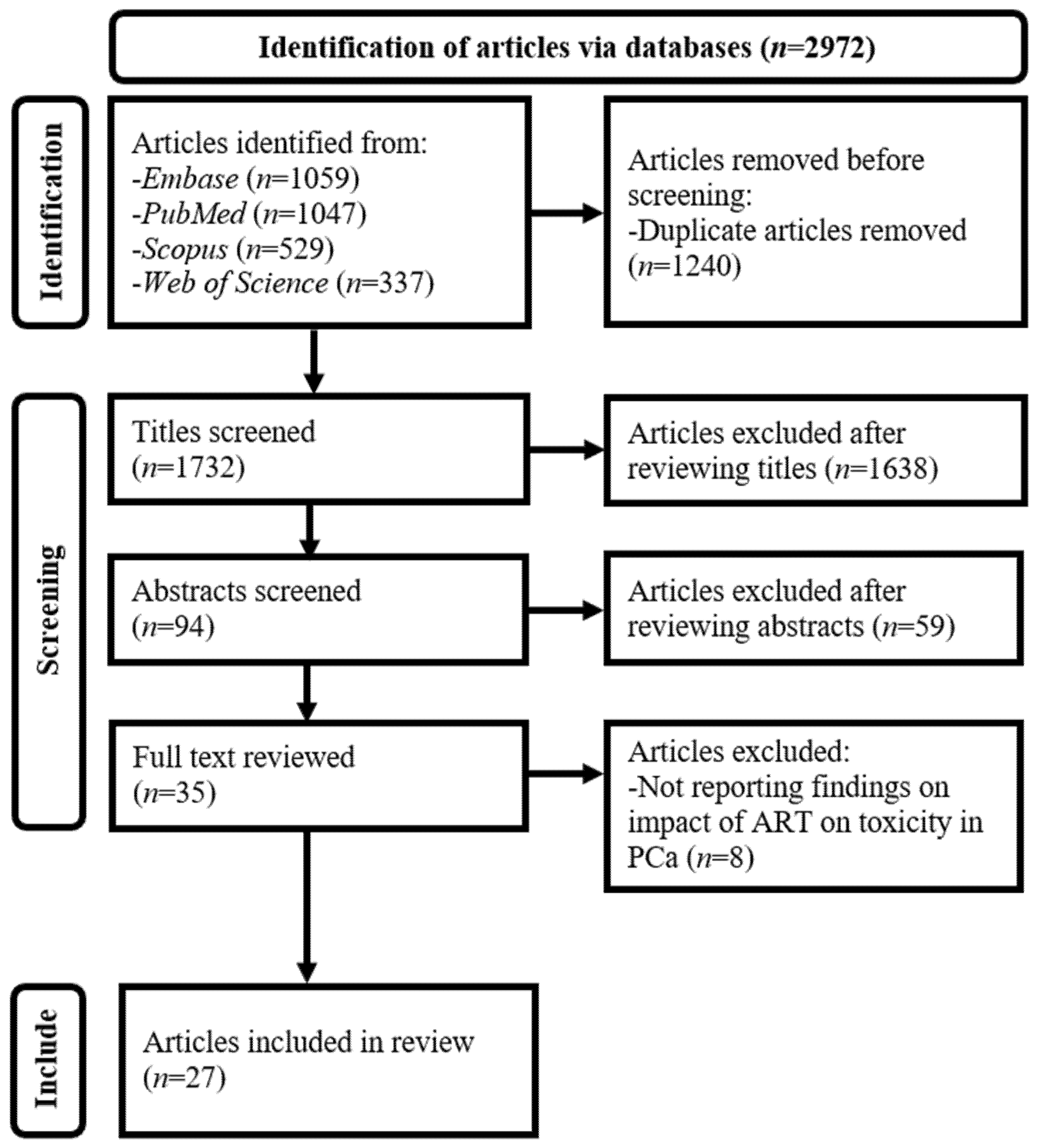
2.1. Literature Search
2.2. Article Selection
2.3. Data Extraction and Synthesis
- Bibliographic information: Author and year of publication.
- Technical parameters: Image guidance modality (e.g., 1.5 T MRI-guided), ART system (such as Elekta Unity, Stockholm, Sweden), fractionation scheme (e.g., SBRT), total dose (such as 35 Gy), fractional dose (e.g., 7 Gy), and planning TV (PTV) coverage (such as prostate gland ± seminal vesicles + 5 mm).
- Study design and population: Study type (prospective or retrospective), sample size (e.g., 100 patients), tumor, node, and metastasis (TNM) staging of PCa (such as cT1-T2), proportion of patients receiving hormonal therapy, and proportion with rectal spacer or balloon.
3. Results
4. Discussion
5. Conclusions
Author Contributions
Funding
Institutional Review Board Statement
Informed Consent Statement
Data Availability Statement
Conflicts of Interest
Abbreviations
| 2D | Two-dimensional |
| 3D | Three-dimensional |
| ART | Adaptive radiation therapy |
| CBCT | Cone-beam computed tomography |
| CFRT | Conventional fractionated radiation therapy |
| CT | Computed tomography |
| DL | Deep learning |
| Embase | Excerpta Medica Database |
| EORTC | European Organization for Research and Treatment of Cancer |
| EPID | Electronic portal imaging device |
| GI | Gastrointestinal |
| GU | Genitourinary |
| MHFRT | Moderate hypofractionated radiation therapy |
| MRI | Magnetic resonance imaging |
| NA | Not available |
| OARs | Organs at risk |
| PCa | Prostate cancer |
| PRISMA-ScR | Preferred Reporting Items for Systematic Reviews and Meta-Analyses Extension for Scoping Reviews |
| RT | Radiation therapy |
| RTOG | Radiation Therapy Oncology Group |
| SBRT | Stereotactic body radiation therapy |
| SV | Seminal vesicle |
| TNM | Tumor, node, and metastasis |
| TV | Target volume |
Appendix A. Search Strategies Employed for Electronic Scholarly Publication Databases

| Database | Search Statement | Articles Identified (n) |
|---|---|---|
| Excerpta Medica Database (Embase) | (‘prostate cancer’/exp OR ‘prostate tumor’/exp OR ‘prostate cancer’ OR ‘prostate carcinoma’) AND (‘adaptive radiotherapy’/exp OR “adaptive radiotherapy” OR “adaptive radiation therapy” OR ‘adaptive planning’ OR ‘treatment adaptation’ OR ‘online adaptive radiotherapy’ OR ‘offline adaptive radiotherapy’ OR ‘MR guided radiotherapy’ OR ‘MRI guided radiotherapy’ OR ‘MR linac’ OR ‘MRIdian’ OR ‘Unity’ OR ‘CBCT guided’ OR ‘cone beam CT guided’) AND (‘radiotherapy’/exp) AND [english]/lim AND [2000–2025]/py | 1059 |
| PubMed | (“Prostatic Neoplasms”[Mesh] OR “prostate cancer” OR “prostate carcinoma”) AND (“adaptive radiotherapy” OR “adaptive radiation therapy” OR “ART plan” OR “adaptive planning” OR “online adaptive” OR “offline adaptive” OR “replanning” OR “MR-guided” OR “MRI-guided” OR “MR linac” OR “MR-Linac” OR “magnetic resonance-guided” OR MRgRT OR “CBCT-guided” OR “cone-beam CT” OR “cone beam CT”) AND (radiotherapy OR “radiation therapy”) AND (English[lang]) AND (“2000/01/01”[PDAT]: “2025/12/31”[PDAT]) | 1047 |
| Scopus | TITLE-ABS-KEY((prostate OR prostatic) W/2 (cancer OR carcinoma OR tumor OR tumour) AND (adaptive W/3 (radiotherapy OR “radiation therapy”) OR “adaptive radiotherapy” OR “online adaptive radiotherapy” OR “MR-guided” W/2 radiotherapy OR “MRI-guided radiotherapy” OR “MR linac” OR “CBCT-guided radiotherapy”)) AND PUBYEAR AFT 1999 AND (LIMIT-TO(LANGUAGE, “English”)) | 529 |
| Web of Science | TS = ((prostate OR prostatic) NEAR/2 (cancer OR carcinoma OR tumor OR tumour) AND (adaptive NEAR/3 (radiotherapy OR “radiation therapy”) OR “adaptive radiotherapy” OR “adaptive radiation therapy” OR “online adaptive radiotherapy” OR “MR-guided radiotherapy” OR “MRI-guided radiotherapy” OR “MR Linac” OR “CBCT-guided radiotherapy”)) AND LANGUAGE: (English) AND DOCUMENT TYPES: (Article OR Trial) AND Timespan: 2000–2025 | 337 |
Appendix B. Sensitivity and Subgroup Analysis Results for Median Incidence Rates of Acute and Late Toxicities in Adaptive Radiation Therapy (ART)
| Toxicity | All Studies | Without Outlier Studies | Studies with <100 Patients | Studies with ≥100 Patients | ||
|---|---|---|---|---|---|---|
| Timing | System | Grade | ||||
| Acute | GI | 1 | 15.0% | 13.5% | 13.5% | 20.1% |
| 2 | 1.0% | 1.0% | 2.9% | 2.0% | ||
| 3 | 0.0% | 0.0% | 0.0% | 0.0% | ||
| GU | 1 | 47.1% | 47.1% | 47.1% | 47.0% | |
| 2 | 9.6% | 7.7% | 8.3% | 11.0% | ||
| 3 | 0.0% | 0.0% | 0.0% | 0.0% | ||
| Late | GI | 1 | 10.0% | 9.6% | 9.6% | 10.0% |
| 2 | 2.0% | 2.0% | 3.6% | 2.0% | ||
| 3 | 0.0% | 0.0% | 0.0% | 1.0% | ||
| GU | 1 | 29.7% | 29.7% | 28.6% | 30.0% | |
| 2 | 5.0% | 3.2% | 0.0% | 6.8% | ||
| 3 | 0.0% | 0.0% | 0.0% | 1.0% | ||
References
- Prostate Cancer Statistics. Available online: https://www.wcrf.org/preventing-cancer/cancer-statistics/prostate-cancer-statistics/ (accessed on 15 January 2025).
- Cancer in Men: Prostate Cancer Is #1 for 118 Countries Globally. Available online: https://www.cancer.org/research/acs-research-news/prostate-cancer-is-number-1-for-118-countries-worldwide.html (accessed on 15 January 2025).
- Prostate Cancer: 3. Epidemiology and Aetiology. Available online: https://uroweb.org/guidelines/prostate-cancer/chapter/epidemiology-and-aetiology (accessed on 15 January 2025).
- Ng, C.K.C. Performance of commercial deep learning-based auto-segmentation software for prostate cancer radiation therapy planning: A systematic review. Information 2025, 16, 215. [Google Scholar] [CrossRef]
- Wasim, S.; Park, J.; Nam, S.; Kim, J. Review of current treatment intensification strategies for prostate cancer patients. Cancers 2023, 15, 5615. [Google Scholar] [CrossRef]
- Leung, V.W.S.; Ng, C.K.C.; Lam, S.K.; Wong, P.T.; Ng, K.Y.; Tam, C.H.; Lee, T.C.; Chow, K.C.; Chow, Y.K.; Tam, V.C.W.; et al. Computed tomography-based radiomics for long-term prognostication of high-risk localized prostate cancer patients received whole pelvic radiotherapy. J. Pers. Med. 2023, 13, 1643. [Google Scholar] [CrossRef]
- Wong, J.Y.K.; Leung, V.W.S.; Hung, R.H.M.; Ng, C.K.C. Comparative study of Eclipse and RayStation multi-criteria optimization-based prostate radiotherapy treatment planning quality. Diagnostics 2024, 14, 465. [Google Scholar] [CrossRef]
- Hekman, L.; Barrett, A.; Ross, D.; Palaganas, E.; Giridhar, P.; Elumalai, T.; Pragathee, V.; Block, A.M.; Welsh, J.S.; Harkenrider, M.M.; et al. A systematic review of clinical trials comparing radiation therapy versus radical prostatectomy in prostate cancer. Clin. Genitourin. Cancer 2024, 22, 102157. [Google Scholar] [CrossRef] [PubMed]
- Numakura, K.; Kobayashi, M.; Muto, Y.; Sato, H.; Sekine, Y.; Sobu, R.; Aoyama, Y.; Takahashi, Y.; Okada, S.; Sasagawa, H.; et al. The current trend of radiation therapy for patients with localized prostate cancer. Curr. Oncol. 2023, 30, 8092–8110. [Google Scholar] [CrossRef] [PubMed]
- Cox, J.D.; Stetz, J.; Pajak, T.F. Toxicity criteria of the Radiation Therapy Oncology Group (RTOG) and the European Organization for Research and Treatment of Cancer (EORTC). Int. J. Radiat. Oncol. Biol. Phys. 1995, 31, 1341–1346. [Google Scholar] [CrossRef]
- Nikitas, J.; Jamshidian, P.; Tree, A.C.; Hall, E.; Dearnaley, D.; Michalski, J.M.; Lee, W.R.; Nguyen, P.L.; Sandler, H.M.; Catton, C.N.; et al. The interplay between acute and late toxicity among patients receiving prostate radiotherapy: An individual patient data meta-analysis of six randomised trials. Lancet Oncol. 2025, 26, 378–386. [Google Scholar] [CrossRef]
- Pisani, C.; Galla, A.; Loi, G.; Beldì, D.; Krengli, M. Urinary toxicity in patients treated with radical EBRT for prostate cancer: Analysis of predictive factors in an historical series. Bull Cancer 2022, 109, 826–833. [Google Scholar] [CrossRef] [PubMed]
- Kunheri, B.; Lakshmi, J.S.; Ravindran, G.C.; Haridas, M.V. Helical tomotherapy for prostate cancer radiation therapy: An audit of early toxicity and quality of life. J. Can. Res. Ther. 2021, 17, 366–371. [Google Scholar] [CrossRef]
- Yan, D.; Vicini, F.; Wong, J.; Martinez, A. Adaptive radiation therapy. Phys. Med. Biol. 1997, 42, 123–132. [Google Scholar] [CrossRef] [PubMed]
- Yan, D.; Wong, J.; Vicini, F.; Michalski, J.; Pan, C.; Frazier, A.; Horwitz, E.; Martinez, A. Adaptive modification of treatment planning to minimize the deleterious effects of treatment setup errors. Int. J. Radiat. Oncol. Biol. Phys. 1997, 38, 197–206. [Google Scholar] [CrossRef]
- Lavrova, E.; Garrett, M.D.; Wang, Y.F.; Chin, C.; Elliston, C.; Savacool, M.; Price, M.; Kachnic, L.A.; Horowitz, D.P. Adaptive radiation therapy: A review of CT-based techniques. Radiol. Imaging Cancer 2023, 5, e230011. [Google Scholar] [CrossRef]
- Allegra, A.G.; Nicosia, L.; Rigo, M.; Bianchi, N.; Borgese, R.F.; De Simone, A.; Giaj-Levra, N.; Gurrera, D.; Naccarato, S.; Pastorello, E.; et al. MR-guided adaptive radiotherapy in localized prostate cancer. Technol. Cancer Res. Treat. 2025, 24, 15330338241297231. [Google Scholar] [CrossRef]
- Patel, R.K.; Lee, J.; Patel, M.; Zaorsky, N.G.; Zuhour, R.; Kashani, R.; Henke, L.E.; Spratt, D.E.; Price, A.T.; Jia, A.Y. Evolution of image guidance and adaptive radiation therapy for localized prostate cancer. Int. J. Radiat. Oncol. Biol. Phys. 2025, 122, 968–975. [Google Scholar] [CrossRef]
- Leeman, J.E.; Shin, K.Y.; Chen, Y.H.; Mak, R.H.; Nguyen, P.L.; D’Amico, A.V.; Martin, N.E. Acute toxicity comparison of magnetic resonance-guided adaptive versus fiducial or computed tomography-guided non-adaptive prostate stereotactic body radiotherapy: A systematic review and meta-analysis. Cancer 2023, 129, 3044–3052. [Google Scholar] [CrossRef]
- Casillas, J.E.J.; Valle, L.F.; Pham, J.; O’Connell, D.; Qi, X.S.; Lamb, J.M.; Ghafarian, M.; Steinberg, M.L.; Cao, M.; Kishan, A.U. Advancing prostate cancer treatment: A review of CT and MR-guided online adaptive radiotherapy techniques. Semin. Radiat. Oncol. 2025, 35, 342–352. [Google Scholar] [CrossRef] [PubMed]
- Ghilezan, M.; Yan, D.; Martinez, A. Adaptive radiation therapy for prostate cancer. Semin. Radiat. Oncol. 2010, 20, 130–137. [Google Scholar] [CrossRef] [PubMed]
- McPartlin, A.J.; Li, X.A.; Kershaw, L.E.; Heide, U.; Kerkmeijer, L.; Lawton, C.; Mahmood, U.; Pos, F.; van As, N.; van Herk, M.; et al. MRI-guided prostate adaptive radiotherapy—A systematic review. Radiother. Oncol. 2016, 119, 371–380. [Google Scholar] [CrossRef]
- McVicar, N.; Popescu, I.A.; Heath, E. Techniques for adaptive prostate radiotherapy. Phys. Med. 2016, 32, 492–498. [Google Scholar] [CrossRef]
- Posiewnik, M.; Piotrowski, T. A review of cone-beam CT applications for adaptive radiotherapy of prostate cancer. Phys. Med. 2019, 59, 13–21. [Google Scholar] [CrossRef]
- Szablewska, S.; Roszkowski, K. Magnetic resonance-guided adaptive stereotactic radiotherapy in prostate cancer patients: A systematic literature review. Rep. Pract. Oncol. Radiother. 2025, 30, 548–558. [Google Scholar] [CrossRef]
- Hall, W.A.; Kishan, A.U.; Hall, E.; Nagar, H.; Vesprini, D.; Paulson, E.; Van der Heide, U.A.; Lawton, C.A.F.; Kerkmeijer, L.G.W.; Tree, A.C. Adaptive magnetic resonance image guided radiation for intact localized prostate cancer how to optimally test a rapidly emerging technology. Front. Oncol. 2022, 12, 962897. [Google Scholar] [CrossRef]
- Ng, C.K.C.; Leung, V.W.S.; Hung, R.H.M. Clinical evaluation of deep learning and atlas-based auto-contouring for head and neck radiation therapy. Appl. Sci. 2022, 12, 11681. [Google Scholar] [CrossRef]
- Ng, C.K.C. Performance of commercial deep learning-based auto-segmentation software for breast cancer radiation therapy planning: A systematic review. Multimodal Technol. Interact. 2024, 8, 114. [Google Scholar] [CrossRef]
- Hooshangnejad, H.; Chen, Q.; Feng, X.; Zhang, R.; Farjam, R.; Voong, K.R.; Hales, R.K.; Du, Y.; Jia, X.; Ding, K. DAART: A deep learning platform for deeply accelerated adaptive radiation therapy for lung cancer. Front. Oncol. 2023, 13, 1201679. [Google Scholar] [CrossRef]
- Munn, Z.; Peters, M.D.J.; Stern, C.; Tufanaru, C.; McArthur, A.; Aromataris, E. Systematic review or scoping review? Guidance for authors when choosing between a systematic or scoping review approach. BMC Med. Res. Methodol. 2018, 18, 143. [Google Scholar] [CrossRef]
- Mak, S.; Thomas, A. Steps for conducting a scoping review. J. Grad. Med. Educ. 2022, 14, 565–567. [Google Scholar] [CrossRef] [PubMed]
- PRISMA-Scoping Reviews. Available online: https://www.prisma-statement.org/scoping/ (accessed on 9 December 2025).
- Shah, K.A.; Ng, C.K.C. Workplace violence in medical radiation science: A systematic review. Radiography 2024, 30, 440–447. [Google Scholar] [CrossRef]
- Ng, C.K.C. Artificial intelligence for radiation dose optimization in pediatric radiology: A systematic review. Children 2022, 9, 1044. [Google Scholar] [CrossRef] [PubMed]
- Ng, C.K.C. Diagnostic performance of artificial intelligence-based computer-aided detection and diagnosis in pediatric radiology: A systematic review. Children 2023, 10, 525. [Google Scholar] [CrossRef]
- Ng, C.K.C. Generative adversarial network (generative artificial intelligence) in pediatric radiology: A systematic review. Children 2023, 10, 1372. [Google Scholar] [CrossRef]
- Mowbray, F.I.; Manlongat, D.; Shukla, M. Sensitivity analysis: A method to promote certainty and transparency in nursing and health research. Can. J. Nurs. Res. 2022, 54, 371–376. [Google Scholar] [CrossRef] [PubMed]
- Critical Appraisal Tools. Available online: https://jbi.global/critical-appraisal-tools/ (accessed on 28 January 2026).
- Brabbins, D.; Martinez, A.; Yan, D.; Lockman, D.; Wallace, M.; Gustafson, G.; Chen, P.; Vicini, F.; Wong, J. A dose-escalation trial with the adaptive radiotherapy process as a delivery system in localized prostate cancer: Analysis of chronic toxicity. Int. J. Radiat. Oncol. Biol. Phys. 2005, 61, 400–408. [Google Scholar] [CrossRef]
- Vargas, C.; Yan, D.; Kestin, L.L.; Krauss, D.; Lockman, D.M.; Brabbins, D.S.; Martinez, A.A. Phase II dose escalation study of image-guided adaptive radiotherapy for prostate cancer: Use of dose-volume constraints to achieve rectal isotoxicity. Int. J. Radiat. Oncol. Biol. Phys. 2005, 63, 141–149. [Google Scholar] [CrossRef] [PubMed]
- Park, S.S.; Yan, D.; McGrath, S.; Dilworth, J.T.; Liang, J.; Ye, H.; Krauss, D.J.; Martinez, A.A.; Kestin, L.L. Adaptive image-guided radiotherapy (IGRT) eliminates the risk of biochemical failure caused by the bias of rectal distension in prostate cancer treatment planning: Clinical evidence. Int. J. Radiat. Oncol. Biol. Phys. 2012, 83, 947–952. [Google Scholar] [CrossRef]
- Hama, Y.; Kaji, T. Long-term follow-up results of CT-guided daily adaptive radiation therapy for localized prostate cancer. Anticancer Res. 2018, 38, 5959–5962. [Google Scholar] [CrossRef] [PubMed]
- Goddard, L.C.; Cabrera, J.; Tang, J.; Garg, M.K.; Tome, W.A. Real-time adaptive motion management in prostate stereotactic body radiation therapy: Clinical and dosimetric analysis of 25 patients. Cureus 2025, 17, e82197. [Google Scholar] [CrossRef]
- Bruynzeel, A.M.E.; Tetar, S.U.; Oei, S.S.; Senan, S.; Haasbeek, C.J.A.; Spoelstra, F.O.B.; Piet, A.H.M.; Meijnen, P.; Bakker van der Jagt, M.A.B.; Fraikin, T.; et al. A prospective single-arm phase 2 study of stereotactic magnetic resonance guided adaptive radiation therapy for prostate cancer: Early toxicity results. Int. J. Radiat. Oncol. Biol. Phys. 2019, 105, 1086–1094. [Google Scholar] [CrossRef]
- Alongi, F.; Rigo, M.; Figlia, V.; Cuccia, F.; Giaj-Levra, N.; Nicosia, L.; Ricchetti, F.; Sicignano, G.; De Simone, A.; Naccarato, S.; et al. 1.5 T MR-guided and daily adapted SBRT for prostate cancer: Feasibility, preliminary clinical tolerability, quality of life and patient-reported outcomes during treatment. Radiat. Oncol. 2020, 15, 69. [Google Scholar] [CrossRef]
- Alongi, F.; Rigo, M.; Figlia, V.; Cuccia, F.; Giaj-Levra, N.; Nicosia, L.; Ricchetti, F.; Vitale, C.; Sicignano, G.; De Simone, A.; et al. Rectal spacer hydrogel in 1.5T MR-guided and daily adapted SBRT for prostate cancer: Dosimetric analysis and preliminary patient-reported outcomes. Br. J. Radiol. 2021, 94, 20200848. [Google Scholar] [CrossRef]
- de Mol van Otterloo, S.R.; Christodouleas, J.P.; Blezer, E.L.A.; Akhiat, H.; Brown, K.; Choudhury, A.; Eggert, D.; Erickson, B.A.; Daamen, L.A.; Faivre-Finn, C.; et al. Patterns of care, tolerability, and safety of the first cohort of patients treated on a novel high-field MR-linac within the MOMENTUM study: Initial results from a prospective multi-institutional registry. Int. J. Radiat. Oncol. Biol. Phys. 2021, 111, 867–875. [Google Scholar] [CrossRef]
- Poon, D.M.C.; Yuan, J.; Wong, O.L.; Yang, B.; Chiu, S.T.; Cheung, K.Y.; Chiu, G.; Yu, S.K. 1.5T magnetic resonance-guided stereotactic body radiotherapy for localized prostate cancer: Preliminary clinical results of clinician- and patient-reported outcomes. Cancers 2021, 13, 4866. [Google Scholar] [CrossRef]
- Tetar, S.U.; Bruynzeel, A.M.E.; Oei, S.S.; Senan, S.; Fraikin, T.; Slotman, B.J.; Moorselaar, R.J.A.V.; Lagerwaard, F.J. Magnetic resonance-guided stereotactic radiotherapy for localized prostate cancer: Final results on patient-reported outcomes of a prospective phase 2 study. Eur. Urol. Oncol. 2021, 4, 628–634. [Google Scholar] [CrossRef] [PubMed]
- Ugurluer, G.; Atalar, B.; Zoto Mustafayev, T.; Gungor, G.; Aydin, G.; Sengoz, M.; Abacioglu, U.; Tuna, M.B.; Kural, A.R.; Ozyar, E. Magnetic resonance image-guided adaptive stereotactic body radiotherapy for prostate cancer: Preliminary results of outcome and toxicity. Br. J. Radiol. 2021, 94, 20200696. [Google Scholar] [CrossRef] [PubMed]
- Alongi, F.; Rigo, M.; Figlia, V.; Nicosia, L.; Mazzola, R.; Giaj Levra, N.; Ricchetti, F.; Trapani, G.; Attinà, G.; Vitale, C.; et al. 1.5T MR-guided daily-adaptive SBRT for prostate cancer: Preliminary report of toxicity and quality of life of the first 100 patients. J. Pers. Med. 2022, 12, 1982. [Google Scholar] [CrossRef] [PubMed]
- Poon, D.M.C.; Yuan, J.; Yang, B.; Wong, O.L.; Chiu, S.T.; Chiu, G.; Cheung, K.Y.; Yu, S.K.; Yung, R.W.H. A prospective study of stereotactic body radiotherapy (SBRT) with concomitant whole-pelvic radiotherapy (WPRT) for high-risk localized prostate cancer patients using 1.5 tesla magnetic resonance guidance: The preliminary clinical outcome. Cancers 2022, 14, 3484. [Google Scholar] [CrossRef]
- Teunissen, F.R.; Willigenburg, T.; Meijer, R.P.; van Melick, H.H.E.; Verkooijen, H.M.; van der Voort van Zyp, J.R.N. The first patient-reported outcomes from the Utrecht Prostate Cohort (UPC): The first platform facilitating ‘trials within cohorts’ (TwiCs) for the evaluation of interventions for prostate cancer. World J. Urol. 2022, 40, 2205–2212. [Google Scholar] [CrossRef]
- Turkkan, G.; Bilici, N.; Sertel, H.; Keskus, Y.; Alkaya, S.; Tavli, B.; Ozkirim, M.; Fayda, M. Clinical utility of a 1.5 T magnetic resonance imaging-guided linear accelerator during conventionally fractionated and hypofractionated prostate cancer radiotherapy. Front. Oncol. 2022, 12, 909402. [Google Scholar] [CrossRef]
- Gelbart Pridan, O.; Ben David, M.A.; Zalmanov, S.; Lipski, Y.; Grinberg, V.; Levin, D.; Apter, S.; Guindi, M.; Epstein, D.; Radus, R.; et al. Outcome of the first 200 patients with prostate cancer treated with MRI-linac at Assuta MC. Front. Oncol. 2023, 13, 1151256. [Google Scholar] [CrossRef]
- Teunissen, F.R.; Willigenburg, T.; Tree, A.C.; Hall, W.A.; Choi, S.L.; Choudhury, A.; Christodouleas, J.P.; de Boer, J.C.J.; de Groot-van Breugel, E.N.; Kerkmeijer, L.G.W.; et al. Magnetic resonance-guided adaptive radiation therapy for prostate cancer: The first results from the MOMENTUM study-An international registry for the evidence-based introduction of magnetic resonance-guided adaptive radiation therapy. Pract. Radiat. Oncol. 2023, 13, e261–e269. [Google Scholar] [CrossRef]
- Fink, C.A.; Ristau, J.; Buchele, C.; Klüter, S.; Liermann, J.; Hoegen-Saßmannshausen, P.; Sandrini, E.; Lentz-Hommertgen, A.; Baumann, L.; Andratschke, N.; et al. Stereotactic ultrahypofractionated MR-guided radiotherapy for localized prostate cancer—Acute toxicity and patient-reported outcomes in the prospective, multicenter SMILE phase II trial. Clin. Transl. Radiat. Oncol. 2024, 46, 100771. [Google Scholar] [CrossRef] [PubMed]
- Hassan, S.P.; de Leon, J.; Batumalai, V.; Moutrie, Z.; Hogan, L.; Ge, Y.; Stricker, P.; Jameson, M.G. Magnetic resonance guided adaptive post prostatectomy radiotherapy: Accumulated dose comparison of different workflows. J. Appl. Clin. Med. Phys. 2024, 25, e14253. [Google Scholar] [CrossRef] [PubMed]
- Poon, D.M.C.; Yuan, J.; Wong, O.L.; Yang, B.; Tse, M.Y.; Lau, K.K.; Chiu, S.T.; Chiu, P.K.; Ng, C.F.; Chui, K.L.; et al. One-year clinical outcomes of MR-guided stereotactic body radiation therapy with rectal spacer for patients with localized prostate cancer. World J. Urol. 2024, 42, 97. [Google Scholar] [CrossRef]
- Sritharan, K.; Daamen, L.; Pathmanathan, A.; Schytte, T.; Pos, F.; Choudhury, A.; van der Voort van Zyp, J.R.N.; Kerkmeijer, L.G.W.; Hall, W.; Hall, E.; et al. MRI-guided radiotherapy in twenty fractions for localised prostate cancer; results from the MOMENTUM study. Clin. Transl. Radiat. Oncol. 2024, 46, 100742. [Google Scholar] [CrossRef] [PubMed]
- Westley, R.L.; Biscombe, K.; Dunlop, A.; Mitchell, A.; Oelfke, U.; Nill, S.; Murray, J.; Pathmanathan, A.; Hafeez, S.; Parker, C.; et al. Interim toxicity analysis from the randomized HERMES trial of 2- and 5-fraction magnetic resonance imaging-guided adaptive prostate radiation therapy. Int. J. Radiat. Oncol. Biol. Phys. 2024, 118, 682–687. [Google Scholar] [CrossRef]
- Cooper, S.; Westley, R.L.; Biscombe, K.; Dunlop, A.; Mitchell, A.; Oelfke, U.; Nill, S.; Manning, G.; Burnett, S.; Murray, J.; et al. HERMES: Randomized trial of 2-fraction or 5-fraction magnetic resonance imaging-guided adaptive prostate radiation therapy. Int. J. Radiat. Oncol. Biol. Phys. 2025, 123, 773–782. [Google Scholar] [CrossRef]
- Joye, A.A.; Zosso-Pavic, M.; Beckmann, J.; Bonzon, J.; Stolz, S.; Willmann, J.; Ahmadsei, M.; Christ, S.M.; Andratschke, N.; Guckenberger, M.; et al. Implementation of a comprehensive clinical quality assurance system in radiation oncology. Radiat. Oncol. 2025, 20, 69. [Google Scholar] [CrossRef]
- Lalmahomed, T.A.; Willigenburg, T.; van de Pol, S.M.G.; de Groot-van Breugel, E.N.; Snoeren, L.M.W.; Hes, J.; van Melick, H.H.E.; Verkooijen, H.M.; de Boer, J.C.J.; van der Voort van Zyp, J.R.N. Acute toxicity and quality of life after margin reduction using a sub-fractionation workflow for stereotactic radiotherapy of localized prostate cancer on a 1.5 Tesla MR-linac. Radiother. Oncol. 2025, 207, 110845. [Google Scholar] [CrossRef]
- Poon, D.M.C.; Yuan, J.; Wong, O.L.; Yang, B.; Tse, M.Y.; Fung, Y.Y.; Chiu, S.T.; Lin, W.C.; Cheung, K.Y.; Chiu, G.; et al. Post-prostatectomy magnetic resonance-guided radiotherapy on a 1.5 tesla magnetic resonance integrated linear accelerator: Feasibility, toxicity, and preliminary clinical outcomes. Asia Pac. J. Clin. Oncol. 2025, 21, 247–255. [Google Scholar] [CrossRef]
- Eidex, Z.; Ding, Y.; Wang, J.; Abouei, E.; Qiu, R.L.J.; Liu, T.; Wang, T.; Yang, X. Deep learning in MRI-guided radiation therapy: A systematic review. J. Appl. Clin. Med. Phys. 2024, 25, e14155. [Google Scholar] [CrossRef] [PubMed]
- What Is the MR-Linac Consortium? Available online: https://mrlconsortium.org/what-is-the-consortium/ (accessed on 5 December 2025).
- Over Two Decades of AI-Powered Precision: How Synchrony Has Revolutionized Real-Time Adaptive Radiotherapy Delivery. Available online: https://www.accuray.com/blog/over-two-decades-of-ai-powered-precision-how-synchrony-has-revolutionized-real-time-adaptive-radiotherapy-delivery/ (accessed on 5 December 2025).

| Inclusion Criteria | Exclusion Criteria |
|---|---|
|
|
| Author and Year | Image Guidance Modality | ART System | Fractionation Scheme | Total Dose (Gy) | Fractional Dose (Gy) | Planning Target Volume Coverage | Study Type | Sample Size (Number of Patients) | Tumor, Node, and Metastasis Staging | Proportion of Patients Receiving Hormonal Therapy | Proportion with Rectal Spacer/Balloon | Article Quality |
|---|---|---|---|---|---|---|---|---|---|---|---|---|
| Computed Tomography (CT)-Guided | ||||||||||||
| Brabbins et al. (2005) [39] | CT-guided | NA | CFRT | 70.2–79.2 | 1.8 | Prostate gland +/− SV (for intermediate-risk) + 5 mm (3 mm posteriorly) | Prospective | 280 | T1b-T3, NX-N0, M0 | 21.4% | NA | 75.0% |
| Vargas et al. (2005) [40] | CT-guided | NA | CFRT | 70.2–79.2 | 1.8 | Prostate gland +/− 1 cm/2 cm of SV | Prospective | 331 | T1a-T3c, N0, M0 | 22.0% | NA | 77.3% |
| Park et al. (2012) [41] | CT-guided | NA | CFRT | 70.2–79.2 | 1.8 | NA | Retrospective | 962 | NA | 23.7% | NA | 72.2% |
| Hama and Kaji (2018) [42] | CT-guided | NA | CFRT | 76.0 | 2.0 | Prostate gland +/− SV + (5 mm–10 mm) | Prospective | 26 | cT1c-T3a, N0, M0 | Neoadjuvant hormone therapy: 76.9%; adjuvant hormone therapy: 42.3% | NA | 61.1% |
| Goddard et al. (2025) [43] | CT-guided | Radixact Synchrony | SBRT | 40.0 | 8.0 | Prostate gland +/− SV (for intermediate-risk) + 5 mm (3 mm posteriorly) | Retrospective | 25 | Stage 1 to 2c | NA | 92.0% | 55.6% |
| Magnetic Resonance Imaging (MRI)-Guided | ||||||||||||
| Bruynzeel et al. (2019) [44] | 0.35 T MRI-guided | ViewRay MRIdian | SBRT | 36.3 | 7.3 | Prostate gland +/− SV (for intermediate-risk) + 5 mm (3 mm posteriorly) | Prospective | 101 | cT1c-T3b, N0, M0 | 82.2% | NA | 83.3% |
| Alongi et al. (2020) [45] | 1.5 T MRI-guided | Elekta Unity | SBRT | 35.0 | 7.0 | Prostate gland +/− SV (for intermediate-risk) + 5 mm (3 mm posteriorly) | Prospective | 25 | cT1-T2, N0, M0 | 36.0% | NA | 87.5% |
| Alongi et al. (2021) [46] | 1.5 T MRI-guided | Elekta Unity | SBRT | 35.0 | 7.0 | Prostate gland +/− SV (for intermediate-risk) + 5 mm (3 mm posteriorly) | Retrospective-prospective | 20 (retrospective) and 20 (prospective) | cT1-T2 | 30.0% (retrospective) and 20.0% (prospective) | 100.0% (retrospective) and 0.0% (prospective) | 90.0% |
| de Mol van Otterloo et al. (2021) [47] | 1.5 T MRI-guided | Elekta Unity | CFRT and SBRT | 60.0 (CFRT) and 36.3/38.6 (SBRT) | 3.0 (CFRT) and 7.3/7.7 (SBRT) | NA | Prospective | 223 | NA | NA | NA | 87.5% |
| Poon et al. (2021) [48] | 1.5 T MR-guided | Elekta Unity | SBRT | 36.3/40.0 | 7.3/8.0 | Prostate gland +/− SV (for intermediate-/high-risk) + 5 mm (3 mm posteriorly) | Prospective | 51 | T1 (7.8%), T2 (74.5%), T3 (17.6%), N0, M0 | 58.8% | 19.6% | 80.0% |
| Tetar et al. (2021) [49] | 0.35 T MR-guided | ViewRay MRIdian | SBRT | 36.3 | 7.3 | Prostate gland +/− SV (for intermediate-/high-risk) + 3 mm | Prospective | 101 | T1–3b, N0, M0 | 83.2% | 0.0% | 87.5% |
| Ugurluer et al. (2021) [50] | 0.35 T MR-guided | ViewRay MRIdian | SBRT | 36.3 | 7.3 | Prostate gland +/− SV (for intermediate-/high-risk) + 3 mm | Retrospective | 50 | T1 (60.0%), T2 (36.0%), T3 (4.0%), N0, M0 | 28.0% | NA | 87.5% |
| Alongi et al. (2022) [51] | 1.5 T MR-guided | Elekta Unity | SBRT | 35.0 (low-risk/M1); 36.3 (intermediate-/high-risk) | 7.0 (low-risk/M1); 7.3 (intermediate-/high-risk) | Prostate gland +/− SV (for M1, intermediate- and high-risk) + 5 mm (3 mm posteriorly) | Prospective | 100 | cT1-cT3b, N0, M0 | 32.0% | 37.0% | 87.5% |
| Poon et al. (2022) [52] | 1.5 T MR-guided | Elekta Unity | SBRT | 40.0 | 8.0 | Prostate gland +/− base of SV (for cT3b, entire SV) + 5 mm (3 mm posteriorly) | Prospective | 42 | cT2a-T3b, N0, M0 | 100.0% | 31.0% | 70.0% |
| Teunissen et al. (2022) [53] | MR-guided | Elekta Unity | CFRT and SBRT | 62.0 (CFRT) and 36.3 (SBRT) | 3.1 (CFRT) and 7.3 (SBRT) | NA | Prospective | 293 | cT1 (51.4%), cT2 (43.8%), cT3 (4.5%), cT4 (0.3%) | 13.7% | NA | 80.0% |
| Turkkan et al. (2022) [54] | 1.5 T MR-guided | Elekta Unity | CFRT, MHFRT, and SBRT | 66.0–78.0 (CFRT), 70.0 (MHFRT), and 36.3 (SBRT) | 1.8–2.0 (CFRT), 2.5 (MHFRT), and 7.3 (SBRT) | NA | Retrospective | 14 | NA | 71.0% | NA | 88.9% |
| Gelbart Pridan et al. (2023) [55] | 0.35 T MR-guided | ViewRay MRIdian | SBRT | 36.3 | 7.3 | Prostate gland + 3 mm | Retrospective | 200 | NA | 28.0% | 0.0% | 70.0% |
| Teunissen et al. (2023) [56] | 1.5 T MR-guided | Elekta Unity | SBRT | 36.3 | 7.3 | Prostate gland +/− SV + 5 mm (for 2 patients, 3 mm posteriorly) | Prospective | 425 | cT1 (39.2%), cT2 (55.7%), cT3 (5.1%) | 18.4% | NA | 85.0% |
| Fink et al. (2024) [57] | 0.35 T MR-guided | ViewRay MRIdian | SBRT | 37.5 | 7.5 | Prostate gland +/− SV (for intermediate-/high-risk) + 3 mm | Prospective | 69 | <T3a, N0, M0 | 12.0% | 0.0% | 88.9% |
| Hassan et al. (2024) [58] | 1.5 T MR-guided | Elekta Unity | CFRT | 70.0–72.0 | 3.0 | NA | Prospective | 7 | NA | 0.0% | NA | 87.5% |
| Poon et al. (2024) [59] | 1.5 T MR-guided | Elekta Unity | SBRT | 36.3/40.0 | 7.3/8.0 | NA | Prospective | 34 | T2 (76.5%); T3 (23.5%), N0, M0 | 76.5% | 100.0% | 77.3% |
| Sritharan et al. (2024) [60] | 1.5 T MR-guided | Elekta Unity | CFRT | 60.0 | 3.0 | Prostate gland +/− SV + 5 mm (3 mm/0 mm posteriorly) | Prospective | 140 | NA | 58.0% | NA | 70.0% |
| Westley et al. (2024) [61] | 1.5 T MR-guided | Elekta Unity | SBRT | 24.0 (2-fraction) and 36.3 (5-fraction) | 12.0 (2-fraction) and 7.3 (5-fraction) | Prostate gland +/− 1 cm/2 cm of SV + 3 mm | Prospective | 20 | T2 (70%), T3a (30%) (2-fraction) and T2 (80%), T3a (20%) (5-fraction) | 100.0% | 0.0% | 90.0% |
| Cooper et al. (2025) [62] | 1.5 T MR-guided | Elekta Unity | SBRT | 24.0 (2-fraction) and 36.3 (5-fraction) | 12.0 (2-fraction) and 7.3 (5-fraction) | Prostate gland +/− 2 cm of SV + 3 mm (2-fraction) and prostate gland +/− 1 cm of SV + 3 mm (5-fraction) | Prospective | 23 (2-fraction) and 24 (5-fraction) | cT1-T3a (2-fraction) and cT1c-T3a (5-fraction) | 100.0% | NA | 90.0% |
| Joye et al. (2025) [63] | 0.35 T MR-guided | ViewRay MRIdian | SBRT | 36.3/37.5 | 7.3/7.5 | NA | Prospective | 209 | cT1a-T3b, N0, M0 | NA | NA | 80.0% |
| Lalmahomed et al. (2025) [64] | 1.5 T MR-guided | Elekta Unity | SBRT | 36.3 | 7.3 | Prostate gland +/− SV + 2–3 mm (tight margin group) and prostate gland +/− SV + 5 mm (standard margin group) | Prospective | 106 (tight margin group) and 106 (standard margin group) | NA | 5.0% (tight margin group) and 6.0% (standard margin group) | NA | 81.8% |
| Poon et al. (2025) [65] | 1.5 T MR-guided | Elekta Unity | CFRT | 70.0 | 2.0 | NA | Prospective | 30 | pT2b-pT4, pNX-pN0, M0 | 96.7% | 0.0% | 88.9% |
| Toxicity | ART | CT-Guided ART | MRI-Guided ART | |||||
|---|---|---|---|---|---|---|---|---|
| Timing | System | Grade | Median Incidence Rate (Range) | Study Reference | Median Incidence Rate (Range) | Study Reference | Median Incidence Rate (Range) | Study Reference |
| Acute | GI | 1 | 15.0% (0.0–100.0%) | [44,45,46,48,49,50,51,52,53,54,55,57,58,59,60,61,62,63,64,65] | NA | NA | 13.6% (0.0–100.0%) | [44,45,46,48,49,50,51,52,53,54,55,57,58,59,60,61,62,63,64,65] |
| 2 | 1.0% (0.0–33.6%) | [40,44,45,46,48,49,50,51,52,53,54,55,57,58,59,60,61,62,63,64,65] | 33.6% 1 | [40] | 1.0% (0.0–17.0%) | [44,45,46,48,49,50,51,52,53,54,55,57,58,59,60,61,62,63,64,65] | ||
| 3 | 0.0% (0.0–4.0%) | [40,44,45,46,48,49,50,51,52,53,54,55,57,58,59,60,61,62,63,64,65] | 0.9% 1 | [40] | 0.0% (0.0–4.0%) | [44,45,46,48,49,50,51,52,53,54,55,57,58,59,60,61,62,63,64,65] | ||
| GU | 1 | 47.1% (1.0–100.0%) | [43,44,45,46,47,48,49,50,51,52,54,55,56,57,58,59,60,61,62,63,64,65] | 56.0% 1 | [43] | 47.1% (1.0–100.0%) | [44,45,46,47,48,49,50,51,52,54,55,56,57,58,59,60,61,62,63,64,65] | |
| 2 | 9.6% (0.0–36.0%) | [43,44,45,46,47,48,49,50,51,52,54,55,56,57,58,59,60,61,62,63,64,65] | 12.0% 1 | [43] | 9.1% (0.0–36.0%) | [44,45,46,47,48,49,50,51,52,54,55,56,57,58,59,60,61,62,63,64,65] | ||
| 3 | 0.0% (0.0–10.0%) | [43,44,45,46,47,48,49,50,51,52,54,55,56,57,58,59,60,61,62,63,64,65] | 0.0% 1 | [43] | 0.0% (0.0–10.0%) | [44,45,46,47,48,49,50,51,52,54,55,56,57,58,59,60,61,62,63,64,65] | ||
| Late | GI | 1 | 10.0% (0.0–43.0%) | [39,40,41,42,49,50,51,52,56,58,60,62,65] | 26.7% (15.4–38.0%) | [39,42] | 9.1% (0.0–43.0%) | [49,50,51,52,56,58,60,62,65] |
| 2 | 2.0% (0.0–26.0%) | [39,40,41,42,49,50,51,52,56,58,60,62,65] | 17.2% (3.8–26.0%) | [39,40,41,42] | 1.6% (0.0–22.0%) | [49,50,51,52,56,58,60,62,65] | ||
| 3 | 0.0% (0.0–10.0%) | [39,40,41,42,49,50,51,52,56,58,60,62,65] | 2.9% (0.0–10.0%) | [39,40,41,42] | 0.0% (0.0–3.0%) | [49,50,51,52,56,58,60,62,65] | ||
| GU | 1 | 29.7% (3.8–70.0%) | [39,42,49,50,51,52,56,60,62,65] | 20.9% (3.8–38.0%) | [39,42] | 29.7% (13.0–70.0%) | [49,50,51,52,56,60,62,65] | |
| 2 | 5.0% (0.0–15.5%) | [39,41,42,49,50,51,52,56,60,62,65] | 6.0% (0.0–15.5%) | [39,41,42] | 4.2% (0.0–14.0%) | [49,50,51,52,56,60,62,65] | ||
| 3 | 0.0% (0.0–9.0%) | [39,41,42,49,50,51,52,56,60,62,65] | 4.3% (0.0–9.0%) | [39,41,42] | 0.0% (0.0–3.0%) | [49,50,51,52,56,60,62,65] | ||
| Toxicity | Adaptive CFRT | Adaptive SBRT | ||||
|---|---|---|---|---|---|---|
| Timing | System | Grade | Median Incidence Rate (Range) | Study Reference | Median Incidence Rate (Range) | Study Reference |
| Acute | GI | 1 | 17.5% (0.0–46.7%) | [53,58,60,65] | 14.0% (0.0–100.0%) | [44,45,46,48,49,50,51,52,54,55,57,59,61,62,63,64] |
| 2 | 1.7% (0.0–14.3%) | [53,58,60,65] | 1.0% (0.0–33.6%) | [44,45,46,48,49,50,51,52,54,55,57,59,61,62,63,64] | ||
| 3 | 0.0% (0.0%) | [53,58,60,65] | 0.0% (0.0–4.0%) | [44,45,46,48,49,50,51,52,54,55,57,59,61,62,63,64] | ||
| GU | 1 | 56.5% (14.3–73.3%) | [56,58,60,65] | 47.0% (1.0–100.0%) | [43,44,45,46,47,48,49,50,51,52,54,55,57,59,61,62,63,64] | |
| 2 | 2.4% (0.0–7.0%) | [56,58,60,65] | 11.9% (0.0–36.0%) | [43,44,45,46,47,48,49,50,51,52,54,55,57,59,61,62,63,64] | ||
| 3 | 0.0% (0.0%) | [56,58,60,65] | 0.0% (0.0–10.0%) | [43,44,45,46,47,48,49,50,51,52,54,55,57,59,61,62,63,64] | ||
| Late | GI | 1 | 10.4% (5.0–38.0%) | [39,40,41,42,56,58,60,65] | 9.1% (0.0–43.0%) | [49,50,51,52,62] |
| 2 | 2.5% (0.0–26.0%) | [39,40,41,42,56,58,60,65] | 2.0% (0.0–22.0%) | [49,50,51,52,62] | ||
| 3 | 0.0% (0.0–10.0%) | [39,40,41,42,56,58,60,65] | 0.0% (0.0–3.0%) | [49,50,51,52,62] | ||
| GU | 1 | 28.3% (3.8–63.3%) | [39,41,42,56,60,65] | 33.0% (13.0–70.0%) | [49,50,51,52,62] | |
| 2 | 4.2% (0.0–15.5%) | [39,41,42,56,60,65] | 5.9% (0.0–14.0%) | [49,50,51,52,62] | ||
| 3 | 0.0% (0.0–9.0%) | [39,41,42,56,60,65] | 0.0% (0.0–3.0%) | [49,50,51,52,62] | ||
Disclaimer/Publisher’s Note: The statements, opinions and data contained in all publications are solely those of the individual author(s) and contributor(s) and not of MDPI and/or the editor(s). MDPI and/or the editor(s) disclaim responsibility for any injury to people or property resulting from any ideas, methods, instructions or products referred to in the content. |
© 2026 by the authors. Licensee MDPI, Basel, Switzerland. This article is an open access article distributed under the terms and conditions of the Creative Commons Attribution (CC BY) license.
Share and Cite
Li, M.; Ching, J.C.F.; Tong, J.T.; Man, J.Y.K.; Hung, R.H.M.; Leung, V.W.S.; Ng, C.K.C. Impact of Adaptive Radiation Therapy on Toxicity in Prostate Cancer: A Scoping Review. Biomedicines 2026, 14, 370. https://doi.org/10.3390/biomedicines14020370
Li M, Ching JCF, Tong JT, Man JYK, Hung RHM, Leung VWS, Ng CKC. Impact of Adaptive Radiation Therapy on Toxicity in Prostate Cancer: A Scoping Review. Biomedicines. 2026; 14(2):370. https://doi.org/10.3390/biomedicines14020370
Chicago/Turabian StyleLi, Miao, Jerry C. F. Ching, Julian T. Tong, Jacky Y. K. Man, Rico H. M. Hung, Vincent W. S. Leung, and Curtise K. C. Ng. 2026. "Impact of Adaptive Radiation Therapy on Toxicity in Prostate Cancer: A Scoping Review" Biomedicines 14, no. 2: 370. https://doi.org/10.3390/biomedicines14020370
APA StyleLi, M., Ching, J. C. F., Tong, J. T., Man, J. Y. K., Hung, R. H. M., Leung, V. W. S., & Ng, C. K. C. (2026). Impact of Adaptive Radiation Therapy on Toxicity in Prostate Cancer: A Scoping Review. Biomedicines, 14(2), 370. https://doi.org/10.3390/biomedicines14020370