Article

Evaluation of Cytokine Levels in Cardiac Transthyretin and Immunoglobulin Light Chain Amyloidosis and Their Correlation with Myocardial Inflammatory Cells and MACE

1
Department of Cardiology, Angiology and Intensive Care Medicine, Deutsches Herzzentrum der Charité, Hindenburgdamm 30, 12203 Berlin, Germany
2
Cardiopathology, Institute for Pathology and Neuropathology, University Hospital Tübingen, 72076 Tübingen, Germany
3
Core Unit for Bioinformatics (CUBI), Berlin Institute of Health (BIH) at Charité—Universitätsmedizin Berlin, 10117 Berlin, Germany
4
Berlin Institute of Health (BIH) at Charité—Universitätsmedizin Berlin, 10117 Berlin, Germany
*
Author to whom correspondence should be addressed.
Biomedicines 2025, 13(9), 2254; https://doi.org/10.3390/biomedicines13092254
Submission received: 12 August 2025 / Revised: 3 September 2025 / Accepted: 9 September 2025 / Published: 12 September 2025
(This article belongs to the Section Cell Biology and Pathology)

Abstract

Aims: Myocardial inflammation in cardiac amyloidosis is associated with poor clinical outcomes. This study aimed to (a) investigate the relationship between peripheral blood cytokine levels and the presence of inflammatory cells within the myocardium, and to (b) evaluate the potential of cytokines as predictors of major adverse cardiovascular events (MACE) in transthyretin (ATTR) and immunoglobulin light chain (AL) cardiac amyloidosis. Methods: Peripheral blood samples were collected from 50 patients with cardiac ATTR or AL amyloidosis between 2018 and 2023 at baseline and every three months during follow-up visits. Cytokine analysis was performed using Olink’s Proximity Extension Assay. For MACE prediction analysis, only patients with MACE occurring within ±14 days of a study visit were included (n = 16). Associations were evaluated using linear models. Results: No significant associations were identified between the EMB-confirmed myocardial presence of inflammatory cells and cytokine levels. There was a trend of weak-to-moderate associations between serial blood cytokine levels and MACE, albeit this was non-significant after adjustment for multiple testing (FDR): r2 = 0.28 for PON3 (p = 0.00075, FDR = 0.28), SIGLEC1 (p = 0.00077, FDR = 0.28), and IL-6 (p = 0.00086, FDR = 0.31). Conclusions: Peripheral blood cytokine levels were not reliable biomarkers for the myocardial presence of inflammatory cells. PON3, SIGLEC1, and IL-6 demonstrated a statistically non-significant trend of a weak-to-moderate association with MACE in cardiac amyloidosis. Since we recently demonstrated that amyloidosis with an inflammatory component is associated with poor outcomes, these additional findings underscore the need for alternative approaches to identify and manage inflammation in this patient population.

Graphical Abstract

1. Introduction

Cardiac transthyretin (ATTR) and immunoglobulin light chain (AL) amyloidosis are both diseases characterized by the accumulation of misconfigured proteins, which can lead to infiltrative cardiomyopathy and heart failure [1]. With the availability of disease-modifying drugs such as tafamidis [2,3], vutrisiran [4], or the NI006-antibody [5], the interest in cardiac amyloidosis has been rapidly increasing in the scientific community [6,7,8].
Recent studies have highlighted that cardiomyopathies of various etiologies, including amyloidosis, are often accompanied by cardiac inflammation [9,10,11]. There is growing evidence to suggest that this inflammation may contribute to heart failure [9,11,12,13,14,15,16]. As such, understanding the inflammatory processes in cardiac amyloidosis is becoming an increasingly important research focus.
In a study of 50 patients with ATTR and AL cardiac amyloidosis, endomyocardial biopsies (EMBs) revealed that 42% of these patients exhibited myocardial infiltration by CD3+ T cells and CD68+ macrophages [17]. Since myocardial inflammation has been reported to be associated with poor clinical outcomes in ATTR and AL cardiac amyloidosis [10,11], it is critical to better understand the underlying inflammatory mechanisms. Specifically, it remains unclear whether inflammation is simply a byproduct of cardiac amyloidosis or whether it actively drives disease progression. Additionally, to advance personalized therapies in cardiac amyloidosis, it is essential to identify patients who may benefit from targeted anti-inflammatory treatments. We therefore aimed to (a) investigate the relationship between peripheral blood cytokine levels and the presence of inflammatory cells within the myocardium confirmed by EMB, and to (b) evaluate the potential of cytokines as predictors of MACE in ATTR and AL cardiac amyloidosis.

2. Materials and Methods

2.1. Study Design, Study Participants, Recruitment, and Pre-Analytics

To investigate the association between peripheral blood cytokine levels and the presence of inflammatory cells within the myocardium and MACE, ethylenediaminetetraacetic acid (EDTA) plasma samples from 50 patients with cardiac ATTR or AL amyloidosis were collected between the years 2018 and 2023 and stored in the biobank of the Deutsches Herzzentrum der Charité, Campus Benjamin Franklin. Blood was obtained from each patient at the baseline visit and during study visits every three months. Blood samples were collected in EDTA tubes, and EDTA plasma was separated from the samples by centrifugation and stored at −80 °C.
An EMB was only performed when clinically indicated [1]. In order to focus on samples from patients with a definitive diagnosis of myocardial inflammation, only patients for whom an EMB sample was available were included in the correlation analysis of peripheral cytokine levels with myocardial immune cell infiltration.
For analyzing whether peripheral blood cytokine levels predict MACE, only patients with MACE occurring within ±14 days of a study visit were included. MACE were defined as at least one of the following: all-cause mortality, cardiovascular mortality, hospitalization for heart failure, or acute decompensated heart failure.

2.2. Endomyocardial Biopsies

EMB samples were obtained during routine diagnostic workup as indicated by the European Society of Cardiology (ESC) position statement on the diagnosis and treatment of cardiac amyloidosis [1]. The EMB samples were sent to, and analyzed by, a specialized cardiopathologist. Samples were stained for CD3, CD20, and CD68.

2.3. Cytokine Analysis

For analyzing peripheral blood cytokine levels in EDTA plasma samples, we utilized the Proximity Extension Assay (PEA) by Olink. This method is based on two antibodies carrying unique oligonucleotide sequences that bind to the target proteins. The proximity of the two antibodies leads to hybridization of the oligonucleotide sequences that they carry and makes them accessible for deoxyribonucleic acid (DNA) polymerases, which extend the strands. The DNA strands can then be quantified by a real-time polymerase chain reaction (PCR).
Olink’s PEA technology empowers high-throughput and highly specific proteomics and is therefore a suitable approach for biomarker development [18]. We chose the Olink Explore 384—Inflammation panel for our analyses, enabling us to include 368 proteins associated with inflammation. While the panel is called “Olink Explore 384” based on its 384-plex capacity, it measures 368 inflammation-related proteins, with the remaining assay space used for internal controls and quality assurance. Supplemental File S1 lists all of the cytokines included in the Olink Explore 384—Inflammation panel.
Library preparation, randomization, quality control, and data processing were conducted using Olink’s standard processes (see https://www.olink.com for more information, accessed on 1 July 2025). Normalized protein expression (NPX) represents the data and provides a relative quantification of the samples within our patient cohorts.

2.4. Statistical Analysis

Statistical analysis was conducted using R (version 4.2.1). We used standard linear models to analyze the association between predictor and response variables. In the first analysis, the predictors were the myocardial presence of inflammatory cells, and the response variables were cytokine levels as NPX values. In the second analysis, cytokine levels as NPX values were used as predictors, and MACE were the response variables. For repeated measures analysis, we used the packages lme4 (v. 1.1) and lmerTest (v. 3.1). The obtained p-values were corrected for the false discovery rate (FDR) using the Benjamini–Hochberg method. For the analysis of baseline patient characteristics, categorical variables were summarized as counts and percentages and analyzed with the chi-square test. Continuous variables were presented as medians with interquartile ranges (IQR) and compared using the Mann–Whitney U test.

2.5. Ethics

This investigation conforms with the principles outlined in the Declaration of Helsinki [19]. Furthermore, this study was reviewed and approved by the ethics committee of Charité—Universitätsmedizin Berlin. All patients provided their written informed consent.

3. Results

3.1. Study Population

We enrolled 50 patients with cardiac ATTR or AL amyloidosis in this study (Table 1).
EMB samples were obtained from 27 of these 50 patients. In the remaining patients, a diagnosis of cardiac amyloidosis was established non-invasively, in line with ESC recommendations, using 99mTc-DPD scintigraphy together with serum/urine electrophoresis and immunofixation [1]. These 27 cases with an EMB-based diagnosis were used to investigate the relationship between peripheral blood cytokine levels and the presence of inflammatory cells within the myocardium. Among them, 10 patients who showed no signs of myocardial inflammation on immunohistology served as the control group for the first analysis. There were no significant differences in baseline characteristics between the cohorts in this first analysis, including type of amyloidosis, age, sex, N-terminal pro b-type natriuretic peptide (NT-proBNP), or creatinine (Table 1).
For analyzing whether peripheral blood cytokine levels predict MACE, only patients with MACE occurring within ±14 days of a study visit were included, leading to 16 patients being included in the second analysis. A total of 21 patients were included, for which no MACE were recorded. Therefore, these patients served as the control group for the second analysis. Although a significant difference in baseline creatinine levels was observed between the cohorts (1.3 mg/dL [1.0–1.6] vs. 1.2 mg/dL [1.1–1.4]; p = 0.049), this difference is not considered to be clinically relevant (Table 1).

3.2. Cytokine Levels and Their Association with the Myocardial Presence of Inflammatory Cells in Cardiac Amyloidosis

To analyze whether the EMB-verified presence of inflammatory cells within the myocardium causes changes in the levels of specific cytokines, we performed a differential analysis using a linear model. After correction for multiple testing, no cytokines were significantly associated with the EMB-verified presence of inflammatory cells within the myocardium (Figure 1, Table 2).

3.3. Cytokine Levels as Predictors of MACE in Cardiac Amyloidosis

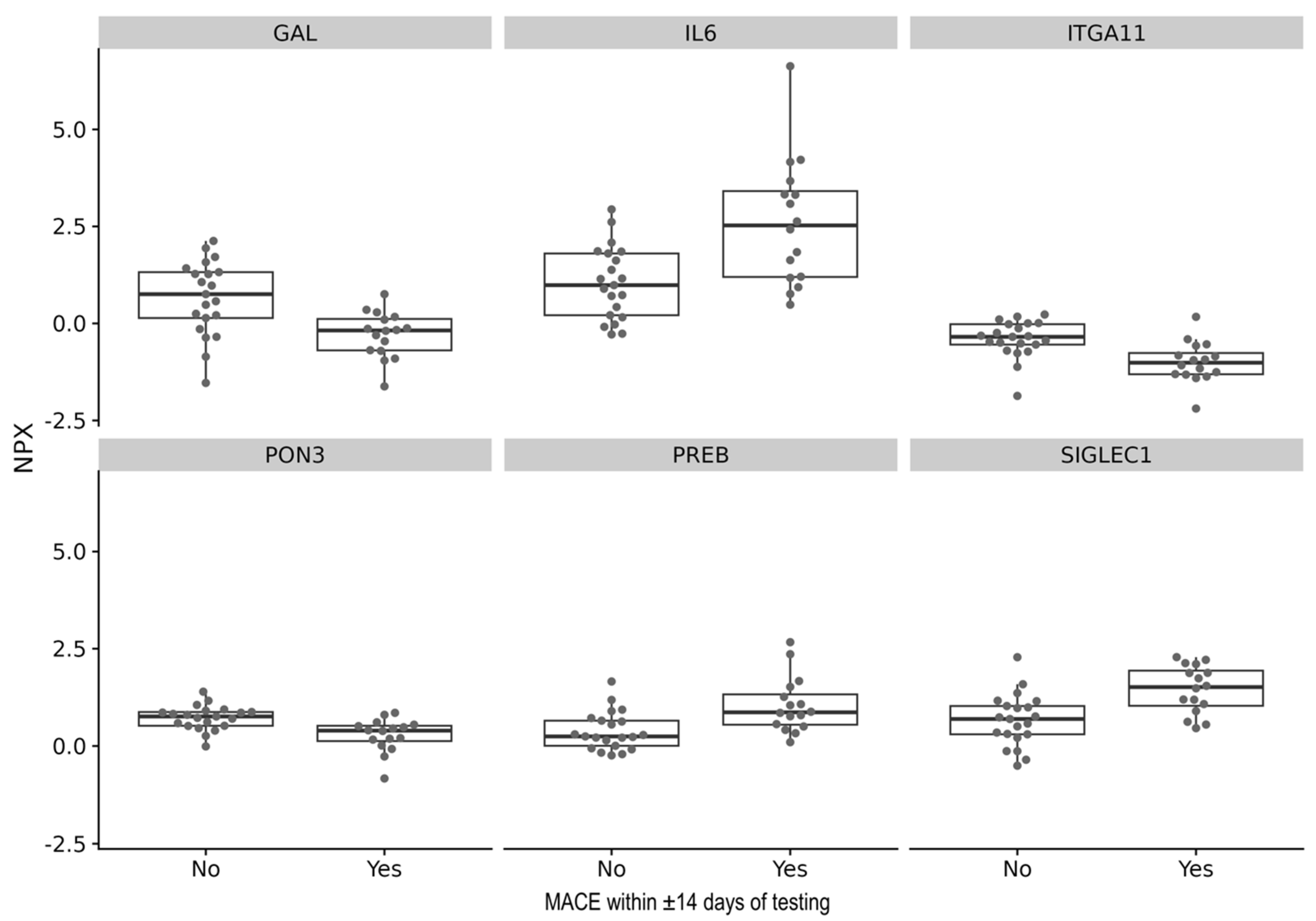
A linear model was used to analyze whether cytokines can predict MACE within ±14 days of testing. Although there were no cytokines significantly associated with MACE after correction for multiple testing, there was a trend of a weak-to-moderate associations between serial blood cytokine levels of Paraoxonase 3 (PON3) (p = 0.00075, FDR = 0.28), Sialic-Acid-Binding Ig-Like Lectin 1 (SIGLEC1) (p = 0.00077, FDR = 0.28), and Interleukin 6 (IL-6) (p = 0.00086, FDR = 0.31) and MACE (Figure 2, Table 3).

4. Discussion

There is increasing evidence that myocardial inflammation in cardiac amyloidosis may play a relevant role for prognostic outcomes [10,11,17]. Therefore, we investigated the clinical implications of testing for myocardial inflammation by studying (a) the relationship between peripheral blood cytokine levels and the presence of inflammatory cells within the myocardium confirmed by EMB, and (b) the potential of cytokines as predictors of MACE in ATTR and AL cardiac amyloidosis. We hypothesized that cardiac background inflammation characterized by infiltration of inflammatory cells into the myocardium alters cytokine levels, serving as biomarkers for diagnosis, and that cytokines predict MACE in cardiac amyloidosis.
In our analysis, which may have been limited by the small sample size, no significant associations were found between serial blood cytokine levels and the status of myocardial immune cell infiltration. Given that we included all patients with myocardial inflammation, even those who did not meet the criteria outlined in the ESC position statement on myocarditis [20], it is also possible that, in cases of starker cardiac inflammation in amyloidosis, the effect on peripheral cytokine levels could be more pronounced.
The rationale behind our approach was the scientific discussion concerning to what extent the healthy heart contains immune cells and what the cut-off value is for defining cardiac inflammation in amyloidosis, and more generally, in cardiomyopathies caused by etiologies other than myocarditis [21]. Additionally, since an EMB provides only a snapshot in time, it remains unclear as to whether the biopsies from the patients of our cohort were taken during active inflammation or potentially a period of remission [9]. A key question in cardiac amyloidosis remains the timing and trajectory of inflammation, which also could have influenced our data.
Some studies suggest that during the chronic phase of myocardial inflammation, the overall density of immune cells within the myocardium decreases, while the proportion of T cells increases [9,22]. This may also influence peripheral cytokine levels, which might render them only an indirect measure of the status of myocardial inflammation at the time of assessment. We have previously demonstrated that in chronic myocarditis, inflammatory markers in the blood including C-reactive protein and troponin frequently normalize, while inflammation in the myocardium continues to progress [23].
This complexity in detecting myocardial inflammation is further compounded by the fact that an EMB can yield false negatives if the sample is taken from a region distant from areas of focal inflammation, as inflammation may not be uniformly distributed throughout the myocardium. In this regard, cardiac magnetic resonance (CMR) imaging, while considered the non-invasive gold standard in diagnosing myocarditis [24], might not be a suitable method in detecting myocardial inflammation in cardiac amyloidosis as the currently available data are limited and inconsistent [25]. Furthermore, it is known from myocarditis that CMR imaging has a limited diagnostic window of a few weeks for detecting myocardial inflammation [26], which may reduce its utility in the context of the chronic clinical trajectory of cardiac amyloidosis.
While our study may have been underpowered to detect robust associations for serial blood levels of cytokines and MACE, several cytokines, such as IL-6, demonstrated weak-to-moderate effect sizes and explained variance in relation to MACE. Nonetheless, none of these associations withstood false discovery rate (FDR) correction, underscoring the need for cautious interpretation. Importantly, while previous studies have reported associations between elevated IL-6 levels and adverse outcomes in amyloidosis [27,28], our findings may have been constrained by the limited sample size, emphasizing the need for validation in larger, adequately powered cohorts.
Previous studies using adult cardiomyocyte (AC16)-based models have demonstrated that AC16 cells treated with ATTR exhibit an enhanced response to pro-inflammatory cytokines and increased apoptosis [29]. Similarly, in AC16-based models of AL, there is an upregulation of IL-6 expression [30]. While interactions between these systems appear to influence clinical outcomes, they do not seem to be directly measurable in our cohort in terms of their immediate effect on MACE.
From a clinical perspective, evidence suggests that amyloidosis is associated with inflammation, which in turn leads to worse clinical outcomes [10,11]. More mechanistic studies are needed to stratify the role of inflammation in cardiac amyloidosis pathogenesis and the influence on the clinical trajectory of the patients, especially on the progression to heart failure. Furthermore, the insufficiency of cytokines in peripheral blood samples in assessing the status of cardiac inflammation in our cohort highlights the need for novel methods to detect cardiac inflammation and identify patients who might benefit from anti-inflammatory treatment. One promising tool could be the measurement of the cardiac magnetic field using magnetocardiography [31,32]. With the latest implementation of superconducting quantum interference device (SQUID) sensors, magnetocardiography detects myocardial alterations with increased sensitivity and has been successfully applied in inflammatory cardiomyopathy and cardiac amyloidosis research [32,33].

Supplementary Materials

The following supporting information can be downloaded at: https://www.mdpi.com/article/10.3390/biomedicines13092254/s1, Table S1: Olink Explore 384—Inflammation panel.

Author Contributions

Conceptualization, N.M., P.S., M.M., M.V., J.W.3rd, D.B. and B.H.; Methodology, N.M., P.S., M.M., M.V., K.K., J.W.3rd, D.B. and B.H.; Software, J.W.3rd and D.B.; Validation, N.M., P.S., M.M., J.W.3rd, D.B. and B.H.; Formal analysis, N.M., P.S., M.M., M.V., J.W.3rd, D.B. and B.H.; Investigation, N.M., P.S., M.M., M.V., K.K., J.W.3rd, D.B. and B.H.; Resources, D.B., U.L. and B.H.; Data curation, N.M., P.S., M.M., K.K., J.W.3rd, D.B. and B.H.; Writing—original draft, N.M., J.W.3rd and B.H.; Writing—review & editing, N.M., P.S., M.M., M.V., K.K., J.W.3rd, D.B., U.L. and B.H.; Visualization, N.M., P.S., K.K., J.W.3rd and B.H.; Supervision, U.L. and B.H.; Project administration, B.H.; Funding acquisition, U.L. and B.H. All authors have read and agreed to the published version of the manuscript.

Funding

This project was supported by a grant from Pfizer Inc. Additional funding was provided by a project grant from the Swiss National Science Foundation issued to Bettina Heidecker, MD (money follows researcher grant). Nicolas Musigk, Phillip Suwalski, and Bettina Heidecker received grant funding from the Deutsche Herzstiftung e.V.

Institutional Review Board Statement

The study was conducted in accordance with the Declaration of Helsinki, and approved by the Ethics Committee of Charité—Universitätsmedizin Berlin (EA4/163/21, approval date: 15 July 2021).

Informed Consent Statement

Informed consent was obtained from all subjects involved in the study.

Data Availability Statement

Data presented in this study is contained within the article and Supplementary Material. Further inquiries can be directed to the corresponding author.

Acknowledgments

We thank Xiaomin Wang for her help with sample collection and sample processing. Nicolas Musigk is supported by a stipend from the Deutsche Herzstiftung e.V. Bettina Heidecker is a participant in the BIH-Charité Advanced Clinician Scientist Pilot program funded by the Charité—Universitätsmedizin Berlin and the Berlin Institute of Health.

Conflicts of Interest

BH is inventor on patents that use RNA for the diagnosis of myocarditis. Patent protection is in process for MCG for the diagnosis and measurement of therapy response in inflammatory cardiomyopathy. BH, JW, DB, UL: Patent protection is in process for cytokines for targeted therapy in inflammatory cardiomyopathy and heart failure.

References

  1. Garcia-Pavia, P.; Rapezzi, C.; Adler, Y.; Arad, M.; Basso, C.; Brucato, A.; Burazor, I.; Caforio, A.L.P.; Damy, T.; Eriksson, U.; et al. Diagnosis and treatment of cardiac amyloidosis: A position statement of the ESC Working Group on Myocardial and Pericardial Diseases. Eur. Heart J. 2021, 42, 1554–1568. [Google Scholar] [CrossRef]
  2. Maurer, M.S.; Schwartz, J.H.; Gundapaneni, B.; Elliott, P.M.; Merlini, G.; Waddington-Cruz, M.; Kristen, A.V.; Grogan, M.; Witteles, R.; Damy, T.; et al. Tafamidis Treatment for Patients with Transthyretin Amyloid Cardiomyopathy. N. Engl. J. Med. 2018, 379, 1007–1016. [Google Scholar] [CrossRef]
  3. Rapezzi, C.; Elliott, P.; Damy, T.; Nativi-Nicolau, J.; Berk, J.L.; Velazquez, E.J.; Boman, K.; Gundapaneni, B.; Patterson, T.A.; Schwartz, J.H.; et al. Efficacy of Tafamidis in Patients With Hereditary and Wild-Type Transthyretin Amyloid Cardiomyopathy: Further Analyses From ATTR-ACT. JACC Heart Fail. 2021, 9, 115–123. [Google Scholar] [CrossRef]
  4. Fontana, M.; Berk, J.L.; Gillmore, J.D.; Witteles, R.M.; Grogan, M.; Drachman, B.; Damy, T.; Garcia-Pavia, P.; Taubel, J.; Solomon, S.D.; et al. Vutrisiran in Patients with Transthyretin Amyloidosis with Cardiomyopathy. N. Engl. J. Med. 2025, 392, 33–44. [Google Scholar] [CrossRef]
  5. Garcia-Pavia, P.; Aus dem Siepen, F.; Donal, E.; Lairez, O.; van der Meer, P.; Kristen, A.V.; Mercuri, M.F.; Michalon, A.; Frost, R.J.A.; Grimm, J.; et al. Phase 1 Trial of Antibody NI006 for Depletion of Cardiac Transthyretin Amyloid. N. Engl. J. Med. 2023, 389, 239–250. [Google Scholar] [CrossRef] [PubMed]
  6. Musigk, N.; Heidecker, B. Transthyretin amyloidosis: The picture is getting clearer. Eur. J. Heart Fail. 2022, 24, 1697–1699. [Google Scholar] [CrossRef] [PubMed]
  7. Rubin, J.; Maurer, M.S. Cardiac Amyloidosis: Overlooked, Underappreciated, and Treatable. Annu. Rev. Med. 2020, 71, 203–219. [Google Scholar] [CrossRef] [PubMed]
  8. Müller, M.L.; Butler, J.; Heidecker, B. Emerging therapies in transthyretin amyloidosis—A new wave of hope after years of stagnancy? Eur. J. Heart Fail. 2020, 22, 39–53. [Google Scholar] [CrossRef]
  9. Musigk, N.; Suwalski, P.; Golpour, A.; Fairweather, D.; Klingel, K.; Martin, P.; Frustaci, A.; Cooper, L.T.; Lüscher, T.F.; Landmesser, U.; et al. The inflammatory spectrum of cardiomyopathies. Front. Cardiovasc. Med. 2024, 11, 1251780. [Google Scholar] [CrossRef]
  10. Siegismund, C.S.; Escher, F.; Lassner, D.; Kühl, U.; Gross, U.; Fruhwald, F.; Wenzel, P.; Münzel, T.; Frey, N.; Linke, R.P.; et al. Intramyocardial inflammation predicts adverse outcome in patients with cardiac AL amyloidosis. Eur. J. Heart Fail. 2018, 20, 751–757. [Google Scholar] [CrossRef]
  11. Müller, M.L.; Brand, A.; Mattig, I.; Spethmann, S.; Messroghli, D.; Hahn, K.; Violano, M.; Mitchell, J.D.; Hare, J.M.; Frustaci, A.; et al. Myocardial Inflammation in Cardiac Transthyretin Amyloidosis: Prevalence and Potential Prognostic Implications. Circ. Heart Fail. 2025, 18, e012146. [Google Scholar] [CrossRef] [PubMed]
  12. Levine, B.; Kalman, J.; Mayer, L.; Fillit, H.M.; Packer, M. Elevated circulating levels of tumor necrosis factor in severe chronic heart failure. N. Engl. J. Med. 1990, 323, 236–241. [Google Scholar] [CrossRef] [PubMed]
  13. Mann, D.L. Innate immunity and the failing heart: The cytokine hypothesis revisited. Circ. Res. 2015, 116, 1254–1268. [Google Scholar] [CrossRef] [PubMed]
  14. Adamo, L.; Rocha-Resende, C.; Prabhu, S.D.; Mann, D.L. Reappraising the role of inflammation in heart failure. Nat. Rev. Cardiol. 2020, 17, 269–285. [Google Scholar] [CrossRef]
  15. Pieske, B.; Tschöpe, C.; de Boer, R.A.; Fraser, A.G.; Anker, S.D.; Donal, E.; Edelmann, F.; Fu, M.; Guazzi, M.; Lam, C.S.P.; et al. How to diagnose heart failure with preserved ejection fraction: The HFA–PEFF diagnostic algorithm: A consensus recommendation from the Heart Failure Association (HFA) of the European Society of Cardiology (ESC). Eur. Heart J. 2019, 40, 3297–3317. [Google Scholar] [CrossRef]
  16. Van Linthout, S.; Tschöpe, C. Inflammation-Cause or Consequence of Heart Failure or Both? Curr. Heart Fail. Rep. 2017, 14, 251–265. [Google Scholar] [CrossRef]
  17. Klingel, K.; Hehn, A.; Sauter, M. P5403The impact of inflammation on the outcome of cardiac amyloidosis. Eur. Heart J. 2017, 38 (Suppl. S1), ehx493.P5403. [Google Scholar] [CrossRef]
  18. Lundberg, M.; Eriksson, A.; Tran, B.; Assarsson, E.; Fredriksson, S. Homogeneous antibody-based proximity extension assays provide sensitive and specific detection of low-abundant proteins in human blood. Nucleic Acids Res. 2011, 39, e102. [Google Scholar] [CrossRef]
  19. Rickham, P.P. HUMAN EXPERIMENTATION. CODE OF ETHICS OF THE WORLD MEDICAL ASSOCIATION. DECLARATION OF HELSINKI. Br. Med. J. 1964, 2, 177. [Google Scholar] [CrossRef]
  20. Caforio, A.L.; Pankuweit, S.; Arbustini, E.; Basso, C.; Gimeno-Blanes, J.; Felix, S.B.; Fu, M.; Heliö, T.; Heymans, S.; Jahns, R.; et al. Current state of knowledge on aetiology, diagnosis, management, and therapy of myocarditis: A position statement of the European Society of Cardiology Working Group on Myocardial and Pericardial Diseases. Eur. Heart J. 2013, 34, 2636–2648, 2648a–2648d. [Google Scholar] [CrossRef]
  21. Halushka, M.K.; d’Amati, G.; Bois, M.C.; De Gaspari, M.; Giordano, C.; Klingel, K.; Leduc, C.; Ohta-Ogo, K.; Ozcan, I.; Rizzo, S.; et al. The frequency of CD3+ lymphocytes in non-myocarditis endomyocardial biopsies. Cardiovasc. Pathol. 2025, 77, 107733. [Google Scholar] [CrossRef]
  22. Fairweather, D.; Kaya, Z.; Shellam, G.R.; Lawson, C.M.; Rose, N.R. From infection to autoimmunity. J. Autoimmun. 2001, 16, 175–186. [Google Scholar] [CrossRef] [PubMed]
  23. Berg, J.; Kottwitz, J.; Baltensperger, N.; Kissel, C.K.; Lovrinovic, M.; Mehra, T.; Scherff, F.; Schmied, C.; Templin, C.; Lüscher, T.F.; et al. Cardiac Magnetic Resonance Imaging in Myocarditis Reveals Persistent Disease Activity Despite Normalization of Cardiac Enzymes and Inflammatory Parameters at 3-Month Follow-Up. Circ. Heart Fail. 2017, 10, e004262. [Google Scholar] [CrossRef] [PubMed]
  24. Schulz-Menger, J.; Collini, V.; Gröschel, J.; Adler, Y.; Brucato, A.; Christian, V.; Ferreira, V.M.; Gandjbakhch, E.; Heidecker, B.; Kerneis, M.; et al. 2025 ESC Guidelines for the management of myocarditis and pericarditis: Developed by the task force for the management of myocarditis and pericarditis of the European Society of Cardiology (ESC)Endorsed by the Association for European Paediatric and Congenital Cardiology (AEPC) and the European Association for Cardio-Thoracic Surgery (EACTS). Eur. Heart J. 2025, ehaf192. [Google Scholar] [CrossRef]
  25. Kottam, A.; Hanneman, K.; Schenone, A.; Daubert, M.A.; Sidhu, G.D.; Gropler, R.J.; Garcia, M.J.; on behalf of the American Heart Association Council on Cardiovascular Radiology and Intervention. State-of-the-Art Imaging of Infiltrative Cardiomyopathies: A Scientific Statement From the American Heart Association . Circ. Cardiovasc. Imaging 2023, 16, e000081. [Google Scholar] [CrossRef]
  26. Lurz, P.; Luecke, C.; Eitel, I.; Föhrenbach, F.; Frank, C.; Grothoff, M.; Waha, S.d.; Rommel, K.-P.; Lurz, J.A.; Klingel, K.; et al. Comprehensive Cardiac Magnetic Resonance Imaging in Patients With Suspected Myocarditis. JACC 2016, 67, 1800–1811. [Google Scholar] [CrossRef]
  27. Hein, S.J.; Knoll, M.; Aus dem Siepen, F.; Furkel, J.; Schoenland, S.; Hegenbart, U.; Katus, H.A.; Kristen, A.V.; Konstandin, M. Elevated interleukin-6 levels are associated with impaired outcome in cardiac transthyretin amyloidosis. World J. Cardiol. 2021, 13, 55–67. [Google Scholar] [CrossRef]
  28. Lugitsch, J.; Hoeller, V.; Schwegel, N.; Santner, V.; Gollmer, J.; Kolesnik, E.; Lipp, R.; Niedrist, T.; Rainer, P.; Zirlik, A.; et al. Interleukin-6 plasma levels predict mortality and heart failure events in cardiac transthyretin amyloidosis. Eur. Heart J. 2023, 44 (Suppl. 2), ehad655.1884. [Google Scholar] [CrossRef]
  29. Ghosh, S.; Villacorta-Martin, C.; Lindstrom-Vautrin, J.; Kenney, D.; Golden, C.S.; Edwards, C.V.; Sanchorawala, V.; Connors, L.H.; Giadone, R.M.; Murphy, G.J. Mapping cellular response to destabilized transthyretin reveals cell- and amyloidogenic protein-specific signatures. Amyloid 2023, 30, 379–393. [Google Scholar] [CrossRef]
  30. Jordan, T.L.; Maar, K.; Redhage, K.R.; Misra, P.; Blancas-Mejia, L.M.; Dick, C.J.; Wall, J.S.; Williams, A.; Dietz, A.B.; van Wijnen, A.J.; et al. Light chain amyloidosis induced inflammatory changes in cardiomyocytes and adipose-derived mesenchymal stromal cells. Leukemia 2020, 34, 1383–1393. [Google Scholar] [CrossRef]
  31. Golpour, A.; Suwalski, P.; Landmesser, U.; Heidecker, B. Case report: Magnetocardiography as a potential method of therapy monitoring in amyloidosis. Front. Cardiovasc. Med. 2023, 10, 1224578. [Google Scholar] [CrossRef]
  32. Brala, D.; Thevathasan, T.; Grahl, S.; Barrow, S.; Violano, M.; Bergs, H.; Golpour, A.; Suwalski, P.; Poller, W.; Skurk, C.; et al. Application of Magnetocardiography to Screen for Inflammatory Cardiomyopathy and Monitor Treatment Response. J. Am. Heart Assoc. 2023, 12, e027619. [Google Scholar] [CrossRef]
  33. Müller, M.L.; Suwalski, P.; Wilke, F.; Latinova, E.; Satilmis, G.; Musigk, N.; Brand, A.; Mattig, I.; Knebel, F.; Messroghli, D.; et al. Magnetocardiography for the non-invasive diagnosis of myocardial inflammation in cardiac amyloidosis: A proof-of-concept study. ESC Heart Fail. 2025. [Google Scholar] [CrossRef]
Figure 1. Relative protein levels of the top 6 cytokines associated with myocardial presence of inflammatory cells in cardiac ATTR and AL amyloidosis.
Biomedicines 13 02254 g001
Figure 2. Relative protein levels of the top 6 cytokines associated with MACE in cardiac ATTR and AL amyloidosis.
Biomedicines 13 02254 g002
Table 1. Patient characteristics.
Analysis 1: Correlation of Cytokines with Myocardial
Presence of Inflammatory Cells
Analysis 2: Correlation of Cytokines with MACE
EMB-Positive for Myocardial Presence of Inflammatory Cells
(n = 17)
EMB-Negative for Myocardial Presence of Inflammatory Cells
(n = 10)
p-ValueMACE ± 14 Days of a Study Visit
(n = 16)
No MACE ± 14 Days of a Study Visit
(n = 21)
p-Value
ATTR (n, %)10 (58.8%)7 (70.0%)0.5747 (43.8%)15 (71.4%)0.574
AL (n, %)7 (41.2%)3 (30.0%)9 (56.2%)6 (28.6%)
Age in years
(median, IQR)
77.0 (73.0–83.0)76.5 (72.8–81.8)178.0 (74.5–83.0)78.0 (68.0–82.0)0.684
Sex female
(n, %)
5 (29.4%)2 (20.0%)0.9325 (31.3%)4 (19.0%)0.933
BMI in kg/m2
(median, IQR)
23.9 (22.6–25.3)25.4 (22.4–26.7)0.35924.6 (21.3–27.3)23.9 (21.8–25.9)0.255
hs-cTn in ng/L
(median, IQR)
69.0 (44.0–159.0)55.0 (41.0–78.0)0.59081.5 (57.3–129.0)57.0 (35.0–79.5)0.182
NT-proBNP in ng/L
(median, IQR)
5008.0 (2553.0–9448.0)2112.5 (1163.3–5370.0)0.2046428.5 (3673.5–16,363.5)2114.0 (1010.0–5008.0)0.132
Creatinine
in mg/dL
(median, IQR)
1.2 (1.1–1.4)1.3 (1.1–1.5)0.5301.3 (1.0–1.6)1.2 (1.1–1.4)0.049
AL (immunoglobulin-light-chain amyloidosis), ATTR (transthyretin amyloidosis), BMI (body mass index), hs-cTn (high-sensitivity cardiac troponin), EMB (endomyocardial biopsy), IQR (interquartile range), MACE (major adverse cardiovascular event), NT-proBNP (N-terminal pro-B-type natriuretic peptide).
Table 2. Top 10 cytokines associated with myocardial presence of inflammatory cells in cardiac ATTR and AL amyloidosis.
AssayEstimateR2p-ValueFDR
CD244−0.470.280.00451
ADGRE2−0.490.240.01011
AOC1−1.130.220.01341
CLEC4D−1.010.220.01421
IL1RL2−0.410.220.01431
CD84−0.370.210.01661
PRKAB1−0.760.210.01671
OSCAR−0.570.200.02071
RGS8−1.040.180.02511
CD200−0.310.180.02851
There was no significant difference in the expression of the top 10 cytokines between patients with and without inflammatory cells in the myocardium. The selected cytokines are the ten with the highest correlation to the myocardial presence of inflammatory cells, as determined by differential analysis using a linear model. ADGRE2 (Adhesion G-Protein-Coupled Receptor E2), AOC1 (Amine Oxidase Copper Containing 1), CD200 (Cluster of Differentiation 200), CD244 (Cluster of Differentiation 244, Natural Killer Cell Receptor 2B4), CD84 (Cluster of Differentiation 84), CLEC4D (C-Type Lectin Domain Family 4 Member D, Dectin-3), IL1RL2 (Interleukin 1 Receptor-Like 2, IL-36 Receptor), OSCAR (Osteoclast-Associated Immunoglobulin-Like Receptor), PRKAB1 (Protein Kinase AMP-Activated Non-Catalytic Subunit Beta 1), RGS8 (Regulator of G-Protein Signaling 8), Estimate (effect size estimate), FDR (false discovery rate calculated using Benjamini–Hochberg correction), r2 (coefficient of determination).
Table 3. Top 10 cytokines associated with MACE in cardiac ATTR and AL amyloidosis.
AssayEstimateR2p-ValueFDR
PON3−0.450.280.000750.28
SIGLEC10.790.280.000770.28
IL61.550.280.000860.31
ITGA11−0.590.270.001070.39
GAL−0.950.260.001360.49
PREB0.660.240.001930.70
PRKCQ0.730.240.001960.71
EPCAM−0.810.230.002500.90
PNPT11.040.220.003201.00
TNFRSF13C0.800.220.003301.00
There was no significant difference in the expression of the top 10 cytokines between patients with and without MACE within ±14 days of testing. The selected cytokines are the ten with the highest correlation to MACE ±14 days of testing, as determined by differential analysis using a linear model. EPCAM (Epithelial Cell Adhesion Molecule), GAL (Galanin and GMAP Prepropeptide), IL6 (Interleukin 6), ITGA11 (Integrin Subunit Alpha 11), PON3 (Paraoxonase 3), PNPT1 (Polyribonucleotide Nucleotidyltransferase 1), PREB (Prolactin Regulatory Element Binding), PRKCQ (Protein Kinase C Theta), SIGLEC1 (Sialic-Acid-Binding Ig-Like Lectin 1), TNFRSF13C (Tumor Necrosis Factor Receptor Superfamily Member 13C), Estimate (effect size estimate), FDR (false discovery rate calculated using Benjamini–Hochberg correction), r2 (coefficient of determination).
Disclaimer/Publisher’s Note: The statements, opinions and data contained in all publications are solely those of the individual author(s) and contributor(s) and not of MDPI and/or the editor(s). MDPI and/or the editor(s) disclaim responsibility for any injury to people or property resulting from any ideas, methods, instructions or products referred to in the content.